Báo cáo khoa học: Molecular characterization of a novel nuclear transglutaminase that is expressed during starfish embryogenesis ppt
Bạn đang xem bản rút gọn của tài liệu. Xem và tải ngay bản đầy đủ của tài liệu tại đây (254.09 KB, 11 trang )
Molecular characterization of a novel nuclear transglutaminase
that is expressed during starfish embryogenesis
Hiroyuki Sugino*, Yudai Terakawa, Akiko Yamasaki, Kazuhiro Nakamura, Yoshiaki Higuchi,
Juro Matsubara, Hisato Kuniyoshi and Susumu Ikegami
Department of Applied Biochemistry, Hiroshima University, Japan
We report the constitution and molecular characterization
of a novel tran sglutaminase (EC 2.3.2.13) that starts to
accumulate specifically in the nucleus in the starfish (Asterina
pectinifera) embryo after progression through the early
blastula stage. The cDNA for the nuclear transglutaminase
was cloned and the cDNA-deduced sequence defines a single
open reading frame encoding a protein with 737 amino acids
and a predicted molecular mass of 83 kDa. A comparison of
this transglutaminase with other members of the gene family
revealed an overall sequence identity of 33–41%. A special
sequence feature of this transglutaminase, which is not found
in other transglutaminases, is t he presence of nuclear local-
ization signal-like sequences in the N-terminal region.
Microinjection of hybrid constructs that encode the N-ter-
minal segment fused to reporter proteins into the germinal
vesicle of an oocyte produced chimeric proteins by
transcription-coupled translation. It was foun d that the
N-terminal segmen t alone was sufficient t o effect nuclear
accumulation of an otherwise cytoplasmic protein. These
results suggest that the nuclear accumulation of the trans-
glutaminase may play an important role in nuclear remod-
eling during early starfish embryogenesis.
Keywords: transglutaminase; nucleus; starfish; e mbryo;
cloning.
The class of enzymes that are commonly referred to as
transglutaminases (TG) (EC 2.3.2.13) are known mostly for
their role in the post-translational remodeling of proteins
(reviewed in [1]). These enzymes catalyze protein cross-
linking reactions via the formation of e-(c-glutamyl)lysine
bonds between the c-carboxyl group of a Gln residue in one
polypeptide chain and the e-amino group of a Lys residue in
a second polypeptide chain. Well-documented examples of
TG are p lasma factor X IIIa [2], keratinocyte TG [ 3],
epidermalTG[4],tissueTG[5],andprostaticTG[6].
Recent findings have shown that, apart f rom their protein
modifying capabilities, tissue TG is also able to function as a
component of the signal-transducing G protein complex [7].
The cDNA of G
ha
, involved in the transmission of
adrenergic stimuli, is identical to that of tissue TG of
human endothelial cells [7]. Tissue TG is localized mainly in
the cytosol, but detectable tissue TG expression has been
reported in the nucleus [8–10]. However, TG a ctivity in the
nucleus and the mechanisms of its translocation is not well
understood, and nucleus-specific TG has not been reported.
It is accepted that many proteins are able to cross nuclear
membranes and accumulate against gradients to c oncen-
trate in the nucleus [11,12]. The nuclear translocation of
proteins via the nuclear pore complex is dependent on a
nuclear localization signal in the protein, which is rich in
basic amino acids and may be bipartite [13–1 5]. T he
functional assays of such nuclear localization signals are
usually based on the ability of a signal to confer nuclear
localization to an otherwise non-nuclear protein.
The present paper describes the occurrence of a novel TG
that is localized exclusively in the nucleus of starfish
(Asterina pectinifera) embryonic cells and is designated
nuclear TG (nTG). The amino-acid sequence derived from
the cDNA sequence contains putative nuclear localization
signals [15] in the N -terminal region. We demonstrate here
that the N-terminal region promotes the nuclear accumu-
lation of an otherwise cytoplasmic protein, namely pyruvate
kinase (PK), in t he A. pectinifera oocyte system. This
finding suggests that nuclear localization signals in the
N-terminal region of nTG are functional in the starfish
embryonic cells. Northern b lot analyses carried out in this
study demonstrate that nTG mRNA appears at the early
blastula stage a nd increases thereafter. The nTG protein
level inc reases in parallel w ith m RNA levels. These results
suggest that nTG is, directly or indirectly, involved in the
modification of the nuclear structure or intranuclear
signaling pathways during starfish embryogenesis [16–18].
MATERIALS AND METHODS
Cultivation of embryos
Specimens of the starfish, A. pec tinifera, were collected from
coastal waters off Japan during their breeding season and
maintained in artificial sea water in laboratory aquaria at
Correspondence to S. Ikegami, Department of Applied Biochemistry,
Hiroshima University, 1-4-4 Kagamiyama, Higashi-hiroshima,
Hiroshima 739-8528, Japan.
Fax: + 81 824 22 7059, Tel.: + 81 824 24 7948,
E-mail:
Abbreviations: nTG, nuclear transglutaminase; GFP, g reen fluorescent
protein; PK, pyruvate kinase; TG, transglutaminase.
*Present address: Department of Applied Life Science, Faculty of
Engineering, Sojo University, Japan.
Note: the nucleotide sequence reported in this paper has been sub-
mitted to the DDBJ Data Bank with accession number AB036064.
(Received 26 October 2001, revised 8 February 2002, accepted 20
February 2002)
Eur. J. Biochem. 269, 1957–1967 (2002) Ó FEBS 2002 doi:10.1046/j.1432-1033.2002.02847.x
15 °C. Eggs and sperm were obtained as described previ-
ously [18–20]. Eggs were fertilized and embryos were
cultured in artificial s ea water that contained 5 mgÆmL
)1
streptomycin sulfate and 50 lgÆmL
)1
penicillin G. Cultures
were maintained in jars at a density of < 5000 embryos per
mL with gentle stirring. Only cultures w ith a fertilization
rate in excess of 95% and normal morphological develop-
ment were used for experimentation.
RT-PCR
Poly(A)
+
RNA was prepared from blastulae (packed vol-
ume, 300 lL) using a QuickPrep micro mRNA purification
kit (Amersham Pharmacia B iotech). RNA (0.1 lg) was
reverse-transcribed into cDNA in a total volume of 20 lL
using the RNA LA PCR kit (Takara, Tokyo, Japan) with
oligo(dT) primer. cDNA coding for tissue TG of bovine
endothelial cells [21] was used as a n internal c ontrol for
PCR. PC R w as carried out with 1.25 U of KOD DNA
polymerase (Toyobo, Osaka, Japan) in the reaction mixture
(50 lL) that contained 120 m
M
Tris/HCl (pH 8.0), 10 m
M
KCl, 6 m
M
(NH
4
)
2
SO
4
, 0.1% Triton X-100, 0.001% BSA,
1m
M
MgCl
2
,0.2m
M
each of four deoxyribonucleoside
5¢-triphosphates, and 4 l
M
each of the TG-specific degen-
erate oligonucleotide primers, TG5 (5¢-TAYGGNCARTG
YTGGGT-3¢;N¼ A, C, G or T; Y ¼ CorT;R¼ Aor
G) and TG 3V (5¢-CCANACRTGRAARTTCCA-3¢). The
PCR cycles were 15 s at 98 °C, 2 s at 55 °C, and 10 s at
74 °C. A total of 25 cycles were run, with the first cycle
containing an extended denaturation p eriod (2 min). The
195-bp PCR product was gel-purified and sequenced by
means of the dideoxy chain termination method using the
Thermo sequenase II dye terminator cycle sequencing kit
(Amersham Pharmacia Biotech) with TG5 and TG3V
primers.
Isolation of cDNA clones and DNA sequencing
Adaptor-ligated double stranded cDNA was prepared from
poly(A)
+
RNA of A. pectinifera blastulae using the Mara-
thon cDNA amplification kit (Clontech) in conjunction
with the oligo(dT) primer and Marathon cDNA adaptor.
TG sequences were amp lified by PCR in both directions
using TG-specific oligonucleotide primers VG5-3 (5¢-ACCC
TCCTCCAGATCGGG-3¢)andTG3-1(5¢-GGACTGTG
CAGAAGTCT-3¢), and the adaptor-specific primer AP1
(5¢-CCATCCTAATACGACTCACTATAGGGC-3¢). The
PCR cycles were 15 s at 98 °C, 2 s at 55 °C, and 30 s at
74 °C. A total of 40 cycles were run, w ith the fi rst cycle
containing an extended denaturation period (2 min). Nested
PCR reactions were performed using the product of the first
PCR under the conditions described above with adaptor
specific primer AP2 (5¢-ACTCACTATAGGGCTCGAGC
GGC-3¢), and internal TG-specific primers TG5-4 (5¢ CCA
TCCAGCAGTCATTCC-3¢)andTG3-2(5¢-AATTTTGC
CTCGGCTCA-3¢). The PCR products were gel-purified
using an Ultraclean DNA purification kit (Mo Bio Labo-
ratories), cloned, and both strands were sequenced from
both d irections under the conditions described a bove. The
deduced cDNA sequence was devoid of a termination
codon. To isolate an oligonucleotide that codes for the
C-terminal region of nTG, the 3¢-RACE approach was
carried out using TG3-3 (5¢-ATCGTGTCGCTGACCAA
C-3¢)andTG3-4(5¢-CC ATTGCCGTACCCGCTG-3¢), the
sequences of which were d erived from the determined
internal region, and the adaptor-specific primers AP1 and
AP2. TG-specific primers designed from the 5¢ and 3 ¢ ends
of the obtained products, 5¢GSP1 (5¢-CGATTACAGTCG
TGGTCAGAGCTG-3¢), 5¢GSP2 (5¢-TCGTGGTCAGAG
CTGTTGTTTGTG-3¢), 3¢GSP1 (5¢-CAAGGACTGACC
TTCACTGAGATG-3¢)and3¢GSP2 (5¢-GTGGCGTTGG
GATGCAACATTGTG-3¢), were used to amplify the full-
length cDNA, and the BamHI (5¢-GCGGATCCATGGTT
CGTCGATCCACTCGC-3¢)andNotI primers (5¢-CT
GCGGCCGCTTAAGCACTCTTGACATTGAG-3¢)to
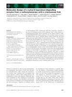
amplify the coding sequence (Fig. 1).
RNA isolation and Northern blot hybridization
Samples of poly(A)
+
RNA (0.5 lg) were prepared from
staged embryos as described previously [22]. They were
denatured and separated by formaldehyde gel electropho-
resis and transferred to nylon filters (Amersham Pharmacia
Biotech). The blots were hybridized overnight at 42 °Cin
hybridization buffer with a probe and washed according to
the manufacture’s recommended protocol. Digoxygenin-
labeled antisense RNA p robes were p repared from a
linearized plasmid DNA template, which contained a
0.27-kbp StuI–NotI restriction fragment of nTG cDNA or
0.15-kbp BamHI–EcoRI restrictio n fragmen t of A. pecti-
nifera ubiquitin cDNA (H. Sugino, unpublished data) using
the digoxygenin-RNA labeling kit (Roche Molecular Bio-
chemicals). D igoxygenin-labeled RNA probes were immu-
nodetected with an Fab fragment of anti-digoxygenin Ig
conjugated to alkaline phosphatase. The bound Ig conju-
gate was then visualized with the chemiluminescent sub-
strate CDP-Star (Roche Molecular Biochemicals).
Expression and purification of glutathione
S
-transferase-conjugated nTG
To generate a recombinant protein of nTG with
N-terminally placed glutathinone S-transferase, the 2214-bp
BamHI–NotI fragment, which contained the entire coding
region of nTG (nTG fragment), was inserted between the
BamHI and NotI sites of p GEX-4T-1(Amersham Pharma-
cia Biotech). Escherichia coli [strain BL21 (DE3)] were
transformed and transcription was induced with 0.5 m
M
10 2 3 (kb)
5' 3'
TG3-3 + AP1 / TG3-4 + AP2
BamHI primer + NotI primer
TG5 + TG3V
5'GSP1 + 3'GSP1 / 5'GSP2 + 3'GSP2
TG3-1 + AP1 / TG3-2 + AP2
TG5-3 + AP1 / TG5-4 + AP2
Fig. 1. PCR strategy for amplification of nTG cDNA. The horizontal
bar i ndicates the n TG cDNA. Thic k horizontal b ars indicate the
sequences of PCR-amplified clones. T he p rimers used for PCR a re
given on the right. The sequences of the oligonucleotide primers are
given in Materials and methods.
1958 H. Sugino et al. (Eur. J. Biochem. 269) Ó FEBS 2002
isopropyl thio-b-
D
-galactoside. Bacteria were lysed in 1%
Triton X-100 in NaCl/P
i
, sonicated with six bursts of 10 s,
and incubated at 4 °C for 1 h. Insoluble materials were
removed by centrifugation at 13 000 g for 10 min. Gluta-
thione S-transferase-conjugated nTG w as purified from th e
supernatant using glutathione–Sepharose 4B beads (Amer-
sham Pharmacia Biotech) essentially following the protocol
provided by the manufacturer.
Biochemical fractionation of embryos
Embryos were washed with ice-cold solution 1 [0.25
M
sucrose, 10 m
M
Tris/HCl (pH 8.0), and 0 .1 m
M
EDTA].
They were then resuspended in the same volume of solution
1, to which had been added 0.15 m
M
spermine and 0.5 m
M
spermidine. The suspension was homogenized by 10 strokes
with a Dounce homogenizer. To the homogenate was added
1.3 v ol. of 2.0
M
sucrose, 65 m
M
KCl, 15 m
M
NaCl, 15 m
M
Tris/HCl (pH 8.0), 0.15 m
M
spermine, 0.5 m
M
spermidine,
10 m
M
2-mercaptoethanol, and 0.1 m
M
phenylmethane-
sulfonyl fluoride. The mixture was centrifuged for 50 min at
50 000 g, to give the nuclear fraction in the form of a pellet.
Subnuclear fractionation was carried out according to the
method described by Singh et al. [8]. In brief, the nuclear
suspension was suspended in 10% sucrose, 10 m
M
trieth-
anolamine/HCl (pH 7.5), and 0.1 m
M
MgCl
2
. The suspen-
sion was treated with 5 lgÆmL
)1
of deoxyribonuclease I
(Worthington Biochemical) and 2 lgÆmL
)1
of ribonuc-
lease A (Sigma Chemicals) for 15 min at 22 °C, followed by
centrifugation for 1 0 m in at 4 °C (20 000 g). The superna-
tant was collected and designated as Sup1. The pellet
obtained after this step was treated with 1% Triton X-100
and recentrifuged. T he supernatant was separated and
designated as Sup2. The pellet was resuspended in 25 m
M
Tris/HCl (pH 7.5), 1% Triton X-100, and 0.5
M
NaCl. This
suspension was incubated for 30 min at 4 °Candthen
centrifuged f or 10 min a t 20 000 g. T he supernatant was
separated and designated as Sup3. The pellet was resus-
pended in 10S buffer [50 m
M
Hepes/HCl (pH 7.2), 10 m
M
sodium phosphate, 250 m
M
NaCl, 0.3% Nonidet P-40,
0.1% Triton X-100, 0.005% SDS, 1 m
M
NaF, 0.5 m
M
dithiothreitol, a nd 0.1 m
M
phenylmethanesulfonyl fluoride]
and the suspension incubated for 30 min, followed by
centrifugation for 10 m in at 17 000 g. The supernatant,
designated as Sup4, was separated from the pellet. For the
immunoprecipitation experiment, Sup4 was concentrated to
1 : 26 of the original volume using Centricon-10 (Amicon).
Proteins were determined by the modified method of
alkaline copper (Lowry) protein assay [23] using BSA as the
standard.
Transglutaminase activity assays
TG activity was assayed by fluorometric measurement of
monodansylcadaverine conjugation to N,N-dimethylcasein
[24]. S tandard reaction mixtures contained 2.5 mgÆmL
)1
N,N-dimethylcasein,0.5 m
M
monodansylcadaverine,10 m
M
Tris/HCl (pH 7.5), 5 m
M
CaCl
2
,and5m
M
dithiothreitol in
400 lL. Incubation was c arried out at 37 °C for 30 min.
Reactions were quenched by the addition of 400 lLof10%
(w/v) trichloroacetic acid and the suspension was chilled on
ice for 20 min. Precipitated protein was collected by
centrifugation for 20 min at 16 000 g,andwashedthree
times with cold ethanol/diethyl ether (1 : 1, v/v), before
solubilization in 4 mL of 50 m
M
Tris/HCl (pH 7.5), 8
M
urea, and 0.5% (w/v) SDS. The amount of incorporated
monodansylcadaverine was determined by measuring the
fluorescence o f the solubilized protein using a Shimazu
RF-540 fluorescence spectrophotometer with an excitation
wavelength of 340 nm, emission wavelength of 525 nm, and
a 5-nm slit. The instrument was calibrated with m ono-
dansylcadaverine in 50 m
M
Tris/HCl (pH 7.5), 8
M
urea,
and 0.5% (w/v) SDS prior to each run. One unit of enzyme
activity defined as AIU (amine incorporation unit per min)
was calculated as described previously [24].
Preparation of nTG-specific antibodies
Two portions of the putative amino acid sequence of nTG,
Leu-Asp-Tyr-His-Tyr-Asp-Glu-Asn-Ser-Glu-Pro-Leu-Asp-
Asp and Arg-Arg-Ser-Thr-Arg-Thr-Arg-Ser-Thr-Pro-Thr-
Arg-Phe-Gly-Tyr-Thr-Asp-Arg, were used to produce
nTG-specific polyclonal antibodies, anti-(nTG-M) Ig and
anti-(nTG-N) Ig, respectively. The peptides were synthe-
sized such that each of them contained an artificial Cys
residue at the N- or C-terminus, respectively, for coupling
purposes. E ach s ynthesized peptide was conjugated to
maleimide-activated keyhole limpet hemocyanin (Amer-
sham Pharmacia B iotech) a ccording to manufacturer’s
instructions. New Zealand White rabbits were then immu-
nized with a keyhole limpet hemocyanin-conjugated peptide
(0.5 mg for each injection). Anti-nTG Ig in the antisera were
affinity purified on the antigenic peptide cross-linked to
2-fluoro-1-methylpyridinium-toluene-4-sulfonate-activated
cellulose (Seikagaku Kogyo, Tokyo, Japan). The bound
nTG-specific Ig were e luted with 100 m
M
glycine-HCl
(pH 2.5). The eluates were neutralized with 1
M
Tris, and
stored at )80 °C.
Polyacrylamide gel electrophoresis and immunoblotting
SDS/PAGE was c arried out accor ding to the method
described by Laemmli [25]. Immunoblotting was performed
on poly(vinylidene difluoride) membranes using anti-
(nTG-M) I g (1.1 lgÆmL
)1
), and horseradish peroxidase-
coupled goat a nti-(rabbit I gG) I g (Bio-Rad). D etection o f the
peroxidase was carried out with 3,3¢-diaminobenzidine and
H
2
O
2
. A control experiment w as performed using the anti-
(nTG-M) Ig t hat h ad been preincubated for 1 h at37 °Cwith
the antigenic peptide (0.65 lgÆmL
)1
of affinity-purified Ig).
Immunoprecipitation
Concentrated Sup4 (10 lL) was incubated with the affinity-
purified anti-(nTG-N) Ig (3 lg) for 3 h at 4 °C in 400 lLof
IP buffer [50 m
M
Tris/HCl (pH 7.5), 150 m
M
NaCl, 0 .5%
Triton X-100, and 0.1% SDS]. After the incubation, 100 lL
of protein A–Sepharo se that had be en equilibrated in IP
buffer was added, and t he mixture w as then rotated
moderately for 1 h at 4 °C. Following centrifugation and
removal of the supernatant, the pellets were washed twice
with IP buffer, and resuspended with 4 00 lL of IP buffer
(total volume, 500 lL). C ontrol experiments were per-
formed using the affinity-purified anti-ANOC Ig, which was
raised against the C-terminal portion of ANO 39, a starfish
protein unrelated to nTG [22].
Ó FEBS 2002 Nuclear transglutaminase in starfish embryos (Eur. J. Biochem. 269) 1959
CGATTACAGTCGTGGTCAGAGCTGTTGTTTGTGTTCCTTGTAAATCGTAATCATCCAAA 59
ATGGTTCGTCGATCCACTCGCACCCGCAGCACCCCTACCCGCTTCGGCTACACCGACCGG 119
M V R R S T R T R S T P T R F G Y T D R
TTTGAGCCGTATGCCCGCAAGCCTAAACGGGAAACGACGCGCACAGAGGGGCGACGCTAC 179
F E P Y A R K P K R E T T R T E G R R Y
GTACCCGCCACACCACTGACTCTGCCTACGCTGAAAGAAAAAAAGACGCAACTCAAGGTG 239
V P A T P L T L P T L K E K K T Q L K V
GTGTCAGTTGATCTATGTGTGGAGCGAAACCAGCAGGAGCATAAGACCAGCAAGTACAAG 299
V S V D L C V E R N Q Q E H K T S K Y K
GTTGACAATCTGGTCCTGCGTCGTGGTCAACCGTTCCACCTCAATGTCAAGTTTGACCGA 359
V D N L V L R R G Q P F H L N V K F D R
GACTTCAAGCCGAGTACCGATGAACTTGTATTGGAATTACGAATGGGCAGCCGTGCCAAC 419
D F K P S T D E L V L E L R M G S R A N
GTGACCAAGGGCACACGCTGTGTGGCCCCCGTGGTAACGTCAGCCCCCGACCACGACGAT 479
V T K G T R C V A P V V T S A P D H D D
TGGGGCATTAAGGTGGAGAGTGCCAAAGGCGCCAACGTGACGCTGAAGGTCTTCTGTAGT 539
W G I K V E S A K G A N V T L K V F C S
TCGGAGGCTCTTATTGGCTACTACAATCTGTACATCTTGACGATGAGCGGTGGGGATGAA 599
S E A L I G Y Y N L Y I L T M S G G D E
TACGAGTATGAATCTCCTAAGGAGCTCATCATGCTGTTCAACGCCTGGTGCAAAGATGAT 659
Y E Y E S P K E L I M L F N A W C K D D
GATGTGTATATGGCTGATGAGGTGAAACGGCAGGAGTACGTCATGGGCGAAGTCAGCCTG 719
D V Y M A D E V K R Q E Y V M G E V S L
TACTTCTATGGTTCCAAGTATCGCATCGGCTCATCCCCATGGAACTACGGGCAGTTTGAG 779
Y F Y G S K Y R I G S S P W N Y G Q F E
AAAATGTCGTTGGACTGTGCCCTGTATTTGCTGCAGAAGTCCGGCATGCCCGACTCTAGC 839
K M S L D C A L Y L L Q K S G M P D S S
CGCAAGAGCCCCATCCAGGTTTCCAGGGTTTTATCTGCCTTGGTCAATGCCCAAGATGAT 899
R K S P I Q V S R V L S A L V N A Q D D
GACGGAGTTCTCGTGGGAAGATGGGATGGGGAGTATGACGACGGCATTTCCCCTACCACC 959
D G V L V G R W D G E Y D D G I S P T T
TGGACTGGGAGCATCGCCATCTTGTCCCAGTACATGAAGACTCGGGAATCGGTCAAATAC 1019
W T G S I A I L S Q Y M K T R E S V K Y
GGCCAGTGTTGGGTGTTCGGGAGTCTGCTCACTGGACTGTGCAGAAGTCTGGGTCTACCC 1079
G Q C W V F G S L L T G L C R S L G L P
ACCCGGACCATCACCAATTTTGCCTCGGCTCACGACACCGATGGCAACCTGACTCTTGAC 1139
T R T I T N F A S A H D T D G N L T L D
TACCACTACGATGAGAACTCGGAACCGTTGGATGACTATGACGAAGATAGTATCTGGAAT 1199
Y H Y D E N S E P L D D Y D E D S I W N
TTCCACGTATGGAATGACTGCTGGATGGCTAGACCCGATCTGGAGGAGGGTTACGGGGGC 1259
F H V W N D C W M A R P D L E E G Y G G
TGGCAGGCCGTGGACGCAACCCCTCAGGAAACAAGCAACGGTGTGTACTGCATGGGACCT 1319
W Q A V D A T P Q E T S N G V Y C M G P
ACCTCTCTGCGCGCCATCAAGCAGGGTCACGTGTACATGCAGTATGACACCAAGTTTGCC 1379
T S L R A I K Q G H V Y M Q Y D T K F A
TTTGCTGAGGTCAACGCTGAAAAGGTCTACTGGAAGGTCTTCACGAAATCTAGAAAGGCC 1439
F A E V N A E K V Y W K V F T K S R K A
CCGGAGGTCATAGACATTGACTCCGATGATGTCGGATGCAAGATCAGCACCAAAGCCGTC 1499
P E V I D I D S D D V G C K I S T K A V
GGCAAATTTGAGCGTGAGGACATCACTGAGCAGTACAAGTACAAGGAAGGAACGGAGTTG 1559
G K F E R E D I T E Q Y K Y K E G T E L
GAGCGCATCGCCGTCAGAGAAGCCAGCCGTCATGTACGCAAAGCAAAGAGAATTCTCAAG 1619
E R I A V R E A S R H V R K A K R I L K
AACCTTGTCCGCGACGTGGACTTTGACGTGGACATGGCGGAGGAGTTCCCCATTGGGAAA 1679
N L V R D V D F D V D M A E E F P I G K
GATATCAAGTTCACTATCACTATGGTGAATAAGTCACAACAGACACGTAATGTCTTTCTG 1739
D I K F T I T M V N K S Q Q T R N V F L
GGTGTGACAGGAAGCACCGTGTACTACACAGGTGTTAAGAAGGCCAAGGTGTCATCCTAC 1799
G V T G S T V Y Y T G V K K A K V S S Y
AATGGCACCCTGCCACTGAAGGCAAAGGAAACGCGAGTGATTCCTGTGACTGTACCTGCG 1859
N G T L P L K A K E T R V I P V T V P A
TCTGACTACCTGCCGCAGCTCACTGACTATGCTGGCGTAACGTTCTTCATCATGGCTTCC 1919
S D Y L P Q L T D Y A G V T F F I M A S
GTCAAGGAGACCAAGCAACCATTCAGCAGGCAGTATGACGCCGTGCTTGATAAGCCTGAC 1979
V K E T K Q P F S R Q Y D A V L D K P D
CTGGAGGTCAAGACGGAGGGGCCCATTGTGCGTGGCAAGCCGTTCACAGCTATCGTGTCG 2039
L E V K T E G P I V R G K P F T A I V S
CTGACCAACCCATTGCCGTACCCGCTGACTGACTGCAGCCTACTTATGGAGGGGTCCATC 2099
L T N P L P Y P L T D C S L L M E G S I
ATTGAGGGCGCCAAACGGGTCAAAGCTCCACATGTTCCAGTGAACGGTAAGATGGCCCAG 2159
I E G A K R V K A P H V P V N G K M A Q
CGAGTGCAGCTGACACCCAAGACTGCTGGATCGTGCGACCTCATCGTCAGCTTCAGTTCC 2219
R V Q L T P K T A G S C D L I V S F S S
CCGCAGCTCAGTGGTGTCAAGGCCCATGTCACACTCAATGTCAAGAGTGCTTAATTTGCT 2279
P Q L S G V K A H V T L N V K S A *
ATGCGAGGTCAGCATTTATCCAACCAGAAGCTTCACGGAGCTAGCTGGGCAAGGAAATTT 2339
GATAATCGCAAGAAATAATTTCCCCCCAAAAACAAAAGGTTGTTGGCTGAAAATACTTCT 2399
ACATGTACATGTATATCACTTTGAACTGGTTTTCATTAAAAAAAAAAAACCATCAATTTG 2459
AGAAGAAACAATTACTTCTTAAGTCAATTAATTTTTCTAGAAATGCAAAAGATATTCCCC 2519
TTAACAGCTGTTTGAAATGAGGCCTCGGTCTCAAGTTTAAGAGTGCCCCCATATGTAAGC 2579
TAAAAAGCTCCAGGAAGTTGACCCAGAAGAAATTTGTTAAGAGTTCACGGATAAGCAAGG 2639
TATTTGGATAAGGTGCATTTGTACATTTTGTGTGTACTGGTTTAGTGTAGAATTTAATTT 2699
TTTTTGGTTAATTCTGTCACAAGAACATAATTCTATGGTTACTACACAATGTTGCATCCC 2759
AACGCCACCTTTTTATTTTTAATCATATATCATCTCAGTGAAGGTCAGTCCTTG 2813
A
1960 H. Sugino et al. (Eur. J. Biochem. 269) Ó FEBS 2002
To measure TG activity r ecovered in e ach fraction,
aliquots (200 lL) of the supernatants or the resuspend ed
pellets were incubated in the same condition as described
above, except that incubation was carried out for 1 h.
Immunofluorescence microscopy
Embryos were processed for immunofluorescence as
whole mounts. In some experiments, embryos were
dissociated by the method described by Kaneko &
Dan-Sohkawa [26]. The whole embryos or dissociated
cells were fixed with 3.5% formaldehyde for 30 min at
room temperature. After washing in NaCl/P
i
without
divalent cations, the cells were incubated in 1% T riton
X-100 in NaCl/P
i
,theninNaCl/P
i
alone, then in acetone
()20 °C), and finally in NaCl/P
i
again. The samples were
blocked with 3% BSA in NaCl/P
i
for 30 min at 37 °C.
Incubations wi th primary and secondary antibodies were
carried out for 2 h at 37 °C. Monospecific anti-(nTG-M)
Ig (1.9 lgÆmL
)1
), which had been preincubated with the
antigenic peptide (1.1 lgÆmL
)1
of affinity-purified Ig), was
used as the negative control. The secondary antibody was
cEry MGGP 4
lHem MYGFGRGNMFRNRSTRYRRRPRYRAENYHSYMLDLLENMNEEFGRNWWGTPESHQPDS 58
nTG MVRRSTRTRSTPTRFGYTDRFEPYARKPKRETTRTEGRRYVPATPLTL 48
hKer MMDGPRSDVGRWGGNPLQPPTTPSPEPEPEPDGRSRRGGGRSFWARCCGCCSCRNAADDDWGPEPSDSRGRGSSSGTRRPGSRGSDSRRPVSRGSGVNAA 100
gpLiv MAEDLILERCDLQLEV NGRDHRTADLCRERLVLRRGQPFWLTLHFEGRGYEAGVDTLTFNAVTGPDPSEEAGTMARFSLSSAV EGGTW 88
cEry GPDGTMAEELVLETCDLQCER NGREHRTEEMGSQQLVVRRGQPFTITLNFAGRGYEEGVDKLAFDVETGPCPVETSGTRSHFTLTDCP EEGTW 97
lHem GPSSLQVESVELYTRDNAREH NTFMYDLVDGTKPVLILRRGQPFSIAIRFK-RNYNPQQDRLKLEIGFGQQPLITKGTLIMLPVSGSDTFTKDKTQW 154
nTG PTLKEKKTQLKVVSVDLCVER NQQEHKTSKYKVDNLVLRRGQPFHLNVKFD-RDFKPSTDELVLELRMGSRANVTKGTRCVAPVVTSAP DHDDW 141
hKer GDGTIREGMLVVNGVDLLSSRSDQNRREHHTDEYEYDELIVRRGQPFHMLLLLS RTYESSDRITLELLIGNNPEVGKGTHVIIPVGKGG SGGW 193
hPro MMDASKELQVLHIDFLNQD NAVSHHTWEFQTSSPVFRRGQVFHLRLVLN QPLQSYHQLKLEFSTGPNPSIAKHTLVVLDPRTPS DHYNW 89
. . * . * . . **** * . . . . . . * . * *
gpLiv SASAVDQQDSTVSLLLSTPADAPIGLYRLSLEASTGYQG SSFVLGHFILLYNPRCPADAVYMDSDQERQEYVLTQQGFIYQGSAKFINGIPWN 181
cEry SAVLQQQDGATLCVSLCSPSIARVGRYRLTLEASTGYQG SSFHLGDFVLLFNAWHPEDAVYLKEEDERREYVLSQQGLIYMGSRDYITSTPWN 190
lHem DVRLRQHDGAVITLEIQIPAAVAVGVWKMKIVSQLTSEEQPNVSAVTHECKNKTYILFNPWCKQDSVYMEDEQWRKEYVLSDVGKIFTGSFKQPVGRRWI 254
nTG GIKVESAKGANVTLKVFCSSEALIGYYNLYILTMSGGDE YEYESPKELIMLFNAWCKDDDVYMADEVKRQEYVMGEVSLYFYGSKYRIGSSPWN 235
hKer KAQVVKASGQNLNLRVHTSPNAIIGKFQFTVRTQSDAGEFQLP FDPRNEIYILFNPWCPEDIVYVDHEDWRQEYVLNESGRIYYGTEAQIGERTWN 289
hPro QATLQNESGKEVTVAVTSSPNAILGKYQLNVKTGNHILK SEENILYLLFNPWCKEDMVFMPDEDERKEYILNDTGCHYVGAARSIKCKPWN 180
. . . . . .* . . .*.*. * * . *.** . . . *. *
gpLiv FGQFEDGILDICLMLLDTNPKFLKNAGQDCSRRSRPVYVGRVVSAMVNCND-DQGVLQGRWDNNYSDGVSPMSWIGSVDILRRWKDYGCQRVKYGQCWVF 280
cEry FGQFEDEILAICLEMLDINPKFLRDQNLDCSRRNDPVYIGRVVSAMVNCNDEDHGVLLGRWDNHYEDGMSPMAWIGSVDILKRWRRLGCQPVKYGQCWVF 290
lHem FGQFTDSVLPACMLILER S-GLDYTARSNPIKVVRAISAMVNNID-DEGVLEGRWQEPYDDGVAPWMWTGSSAILEKYLKTRGVPVKYGQCWVF 346
nTG YGQFEKMSLDCALYLLQKS G MPDSSRKSPIQVSRVLSALVNAQD-DDGVLVGRWDGEYDDGISPTTWTGSIAILSQYMKTRES-VKYGQCWVF 326
hKer YGQFDHGVLDACLYILDRR G MPYGGRGDPVNVSRVISAMVNSLD-DNGVLIGNWSGDYSRGTNPSAWVGSVEILLSYLRTGYS-VPYGQCWVF 380
hPro FGQFEKNVLDCCISLLTES SLKPTDRRDPVLVCRAMCAMMSFEK-GQGVLIGNWTGDYEGGTAPYKWTGSAPILQQYYNTKQA-VCFGQCWVF 271
.*** . * .* * *. . *. . . *** *.* * * * * ** ** . * .******
gpLiv AAVACTVLRCLGIPTRVVTNFNSAHDQNSNLLIEYFRNESGE-IEGNKSEMIWNFHCWVESWMTRPDLEPGYEGWQALDPTPQEKSEGTYCCGPVPVRAI 379
cEry AAVACTVMRCLGVPSRVVTNYNSAHDTNGNLVIDRYLSETGM-EERRSTDMIWNFHCWVECWMTRPDLAPGYDGWQALDPTPQEKSEGVYCCGPAPVKAI 389
lHem AGVANTVSRALGIPSRTVTNYDSAHDTDDTLTIDKWFDKNGDKIEDATSDSIWNFHVWNDCWMARPDLPTGYGGWQAYDSTPQETSEGVYQTGPASVLAV 446
nTG GSLLTGLCRSLGLPTRTITNFASAHDTDGNLTLDYHYDENSEPLDDYDEDSIWNFHVWNDCWMARPDLEEGYGGWQAVDATPQETSNGVYCMGPTSLRAI 426
hKer AGVTTTVLRCLGLATRTVTNFNSAHDTDTSLTMDIYFDENMKPLEHLNHDSVWNFHVWNDCWMKRPDLPSGFDGWQVVDATPQETSSGIFCCGPCSVESI 480
hPro AGILTTVLRALGIPARSVTGFDSAHDTERNLTVDTYVNENGKKITSMTHDSVWNFHVWTDAWMKRPDLPKGYDGWQAVDATPQERSQGVFCCGPSPLTAI 371
. *.** * .* **** . .* . .**** * ** **** *. ***. *.**** *.* . **
gpLiv KEGHLNVKYDAPFVFAEVNADVVNWIRQK DGSLRKSIN-HLVVGLKISTKSVGRDE REDITHTYKYPEGSEEEREAFVRANHLNKLATKE- 468
cEry KEGDLQVQYDIPFVFAEVNADVVYWIVQS DGEKKKSTH-SSVVGKNISTKSVGRDS REDITHTYKYPEGSEKEREVFSKAEHEKSSLG 476
lHem QRGEIGYMFDSPFVFSEVNADVVHWQEDDSS-ETGYKKLKIDSYRVGRLLLTKKIGVDDDFGDADAEDITDQYKNKEGTDEERMSVLNAARSSGFNYAFN 545
nTG KQGHVYMQYDTKFAFAEVNAEKVYWKVFTKS-RKAPEVIDIDSDDVGCKISTKAVGKFE REDITEQYKYKEGTELERIAVREASRHVRKAKR 517
hKer KNGLVYMKYDTPFIFAEVNSDKVYWQRQD DGSFKIVYVEEKAIGTLIVTKAISSNMR EDITYLYKHPEGSDAERKAVETAAAHGSKPNVYA 571
hPro RKGDIFIVYDTRFVFSEVNGDRLIWLVKMVNGQEELHVISMETTSIGKNISTKAVGQDR RRDITYEYKYPEGSSEERQVMDHAFLLLSSERE 463
* . .* * *.*** . * .* . ** .*** ** **. ** . *
gpLiv -EAQEETGVAMRIRVGQNMTMGSDFDIFAYITNGTAESHECQLLLCARIVSYNGVLGPVCSTNDLLNLTLDPFSENSIPLH-ILYEKYGDYLTESNLIKV 566
cEry EQEEGLHMRIKLSEGANNGSDFDVFAFISNDTDKERECRLRLCARTASYNGEVGPQCGFKDLLNLSLQPHMEQSVPLR-ILYEQYGPNLTQDNMIKV 572
lHem LPSPEKEDVYFNLLDIEKIKIGQPFHVTVNIENQSSETRRVSAVLSASSIYYTGITGRKIKRENGN-FSLQPHQKEVLSIE-VTPDEYLEKLVDYAMIKL 643
nTG ILKNLVRDVDFDVDMAEEFPIGKDIKFTITMVNKSQQTRNVFLGVTGSTVYYTGVKKAKVSSYNGT-LPLKAKETRVIPVT-VPASDYLPQLTDYAGVTF 615
hKer N-RGSAEDVAMQVEAQDAVMG-QDLMVSVMLINHSSSRRTVKLHLYLSVTFYTGVSG-TIFKETKKEVELAPGASDRVTMP-VAYKEYRPHLVDQGAMLL 667
hPro HRRPVKENFLHMSVQSDDVLLGNSVNFTVILKRKTAALQNVNILGSFELQLYTGKKMAKLCDLNKTSQIQGQVSEVTLTLDSKTYINSLAILDDEPVIRG 563
. . . . . . . *.* . * . .
gpLiv RGLLIEPAANSYVLAERDIYLENPEIKIRVLGEPKQNRKLIAEVSLKNPLPVPLLGCIFTVEGAGLTKDQKSVEVPDPVEAGEQAKVRVDLLPTEVGLHK 666
cEry VALLTEYETGDSVVAIRDVYIQNPEIKIRILGEPMQERKLVAEIRLVNPLAEPLNNCIFVVEGAGLTEGQRIEELEDPVEPQAEAKFRMEFVPRQAGLHK 672
lHem YAIATVKETQQTWSEEDDFMVEKPNLELEIRGNLQVGTAFVLAISLTNPLKRVLDNCFFTIEAPGVTGAFR VTNRDIQPEEVAVHTVRLIPQKPGPRK 741
nTG FIMASVKETKQPFSRQYDAVLDKPDLEVKTEGPIVRGKPFTAIVSLTNPLPYPLTDCSLLMEGSIIEGAKR VKAPHVPVNGKMAQRVQLTPKTAGSCD 713
hKer NVSGHVKESGQVLAKQHTFRLRTPDLSLTLLGAAVVGQECEVQIVFKNPLPVTLTNVVFRLEGSGLQRPKI LNVGDIGGNETVTLRQSFVPVRPGPRQ 765
hPro FIIAEIVESKEIMASEVFTSFQYPEFSIELPNTGRIGQLLVCNCIFKNTLAIPLTDVKFSLESLGISSLQT SDHGTVQPGETIQSQIKCTPIKTGPKK 661
. . * . . *.* * . . .*. . . * * .
gpLiv LVVNFECDKLKAVKGYRNVIIGPA 690
cEry LMVDFESDKLTGVKGYRNVIIAPLPK 698
lHem IVATFSSRQLIQVVGSKQVEVLD 764
nTG LIVSFSSPQLSGVKAHVTLNVKSA 737
hKer LIASLDSPQLSQVHGVIQVDVAPAPGDGGFFSDAGGDSHLGETIPMASRGGA 817
hPro FIVKLSSKQVKEINAQKIVLITK 684
. . . .
B
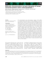
Fig. 2. Nucleotide and deduced amino acid sequences of nTG. (A) The nucleotide sequence of the cDNA clone which encodes nTG and the amino
acid sequence deduced therefrom. (B) D educed amino-acid sequences for guinea pig liver TG (gpLiv) [ 5], chicken erythrocyte TG (cEry) [30],
Limulus hemocyte TG (lHem) [31], human keratinocyte TG (hKer) [3], human prostate TG (hPro) [6], and nTG are shown using the single letter
amino acid codes. Gaps have been inserted to achieve maximum similarity. Asterisks and dots at the bottom of the aligned sequences indicate
positions that are occupied by identical or chemically similar amino acids in all TG. The arrowhead indicates the active site Cys residue [31]. The
arrows indicate the positions of the H is and Asp residues of the c atalytic triad [35]. Putative nuclear localization signals [11] are underlined.
Ó FEBS 2002 Nuclear transglutaminase in starfish embryos (Eur. J. Biochem. 269) 1961
donkey anti-(rabbit IgG) Ig labeled with fluorescein
(Amersham Pharmacia Biotech). Specimens were observed
with a Nikon Eclipse E600 equipped with d ifferential
interphase and epifluorescence optics.
Generation of green fluorescent protein–fusion protein
constructs
The 500 -bp BamHI–HindIII fragment, which contained the
Drosophila heat shock protein promoter, was inserted into
the BglII–HindIII site of pEGFP-1 (Clontech) t o generate
pHEG. To generate the fusion protein between the green
fluorescent protein (GFP) and nTG, the 800-bp KpnI–
BamHI fragment that contained the GFP gene (GFP
fragment) was first generated by subclo ning the 800-bp
Eco47III–PstI fragment of pEGFP-C1 ( Clontech) into the
HincII–PstI sites of pBluescriptII KS(+) (Toyobo), and
then digested with KpnIandBamHI. The 2214-bp BamHI –
NotI fragment, which contains the entire coding region of
nTG (nTG f ragment), was generated via PCR with BamHI
and NotI primers. The 2043-bp BamHI–NotIfragment,
which contains the coding region of nTG, bu t without the
N-terminal 57 amino-acids residue (nTGDN57 fragment),
was generated via PCR with BamHI-2 (5¢-AGGGATCCCT
CAAGGTGGTGTCAGTTGATC-3¢)andNotI primers.
The 171-bp BamHI–XhoI fragment, which contains the
N-terminal 57 amino acid residues of nTG (N57 fragment),
was generated via PCR with BamHI and XhoI(5¢-GACTC
GAGTTGCGTCTTTTTTTCTTTCAGC-3¢) primers. The
1596-bp BamHI–NotI fragment, which contains the en tire
coding region of rat muscle P K [27] ( PK fragment), was
generatedviaPCRwithPK-N(5¢-GCCGGATCCGGC
CTCGAGATGCCCAAGCCAGACAGC-3¢)andPK-C
(5¢-GAGCGGCCGCTCATCAGCCGAGCTCTGGTAC
AGGCACTAC-3¢) primers. The PK fragment was digested
with XhoI and ligated with N57 fragment to give the
N57PK fragment. The nTG, nTGDN57, PK, and N57PK
fragments were separately ligated with the GFP fragment
and the vector fragment derived from KpnI/NotI-digestion
of pHEG to produce pHE-TG, pHE-TGDN57, pHE-PK,
and pHE-N57PK, respectively.
Expression of cloned cDNAs
in vivo
The constructs were separately dissolved in 10 m
M
Tris/HCl
(pH 8.5) to g ive a final concentration o f 200 ngÆlL
)1
.
Twenty picoliters of the solution, along with a small amount
of KF96 silicone oil (Shin-Etsu Chemical, Tokyo, Japan),
were then microinjected into the germinal vesicle of an
oocyte as described previously [22]. T hree to four hours
later, the injected oocytes we re examined for localization
of fluorescent proteins under a fluorescence microscope
equipped with differential interphase and epifluorescence
optics.
RESULTS
Molecular cloning of
A. pectinifera
transglutaminase
Comparison of the amino-acid sequences among already-
known TGs shows highly conserved regions, including the
TG active site [28], in t he middle portions of the polypep-
tides. Based on the sequence of the con served regions, a
single set of degenerate oligonucleotide primers, TG5 and
TG3V, were designed and used for an RT-PCR experiment.
Using poly(A)
+
RNA f rom A. pectinifera embryos a t the
early b lastula stage as a template, a single PCR product
which encodes a 65-amino-acid sequence similar to that of
the catalytic site-containing region of the other TGs was
amplified. To obtain further sequences upstream of primer
TG5 and downstream of primer TG3V, we used the r apid
amplification of cDNA ends (RACE) approach with the
strategy summarized in Fig. 1. Finally, a 2813-bp cDNA
was amplified utilizing the primers 5¢GSP1,2 and 3¢GSP1,2,
which correspond to the 5¢ or 3¢ edges, respectively, of
B
200 -
97.2 -
66.2 -
45.0 -
31.0 -
21.5 -
14.4 -
6.5 -
321
112.0 -
81.0 -
645
A
Fig. 3. Western blot analysis of nTG during embryogenesis. (A) Cyto-
solic fractions (lanes 1–3 and 7–9) and nuclear fractions (lanes 4–6 and
10–12) we re prepared from 8-h-old early b lastulae (lanes 1, 4, 7, and
10), 12-h-old mid-blastulae (lanes 2, 5, 8, and 11), and 24-h-old mid-
gastrulae (lanes 3, 6, 9, and 12). An a liquot of each fraction (60 lg
bovine serum albumin-equivalent per lane) was separated by SDS/
PAGE, and the gel was stained with Coomasssie blue (lanes 1–6) or
transferred to poly(vinylidene difluoride) membrane, followed by
staining using anti-(nTG-M) Ig as a probe (lanes 7–12). Sizes of the
molecular mass marker proteins in kDa are shown to the left. (B)
Nuclear fractions were prepared from 29- h-old midgastrulae (lan es 1
and 4), 40-h-old late gastrulae (lanes 2 and 5), and 51-h-old bipinnariae
(lanes 3 and 6). An aliquot of each fraction (3000 embryos-equivalent
per lane) was separated by SDS/PAGE, and the gel was stained with
Coomasssie blue (lanes 1–3) or transferred to poly(vinylidene difluo-
ride) membrane, followed b y s taining using an ti-(nTG-N) Ig a s a p robe
(lanes 4–6). S izes of the molecular mass marker prote ins in kD a are
shown to the left.
1962 H. Sugino et al. (Eur. J. Biochem. 269) Ó FEBS 2002
the sequence obtained by t he RACE experiments. The
cDNA contained a single open reading fram e ( ORF)
beginning with an ATG codon in an adequate context for
the initiation of translation (Fig. 2A); the sequence
CCAAAATGG s urrounding the A TG fits t he consensus
sequence CC(G/A)CCATGG f or the eukaryotic initiator
site [29]. The predicted protein consists of 737 amino acids,
with a molecular mass of 83 105 Da and an isoelectric point
of 7.9. Neither a polyadenylation signal (AATAAA) nor a
poly(A)
+
tail was found in the 540-bp untranslated region
following the termination codon (TAA), suggesting that the
cDNA might not be full-length.
Figure 2B shows the alignment of t he deduced amino-
acid sequence w ith those o f t he other known TGs from
various species [2,3,5,6,30,31]. The predicted protein exhi-
bited 33–41% identity with other TGs. The residues
comprising the catalytic triad are perfectly conserved
(Cys323, His382, Asp405; Fig. 2B). Three acidic residues,
Glu447, Glu496, and Glu501, which could act as a Ca
2+
-
binding site [32], were also conserved. The sequence
surrounding His351, i.e. Ser349-Ala-His-Asp352, was con-
served, suggesting its interaction with Glu443 by analogy
with the crystallography data on factor XIIIa [32] (Fig. 2B).
On the other hand, residues for the putative GTP-binding
region [33] found in tissue TGs were not well conserved.
To confirm whether the predicted protein carries TG
activity, a bacterially expressed protein in which the putative
ORF was fused in-frame at its N-terminal end to glutathi-
one–S-transferase was prepared, and assayed for TG acti-
vity. The recombinant protein catalyzed the incorporation
of monodansylcadaverine into N,N-dimethylc asein with K
m
and V
max
values of 0.35 m
M
and 13.3 n molÆmin
)1
Æmg
)1
,
respectively, indicating that the predicted protein is a
transglutaminase.
Subcellular localization of
A. pectinifera
transglutaminase
A major characteristic feature of the A. pectinifera TG is the
presence of two putative nucle ar localization signals in the
N-terminal region, a monopartite (residues 26–30) and a
bipartite (residues 38–39 and 52–55) ones [12–14], suggest-
ing nuclear localization of this protein. To examine if the
A. pectinifera TG is a nuclear protein, we raised an
B
ab
cd
ef
gh
C
ab
cd
ef
gh
a
b
A
Fig. 4. Subcellular localization of nTG in embryos. (A) The distribution of nTG in a mid gastrula, as detected by indirect immuno fluorescen ce
microscopy using a rabbit anti-(nTG-M) Ig and a fluorescein-conjugated secondary antibody. Immunofluorescence micrographs (a) and corre-
sponding Normaski differential interference-contrast images (b). Bar, 50 lm (B) The distribution of nTG in cells dissociated from 24-h-old
midgastrulae, as detected by indirect immunofluorescence microscopy using a rabbit anti-(nTG-M) Ig and a fluoresce in-conjugated secondary Ig
(a,b). As a negative control, parallel immunofluorescence was performed using the anti-(nTG -M) Ig preincubated with the antigenic peptide (c,d) or
preimmune sera (e,f), or omitting the primary antibody (g,h). Immunofluorescence micrographs (a,c,e,g) and the corresponding Nomarski
differential interference-contrast micrographs (b,d,f,h). Bar, 5 lm (C) Subcellular localization of nTG during embryogenesis. The distribution of
nTG in cells dissociated from 8-h- (a,b), 12-h- (c,d), 15-h- (e,f), and 24-h-old embryos (g,h) as detected by indirect immunofluorescence microscopy
as described abo ve. Immu nofluorescenc e micrographs (a,c,e ,g), and th e corresponding Nomarski differential interference-contrast micrographs
(b,d,f,h). Bar, 5 lm.
Ó FEBS 2002 Nuclear transglutaminase in starfish embryos (Eur. J. Biochem. 269) 1963
antibody, d esignated anti-(nTG-M) Ig, against the peptide
whose sequence (Leu359–Asp372) was deduced from the
nucleotide sequence of cloned cDNA, and used it for
Western blot analysis (Fig. 3A) and immunocytochemistry
(Fig. 4B). On blots shown in Fig. 3A, this antibody
specifically reacted with a single 90-kDa protein of the
nuclear fraction which was prepared from mid-blastulae
(12 h after fertilization: lane 11) or midgastrulae (24 h after
fertilization: lane 12) whereas n o band was detected in the
cytosol fraction (lane 8, 9). When formalin-fixed prepara-
tions of single cel ls, which had been dissociated from
the midgastrulae, were stained with the same antibody, the
signal was limited to the nucleus (Fig. 4 B, a and b). The
staining was fully blocked by preincubation of the antibody
with the antigenic peptide (Figs 4B, c and d), demonstrating
that the observed fluorescence was not derived from
nonspecific labeling of the nucleus. These results collectively
indicate that the protein encoded by the cloned cDNA is
localized to the nucleus. Hence, we designated the protein
Ônuclear transglutaminase (nTG) Õ.
Expression pattern of nTG during embryogenesis
Early starfish development may be directed by two sources
of mRNA: (a) a pool stored in the immature oocyte
transcribed from the maternal genome during oogenesis
such as ANO39 mRNA [22], and (b) newly synthesized
mRNA transcribed from the embryonic genome. Northern
blot hybridization on poly(A)
+
RNA from blastulae and
gastrulae showed a gradual increase in the signal at 5.0-kb
during the progression of embryonic development (Fig. 5),
whereas hybridization on poly(A)
+
RNA from f ertilized
eggs showed little or no signals, s uggesting that nTG
mRNA belongs to the latter. The developmental Western
blot analysis revealed that the 90-kDa band corresponding
to the nTG protein was first detected in the mid-blastula
embryo and that the level of the band increased by t he
bipinnaria stage (Fig. 3A, lanes 10–11, Fig. 3B, lanes 4–6).
Therefore, the nTG protein s ynthesis starts at mid-blastula
stage and continues thereafter.
Immunostaining o f t he dissociated cells of embryos at
different developmental stages revealed a specific pattern of
accumulation. At the 8- to 12-h-old early blastula stages,
nTG was undetectable in the nucleus (Fig. 4C, a–d). The
nucleus of the early blastula is larger and looser than that of
embryos collected at later developmental stages (Figs 4C,
b,d,f,h). nTG starts to accumulate in the compact nucleus of
the 15-h-old mid-blastula (Figs 4C, e,f) and its amount
increases over the next 9 h (Figs 4C, g,h).
To identify the cells expressing nTG, formalin-fixed
whole-mount embryos at the midgastrula stage were stained
with the anti-(nTG-M) Ig. Staining was not limited to
specific areas but was observed in cells of all the germ layers
(Fig. 4 A).
Extraction of nTG from midgastrulae
We measured the TG activity in nuclear preparations
obtained from 8-h-old early blastulae, 12-h-old mid-blast-
ulae, and 24-h-old midgastrulae. As the total TG activity
(Fig. 6A) as well as the amount of nTG protein (Fig. 3A)
was the highest in the nuclear fraction prepared from
midgastrulae, we e xtracted nTG f rom this preparation
according to the methods of Singh et al. [8]. After treatment
with deoxyribonuclease I and ribonuclease A (Sup1), the
nuclear preparation was subjected to extraction with 1%
Triton X-100 t o afford Sup2 (nuclear membrane fraction),
andthenwithacombinationof1%TritonX-100and0.5
M
NaCl to afford Sup3 (nuclear pore–lamina complex
fraction). Substantial TG activity remained insoluble after
extraction of the nuclear pore–lamina complex. Extraction
of the pellet with 10S buffer, which contained 0.005% SDS
along with a nonionic detergent, successfully solubilized the
enzyme; the total activity recovered in Sup4 was nearly twice
as large as that in the nuclear fraction (Fig. 6B). The
apparent activation of the enzymatic activity recovered in
Sup4 could be due to the r emoval of putative i nhibitors
during subnuclear fractionation.
SDS/PAGE of Sup4 resulted in a prominent band with
an apparent molecular mass of 90 kDa (Fig. 6C, lane 1),
which was re cognized by the anti-(nTG-M) Ig in Western
blot analysis (Fig. 6C, lane 3). To determine if the TG
activity in Sup4 results from t he nTG protein, Sup4 was
subjected to immunoprecipitation with the antibody raised
against the N-terminal portion (Arg3–Arg20) of nTG [anti-
(nTG-N) Ig]. As a result, the TG activity was mainly
recovered in the immunoprecipitate (Fig. 7), showing t hat
the molecule, which predominantly generates the TG
activity in Sup4, is the nTG.
Identification of the segment containing nuclear
localization signals in nTG
To identify the elements(s) in nTG that determine nucleus-
specific topogenesis, we examined the localization of the
12345
10.0 -
4.0 -
3.0 -
6.0 -
Fig. 5. Expression of nTG gene. Northern blots of poly(A)
+
RNA
from fertilized eggs (lane 1), 8-h-old early blastulae (lane 2), 12-h-old
mid-blastulae (lane 3), 15-h-old late b lastulae (lane 4), and 24-h-old
midgastrulae (lane 5). The filter was hybridized with a digoxygenin-
labeled RNA probe obtained from the c DNA of nTG (upper pane l)
and of A. pectinifera ubiquitin (lower panel). Each lane was loaded
with 0.5 lgofpoly(A)
+
RNA. Sizes of the transcripts expressed in kb
were determined by comparison to the relative migration of RNA
markers.
1964 H. Sugino et al. (Eur. J. Biochem. 269) Ó FEBS 2002
GFP–nTG fusion protein produced in an oocyte by
microinjection of pHE-TG, which c ontains the Drosophila
heat shock protein promoter and a gene that encodes the
fusion protein (Fig. 8A), into the germinal vesicle, a nucleus
that is arrested in prophase of division I of meiosis [22].
Transcription-coupled translation produced the fluorescent
fusion protein and the major fraction was accumulated in
the germinal vesicle as shown by fluorescence microscopy
(Fig. 8B, a,e). On the other hand, microinjection of pHE-
TGDN57, which contains the Drosophila heat shock protein
promoter and the gene encoding nTG, in which the
N-terminal 57 amino-acid residues had been deleted and
fused with GFP, led to the formation of a fluorescent
protein which was not located in the nucleus but, rather,
Fig. 6. Extraction of nTG from the nuclear fraction. (A) TG activity in
the nuclear fraction during emb ryogenesis. Enzyme activity was
measured on the nuclear fractions prepared from 8-h-old early blast-
ulae, 12-h-old mid-blastulae, and 24-h-old midgastrulae. The activity is
expressed as the percentage of maximum activity observed in 24-h-old
midgastrulae. (B) TG activity extracted from the n uclear fraction.
Sup1, Sup2, Sup3, and Sup4 ( S1, S2, S3, and S4, respective ly) were
prepared from the nuclear fraction of 24-h-old midgastrulae as
described in Materials and methods, and assayed for TG activity.
Results are shown as t he percent age activity relative to th e total activity
in the nuclear fraction. (C) Western blot analysis of nTG r ecovered
in Sup4. A n a liquot of Sup4 prepared from the nuclear fraction of
24-h-old midgastrulae (400 embryos-equivalent per lane) was sepa-
rated by SDS/PAGE, and the gel was stained with Coomasssie blue
(lane 1) or transferred to poly(vinylidene difluoride) membrane, fol-
lowed by staining using anti-(nTG-M) Ig as a p robe (lanes 3). As a
negative control, parallel immunoblotting was performed using anti-
(nTG-M) Ig preincubate d with the peptid e antigen (lane 2) . Sizes of the
molecular mass marker proteins in kDa are shown to the left.
100
50
0
Input
TG activity (% control)
SIPSIP
anti-nTG-N Control Ig
Fig. 7. Immunoprecipitation of nTG recovered in Sup4. Ten microliters
of concentrated Sup4 were subjected to immunoprecipitation wit h
anti-(nTG-N) Ig or control Ig (anti-ANOC Ig [22]). Total TG activity
recovered in the supernatants (S) or the immunoprecipitates (IP) was
measured, and is expressed as the percentage of the total activity in the
10 lL of concentrated Sup4 (Input). The results shown are the aver-
ages of three experiments. Error bars indicate plus one SEM.
A
nTGGFPhsp
nTG∆N57
hsp GFP
PKhsp GFP
N57hsp GFP PK
pHE-TG
pHE-TG∆N57
pHE-PK
pHE-N57PK
B
a
b
c
d
e
f
g
h
Fig. 8. Subcellular localization of the green fluorescent protein-nTG
fusion protein after express ion in oocytes. (A) Schematic drawings of
constructs pHE-TG, pHE-TGDN, pHE-PK, and pHE-N57PK, which
encode fusion proteins, GFP–nTG, GFP–N57-delete d nTG, GFP–
PK, and GF P–N5 7PK, respectively. hsp, the Drosophila heat sho ck
protein promoter; GFP, green fluorescent protein; N57, N-terminal
region (residues 1–57); nTGDN57, N57-deleted nTG; PK, rat pyruvate
kinase. (B) Subcellular localization of hybrid proteins after expression
in oocytes. Four picograms each of pHE-TG (a,e), pHE-TGDN57
(b,f), pHE-PK (c,g), and pHE-N57PK (d,h) were separately microin-
jected into the germinal vesicle of an Asterina pectinifera oocyte. Three
to four hours later, the injected oocytes were examined for localization
of fluorescent proteins with a fluorescence microscope (a–d) and with
Nomarski differential interphase-contrast optics (e–h). Bar, 50 lm.
Ó FEBS 2002 Nuclear transglutaminase in starfish embryos (Eur. J. Biochem. 269) 1965
almost exclusively in the cytoplasm (Fig. 8B, b,f). The
possibility that the N-terminal 57 amino-acid resid ues (N57)
act as an autonomous signal that is capable of specifying
nuclear translocation was tested by directly transferring it to
the N-terminus of PK, a cytoplasmic protein. A cDNA
encoding rat muscle PK [27] was engineered to include the
GFP sequence a nd the s equence of N57 that precedes the
fusion junction with PK. The construct, pHE-N57PK, was
microinjected into the germinal vesicle of an oocyte and the
subcellular localization of the expressed protein was moni-
tored. The results, as shown in Fig. 8B, clearly demonstrate
the ability of N57 to promote the nuclear accumulation of
PK (Figs 8B, d,h). Without N57, the expressed GFP–PK
fusion protein is located exclusively in the cytoplasm
(Figs 8 B, c,g).
DISCUSSION
During the early development of A. pectinifera, the embryo
undergoes e xtremely rapid cellular replication [16,18].
Slower rates of cell division characterize the embryo from
the early to mid-blastula stages. Concomitant with this rate
reduction, an increase in embryonic t ranscriptional activity
is also observed. The large swollen nuclei become smaller
and more compact, and dispersed chromatin becomes more
condensed. The present study demonstrates that nTG
initially appears in A . pectinifera embryos at t he mid-
blastula transition and that the level of the enzyme protein
increases gradually thereafter (Figs 3 and 4).
nTG is similar to t he TG of vertebrates and arthropods
[34]. Its molecular mass is within the 75–90-kDa range
known for the TG of these organisms [34]. The most unique
property of nTG, not found in other TGs, is that its
distribution is confined to the nucleus. Nuclear localization
of TG has been reported in the studies on tissue TG [8,9]. A
nuclear transport protein, importin-a3, has been shown to
be involved in the active transport of tissue TG into the
nucleus in NCI-H596 cells [10]. Recently, it has b een
demonstrated that tissue TG interacts with histone H2B in
lysates of neural cells which had been committed to
apoptosis and that this interaction might takes place
in vivo, as indicated b y the subcellular localization of the
enzyme in the nuclear matrix [35]. Furthermore, retinoblas-
toma protein has been identified as a nuclear substrate of the
TG activity of tissue TG in promonocytic cells undergoing
apoptosis [36]. These studies have revealed that tissue TG
translocates to the nucleus of mammalian cells and catalyzes
transamidation under certain circumstances. However, the
amount of tissue TG in the nucleus is lower than that in the
cytosol of normally growing cells [9]. On the other hand,
nTG is located exclusively in the nucleus of starfish embryos
(Figs 3 and 4). This could be due to the presence of
functional nuclear localization signals in the N-terminal
region, which are not found in other TGs (Fig. 2B). The
results of in vivo transcription-coupled translation experi-
ments using a series of mutants within the nTG coding
region in-frame with the GFP established that the
N-terminal region is strictly necessary for nuclear targeting
(Fig. 8), implying that nTG has to be transported as other
nuclear proteins across the nuclear membrane.
We recently reported the occurrence of a novel histone
modification in A. pe ctinifera sperm, wh ich invo lves an
e-(c-glutamyl)lysine cross-link between a Gln residue o f
histone H2B and a Lys residue of histone H4 to form a
histone dimer, which has been designated p28 [37,38].
Experimental data not described in t he present paper
indicate that a significant a mount of p28 is p roduced in
A. pectinifera embryos at the mid-blastula stage but not at
earlier stages (T. Shimizu & S. Ikemagi, unpublished
results). Although the formation of an e-(c-glutamyl)
lysine cross-link could be accounted for by several mech-
anisms such as the activation of a c-carbonyl of histone
H2B by esterification, followed by a nucleophilic attack
by an e-amino group of Lys residue of histone H4 [38], the
fact that the cross-link is formed between Gln9 of H2B
and Lys5 of H4 strongly suggests that p28 is produced
by a transamidation reaction catalyzed by TG. Although
the possibility that p28 is produced in the cytoplasm and
then translocated into the nucleus cannot be excluded, our
data show the simultaneous appearance of both n TG
and p28 in the nucleus of embryonic cells during the
progression of embryogenesis. This finding is consistent
with nTG being involved in the histone dimerization
reaction.
We have shown that the treatment of A. pectinifera
embryos with trichostatin A, a potent a nd selective
inhibitor of histone d eacetylase [39], induces hyperacetyla-
tion of histone H4 and causes developmental arrest at the
early gast rula stage [18]. Trichostatin A treatment causes
suppression of the appearance of p28 in A. pectinifera
embryos (T. Shimizu & S. Ikemagi, unpublished results).
The acetylation of Lys5 of histone H4 competes with the
TG-catalyzed histone dimerization reaction because a n
acetylated lysine re sidue is not a functional amine donor
substrate for TG. Deprivation of the amine donor for the
TG reaction to produce p28 could be the cause of
developmental arrest. However, this issue will only be
settled if a very specific inhibitor of the TG activity can b e
obtained and p roduce similar d evelopmental a rrest as
observed b y t richostatin A -treated embryos which are
devoid of p28 but whose histone H4 is in the normal
acetylation-deacetylation c ycle. Such investigations are
currently in progress in our laboratory.
ACKNOWLEDGEMENTS
We thank D rs S. Hirose (Tokyo Institute of Technology), K. Okano
(Akita Prefectural University), and T . Noguchi (Nagoya U niversity) for
the plasmids harboring bovine endotherial TGase, the Drosophila heat
shock protein p romoter, and rat muscle pyruvate kinase, respectively.
This work was supported, in part, by a Grant-in-Aid for Scientific
Research from the Ministry of Education, Science, Sports and Culture,
Japan, and by Special Coordination Funds for Promoting Science and
Technology of t he Science and Technology Agency of the Japanese
Government.
REFERENCES
1. Folk, J.F. (1980) Transglutaminases. Annu. Rev. B iochem . 49,
517–531.
2. Ichinose, A., Henderickson, L.E., Fujiwara, K. & Davie, E.W.
(1986) Amino acid sequence the a subunit of human factor XIII.
Biochemistry 25, 6900–6906.
3. Phillips, M.A., Stewart, B.I., Qin, Q., Charkravarty, R., Floyd,
E.E., J etten, A.M. & Rice, R.H. (1990) Primary structure of
keratinocyte transglutaminase. Proc.NatlAcad.Sci.USA87,
9333–9337.
1966 H. Sugino et al. (Eur. J. Biochem. 269) Ó FEBS 2002
4. Kim, I.G., G orman, J.J., Park, S .C., Chung, S.I. & Steinert,
P.H. (1993) The d educed se quence of the n ovel
protransglutaminase E (TGase3) of human and mouse. J. Biol.
Chem. 268, 12682–12690.
5. Ikura, K., Nasu, T., Yokota, H ., Tsuchiya, Y., Sasaki, R. &
Chiba, H. (1988) Amino acid sequence of guinea pig liver trans-
glutaminase from i ts cDNA sequence. Biochemistry 27 , 2898–
2905.
6. Ho, K.C., Quarmby, V.E., French, F.S. & Wilson, E.M. (1992)
Molecular cloning of rat prostate transglutamin ase com-
plementary DNA. The major androgen-regulated protein DP1 of
rat dorsal prostate and coagul ating gland. J. Biol. Chem. 267,
12660–12667.
7. Nakaoka, H., Perez, D.M., Baek, K.J., Das, T., Husain, A.,
Misono, K., Im, M J. & Graham, R.M. (1994) Gh: a GTP-
binding protein with transglutaminase activity and receptor sig-
naling function. Science 264, 1593–1596.
8. Singh, U.S., Erickson, J.W. & Cerione, R.A. (1995) Identification
and biochemical characterization of an 80 kilodalton GTP-bind-
ing/transglutaminase from r abbit liver nuclei. B i oche mistr y 34,
15863–15871.
9. Lesort, M.A., Hanavanich, K., Zhang, J. & Johnson, G.V. (1998)
Tissue transglutaminase is increased in Huntington’s disease brain.
J. Biol. Chem. 27 3, 11991–11994.
10. Peng, X., Zhang, Y., Zhang, H., Graner, S., Williams, J.F., Levitt,
M.L. & Lokshin, A. (1999) Interaction of tissue transglutaminase
with nuclear transport protein importin-alpha3. FEBS Lett. 446,
35–39.
11. Kalderson, D., Roberts, B.L., Richardson, W.D. & Smith, A.E.
(1984) A short amino acid sequence able to specify nuclear loca-
tion. Cell 39, 499–509.
12. Dingwall, C., Sharnick, S.V. & Laskey, R.A. (1982) A polypeptide
domain that specifies migration of nucleoplasmin into the nucleus.
Cell 30, 449–458.
13. Dingwall, C., Robbins, J., Dilworth, S.M., Roberts, B. &
Richardson, W.D. (1988) The nucleoplasmin nuclear location
sequence is larger and more complex than that of SV-40 large T
antigen. J. Cell. Biol. 107, 841–849.
14.Kleinschidt,J.A.&Seiter,A.(1988)Identificationofdomains
involved in n uclear uptake and hist one binding of protein N1 of
Xenopus laevis. EMBO J. 7, 1605–1614.
15. Messmer, B. & Dreyer, C. (1993) Requirements for nuclear
translocation and nucleolar accumulation of nucleolin of Xenopus
laevis. Eur. J. Cell Biol. 61, 369–382.
16. Kominami, T. & Satoh, N. (1980) Temporal and c ell-numerical
organization of embryos in the starfish, Asterina pectinifera. Zool.
Mag. 89, 244–251.
17. Shimizu, T., Hamada, K., Isomura, H., Myotoishi, Y., Ikegami,
S., Kaneko, H. & Dan-Sohkawa, M. (1995) Selective inhibition of
gastrulationinthestarfishembryobyalbusideB,aninosine
analogue. FEBS Lett. 369, 221–224.
18. Ikegami, S., Ooe, Y., Shimizu, T., Kasahara, T., T sur uta, T.,
Kijima, M ., Yoshida, M. & Beppu, T. (1993) Accumulation of
multiacetylated forms of histones by trichost atin A and its devel-
opmental consequences in early starfish embryos. Roux’s Arch.
Dev. Biol. 202, 144–151.
19. Tsuchimori, N., Miyashiro, S., Shibai, H . & Ikegami, S. (1988)
Adenosine induces dormancy in starfish blastulae. Development
103, 345–351.
20. Isomura, H., Itoh, N. & Ikegami, S. (1989) RNA synthesis in
starfish embryos: developmental consequences of its inhibition by
formycin. Biochim. Biophys. Acta 1007, 343–349.
21. Nakanishi, K., Nara, K., Hagiwara, H., Aoyama, Y., Ueno, H.
& Hirose, S. (1991) Cloning a nd sequence analysis of cD NA
clones for bovine aortic-endothelial-cell transglutaminase. Eur. J.
Biochem. 202, 15–21.
22. Nakajima, H., Matoba, K., Matsumoto, Y., Hongo, T., Kiritaka,
K., Sugino, H., N agamatsu, Y., Hamaguchi, Y. & Ikegami, S.
(2000) Molecular characterization of a novel nucleolar protein in
starfish oocytes which is phosphorylated before and during oocyte
maturation. Eur. J. Biochem. 267, 295–304.
23. Stoscheck, C.M. (1990) Quantitation of protein. Methods
Enzymol. 182, 50–68.
24. Lorand, L. & Gotoh, T. (1970) Fibrinoligase. Meth ods Enzymol .
19, 770–782.
25. Laemmli, U.K. (1970) Cleavage of structural proteins during
the assembly of the head of bacteriophage T4. Nature 227, 680–
685.
26. Kaneko, H. & Dan-Sohkawa, M. (1990) Acellularity of starfish
embryonic mesenchyme cells as shown in vitr o. Development 109,
129–138.
27. Noguchi, T., Inoue, H. & Tanaka, T. (1986) The M1- and
M2-type isozymes of rat pyruvate kinase are produced from the
same gene by alternative RNA splicing. J. Biol. Chem. 261,
13807–13812.
28. Ichinose, A., Bottenus, R.E. & Davie, E.W. (1990) Structure of
transglutaminases. J. Biol. Chem. 265, 13411–13414.
29. Kozak, M. (1977) Nucleotide sequences of 5¢-terminal ribosome-
protected initiation regions from two r eovirus messages. Nature
269, 391–294.
30. Weraarchakul-Boonmark, N., Jeong, J.M., Murthy, S.N.P.,
Engel, J.D. & Lorand, L. (1992) Cloning and expression of
chicken erythrocyte transglutaminase. Proc. Natl Acad. Sci. USA
89, 9804–9808.
31. Tokunaga, F., Muta, T., Iwanaga, S., Ichinose, A., Davie, E.N.,
Kuma, K. & Miyata, T. (1993) Limulus hemocyte transglutami-
nase. cDNA cloning, amino acid sequence, and tissue localization.
J. Biol. Chem. 26 8, 262–268.
32. Iismaa, S.E., Chung, L., Wu, M.J., Teller, D.C., Yee, V.C. &
Graham, R.M. (1997) The core domain of the tissue transgluta-
minase Gh hydrolyzes GTP a nd ATP. Biochemistry 36, 11655–
11664.
33. Iismaa,S.E.,Wu,M.J.,Nanda,N.,Church,W.B.&Graham,
R.M. (2000) GTP binding and signaling by Gh/transglutaminase
II involves distinct residues in a unique GTP-binding p ocket .
J. Biol. Chem. 27 5, 18259–18265.
34. Mottahedeh, J. & Marsh, R. (1998) Characterization of 101-kDa
transglutaminase from Physarum polycephalum and identification
of LAV1-2 as substrate. J. Biol. Chem. 273, 29888–29895.
35. Piredda, L., Farrace, M.G., Bello, M.L., Malorni, W., Melino, G.,
Petruzzelli, R. & Placentini, M. (1999) Identification of ÔtissueÕ
transglutaminase binding proteins in neural cells committed to
apoptosis. FASEB J. 13, 355–364.
36. Oliverio,S.,Amendola,A.,DiSano,F.,Farrace,M.G.,Fesus,L.,
Nemes, Z., Piredda, L., Spinedi, A. & Piacentini, M. (1997) Tissue
transglutaminase-dependent posttranslational modification of the
retinoblastoma gene product in promonocytic cells undergoing
apoptosis. Mol. Cell . Biol. 17, 6040–6048.
37. Shimizu, T., Hozumi, K., Horiike, S., Nunomura, K., Ikegami, S.,
Takao, T. & Shimonishi, Y. (1996) A covalently crosslinked h is-
tone. Nature 380, 32.
38. Shimizu, T., Takao, T., Hozu mi, K., Nunomura, K., Ohta, S.,
Shimonishi, Y. & Ikegami, S. (1997) Structure of a covalently
cross-linked form of core histones present in the starfish sperm.
Biochemistry 36, 12071–12079.
39. Yoshida, M ., Kijima, M., Akita, M. & Beppu,T. (1990) Potent and
specific inhibition of mammalian histone deacetylase both in vivo
and in vitro by trichostatin A. J. Biol. Chem. 265, 17174–17179.
Ó FEBS 2002 Nuclear transglutaminase in starfish embryos (Eur. J. Biochem. 269) 1967


![Tài liệu Báo cáo khoa học: Specific targeting of a DNA-alkylating reagent to mitochondria Synthesis and characterization of [4-((11aS)-7-methoxy-1,2,3,11a-tetrahydro-5H-pyrrolo[2,1-c][1,4]benzodiazepin-5-on-8-oxy)butyl]-triphenylphosphonium iodide doc](https://media.store123doc.com/images/document/14/br/vp/medium_vpv1392870032.jpg)