Báo cáo khoa học: X-ray crystallography, CD and kinetic studies revealed the essence of the abnormal behaviors of the cytochrome b5 Phe35fiTyr mutant pdf
Bạn đang xem bản rút gọn của tài liệu. Xem và tải ngay bản đầy đủ của tài liệu tại đây (818.12 KB, 10 trang )
X-ray crystallography, CD and kinetic studies revealed the essence
of the abnormal behaviors of the cytochrome
b
5
Phe35fiTyr mutant
Ping Yao
1
, Jian Wu
2
, Yun-Hua Wang
1
, Bing-Yun Sun
1
, Zong-Xiang Xia
2
and Zhong-Xian Huang
1
1
Chemical Biology Laboratory, Department of Chemistry, Fudan University, Shanghai, People’s Republic of China;
2
State Key Laboratory of Bio-organic and Natural Products Chemistry, Shanghai Institute of Organic Chemistry,
Chinese Academy of Sciences, Shanghai, People’s Republic of China
Conserved phenylalanine 35 is one of the hydrophobic patch
residues on the surface of cytochrome b
5
(cyt b
5
). This patch
is partially exposed on the surface of cyt b
5
while its buried
face is in direct van der Waals’ contact with heme b. Resi-
dues Phe35 and Phe/Tyr74 also form an aromatic channel
with His39, which is one of the axial ligands of heme b. By
site-directed mutagenesis we have produced three mutants of
cyt b
5
:Phe35fiTyr, Phe35fiLeu, and Phe35fiHis. We
found that of these three mutants, the Phe35fiTyr mutant
displays abnormal properties. The redox potential of the
Phe35fiTyr mutant is 66 mV more negative than that of the
wild-type cyt b
5
and the oxidized Phe35fiTyr mutant is
more stable towards thermal and chemical denaturation
than wild-type cyt b
5
. In this study we studied the most
interesting mutant, Phe35fiTyr, by X-ray crystallography,
thermal denaturation, CD and kinetic studies of heme
dissociation to explore the origin of its unusual behaviors.
Analysis of crystal structure of the Phe35fiTyr mutant
shows that the overall structure of the mutant is basically the
same as that of the wild-type protein. However, the intro-
duction of a hydroxyl group in the heme pocket, and the
increased van der Waals’ and electrostatic interactions
between the side chain of Tyr35 and the heme probably
result in enhancement of stability of the Phe35fiTyr mutant.
The kinetic difference of the heme trapped by the heme
pocket also supports this conclusion. The detailed confor-
mational changes of the proteins in response to heat have
been studied by CD for the first time, revealing the existence
of the folding intermediate.
Keywords: cytochrome b
5
; folding; mutagenesis; stability;
structure.
Cytochrome b
5
(cyt b
5
) is a membrane-bound hemoprotein.
It consists of a water-soluble, heme-containing domain and
a short hydrophobic tail of approximate 40 amino acid
residues that anchors the protein to the microsomal
membrane [1]. The water-soluble domain functions as an
electron mediator in the cytochrome P450 reductase system
[2] and in the fatty acid desaturation system [3], etc. In
erythrocytes, cyt b
5
also exists as a soluble heme-binding
protein lacking the hydrophobic tail where its physiological
role is to reduce methemoglobin [4].
On the surface of cyt b
5
, there is a cluster of negatively
charged residues surrounding the exposed heme edge. These
acidic residues have been proved to bind to the basic
residues of the protein redox partners, such as cytochrome c
[5,6], cytochrome P450 [7], metmyoglobin [8] and methe-
moglobin [9]. On the surface of cyt b
5
, there is also a
hydrophobic patch of 350 A
˚
2
that is surrounded by
negatively charged residues [10]. The patch consists of the
hydrophobic residues, Phe35, Pro40, Leu70 and Phe/Tyr74
and is totally conserved among different species. This patch
is partially exposed to the surface of cyt b
5
, while its buried
part is in direct van der Waals’ contact with the heme [11].
Residues Phe35 and Phe/Tyr74 also form an aromatic
channel with His39, which is one of the axial ligands of
heme b. In addition, it has been reported that Phe35 as well
as Phe58 stabilizes the heme binding through aromatic
interactions with the heme ring system [12].
To illustrate the possible roles of the negative patch as
well as the aromatic channel, we previously designed and
constructed three Phe35 mutants of cyt b
5
,Phe35fiTyr,
Phe35fiLeu, and Phe35fiHis [13]. In that study we found
that of the three mutants, the Phe35fiTyr mutant displayed
abnormal properties. The redox potential of the Phe35fi
Tyr mutant is 66 mV more negative than that of the wild-
type cyt b
5
[14], and the oxidized Phe35fiTyr mutant is
obviously more stable towards heat and chemical denatur-
ationthanwild-typecytb
5
[13]. We also studied electron
transfer reactions of cyt b
5
Phe35fiTyr and Phe35fiLeu
variants with cytochrome c, with the wild-type and the
Tyr83Phe, Tyr83Leu variants of plastocyanin, and with the
inorganic complexes [Fe(EDTA)]
–
,[Fe(CDTA)]
–
and
[Ru(NH
3
)
6
]
3+
. The change at Phe35 of cyt b
5
did not affect
the second-order rate constants of the electron transfer
Correspondence to Z X.Huang,ChemicalBiologyLaboratory,
Department of Chemistry, Fudan University, Shanghai 200433,
China. Fax: + 86 21 65641740, Tel.: + 86 21 65643973,
E-mail:
Z X. Xia, State Key Laboratory of Bio-organic and Natural Products
Chemistry, Shanghai Institute of Organic Chemistry,
Chinese Academy of Sciences, Shanghai 200032, China.
Fax: +86 21 64166128, Tel.: + 86 21 64163300,
E-mail:
Abbreviations:cytb
5
:cytochromeb
5
;Tb
5
: trypsin-solubilized bovine
liver microsomal cytochrome b
5
;Lb
5
: lipase-solubilized bovine liver
microsomal cytochrome b
5
; Mb: myoglobin; r.m.s., root mean square.
Note:P.YaoandJ.Wumadeequalcontributionstothiswork.
Note: the atomic coordinates have been deposited in Protein Data
Bank: PDB ID 1M20.
(Received 16 January 2002, revised 2 July 2002,
accepted 17 July 2002)
Eur. J. Biochem. 269, 4287–4296 (2002) Ó FEBS 2002 doi:10.1046/j.1432-1033.2002.03120.x
reactions. These results show that the invariant aromatic
residues and aromatic channel are not essential for electron
transfer in these systems [15].
Because mutation at Phe35 causes changes in functional
properties, and site-directed mutations rarely leads to
increasing stability, it would be most interesting to reveal
theessentialdifferencebetweenthewild-typeandmutant
proteins and to give a proper interpretation. In this paper,
the secondary structural changes of cyt b
5
and its Phe35fi
Tyr mutant towards heat have been characterized by CD.
Meanwhile, the heme dissociation and transfer reactions also
provide a good means of examining the subtle local
conformation changes around the heme group under natural
conditions. Therefore, the heme dissociation kinetics at
different urea concentrations and the heme transfer reactions
between the wild-type cyt b
5
or its Phe35fiTyr mutant and
apo-myoglobin (Mb) were studied to demonstrate the
affinity changes of the heme with cyt b
5
polypeptide chain.
In this paper the crystal structure of the cyt b
5
Phe35fiTyr
mutant has been determined by X-ray analysis. Based on the
molecular structure and the above detailed studies the
essence of these unusual behaviors is discussed.
MATERIALS AND METHODS
Protein preparation
Bovine liver cyt b
5
and its mutants were prepared and
purified as described previously [13]. The concentrations of
ferricytochrome b
5
and the mutants were determined with
the value of OD
414
¼ 117 m
M
)1
Æcm
)1
[16]. Horse skeletal
Mb was from Sigma and was purified according to the
method described by Hagler et al. [17]. Apo-Mb was
prepared according to the method of La Mar et al.[18].
The concentrations of Mb and apo-Mb were determined
with the values of e
409
¼ 171 m
M
)1
Æcm
)1
[19], and
e
280
¼ 15.2 m
M
)1
Æcm
)1
[20], respectively.
X-ray analysis of cytochrome
b
5
Phe35fiTyr mutant
Crystallization. Single crystals of the Phe35fiTyr mutant
of trypsin-solubilized bovine liver microsomal cytochrome
b
5
(Tb
5
) were grown by the vapor diffusion method in
hanging drops containing 10 mgÆmL
)1
protein solution in
3.1–3.2
M
phosphate buffer (pH 7.5) at 20 °C. This is
similar to the crystallizing condition used for wild-type Tb
5
[21] and lipase-solubilized bovine liver microsomal cyto-
chrome b
5
(Lb
5
) [22]. The typical size of the single crystals
was 0.6 · 0.5 · 0.3 mm. Crystals of wild-type Tb
5
[21]
and the Tb
5
Val61fiHis mutant [23] are isomorphous
belonging to the monoclinic space group C2 with the
following unit cell parameters: a ¼ 70.71 A
˚
,b¼ 40.39 A
˚
,
c ¼ 39.30 A
˚
and b ¼ 111.72°.
The X-ray diffraction data of the Phe35fiTyr mutant
were collected up to 1.8 A
˚
resolution using one single crystal
on the MarResearch Imaging Plate-300 Detector System at
room temperature. Data processing was accomplished with
the programs
DENZO
and
SCALEPACK
[24], giving an R
sym
of
6.1% and data completeness of 94.3%. The crystal data and
the data collection statistics are summarized in Table 1.
Structure solution and crystallographic refinement. The
structure determination and refinement of the cyt b
5
Phe35fiTyr mutant were carried out using the program
packages
X
-
PLOR
[25] and
CNS
[26] successively on a Silicon
Graphics Indigo 2 workstation. All the data up to 1.8 A
˚
were used for structural refinement at the
CNS
refinement
stage. A random sample of 10% of the X-ray data was
excluded from the refinement and was taken as the test data
set, and the agreement between the calculated and observed
structure factors of the test data set was monitored
throughout the course of the refinement. The graphics
software
TURBO
-
FRODO
[27] was used for the model
rebuilding.
The initial structural model of the Phe35fiTyr mutant
was determined using the difference Fourier method based
on the crystal structure of the Val61His mutant of cyt b
5
at
2.1 A
˚
resolution [23], from which all of the solvent
molecules and the side chain of His61 were omitted. Rigid
body refinement, limited to 2.2 A
˚
resolution, yielded an R
factor of 27.1% and an R
free
of 28.4%. The positional
refinement and temperature factor refinement were carried
out for each round using the program
X
-
PLOR
. The program
TURBO
-
FRODO
was used to fit the side chains of Tyr35 and
Val61, and then the model was adjusted manually to
improve the fitting of the model by using the (2Fo-Fc) and
the (Fo-Fc) electron density maps calculated regularly
during the refinement. When the resolution was gradually
extended to 2.0 A
˚
, the solvent molecules were fitted to the
peakshigherthan3 r in the (Fo-Fc) electron density map if
the sites satisfied reasonable distance and geometry criteria.
Those water molecules without a reasonable hydrogen-
bonding environment and with a thermal factor > 50 A
˚
2
were removed from the final model.
The structure was further refined by using the more
powerful program package
CNS
. The simulated annealing
refinement starting from 2500 K with a cooling rate of 25 K
per cycle was carried out, followed by the individual
temperature factor refinement.
Thermal denaturation of cyt
b
5
monitored by CD
CD spectra of cyt b
5
and its variants were recorded with a
Jasco J-715 spectropolarimeter equipped with a Naslab
temperature controller. The path length was 0.1 cm in the
190–250 nm region and 1 cm in the 250–500 nm region,
Table 1. Crystal data and data collection statistics.
Space group C2
Cell dimensions
a(A
˚
) 70.71
b(A
˚
) 40.39
c(A
˚
) 39.30
b (°) 111.72
Number of molecules per asymmetric unit 1
Vm (A
˚
3
ÆDa
)1
) 2.46
Resolution (A
˚
) 1.8
Number of unique reflections 9129
R
sym
(%)
a
6.1 (31.5)
b
Data completeness (%) 94.3 (79.7)
b
h I/r(I) i
c
23.5 (3.9)
b
a
R
sym
¼ SUM (ABS (I- h I i))/SUM (I).
b
The numbers in the
parentheses correspond to the data in the highest resolution shell
(1.80–1.84 A
˚
).
c
Mean signal-to-noise ratio.
4288 P. Yao et al. (Eur. J. Biochem. 269) Ó FEBS 2002
respectively. The ellipticity was recorded at 100 nmÆmin
)1
speed, 0.2 nm resolution, five accumulations, 1.0 nm
bandwidth. Cyt b
5
or its mutant was dissolved in the
phosphate buffer (100 m
M
pH 7.0). The protein concentra-
tions were 25 l
M
in the 190–250 nm region and 12.5 l
M
in
the 250–500 nm region, respectively. At each given tem-
perature, the protein sample was allowed to equilibrate for
20 min before the spectrum was recorded. The temperature
was increased stepwise over the range 30–95 °Candthe
temperature accuracy was within ± 0.1 °C.
Urea- and guanidine hydrochloride-mediated
denaturation of cyt
b
5
variants
For the kinetic study of urea-mediated denaturation of
cyt b
5
and its variants, the time course of the absorbance
increase at 412 nm was recorded immediately after mixing
of 0.3 mL cyt b
5
and 2.7 mL urea or guanidine hydrochlo-
ride at 30 °C. The protein solution was prepared in a
100 m
M
phosphate buffer (pH 7.0). The final concentration
of cyt b
5
was 4 l
M
, and the concentration of urea and
guanidine hydrochloride varied from 0 to 10 and from 0 to
6
M
, respectively. All measurements were carried out on a
HP 8452A diode-array spectrophotometer (Hewlett-Pac-
kard). The kinetics of heme dissociation from cyt b
5
by urea
was analyzed as described in the literature [28,29].
Heme-transfer reaction between cyt
b
5
and apo-myoglobin:
CD spectroscopy. Thetransferofhemefromcytb
5
to apo-
Mb was examined in the 190–250 nm and 250–500 nm
regions separately (10 m
M
sodium acetate buffer, pH 5.5,
room temperature). Equal volumes of cyt b
5
and apo-Mb
were mixed at a final concentrations of 25 l
M
and 30 l
M
for
cyt b
5
and apo-Mb, respectively. The spectrum recording
conditions were the same as described above.
UV–visible spectroscopy. Kinetic analysis of heme disso-
ciation from the wild-type and the mutants of cyt b
5
were
performed as described by Hargrove et al.[30].Theheme
transfer reaction was monitored with a HP 8452A diode-
array spectrophotometer. The temperature was controlled
at ± 0.1 °C with a Neslab RTE-5B circulating bath
instrument. The reaction was initiated by rapidly mixing
equal volumes of solutions containing cyt b
5
and apo-Mb in
a tandem mixing cell with path length of 2 · 0.438 cm. The
final concentrations were 6 l
M
for cyt b
5
and 25 l
M
for
apo-Mb in 10 m
M
sodium acetate buffer (pH 5.5). The
change in absorbance due to the heme transfer from cyt b
5
to apo-Mb was monitored at 408 nm, which is the
maximum difference between cyt b
5
and metMb. The heme
transfer reaction consists of two steps: the first step is the
release of the heme from cyt b
5
, and the second step is
the binding of apo-Mb with the heme b [30]. Because the
second step is very fast (k ¼ 5.8 · 10
5
ÆM
)1
ÆS
)1
)andthe
first step is the rate-determining step for the whole reaction
[31], the heme transfer reaction from cyt b
5
to apo-Mb
could be treated as a first-order reaction. The kinetic trace
can be described mathematically by the equation
DA
t
¼ DA
eq
(1–e
–kt
)whereDA
t
is the increase in absorbance
at time t, DA
eq
is the increase in absorbance at equilibrium,
and k is the rate constants for heme transfer.
The activation energy of the heme transfer reaction
was obtained by measuring the rate constant over the
temperature range of 20–37 °C(10m
M
sodium acetate
buffer pH 5.5). The activation free energy was calculated
from the equation [32,33] k ¼ k
B
T/h exp(– DG°
„
/RT)
where k is the experimental rate of heme dissociation, R is
the gas constant, T is the temperature, h is the Planck
constant, k
B
is the Boltzmann constant, and DG°
„
is the
activation free energy.
RESULTS
Molecular structure of the cyt
b
5
Phe35fiTyr mutant
The final structure of the Phe35fiTyr mutant refined at
1.8 A
˚
resolution gave an R factor of 19.2% and an R
free
of
23.8%. The root mean square (r.m.s.) deviations are
0.010 A
˚
and 1.08° from the ideal bond lengths and bond
angles, respectively. The refinement statistics are summar-
ized in Table 2. All of the nonglycine residues of the final
model are located within the allowed regions (91.7% in the
most favored regions) of the Ramachandran plot obtained
by running the program
PROCHECK
[34]. The Luzzati plot
shows that the estimated error of the refined coordinates is
0.21 A
˚
.
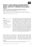
Fig. 1 shows the electron density of Tyr35 and the heme
group in the Phe35fiTyr mutant. The overall structure of
the Phe35fiTyr mutant is basically the same as that of the
wild-type Tb
5
. The r.m.s. deviation for a total of 82 Ca
atoms between the two molecules is 0.07 A
˚
. The secondary
structures of the wild-type protein and its Phe35fiTyr
mutant are the same. Fig. 2A and B shows a part of the
heme-binding pocket of the Phe35fiTyr mutant in two
different views. In wild-type cyt b
5
, the residue Phe35 is
located at helix II, which is a part of the heme-binding
pocket of cyt b
5
, and its side-chain points toward the heme.
The mutation from the nonpolar residue Phe35 to the polar
residue Tyr35 makes slight changes in the side chain
conformation of this residue. The shift of the Ca atom of
Tyr35 of the Phe35fiTyr mutant from that of Phe35 of the
wild-type cyt b
5
is 0.21 A
˚
, within the error limit. The side
chain of Tyr35 of the Phe35fiTyr mutant also points toward
the heme, but the phenol ring shifts away slightly from the
heme plane to avoid the unreasonable contacts with the
heme. The largest shift between the two superimposed
Table 2. Refinement statistics.
No. of amino acid residues 82
No. of prosthetic group 1
No. of solvent molecules 94
No. of reflections used 8870
R factor (%) 19.2
Free R factor (%) 23.8
Root-mean-square deviation
Bond lengths (A
˚
) 0.010
Bond angles (°) 1.08
Mean temperature factors (A
˚
2
)
Main chain 22.11
Side chain 26.29
Heme 23.78
Solvent 42.16
Ó FEBS 2002 Mutation at Phe35 of cytochrome b
5
(Eur. J. Biochem. 269) 4289
aromatic rings is 0.45 A
˚
, i.e., the distance from the atom CZ
(Fig. 1) of Tyr35 to that of Phe35. The crystal structure of
the Phe35fiTyr mutant shows that the side chain of Tyr35
makes strong van der Waals’ contacts with the heme, and
the shortest distance is 3.21 A
˚
, i.e., from the phenol oxygen
atom of Tyr35 to the carbon atom CHB (Fig. 1) of the
heme. In addition, the hydroxyl group of Tyr35 forms a
hydrogen bond (2.86 A
˚
) to a water molecule located outside
the heme pocket, as shown in Fig. 2A. This water molecule
forms another hydrogen bond (2.79 A
˚
) with the atom ND1
of the His26 side chain in a symmetry-related molecule
(Fig. 2A). This water molecule was also found in the
structure of wild-type cyt b
5
as well as in other mutants.
When Phe35 is mutated to Tyr35, this water molecule
moves toward the hydroxyl group of Tyr35 by 0.43 A
˚
,and
the side chain conformation of His26 correspondingly
moves a little bit (for example, the atom ND1 of His26
moves by 0.15 A
˚
) to be closer to the water molecule, which
is shown in Fig. 2B. These hydrogen-bonding interactions
help to stabilize the orientation of Tyr35 side chain. The
Fig. 2. Stereo views of a part of the heme-
binding pocket of the Phe35fiTyr mutant.
These diagrams were prepared using the
graphics program
SETOR
[60]. (A) Helices II,
III, IV, V of Phe35fiTyr are shown as a rib-
bon diagram. Tyr35 and the heme group of
the Phe35fiTyrmutantareshownasthick
lines. The water molecule (Wat) hydrogen
bonded to Tyr35 is shown as a large sphere.
His26 of the symmetry-related molecule (#) is
also shown as thick lines. Hydrogen bonds are
shown as broken lines. Phe35 and the heme
group of wild-type cyt b
5
,shownasthinlines,
are superimposed with Tyr35 and the heme of
the mutant. (B) Tyr35 of the Phe35fiTyr
mutant is superimposed with Phe35 of the
wild-type cyt b
5
. Heme, the water molecule
and His26 # of Phe35fiTyr mutant are su-
perimposed with those of the wild-type cyt b
5
.
Those in the mutant are shown as thick lines
and large spheres, and those in wild-type
cyt b
5
are shown as thin lines and small
spheres. (His26 # of the wild-type cyt b
5
is very
close to that in the mutant and cannot be
seen).
Fig. 1. Stereo view of the (2Fo-Fc) electron
density of Tyr35 and heme in the Phe35fiTyr
mutant, contoured at 1.0 r. The atoms CHB
of heme as well as OH and CZ of Tyr35
are labeled. This diagram was prepared using
the graphics program
TURBO
-
FRODO
.
4290 P. Yao et al. (Eur. J. Biochem. 269) Ó FEBS 2002
conformation of the heme in the Phe35fiTyr mutant is
basically the same as that in wild-type cyt b
5
. One of the two
propionates is hydrogen bonded to the main- and side chain
atoms of Ser64, while the other one extends into the solvent
and does not form any hydrogen bond with the protein
atoms. The former propionate displays the conserved
conformation in the structures of the Phe35fiTyr mutant
and of the wild-type protein as well as other mutants.
However, the conformation of the latter is flexible.
CD spectra of thermal denaturation of cyt
b
5
and its Phe35fiTyr mutant
Fig. 3A shows the CD spectra of wild-type cyt b
5
in the far-
UV region at 30 °C, 65 °C, 70 °Cand95°C, respectively.
The Phe35fiTyrmutantshowssimilarCDspectra(data
not shown). When cyt b
5
and its mutant were subjected to
increasing temperature, the peak at 219 nm decreased
monotonically. At 95 °C, the negative peak at 219 nm
almost disappeared, but a large negative peak appeared at
203 nm. For wild-type cyt b
5
, the peak at 207 nm was still
present at 95 °C, but for the mutant, the peak at 208 nm
changed to a shoulder peak. All of these results suggest that
the a-helix percentage of the protein and its mutant
decreases sharply while the b-sheet percentage also reduces
significantly at high temperature.
Fig. 3B shows the CD spectra of wild-type cyt b
5
in the
250–500 nm region at 30 °C, 67 °C, 69.5 °C, 75 °C, 85 °C,
and 95 °C, respectively. The CD spectra of the Phe35fiTyr
mutant have a similar pattern and are not shown here. In
the Soret region, the peak positions of the two proteins are
basically similar at 30 °C, consistent with those reported in
literatures [35,36]. At the near UV region, the negative CD
peak at 268 nm derived from the four tyrosyl residues of
wild-type cyt b
5
[35] shows a different shape for the
Phe35fiTyr mutant, which has five tyrosyl residues. The
peak at 299.4 nm derived from the single tryptophan
residue for the wild-type protein shifts to 297.6 nm for the
mutant. The spectra in the 267–299.4 nm region are only
slightly different for these two proteins.
Thermal denaturation of wild-type cyt b
5
and its
Phe35fiTyr mutant show similar CD behavior. A negative
peak at 418 nm with strong intensity and a positive peak at
390 nm at room temperature are characteristic of low-spin
state of ferric cyt b
5
[36]. When the temperature was
increased the negative peak at 418 nm was blue-shifted with
a gradual reduction of its intensity. Simultaneously, the
intensity of the positive peak at 390 nm decreased. We
found that with increasing temperature to 69.5 °C, a new
peak around 398 nm with a negative intensity appeared.
The intensity of the peak at 398 nm increased dramatically
from 69.5 to 75 °C, and then gradually decreased from
75 °C to higher temperature. However, even at 95 °C, this
peak does not disappear. Unexpectedly our results are very
different from those of the rabbit liver cyt b
5
reported by
Sugiyama et al. [36]. Their work showed that there was
almost no absorption in the 300–500 nm region of the CD
spectrum when the temperature was 83 °C. This is the
first detailed CD spectrum study on the secondary structure
of cyt b
5
, and characterization of the intermediate
conformation.
The negative peak at 267 nm, which is attributed to
absorption from tyrosyl residues Tyr6, Tyr7, Tyr27 and
Tyr30 [35,37], gradually decreased with increasing tempera-
ture. At 69.5 °C, the peak intensity reduced to almost zero.
A positive peak at this region appeared and its intensity
gradually increased when the temperature changed from
69.5 °Cto75°C, then gradually decreased at higher
temperature. The negative peak at 299.4 nm, which is
assigned to the contribution of Trp22, decreased monotoni-
cally with the increase in temperature. It is known from
X-ray structural analysis of wild-type cyt b
5
[21] that the
core 2 consists of b-strand III (Tyr27–Leu32), b-strand II
(Thr21–Leu25), b-strand I (Lys5–Tyr7) and a-helix I (Thr8–
His15). Trp22, Tyr6, Tyr7, Tyr27 and Tyr30 are the main
aromatic components of the core 2 of cyt b
5
. The pattern of
the absorption changes around 267 nm and 299.4 nm
implies that even though core 2 is largely intact after the
removal of the heme from the protein as reported by
Falzone et al. [38] core 2 experiences significant structural
fluctuation and gradually undergoes complete unfolding.
This study clearly shows the whole process of unfolding and
is an important supplement to the results reported by Pfeil
[39] by means of second derivative spectra and heat capacity
of apo- and holo-cyt b
5
.
Fig. 4A demonstrates the transitional CD curves of
wild-type cyt b
5
monitored at 222 nm, 299 nm, 398.4 nm
and 418.8 nm. The curves of 222 nm, 299 nm and 418.8 nm
possess a similar pattern suggesting that dissociation of the
Fig. 3. CD spectra of the wild-type cyt b
5
from 30 °Cto95°Cat(A)
195–250 nm and (B) 250–500 nm (for clarity of comparison, only part of
the spectra are shown.)
Ó FEBS 2002 Mutation at Phe35 of cytochrome b
5
(Eur. J. Biochem. 269) 4291
Fe–His bond is accompanied by the a-helix unfolding of the
peptide chain and the destroying of Trp22 asymmetrical
environment. Fig. 4B shows the transitional curves of the
Phe35fiTyr mutant, which exhibits a pattern similar to that
of the wild-type protein. All of the CD spectra transitions of
the Phe35fiTyr mutant at 222 nm, 299 nm, 398.4 nm, and
418.8 nm in response to heat are 3 °C higher than those
ofthewild-typecytb
5
, which is consistent with the result of
UV–visible measurement [13]. The results of denaturation
of these proteins by guanidine hydrochloride are also in
agreement with those of urea denaturation. These results
demonstrate that the Phe35fiTyr mutant increases not only
the affinity of the heme to the polypeptide chain but also the
stability of the secondary structure.
The kinetics of the heme dissociation from cyt
b
5
variants mediated by urea
Urea-mediated denaturation of cyt b
5
variants was treated
as a first-order reaction, producing the rate constants of the
heme dissociation at different urea concentrations. The
results are shown in Fig. 5. The rate constants of heme
dissociation reaction increased slightly with the increase in
urea concentration for the wild-type protein. However, it is
interesting to note that cyt b
5
Phe35fiTyrshowsalower
rate. On the contrary, for the Phe35fiLeu mutant the rate
constant increased sharply after the urea concentration
exceeded 5
M
. These results reflect the tightness of the heme
attaching to the polypeptide of cyt b
5
. For the Phe35fiTyr
mutant the heme pocket traps the heme even more strongly
than the wild-type protein. Obviously, for the Phe35fiLeu
mutant the interactions between the heme and its pocket are
much weaker, only a moderate concentration of urea is
needed to speed up the release of heme from the pocket.
The heme transfer from cyt
b
5
or its Phe35fiTyr mutant
to apo-Mb
The kinetic parameters of heme dissociation from cyt b
5
were determined under nondenaturation conditions by
measuring the spontaneous release of the heme from cyt b
5
to apo-Mb, which is used as a heme trap. Although the CD
spectra could show the reaction process clearly, the protein
concentration required is much higher than for the
UV–visible method. Because the high concentration of
protein could cause denaturation of the apo-protein during
the long assay time, all of the heme transfer reactions were
monitored only by the UV–visible spectra. The rates of
heme transfer reaction from the wild-type or the Phe35fi
Tyrmutantofcytb
5
to apo-Mb at 25 ± 0.1 °C show
obviously differences, which can be seen in Fig. 6 and
Table 3. Compared with wild-type cyt b
5
, the heme affinity
of the Phe35fiTyr mutant increased greatly. The activation
free energy and activation energy listed in Table 3, which
were calculated from Eyring plots (Fig. 7) and Arrhenius
plots (data not shown), also show that the mutation has
affected the conformation of the transition state.
The CD spectra of Mb, apo-Mb, wild-type cyt b
5
and
apo-cyt b
5
demonstrate that these proteins have an identical
structure as reported previously [35,36,40,41]. The apo-
cyt b
5
and apo-Mb have no absorption in the Soret band
region because of the lack of the heme prosthetic group. For
the holo-cyt b
5
and holo-Mb, the CD spectra of the Soret
band are entirely different, which illustrates the difference in
the heme environment between cyt b
5
and Mb. In the CD
spectra of cyt b
5
, there is a negative peak at 418 nm with
strong intensity [36]. In contrast, Mb has a strong positive
absorption at 408 nm [41]. Hence, the heme transfer reaction
from cyt b
5
to apo-Mb could be easily and precisely
Fig. 4. The transitional curves of the CD spectra on heating at
222 nm, 299 nm, 398.4 nm, and 418.8 nm. (A) Wild-type cyt b
5
.
(B) Phe35fiTyr mutant of cyt b
5
.
Fig. 5. The rate constants of heme dissociation of cyt b
5
as function of
urea concentration.
4292 P. Yao et al. (Eur. J. Biochem. 269) Ó FEBS 2002
monitored by CD which clearly demonstrates that the heme
transfer reaction under the conditions used proceeded to
completion (data not shown). Meanwhile, from the concen-
tration changes of the holo-Mb in the reaction monitored by
UV–visible spectroscopy, the same conclusion ) that this
reaction is entirely completed ) can be drawn.
DISCUSSION
Protein folding studied by CD spectra
Up to now, CD spectra of cyt b
5
have been studied by only
a few groups [35,36,39]. These CD studies suggested that
there is an increase in disorder and less secondary structure
in the apo-form [35]. However, no detailed information was
provided about the protein’s folding and stability. There is
evidence for the folding of apo-cyt b
5
in vivo prior to the
formation of holo-cyt b
5
[42]. Meanwhile, it is reported that
cyt b
5
consists of two hydrophobic cores. Core 1 is normally
retained by the prosthetic heme group; core 2 comprises
mainly b-sheets. These two cores are well maintained in the
apo-form of the protein [43] and so are especially interesting
for the study of the folding mechanism, intermediates and
stability of the protein by CD spectra.
Usually, the stability of cyt b
5
could be investigated
through the heme dissociation reaction by exposing
the protein to the denaturant or heat. This process was
considered to be a two-state mechanism (H Ð A), in
which only the holo-cyt b
5
(H) and the apo-cyt b
5
(A) are
present at significant concentrations [28,44]. It was
thought that the heme-binding domain of cyt b
5
was
denatured simultaneously with heme dissociation. The
UV–visible spectrum study of cyt b
5
in response to heat
and urea did display several isosbestic points in the
absorbance curves, and the denaturation curves really
showed that the denaturation followed the two-state
mechanism.
The denaturation curves of CD absorption at 222 nm,
299 nm and 418.8 nm shown in Fig. 4A and B indicate that
unfolding of the a-helices, b-sheets and breaking of the His–
Fe bonds of the heme follow the two-state mechanism. It is
noted that a new absorption peak that appeared at
398.4 nm displays slightly different denaturation behav-
iours. Definitely, the absorption at 398.4 nm is derived from
a heme derivative. As heme is a symmetrical chromophore,
it exhibits no inherent optical activity itself [45,46]. Our
experiment also shows that heme in the buffer solution itself
does not exhibit any CD absorption in the region of 250–
500 nm at 30–95 °C. Apo-cyt b
5
has no CD absorption in
the Soret band too, but shows the absorption contributed
from aromatic amino acids in the near UV region [35]. The
concurrent existence of the Soret band absorption at
418.8 nm and 398.4 nm at 69.5 °CshowninFig.3B
indicates that probably there are two types of heme
derivative in the solution; at this stage the heme was not
totally released from the protein heme pocket into the
aqueous environment and part of the low-spin and
six-coordinated heme was changed into the high-spin state
Fig. 7. Eyring plots of the rate constants of heme transfer from cyt b
5
to
apo-myoglobin; (j) Phe35fiTyr mutant (d) wild-type cyt b
5
.
Table 3. The kinetic parameters of the heme-transfer reactions between
apo-Mb and the wild-type and the Phe35fiTyr mutant of cyt b
5
. The
measurements were made in sodium acetate buffer, I ¼ 10 m
M
,
pH 5.5.
Wild-type Phe35fiTyr
k (h
)1
)
a
2.01 (± 0.02) 0.21 (± 0.03)
DG°
„
(kJÆmol
)1
) 91.2 96.8
E
a
(kJÆmol
)1
) 110.4 135.4
a
T ¼ 25 ± 0.1 °C.
Fig. 6. Kinetic traces for heme transfer reaction from the wild-type, or
Phe35fiTyr mutant of cyt b
5
to apo-myoglobin. (A) Experimental data.
(B) Fitted curve.
Ó FEBS 2002 Mutation at Phe35 of cytochrome b
5
(Eur. J. Biochem. 269) 4293
with breaking of the His–Fe bonds. It is known that the
apo-cyt b
5
prepared under mild conditions could generally
maintain the holo-like structures except for some confor-
mational fluctuations observed in the local regions [47].
However, as indicated by molecular dynamics simulations
all a-helices in core 1 are highly mobile, and the tertiary
structure in core 2 of cyt b
5
is rather rigid [48]. Thus, the
denaturation curve of the wild-type protein monitored at
398.4 nm and 67–75 °C by CD implied that there was
probably a collapse of core 1 accompanied by partially
unfolding of the a-helices and breaking of Fe–His bonds.
This temperature region is coincident with the transition
region of cyt b
5
denaturation in response to heat monitored
by UV–visible spectra at 418 nm. At this time, the heme was
still wrapped up in the polypeptide chain of cyt b
5
.
Therefore, the CD absorption at 398.4 nm could be the
result of another form of heme, an intermediate state, in
which the heme is not coordinated by two histidine residues
and does not sit normally in the heme pocket. More
probably it is enveloped by the partially unfolded cyt b
5
polypeptide chain after the collapse of hydrophobic core 1.
From the observations of CD absorption of tryptophan and
tyrosines, however, it is believed at that time the core 2 of
cyt b
5
remains intact. Even at 95 °C, this peak at 398.4 nm
does not disappear completely. Possibly, the heme is still
partially attached to some parts of the random coil of
denatured cyt b
5
polypeptide chain through hydrophobic
interactions. Actually, after we reached this conclusion, we
found that Gray’s group had published a short communi-
cation indicating that in the folding study of cyt b
562
,
normally Ôthe heme iron is ligated axially by the side chains
of Met7 and His102. It is likely that one of these ligands
remains attached to the heme in the unfolded stateÕ [49,50].
Here, we provide the detailed CD spectra evidencing the
existence of the intermediate and a reasonable explanation.
The reason why our results do not agree with those
obtained for the rabbit liver cyt b
5
[36] is not yet known.
But, it is noted that the bovine liver Tb
5
used in this work is
more stable than rabbit liver cyt b
5
.TheT
m
(transition
midpoint of the heat denaturation curve of the UV–visible
spectrum at 412 nm) is 66.9 °C for bovine liver Tb
5
and
55.0 °C for rabbit liver cyt b
5
[13,36]. Maybe a detailed
structural study, similar to the comparison between the
microsomal cyt b
5
and the outer membrane liver mitochon-
dria cyt b
5
[51], is required to reveal the essence of the
different properties.
The stability of cyt
b
5
Phe35 mutants
The wild-type protein usually develops an optimal archi-
tecture to fulfill its biological functions after hundreds and
thousands years of evolution and natural selection. Arti-
ficial site-directed mutagenesis of proteins most often leads
to a decrease in stability: an increase in stability in the
mutant proteins is comparatively rare [52,53]. The main
components of protein stability that could be perturbed by
mutation at interior groups include hydrophobic effects,
van der Waals’ forces, backbone conformation, hydrogen
bonds, local polarity and side chain volume of the
substituted residue. Substitution of tyrosine for phenyl-
alanine should generate 4.8 kJÆmol
)1
destabilization energy
because of the decreased hydrophobic nature of tyrosine,
and may contribute 4–6 kJÆmol
)1
to protein stability if
there is another hydrogen bond generated in the cyt b
5
Phe35fiTyr mutant [53,54]. In our previous study [13], the
Phe35fiTyr mutant of cyt b
5
intheoxidizedstateis
3.3 kJÆmol
)1
more stable than the wild-type protein
towards heat denaturation and is 4.3 kJÆmol
)1
more stable
in urea denaturation. The CD spectra of heat denaturation
also show that the structure transition temperature for the
Phe35fiTyr mutant is higher than that for the wild-type.
Kinetically, the rate constant of heme transfer reactions
from cyt b
5
to apo-Mb for the wild-type protein is 10 times
faster than that for the Phe35fiTyr mutant. The urea-
mediated heme dissociation reactions of various cyt b
5
variants also demonstrate that the heme is trapped in the
heme pocket with different degrees of tightness. Recently,
Silchenko et al. [55] found that cyt b
5
from the outer
mitochondrial membrane of rat liver is substantially more
stable against thermal and chemical denaturation than
bovine liver cyt b
5
. Their study demonstrated that the
enhanced stability of outer mitochondrial membrane cyt b
5
is in large part due to slow heme release, where the heme is
kinetically trapped in the heme pocket of hemoproteins. As
shown in previous work, the residues on the protein surface
were considered to be less important and to have minor
effect on protein stability because these residues exert little
effect on the interactions between the heme and the heme
pocket [6,56]. For the heme pocket residues such as Phe35,
Val61, Val45 and Phe58 the situation is entirely different.
The mutation from the hydrophobic residue Phe35 to a
larger polar residue Tyr35 does not make significant
changes in the overall structure and the local structure
around the mutation site because there is enough space to
accommodate an additional hydroxyl group. However, the
crystal structure of the Phe35fiTyr mutant shows that the
hydroxyl oxygen atom of the side chain of Tyr35 is 3.21 A
˚
away from the atom CHB of the pyrrole group of the
heme, making strong van der Waals’ contacts with
the heme. Obviously, the introduction of hydroxyl group
in the heme pocket strengthens the interactions between
Tyr35 and the heme with the iron in the oxidative state.
The increased van der Waals’ interactions between the side
chain of Tyr35 and the heme can probably make an
obstacle to the departure of the heme from the hydropho-
bic pocket of the protein. The total consequence of this
mutation made ferricytochrome b
5
Phe35fiTyr more
stable compared with the wild-type protein. In the case
of the cyt b
5
Phe35fiHis mutant, besides the increased
hydrophilicity of the histidine residue, the side chain
volume decreases by 36 A
˚
3
compared to the wild-type
cyt b
5
which would effectively reduce the van der Waals’
contact between the histidine and the heme. So, the
Phe35fiHis mutant is 11.8 kJÆmol
)1
less stable than the
wild-type protein [13].
There is a stabilization effect of the heme ring binding to
Phe35 and Phe58 by hydrophobic aromatic interactions. It
has been reported that an edge-to-face orientation between
two aromatic groups is energetically favorable [57].
Sakamoto et al. [45] have studied the effect of amino acids
substitution of hydrophobic residues on heme-binding
properties in the designed two-a-helix peptides. Their
studies demonstrated that the edge-to-face interactions
between the aromatic side chain of the phenylalanine
residues and the porphyrin plane might contribute to the
conformation of peptide–heme conjugates. They also
4294 P. Yao et al. (Eur. J. Biochem. 269) Ó FEBS 2002
proved that the phenylalanine residue located at i ±4
relative to the axial ligand histidine residue in the a-helix was
critical to the edge-to-face interaction between the phenyl-
alanine side chain and the porphyrin ring, providing
stabilization of peptide–heme conjugates [45,46,58]. The
Phe35–His39 of cyt b
5
is consistent with i ± 4 arrangement.
It is clear that the substitution of tyrosine for phenylalanine
at position 35 does not destroy the aromatic interactions
and can also maintain the edge-to-face interaction, provi-
ding the stabilization effect of the heme binding. In the case
of the Phe35fiLeu mutant, however, substitution of leucine
for phenylalanine should break this effect. This is also
supported by the denaturation experiment [13], which
showed that the Phe35fiLeu mutant is 7.8 kJÆmol
)1
less
stable towards heat and 7.9 kJÆmol
)1
less stable towards
urea than the wild-type protein.
Factors affecting redox potential of the Phe35 mutants
The redox potential of the Phe35fiTyr mutant shifts
negatively by 66 mV compared to that of the wild-type
cyt b
5
[13]. As we know, a hydrophilic environment
stabilizes the oxidized state, leading to a lower redox
potential [53]. In particular, the introduction of a polar
hydroxyl group in the Ôlow dielectricÕ interior of the protein
can play a much stronger electrostatic role, stabilizing ferric
iron. The reduction of hydrophobicity reasonably accounts
for the negative shift of redox potential. In addition, the
hydrogen bonding formation between the tyrosine and the
conserved water molecule shown in the crystal structure of
the Phe35fiTyr mutant enhances significantly hydrophilic
influence on the heme causing great alteration of the protein
properties [59].
ACKNOWLEDGMENTS
This work was supported by two grants from the National Natural
Science Foundation of China. We are grateful to Prof. Li-Wen Niu,
Prof. Mai-Kun Teng and Dr Xue-Yong Zhu of the University of
Science and Technology of China for their support and help with the
X-ray data collection.
REFERENCES
1. Spatz, L. & Strittmatter, P. (1971) A form of cytochrome b
5
that
contains an additional hydrophobic sequence of 40 amino acid
residues. Proc. Natl. Acad. Sci. USA 68, 1042–1046.
2. Bonfils, C., Balny, C. & Maurel, P. (1981) Direct evidence for
electron transfer from ferrous cytochrome b
5
to the oxyferrous
intermediate of liver microsomal cytochrome P-450 LM2. J. Biol.
Chem. 256, 9457–9465.
3. Strittmatter, P., Spatz, L., Corcoran, D., Rogers, M.J., Setlow, B.
& Redline, R. (1974) Purification and properties of rat liver
microsomal stearyl coenzyme A desaturase. Proc. Natl Acad. Sci.
USA 71, 4565–4569.
4. Hegesh, E., Hegesh, J. & Kaftory, A. (1986) Congenital methe-
moglobinemia with a deficiency of cytochrome b
5
. N. Engl. J.
Med. 314, 757–761.
5. Mauk, A.G., Mauk, M.R., Moore, G.R. & Northrup, S.H. (1995)
Experimental and theoretical analysis of the interaction between
cytochrome c and cytochrome b
5
. J. Bioenergetic Biomembranes
27, 311–340.
6. Sun,Y.L.,Wang,Y.H.,Yan,M.M.,Sun,B.Y.,Xie,Y.&Huang,
Z.X. (1999) Structure, interaction and electron transfer between
cytochrome b
5
, its E44A and/or E56A mutants and cytochrome c.
J. Mol. Biol. 285, 347–359.
7. Stayton,P.S.,Poulos,T.L.&Sligar,S.G.(1989)Putidaredoxin
competitively inhibits cytochrome b
5
-cytochrome P450
cam
electron
transfer complex. Biochemistry 28, 8201–8215.
8. Livingston, D.J., Mclachlan, S.J., Lamar, G.N. & Brown, W.D.
(1985) Myoglobin: cytochrome b
5
interactions and the kinetic
mechanism of cytochrome b
5
reductase. J. Biol. Chem. 260, 15699–
15707.
9. Poulos, T.L. & Mauk, A.G. (1983) Models for the complexes
formed between cytochrome b
5
and subunits of methemoglobin.
J. Biol. Chem. 258, 7369–7373.
10. Mathews, F.S. & Czerwinski, E.W. (1976) Cytochrome b
5
and
Cytochrome b
5
Reductase from a Chemical and X-ray Diffraction
Viewpoint. Wiley, New York.
11. Lederer, F. (1994) The cytochrome b
5
-fold: an adaptable module.
Biochimie 76, 674–692.
12. Dangi, B., Sarma, S., Yan, C., Banville, D.L. & Guiles, R.D.
(1998) The origin of differences in the physical properties of the
equilibrium forms of cytochrome b
5
revealed through high-
resolution NMR structures and backbone dynamic analyses.
Biochemistry 37, 8289–8302.
13. Yao, P., Wang, Y.H., Sun, Y.L., Huang, Z.X., Xie, Y. & Xiao,
G.T. (1997) Importance of a conserved phenylananine-35 of
cytochrome b
5
to the protein’s stability and redox potential.
Protein Eng. 10, 578–581.
14. Yao, P., Wang, Y H., Xie, Y. & Huang, Z X. (1998) Spectro-
electro-chemical studies of cytochrome b
5
Phe35 mutants.
J. Electroanal. Chem. 445, 197–201.
15. Yao,P.,Wang,Y H.,Sun,B Y.,Xie,Y.,Hirota,S.,Yamauchi,
O. & Huang, Z X. (2002) Kinetic studies on the oxidation of
cytochrome b
5
Phe35 mutants with cytochrome c, plastocyanin
and inorganic complexes. J. Biol. Inorg. Chem. 7, 375–383.
16. Mauk, M.R., Mauk, A.G., Weber, P.C. & Matthew, J.B. (1986)
Electrostatic analysis of the interaction of cytochrome c with
native and dimethyl ester heme substituted cytochrome b
5
.
Biochemistry 25, 7085–7091.
17. Hagler, L., Coppes,R.I. Jr & Herman, R.H. (1979) Metmyoglobin
reductase. Identification and purification of a reduced nicotin-
amide adenine dinucleotide-dependent enzyme from bovine heart
which reduces metmyoglobin. J. Biol. Chem. 254, 6505–6514.
18. La Mar., G.N., Toi, H. & Krishnamoorthi, R. (1984) Proton
NMR investigation of the rate and mechanism of heme rotation in
sperm whale myoglobin: Evidence for intra-molecular reorienta-
tion about a heme twofold axis. J. Am. Chem. Soc. 106, 6395–
6401.
19. Puett, D. (1973) The equilibrium unfolding parameters of horse
and sperm whale myoglobin. Effects of guanidine hydrochloride,
urea, and acid. J. Biol. Chem. 248, 4623–4634.
20. Light, W.R., Rohlfs, R.J., Palmers, G. & Olson, J.S. (1987)
Functional effects of heme orientational disorder in sperm whale
myoglobin. J. Biol. Chem. 262, 46–47.
21. Wu, J., Gan, J.H., Xia, Z.X., Wang, Y.H., Xue, L.L., Xie, Y. &
Huang, Z.X. (2000) Crystal structure of recombinant trypsin-
solubilized fragment of cytochrome b
5
and the structural com-
parison with Val61His mutant. Proteins: Structure, Function Genet
40, 249–257.
22. Durley, R.C.E. & Mathews, F.S. (1996) Refinement and structural
analysis of bovine cytochrome b
5
at 1.5 A
˚
resolution. Acta Crys-
tallogr. D52, 65–76.
23. Xue, L.L. Wang, Y.H. Xie, Y. Yao, P. Wang, W.H. Qian, W.
Huang, Z.X. Wu, J. & Xia. Z.X. (1999) Effect of mutation at
valine 61 on the three-dimensional structure, stability, and redox
potential of cytochrome b
5
. Biochemistry 38, 11961–11972.
24. Otwinowski, Z. & Minor, W. (1997) Processing of X-ray diffrac-
tion data collected in oscillation mode. Methods Enzymol. 276,
307–326.
Ó FEBS 2002 Mutation at Phe35 of cytochrome b
5
(Eur. J. Biochem. 269) 4295
25. Brunger, A.T. (1992) X-PLOR: a System for X-Ray Crystallo-
graphy and NMR. Version 3.1. New Haven: Yale University Press.
26. Brunger, A.T. Adams, P.D. Clore, G.M. et al. (1998) Crystal-
lography and NMR System (CNS): a new software system for
macromolecular structure determination. Acta Crystllogr. D54,
905–921.
27. Roussel, A. & Cambillau, C. (1991) TURBO-FRODO, Silicon
Graphics Partner Geometry Dictionary. Silicon Graphics Inc.
Mountain View, CA, USA.
28. Vergeres, G., Chen, D.Y., Wu, F.F. & Waskell, L. (1993) The
function of tyrosine 74 of cytochrome b
5
. Arch. Biochem. Biophys.
305, 231–241.
29. Matthews, C.R. (1987) Effect of point mutations on the folding of
globular proteins. Methods Enzymol. 154, 498–511.
30. Hargrove, M.S. & Olson, J.S. (1996) The stability of holo-
myoglobin is determined by heme affinity. Biochemistry 35, 11310–
11318.
31. Hargrove, M.S., Singleton, E.W., Quilin, M.L., Ortiz, L.A.,
Philips, G.N. Jr, Olson, J.S. & Mathews, A.J. (1994) Stability of
myoglobin: a model for the folding of heme proteins. J. Biol.
Chem. 269, 4207–4214.
32. Matthew, J.B. & Gurd, F.R.N. (1986) Stabilization and destabi-
lization of protein structure by charge interactions. Methods
Enzymol. 130, 437–453.
33. Smith,M.L.,Paul,J.,Ohlsson,P.I.,Hjortsberg,K.&Paul,K.G.
(1991) Heme-protein fission under nondenaturing conditions.
Proc. Natl. Acad. Sci. USA 88, 882–886.
34. Morris, A.L., MacArthur, M.W., Hutchinson, E.G. & Thornton,
J.M. (1992) Stereochemical quality of protein structure
coordinates. Proteins: Struct. Funct. Genet. 12, 345–364.
35. Huntley, T.E. & Strittmatter, P. (1972) The reactivity of the tyrosyl
residues of cytochrome b
5
. J. Biol. Chem. 247, 4641–4647.
36. Sugiyama, T., Miura, R., Yamano, T., Shiga, K. & Watari, H.
(1980) A reversible spin conversion of cytochrome b
5
at high
temperatures. Biochem. Biophys. Res. Commun. 97, 22–27.
37. Luzzati, P.V. (1952) Traitment statistique des erreurs dans la de-
ternination des structures cristallines. Acta Crystallogr. 5, 802–810.
38. Falzone,C.J.,Mayer,M.R.,Whiteman,E.L.,Moore,C.D.&
Lecomte, J.T. (1996) Design challenges for hemoproteins: the
solution structure of apocytochrome b
5
. Biochemistry 35, 6519–
6526).
39. Pfeil, W. (1993) Thermodynamics of apocytochrome b
5
unfolding.
Protein Sci. 2, 1497–1501.
40. Kawamura-Konishi, Y., Kihara, H. & Suzuki, H. (1988) Recon-
stitution of myoglobin from apoprotein and heme, monitored
by stopped-flow absorption, fluorescence and circular dichroism.
Eur. J. Biochem. 170, 589–595.
41. Hsu, M C. & Woody, R.W. (1971) The origin of the heme Cotton
effects in myoglobin and hemoglobin. J. Am. Chem. Soc. 93, 3515–
3525.
42. Shawver, L.K., Siedel, S.L., Krieter, P.A. & Shires, T.K. (1984) An
enzyme-linked immunoadsorbent assay for measuring cyto-
chrome b
5
and NADPH-cytochrome P-450 reductase in rat liver
microsomal fractions. Evidence for functionally inactive protein.
Biochem. J. 217, 623–632.
43. Moore, C.D. & Lecomte, J.T.J. (1993) Characterization of an
independent structural unit in apocytochrome b
5
. Biochemistry 32,
199–207.
44.Qian,W.,Sun,Y.L.,Wang,Y.H.,Zhuang,J.H.,Xie,Y.&
Huang, Z.X. (1998) The influence of mutation at Glu44 and Glu56
of cytochrome b
5
on the protein’s stabilization and interaction
between cytochrome c and cytochrome b
5
. Biochemistry 37,
14137–14150.
45. Sakamoto, S., Obayaya, I., Ueno, A. & Mihara, H. (1999) Effects
of amino acids substitution of hydrophobic residues on heme-
binding properties of designed two a-helix peptides. J. Chem. Soc.,
Perkin. Trans. 2, 2059–2069.
46. Sakamoto, S., Ueno, A. & Mihara, H. (1998) Design and synthesis
of heme-binding peptides: Relationship between heme-binding
properties and catalytic activities. J.Chem.Soc.,Perkin.Trans.2,
2395–2404.
47. Ihara, M., Takahashi, S., Ishimori, K. & Morishima, I. (2000)
Functions of fluctuation in the heme-binding loops of cytochrome
b
5
revealed in the process of heme incorporation. Biochemistry 39,
5961–5970.
48. Storch, E.M. & Daggett, V. (1996) Structural consequences of
heme removal: molecular dynamics simulations of rat and bovine
apocytochrome b
5
. Biochemistry 35, 11569–11604.
49. Wittung-Stafshede, P., Gray, H.B. & Winkler, J.R. (1997) Rapid
formation of four-helix bundle. Cytochrome b562 folding trig-
gered by electron transfer. J. Am. Chem. Soc. 119, 9562–9563.
50. Pasher, T., Chesick, J.P., Winkler, J.R. & Gray, H.B. (1996)
Protein folding triggered by electron transfer. Science 271, 1558–
1560.
51. Altuve, A., Silchenko, S., Lee, Kyung-Hoon, Kuczera, K.,
Terzyan, S., Zhang, X J., Benson, D.R. & Rivera, M. (2001)
Probing the differences between Rat Liver Mitochondrial
membrane Cytochrome b
5
and Microsomal Cytochrome b
5
.
Biochemistry 40, 9469–9483.
52. Newbold, R.J., Hewson, R. & Whitford, D. (1992) The thermal
stability of the tryptic fragment of bovine microsomal cytochrome
b
5
and a variant containing six additional residues. FEBS Lett.
314, 419–422.
53. Caffrey, M.S. & Cusanovich, M.A. (1994) Site-specific mutagen-
esis studies of cytochrome c. Biochim. Biophys. Acta 1187,277–
288.
54. Sandberg, W. & Terwilliger, T. (1989) Influence of interior pack-
ing and hydrophobicity on the stability of a protein. Science 245,
54–57.
55. Silchenko, S., Sippel, M.L., Kuchment, O., Benson, D.R., Mauk,
A.G., Altuve, A. & Rivera, M. (2001) hemin is kinetically trapped
in cytochrome b
5
from rat outer mitochondrial membrane.
Biochem. Biophys. Res. Commun. 273, 467–472.
56. Hunter, C.L., Lloyd, E., Eltis, L.D., Rafferty, S.P., Lee, H., Smith,
M. & Mauk, A.G. (1997) Role of the heme propionates in the
interaction of heme with apo-myoglobin and apo-cytochrome b
5
.
Biochemistry 36, 1010–1017.
57. Jorgenson, W.L. & Severance, D.L. (1990) Aromatic–aromatic
interactions: free energy profiles for the benzene dimer in water,
chloroform, and liquid benzene. J. Am. Chem. Soc. 112, 4768–
4774.
58. Williamson, D.A. & Benson, D.R. (1998) Remarkable
helix stabilization via-edge-to-face tryptophan–porphyrin inter-
actions in a peptide-sandwiched mesoheme. Chem. Commun. 9,
961–962.
59. Caffrey, M.S. & Cusanovich, M.A. (1991) The effects of surface
charges on the redox potential of cytochrome c
2
from the purple
phototrophic bacterium Rhodobacter capsulatus. Arch. Biochem.,
Biophys. 285, 227–230.
60. Evans, S.V. (1993) SETOR: hardware lighted three-dimensional
solid model representations of macromolecules. J. Mol. Graphics
11, 134–138.
4296 P. Yao et al. (Eur. J. Biochem. 269) Ó FEBS 2002