Báo cáo Y học: What does it mean to be natively unfolded? pptx
Bạn đang xem bản rút gọn của tài liệu. Xem và tải ngay bản đầy đủ của tài liệu tại đây (345.04 KB, 11 trang )
REVIEW ARTICLE
What does it mean to be natively unfolded?
Vladimir N. Uversky
1
Institute for Biological Instrumentation, Russian Academy of Sciences, Pushchino, Moscow, Russia;
2
Department of Chemistry and Biochemistry, University of California, Santa Cruz, CA, USA
Natively unfolded or intrinsically unstructured proteins
constitute a unique group of the protein kingdom. The
evolutionary persistence of such proteins represents strong
evidence in the favor of their importance and raises
intriguing questions about the role of p rotein disorders in
biological processes. Additionally, natively unfolded p ro-
teins, with their lack of ordered structure, represent attractive
targets for the biophysical studies of the unfolded p olypep-
tide chain under physiological conditions in vitro.Thegoalof
this study was to summarize the structural information on
natively unfolded p roteins in o rder to evaluate their major
conformational characteristics. It appeared that natively
unfolded proteins are characterized by low overall hydro-
phobicity and large net charge. They possess hydrodynamic
properties typical of random coils in poor solvent, or pre-
molten globule conformation. These proteins show a low
level of ordered secon dary structure and no tightly packed
core. They are very ¯exible, but may adopt relatively rigid
conformations in the presence of natural ligands. Finally, in
comparison with the globular proteins, natively unfolded
polypeptides possess Ôturn outÕ responses to changes in the
environment, as their structural complexities increase at high
temperature or at e xtreme pH.
Keywords: intrinsically unfolded protein; i ntrinsically
disordered protein; unfolded protein; molten g lobule state;
premolten globule state.
WHAT ARE NATIVELY UNFOLDED
PROTEINS?
Before the phenomenon of natively unfolded p roteins will
be considered, a de®nition of the major players is r equired.
The importance of this issue follows from the fact that many
proteins have been shown to have nonrigid structures under
physiological conditions. These proteins may be separated
in two d ifferent groups. Members of the ®rst group, despite
their ¯ exibility, are rather compact and possess a well-
developed secondary structure, i.e. t hey show properties
typical o f the molten globule [1]. Proteins from the other
group behave almost as random coils [2]. Only members of
the second group will be described below. T hus, to b e
considered as natively unfolded (or intrinsically unstruc-
tured), a protein s hould be extremely ¯exible, essentially
noncompact (extended), and have little or no ordered
secondary structure under physiological conditions.
WHY STUDY INTRINSICALLY
DISORDERED PROTEINS?
The number of p roteins and protein domains, that h ave
been shown in vitro to have little or no ordered structure
under physiological conditions, is rapidly increasing. In fact,
over the past 1 0 years there has been an exponential
increase in the number of such studies, starting from one
paper in 1989, and ending with more than 30 in 2000. The
current list of natively unfolded proteins includes more than
100 e ntries (91 of t hem were tabulated in our recent work
[3]). This collection comprises the full-length proteins and
their domains with chain length of more than 50 amino-acid
residues. Including shorter polypeptides (30±50 residues
long) would probably double this amount.
The growing interest in this class of proteins is for several
reasons. The ®rst issue is the structure±function relationship.
The existence of biologically active but extremely ¯exible
proteins questions the assumption that rigid well-folded
3D-structure is required for functioning. To o vercome this
problem, it has been suggested that the lack of rigid globular
structure under physiological conditions might represent a
considerable functional a dvantage for Ônatively unfoldedÕ
proteins, a s t heir large plasticity a llows them to interact
ef®ciently with several d ifferent targets [4,5]. Moreover, a
disorder/order transition induced in Ônatively unfoldedÕ
proteins during the binding of speci®c targets in vivo might
represent a simple me chanism for regulation of numerous
cellular processes, i ncluding regulation of transcription and
translation, and cell c ycle control. Precise contr ol o ver the
thermodynamics of the binding process may also be achieved
in this way (reviewed in [4,5]). E volutionary con tinuance of
the intrinsically disordered proteins represents additional
Correspondence to V. N. Uversky, Department of Chemistry and
Biochemistry, University of California, Santa Cruz, CA 95064.
Fax: + 831 459 2 935, Tel.: + 831 459 2915,
E-mail:
Abbreviations:NAC,nonamyloidscomponent;AD,Alzheimer's
disease; PD, Parkinson's disease; LB, Lewy body; LN, Lewy neurites;
FTIR, Fourier-transform infrared; SAXS, small angle X-ray scatter-
ing; R
S
, Stokes radius; N, native; MG, molten globule; PMG, pre-
molten globule; U, unfolded; NU, natively unfolded.
(Received 30 May 2001, revised 19 September 2001, accepted 31
October 2001)
Eur. J. Biochem. 269, 2±12 (2002) Ó FEBS 2002
con®rmation of their importance and raises intriguing ques-
tions on the role of protein disorder in biological processes.
Secondly, biomedical aspects are of great importance
too. It has been established t hat d eposition of some
natively unfolded proteins is related to the development of
several neurodegenerative disorders [6,7]. Examples include
Alzheimer's disease [AD; deposition of amyloid-b,tau-
protein, a-synu clein fragment nonamyloids component
(NAC)] [8±11], Niemann-Pick disease type C, subacute
sclerosing panencephalitis, a rgyrophilic grain d isease, myo-
tonic dystrophy, motor neuron disease with neuro®brillary
tangles (accumulation of tau-protein in the form of neuro-
®brillary tangles [10]), Down's syndrome (non®lamentous
amyloid-b deposits [12]), Parkinson's disease (PD), demen-
tia w ith Lewy b ody (LB), LB variant of AD, m ultiple
system atrophy and Hallervorden-Spatz disease (deposition
of a-synuclein in form of LBs and Lewy neurites (LNs) [13±
17]).
Finally, intrinsically unstructured proteins represent a n
attractive subject for the biophysical c haracterization of
unfolded polypeptide chain under the physiolo gical condi-
tions.
The special term Ônatively unfoldedÕ was i ntroduced in
1994 to describe the behavior of tau protein [18], and has
been frequently used ever since. Although large amounts of
experimental data have been accumulated and several
disordered proteins have been rathe r well characte rized
(reviewed in [ 4,5]), the s ystematic analysis o f structural data
for t he family of natively unfolded proteins has not been
made as yet. This lack of methodical inspection of the
conformational behavior of intrinsically unordered proteins
has already lead to some confusion. For example, based on
high thermostability, acidic pI, anomalous electrophoretic
mobility, and t he high c ontent o f turns and random coil
(% 50%), it w as concluded t hat m angan ese stabilizing
protein is natively unfolded [19]. It was also suggested that
the natively unfolded structure of this protein facilitates the
highly effective protein±protein interactions that are neces-
sary for its assembly into photosystem II. However, the
validity of this conclusion was recently questioned [20]. In
fact, more careful analysis of the structural properties of
manganese stabilizing protein showed that it has a rather
compact con formation w ith a well-developed secondary
structure (47% bsheet), i.e. it i s closer t o a molten globule,
than to an unfolded state [20]. Finally, it was reasonably
noted that Ôthe structural feature of a Ônatively unfoldedÕ state
is not the only possibility for conformation al ¯exibility of a
protein to achieve optimal co nditions for interaction with
other proteins. An alternative state with a high potential for
structural adaptability is that of a mo lten globule' [20].
All this demonstrates that a s ystematic analysis of the
structural and conformational properties of the family of
natively unfolded proteins is required.
WHY ARE INTRINSICALLY
DISORDERED PROTEINS UNFOLDED?
It is known that the unique three-dimensional structure of a
globular protein is stabilized by various noncovalent
interactions (conformational forces) of different nature,
namely hydrogen bonds, hydrophobic interactions, van der
Vaals interactions, e tc. F urthermore, a ll the n ecessary
information for the correct folding o f a regular protein into
the r igid biologically active conformation is included in i ts
amino-acid sequence [21]. The a bsence of regular structure
in natively unfolded proteins raises a question about the
speci®c features of their amino-acid sequences. Some of the
sequence peculiarities of these proteins were recognized long
ago. These include the presence of numerous uncompen-
sated charged groups (often negative), i.e. a large net charge
at neutral pH, arising from the extreme pI values in such
proteins [22±24], and a low content of hydrophobic amino-
acid residues [22,23].
The comparison of the overall hydrophobicity and net
charge of native and natively unfolded protein sequences
showed that it is possible to predict whether a given amino-
acid sequence encodes a native (folded ) or an intrinsically
unstructured protein. In fact, this analysis established that the
combination of low mean hydrophobicity and relatively high
net charge r epresents an i mportant prerequisite for t he
absence of compact structure in proteins under physiological
conditions, t hus leading to Ônatively unfoldedÕ proteins [3].
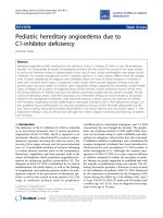
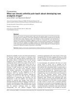
Figure 1 represents the results of this survey and shows that
the natively unfolded proteins are speci®cally localized within
a unique r egion of the charge±hydrophobicity phase space.
The solid line in this ® gure represents the border between
intrinsically unstructured a nd nativeproteins. Ob viously, t his
allows the estimation o f the ÔboundaryÕ mean hydrophobicity
value, <H>
b
, below which a polypeptide chain with a given
mean net charge <R> will be most probably unfolded:
hHi
b
hRi1X151
2X785
1
The v alidity of these predictions has been successfully
shown f or sever al p roteins [ 25]. T his m eans that degree of
compaction of a given polypeptide chain is determined by the
balance in the competition between the charge repulsion
driving unfolding and hydrophobic interactions driving
folding.
In an attempt to understand the relationship between
sequence and disorder, Dunker a nd coauthors have elabo-
rated several neuronal network predictors [5,26±35]. They
assumed that if a protein structure has evolved to have a
functional disordered s tate, then a propensity for disorder
might b e predictable from its amino-acid sequence a nd
composition. The results of such analysis were more than
impressive. It h as been established that disordered r egions
share at least some common sequence features over many
proteins. This includes low sequence complexity, with amino-
acid compositional bias and high predicted ¯exibility [28,29].
Furthermore, the majority of the intrinsically disord ered
proteins, being substantially depleted in I, L, V, W, F, Y, C,
and N, a re enriched in E, K, R, G, Q, S, P, and A [5]. Note
that these f eatures may account for the low o verall hydro-
phobicity and high net charge of the polypetide c hain of
natively unfolded proteins. Interestingly, more than 15 000
proteins in the SwissProt database were identi®ed a s having
long regions of sequence that share these same features [31].
WHAT ARE THE GENERAL
STRUCTURAL CHARACTERISTICS
OF NATIVELY UNFOLDED PROTEINS?
The general conformational properties of intrinsically
unfolded proteins are summarized below. Here we will
mostly focus on the structural characteristics, which m ake
Ó FEBS 2002 Natively unfolded proteins (Eur. J. Biochem. 269)3
such proteins exceptional among others. These a re low
compactness, absence of globularity, low secondary struc-
ture content, and high ¯exibility.
Compactness
The most unambiguous characteristic of the conformational
state of a globular protein is t he hydrodynamic dimensions.
It was noted long ago that h ydrodynamic techniques may
help to r ecognize when a protein has lost all of i ts
noncovalent structure, i.e. when it b ecame unfolded [2].
This is because an essential increase in the hydro dynamic
volume is associated with the unfolding of a protein
molecule. I t is known that globular proteins may exist in
at least four different conformations, native, molten globule,
premolten g lobule a nd unfolded [1,36±39], that may easily
be discriminated by the degree of compactness of the
polypeptide chain. Finally, it has been established that t he
native and unfolded c onformations of globular pr oteins
possess very different molecular mass dependencies of their
hydrodynamic radii (the Stokes radius), R
S
[2,40,41].
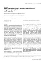
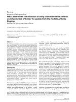
In order to clarify the physical nature of natively unfolded
proteins, Fig. 2 compares log(R
S
)vs.log(M) curves for
these proteins (see Table 1 for details) with same d epen-
dencies for the native, molten globule, premolten globule,
and urea- or GdmCl-unfolded globular proteins (data for
different conformations of globular proteins were taken
from [42]). The log(R
S
)vs.log(M) dependencies for different
conformations of globular proteins might be described by
straight lines:
logR
N
S
À0X204Æ0X0230X357Æ0X005ÁlogM2
logR
MG
S
À0X053 Æ 0X0940X 334 Æ 0X021ÁlogM
3
logR
PMG
S
À0X21 Æ 0X180X392 Æ 0X041ÁlogM
4
logR
Uurea
S
À0X649 Æ 0X0160X521 Æ 0X004ÁlogM
5
logR
UGdmCl
S
À0X723 Æ0X0330X543Æ0X007ÁlogM
6
Where N, native; MG, molten globule; PMG, premolten
globule a nd U(urea) and U(GdmCl) correspond to the
unfolded urea and GdmCl globular proteins, r espectively.
As for natively unfolded proteins, Fig. 2 clearly shows
that in respect of the ir log(R
S
) vs. log(M) dependence they
may be divided in two groups (see Table 1). One group of
the i ntrinsically unstructured proteins behaves as random
coils in poor solvent [denoted as natively unfolded
(NU)(coil)]. Proteins from the other group are essentially
more compact, being c lose with respect to their hydrody-
namic characteristics to premolten globules [denoted as
NU(PMG)]:
logR
NUcoil
S
À0X551 Æ 0X0320X493 Æ 0X008ÁlogM
7
logR
NUPMG
S
À0X239 Æ0X0550X403Æ0X 012ÁlogM
8
This is a very important obse rvation, whic h may help in
understanding the physical natu re of the natively unfolded
proteins. In fact, it is well established that the behavior of
unfolded proteins obeys the theoretical and empirical rules
that apply to linear random coils [1]. Speci®cally, it is known
that the hydrodynamic dimensions of random coils depends
Mean hydrophobicity
0.1 0.2 0.3 0.4 0.5 0.6
Mean net charge
0.0
0.1
0.2
0.3
0.4
0.5
0.6
Fig. 1. Comparison of the mean net charge and the mean hydrophobicity for a set of 275 folded (open circles) and 105 na tivel y unfolded proteins (gra y
circles). The solid line represents the border between intrinsically unstructured and native proteins (see text). Part of the data for this plot is taken
from [3].
4 V. N. Uversky (Eur. J. Biochem. 269) Ó FEBS 2002
essentially on the quality of solvent [2,40,43]. A poor solvent
encourages the attraction of macromolecular segments a nd
hence a chain has to squeeze. Whereas, in a good solvent,
repulsive forces act primarily between the segments a nd the
macromolecule conforms to a loose ¯uctuating c oil [44].
Water is a poor solvent, whereas solutions of urea and
GdmCl are rather good solvents, w ith GdmCl being closer
to the ideal one [2,40]. This difference in solvent quality may
account for the observed divergence in log(R
S
)vs.log(M)
dependencies for the coil-like part o f intrinsically unstruc-
tured proteins. The existence of well-de®ned difference
between the log(R
S
) vs. log(M) dependencies for globular
proteins unfolded by urea and GdmCl also should be noted
in this respect.
Globularity
Another v ery important structural parameter i s the degree
of globularization that re¯ects the p resence or absence of
tightly packed core in the protein molecule. I n f act, it has
been shown that the protein molecules in PMG are
characterized b y low (coil-like) intramolecular packing
density [ 37,38,42,45]. This information could be extracted
from the analysis of small angle X-ray scattering (SAXS)
data (Kratky plot), whose shape is sensitive to the
conformational state of the scattering protein molecules
[45±48]. It has been shown that a scattering curve in the
Kratky plot has a characteristic maximum when the
globular protein is i n the native state o r in the molten
globule state (i.e. has a globular structure). If a protein is
completely unfolded or in a premolten globule con-
formation ( has n o g lobular structure), such a maximum
will be absent on the r espective scattering curve [37,38,42,
45±48].
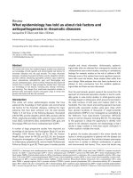
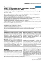
Figure 3A compares the Kratky plots of three natively
unfolded p roteins (a-syn uclein, prothymosin a and c aldes-
mon 636±771 fragment) with t hat of t he rigid g lobular
protein SNase. One can s ee that intrinsically unstructured
proteins give Kratky plots without m axima typical of
folded conformations of globular proteins. The same d ata
has also been reported f or another i ntrinsically unordered
protein, pig calpastatin domain I [49]. Thus, t hese four
natively unfolded proteins are characterized by the absence
of globular structure, or, in other words, they do not have
a tightly packed core under physiological conditions in
vitro. This is a very important observation, which allows
the assumption that all other natively unfolded proteins
may possess the same property. In fact, the analysis of
hydrodynamic data s hows t hat two of the three consid ered
proteins (a-synuclein and prothymosin a) behave as coils in
poor solvent, whereas R
S
of caldesmon 636±771 fragment
is typical of PMG (see Table 1 ). Consequently, r epresen-
tatives of both classes of intrinsically unstructured proteins
(coil-like and PMG-like) have been shown to b e charac-
terized by the absence of rigid globular core. This i s i n
goodagreementwithSAXSdataonconformational
characteristics of t he PMG state of globular proteins
[37,38,42,45].
Secondary structure
Figure 3B presents the far-UV CD s pectra of a-synuclein,
prothymosin a, phosphodiesterase c-subunit and caldes-
mon 636±771 fragment as typical representatives of the
log (M)
3.5 4.0 4.5 5.0 5.5
log (
R
S
)
1.0
1.5
2.0
Fig. 2. Dependencies of the hydrodynamic dime nsions, R
S
, on protein molecular mass, M, for native (gray circles), molten globule (gray reversed
triangles), premolten globule (gray squares), 8
M
urea-unfolded (gray diamonds) and 6
M
GdmCl-unfolded (gray triangles) conformational states of
globular proteins and natively unfolded proteins with coil-like (open circles) and PMG-like properties (open reversed triangles). Thedatausedtoplot
dependencies for native, molten globu le, premolten glo bule a nd GdmCl-unfolded states of globular proteins are taken from [42]. T he data for
natively unfolded p roteins and u rea-unfolded conformation of globular proteins are s umma rized in T ables 1 and 2, respectively. Dashed lines
represent least square ®ts of data earlier obtained for native and urea- or GdmCl-unfolded globular proteins [41].
Ó FEBS 2002 Natively unfolded proteins (Eur. J. Biochem. 269)5
family of natively unfolded p roteins. One can see that these
proteins (as well as all other i ntrinsically unstructured
proteins, whose far-UV CD spectra were studied) possess
distinctive far-UV CD s pectra with characteristic deep
minima in vicinity of 200 nm, and relatively low ellipticity
at 220 nm. The analysis of these spectra yields low content
of ordered secondary structure (a helices and b sheets).
This is also con®rmed b y the Fourier-transform infrared
(FTIR) analysis of secondary structure composition of
natively unfold ed proteins, such as tau protein [18], a-
synuclein [24,50], b-andc-synucleins; a
s
-casein [51], and
cAMP-dependent protein kinase inhibitor [ 52]. Important-
ly, even the caldesmon 636 ±771 fra gment, w hich wa s
shown to have hydrodynamic properties typical of the
PMG (see above), posse sses far-UV CD characteristic of
essentially distorted polypeptide chain. Thus, the low
overall content of ordered secondary structure could be
considered as a general property of intrinsically unstruc-
tured p roteins.
High ¯exibility
The fact that intrinsically unfolded proteins are character-
ized by an increased intramolecular ¯exibility may be easily
derived from a large a mount of NMR studies (summarized
in [4,5,53]). Moreover, recent advances in NMR technology
(especially the use of heteronuclear multidimensional
approach) have even opened the way to detailed structural
and dynamic description o f t hese proteins [4]. Increased
¯exibility o f n atively unfo lded proteins is i ndirectly con-
®rmed by their extremely h igh s ensitivity to protease
degradation in vit ro [4,5,54±59].
Table 1. Hydrodynamic characteristics of the natively unfolded proteins.
M
r
(kDa) R
S
(A
Ê
) Reference
Coil-like proteins
a-Fetoprotein, 447±480 fragment 3.6 15.5 [85]
Vmw65 C-terminal domain 9.3 28 [86]
PDE c 9.7 26
E
m
protein 11.2 28.2 [87]
Apo-cytochrome c 11.7 30 [88]
Prothymosin a 12.1 24.3 [62]
Fibronectin binding domain B 12.3 30.7 [89]
c-Synuclein 13.3 30.4
Fibronectin binding domain A 13.7 31.7 [89]
Ribonuclease A, reduced 13.7 50.6 [41]
b-Synuclein 14.3 32 [90]
a-Synuclein 14.5 32.3 [24,50]
Fibronectin binding domain D 14.7 31.8 [89]
Stathmin 17 33 [91]
CFos-AD domain, 216±380 fragment 17.3 35 [92]
Calf thymus histone 19.8 36.7 [1]
b-Casein 24 41.7 [1]
Phosvitin 24.9 39.9 [1]
Chromatogranin A 48.3 58.5 [76]
Caldesmon 140 91 [93]
MAP-2 220 122 [94]
PMG-like proteins
Osteocalcin 5.4 18.4 [73]
Heat stable protein kinase inhibitor 7.9 22.3 [52]
Caldesmon 636±771 fragment 14 28.1
SNaseD, A90S mutant 14.1 25 [95]
Pf1 gene 5 protein, 1±144 fragment (D4 domain) 15.8 29.5 [96]
PPI-1 20.8 32.3 [97]
DARRP-32 23.1 34 [22]
Manganese stabilizing protein, L245E mutant 26.5 32.7 [98]
Calreticulin, human )41C fragment 40.6 46.2 [59]
Calsequestrin, rabbit 45.2 45 [99]
Calreticulin, huiman 46.8 46.2 [59]
Calreticulin, bovine 47.6 44.2 [59]
Taka-amylase A, reduced 52.5 43.1 [1]
SdrD protein, B1-B5 fragment 64.8 54.7 [75]
Chromatogranin B 77.3 50.3 [77]
Topoisomerase I 90.7 58.5 [100]
Fibronectin 530 115 [101]
6 V. N. Uversky (Eur. J. Biochem. 269) Ó FEBS 2002
ENVIRONMENTAL INFLUENCES
ON THE NATIVELY UNFOLDED
PROTEINS
Temperature effects
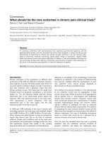
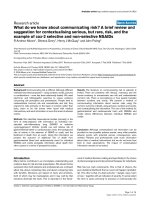
Figure 4A depicts temperature-induced changes i n the far-
UV CD spectra of a-synuclein [50] measured at different
temperatures. At low temperatures, the protein shows a far-
UV CD spectrum typical of an unfolded polypeptide chain.
As the t emperature is increased, the spectrum changes,
consistent with temperature-induced formation of second-
ary structure. Figure 4 B represents the temperature-depen-
dence of [h]
222
for a-synuclein, caldesmon 636±771
fragment, and phosphodiesterase c-subunit. One can see
that for these three proteins major spectral changes occur
over the range of 3 to 30±50 °C. Further heating leads to a
less pronounced effects. Analogous temperature dependen-
cies indicative of heat-induced str ucture formation have
been reported for the receptor extracellular domain of nerve
growth factor [60] and a
s
-casein [61]. Interestingly, it has
been shown that the structural changes induced in all these
proteins by heating are completely reversible. Thus, an
increase in temperature induces the partial folding o f
intrinsically unstructured proteins, rather than the unfolding
typical o f g lobular proteins. The effects of elevated temper-
ature may be attributed to increased strength of the
hydrophobic interaction at higher temperatures, leading
to a stronger hydrophobic driving force for folding.
This observation de®nitely has t o be t aken into account
while discussing conformational behavior of intrinsically
unstructured proteins.
Effect of pH
Figure 4C represents the pH dependence o f [ h]
222
for
a-synu clein and prothymosin a. There is little change i n
the far-UV CD spectra between pH % 9.0 and % 5.5.
However, a decrease in pH from 5.5 to 3.0 results in a
substantial increase in negative intensity in the vicinity of
220 nm. It has also been established that the pH-induced
changes in the far-UV CD spectrum of these t wo proteins
were completely reversible and consistent with the forma-
tion of partially folded PMG-like intermediate conforma-
tion [50,62].
Same pH-induced structural transformations have been
described for pig calpastatin domain I [39], histidine rich
protein I I [63], a nd the naturally occurring human peptide
LL-37 [64]. T hese observations show that a decrease (or
increase) in pH induces partial folding of intrinsically
unordered proteins due to the minimization of their large
net charge present at neutral pH, thereby decreasing
charge/charge intramolecular repulsion and permitting
hydrophobic-driven collapse to the partially folded inter-
mediate.
Effect of counter ions
It was already noted t hat, under physiological pH, intrin-
sically unstructured proteins are unfolded mainly because of
the electrostatic repulsion between the noncompensated
charges of the same sign. To some extent, this resembles the
Fig. 3. Conformational characteristics o f intrinsically disordered pro-
teins. (A) Kratky plots of SAXS data for natively unfolded a-synuclein
(1), prothymosin a (2) a nd caldesmon 636±771 fragment (3). The
Kratky plot of native globular SNase is shown for comparison (4). (B)
Far-UV CD spectra of intrinsically unordered proteins, a-synuclein
(1), prothymosin a (2), caldesmon 636±771 fragment (3) and phos-
phodiesterase c-subunit (4).
Table 2. Hydrodynamic characteristics of 8
M
urea-unfolded p ro teins
without cross-links.
Protein M
r
(kDa) R
S
(A
Ê
) Reference
Insulin 3 14.6 [41]
Ubiquitin 8.5 24.6
Cytochrome c 11.7 4.05
Ribonuclease A 13.7 32.4 [41]
Lysozyme 14.2 33.1 [41]
Hemoglobin 15.5 33.5
Myoglobin 16.9 35.1
b-Lactoglobulin 18.5 37.8 [41]
Chymotrypsinogen 25.7 45 [41]
Carbonic anhydrase B 28.8 47.8 [41]
b-Lactamase 28.8 48.9 [41]
Ovalbumin 43.5 58.8
Serum albumin 66.3 74 [41]
Lactate dehydrogenase 35.3 52
GAP dehydrogenase 36.3 54
Aldolase 40 57
Transferrin 81 81
Thyroglobulin 165 116
Ó FEBS 2002 Natively unfolded proteins (Eur. J. Biochem. 269)7
situation occurring for many proteins at e xtremely low o r
high pH. It has been established that these unfolded proteins
could be transformed into more ordered conformations if
electrostatic repulsion was reduced by binding of oppositely
charged ions [65,66]. Similar s ituation may be expected for
natively unfolded proteins, and, in fact, the metal i on-
stimulated conformational changes have been described for
many intrinsically unstructured proteins.
As an illustration, Fig. 4D represents the [h]
222
depen-
dencies on [ Al
3+
]fora-synuclein. One can s ee that an
increase in the cation content is accompanied by an essential
increase in the intensity of the far-UV CD spectra, re¯ecting
partial folding of the protein. It has been established that
other cations (monovalent, bivalent and trivalent) induce
conformational changes in a-synuclein and transform this
natively unfolded protein into a partially folded intermedi-
ate too. The folding strength of cations increases with the
ionic charge density incre ase [67]. This re¯ects t he effective
screening of the Coulombic charge/charge repulsion. For
polyvalent c ations, an additional important factor could b e
hypothesized, which is the potential capability for cross-
linking or bridging between two or more carboxylates.
Importantly, human antibacterial protein LL-37, a
natively unfolded p rotein with extremely basic net charge,
was shown to be essentially folded in the presence of several
anions [64].
WHAT ELSE IS REQUIRED
FOR INTRINSICALLY UNORDERED
PROTEINS TO FOLD?
Structure forming role of natural ligands
It has been suggested that natively unfolded proteins may
be signi®cantly folded in their normal cellular milieu due
to binding to speci®c targets and ligands (such a s a variety
of small molecules, s ubstrates, cofactors, other proteins,
nucleic acids, membranes, etc.) [3±5,53,68]. The structure-
forming effect of natural partners can be explained by
their in¯uence o n the m ean hydrophobicity and/or net
charge of the natively unfolded polypeptide. In fact, any
interaction of such protein with natural ligand affecting
mean net c harge and/or mean hydrophobicity of the
protein could c hange t hese parameters in such a way that
they will approach values typical of folded native proteins.
This hypothesis has been con®rmed by calculation the
joint mean net charge and mean hydrophobicity of
complexes of several natively unfolded p roteins, ostecalcin,
Fig. 4. Eect of environmental factors on conformational properties of natively unfolded proteins. (A) Heating-induced secondary structure formation
in the n atively unfolded a-synuclein. Representative f ar-UV CD s pectra of the protein measured at dierent temperatures. (B) Temperature-
induced changes in far-UV CD spectrum ([h]
222
vs. temperature depen dence) measured for a-synuc lein (triangles), phosphodiesterase c-subunit
(squares), and caldesmon 636±771 fragment (circles). (C) pH-induced structure formation ([h]
222
vs. pH dependence) in the natively unfolded
a-synuclein (circles) and prothymosin a (triangles). (D) Cation-induced structure formation in natively unfolded a-synuclein. Data for a-synuclein
and protymosin a are taken from [50,67] and [62], respectively.
8 V. N. Uversky (Eur. J. Biochem. 269) Ó FEBS 2002
osteonectin, a-casein, HPV16 E7 protein, calsequestrin,
manganese s tabilizing p rotein and HIV-1 integrase, with
their n atural ligands, metal ions [3]. The e xistence of
pronounced ligand-induced folding has been indeed
established in numerous in vitro studies for many intrin-
sically unstructured proteins. E xamples include: DNA (or
RNA) induced structure f ormation in protamines [69,70],
Max protein [57], high mobility group proteins HMG-14
[71] and HMG-17 [72]; cation-induced folding o f o stecal-
cine [73], osteonectine [ 74], S drd protein [75], chromatog-
ranins A [ 76] and B [77], D131D fragment of SNase [78],
histone H1 [79], protamine [70] and prothymosin-a [80];
folding of cytochrome c inthepresenceofheme[81];
membrane-induced secondary structure formation in para-
thyroid hormone related protein [82]; trimethylamine
N-oxide induced structure formation in glucocorticoid
receptor [83]; h eme-induced folding of histidine-rich pro-
tein II [84], and many others.
CONCLUSIONS
Based on t he data summarized ab ove, a typical na tively
unfolded protein is characterized by: (a) a speci®c amino-
acid sequence with low overall h ydrophobicity and high net
charge; (b) hydrodynamic properties typical of a random
coil in poor solvent, or PMG c onformation; (c) l ow level of
ordered secondary stru cture; (d) t he absence of a tightly
packed core; (e) high conformational ¯exibility; (f) its ability
to adopt relatively rigid c onformation in the presence of
natural ligands; and (g) a Ôturn outÕ response to environ-
mental changes, with the structural complexity increase a t
high temperature or at extreme pH.
ACKNOWLEDGEMENTS
I am grateful to Dr P. Souillac for the careful reading and editing of the
manuscript. This work was s upported in p art by f ellowships from the
Parkinson's Institute and t he National Parkinson's Foundation.
REFERENCES
1. Ptitsyn, O.B. (1995) Molten globule and protein folding. Adv.
Prot. Chem. 47, 83±229.
2. Tanford, C. (1968) Protein denaturation. Adv. Prot . Chem. 23 ,
121±282.
3. Uversky, V.N., Gillespie, J .R. & Fink, A.L. (2000) Why are Ôna-
tively unfoldedÕ proteins unstructured under the physiological
conditions? Proteins 41 , 415±427.
4. Wright, P.E. & Dyson, H.J. (1999) Intrinsically unstructured
proteins: re-assessing the protein s tructure±function p aradigm.
J. Mol. Biol. 293, 321±331.
5. Dunker, A.K., Lawson, J.D., Brown, C.J., W illiams, R.M.,
Romero, P., Oh, J.S., Old®eld, C.J., Campen, A.M., Ratli, C.M.,
Hipps,K.W.,Ausio,J.,Nissen,M.S.,Reeves,R.,Kang,C H.,
Kissinger, C.R., Bailey, R.W., Griswold, M.D., Chiu, W., Garber,
E.C. & Obradovic, A . (2001) In trinsically d isordered protein.
J. Mol. Graph. Model. 19 , 26±59.
6. Uversky, V.N., Talapatra, A., Gillespie, J.R. & Fink, A.L. (1999)
Protein deposits a s the molecular basis of amyloidosis. Part I .
Systemic amyloidoses. Med. Sci. Mon it. 5, 1001±1012.
7. Uversky, V.N., Talapatra, A., Gillespie, J.R. & Fink, A.L. (1999)
Protein deposits as the molecular b asis of amyloidosis. II. Local-
ized amyloidosis and neurodegenerative disordres. Med. Sci.
Monit. 5, 1238±1254.
8. Glenner, G.G. & W ong, C.W. (1984) Alzheimer's-disease and
Down's-syndrome ± sharing of a unique cerebrovascular amyloid
®bril protein. Bio chem. Biophys. R es. Commun. 120 , 885±890.
9. Masters, C.L., Multhaup, G., Simms, G., Pottgiesser, J., Martins,
R.N. & Beyreuther, K. (1 985) Neuronal origin of a cerebral
amyloid: neuro®brillary tangles of Alzheimer's disease contain the
same protein a s the amyloid o f plaque cores an d blood vessels.
EMBO J. 4, 2757±2763.
10. Lee, G .M Y., Balin, B.J., Otvos, L. & Trojanowski. J.Q. (1991)
A68: a major subunit of paired helical ®laments and d erivatized
forms of normal Tau. Science 251, 6 75±678.
11. Ue
Â
da, K., Fukus hima, H., Masliah, E., X ia, Y ., Iwai, A.,
Yoshimoto, M., Otero, D.A., K on do, J ., I hara, Y . & Saitoh, T.
(1993) Mo lecu lar cloning of cDNA e ncodin g an unrecognized
component of amyloid in Alzheimer disease. Proc.NatlAcad.Sci.
USA 90, 11282±11286.
12. Wisniewski, K.E., Dalton, A.J., M cLachlan, C., Wen, G.Y. &
Wisniewski, H .M. (1985) Alzheimer's disease in Down's syn-
drome: clin icopathologic studies. Neurology 35, 957±961.
13. Arawaka, S., Saito, Y., Murayama, S. & Mori, H. (1998) Lewy
body in neurodegeneration with brain iron accumulation type 1 is
immunoreactive for alpha-synuclein. Neurology 51, 887±889.
14. Arima, K ., Ue
Â
da, K., Sunohara, N., Hirai, S., I zumiyama, Y .,
Tonozuka-Uehara, H. & Kawai, M . ( 1998) Imm unoelectron-
microscopic demonstration of NACP/alpha-synuclein-epitopes on
the ®lamentous component of Lewy bodies in Parkinson's disease
and in dementia with L ewy bodies. Brain Res. 808, 93±100.
15. Tu,P.H.,Galvin,J.E.,Baba,M.,Giasson,B.,Tomita,T.,Leight,
S., Nakajo, S., Iwatsubo, T., Trojanowski, J.Q. & Lee, V.M.
(1998) Glial cytoplasmic inclusions in white matter oligodendro-
cytes of multiple system atrophy brains contain insoluble alpha-
synuclein. Ann. Neurol. 44, 415±422.
16. Wakabayashi, K., Yo shimoto, M., Tsuji, S. & Takahashi. H.
(1998) Alpha-synuclein immunoreactivity in glial cytoplasmic in-
clusions in multiple system atrophy. Neurosci. Lett. 249, 180±182.
17. Galvin, J.E., Lee, V.M., Schmidt, M.L., Tu, P.H., Iwatsubo, T. &
Trojanowski, J.Q. (1999) Pat hobiology of the Lewy body. Adv.
Neurol. 80, 313±324.
18. Sch weers, O., Scho
È
nbrunn Hanebeck, E., Marx, A. & Man del-
kow, E. (1994) Structural studies of tau protein an d Alzheimer
paired helical ®laments show n o evidence for be ta-structure.
J. Biol. Chem. 269, 24290±24297.
19. Lidakis-Simantiris, N., Hutchison, R.S., Betts, S.D., Barry, B.A.
& Yocum, C .F. (1999) Manganese s tabilizing protein of photo-
system II is a thermostable, natively unfolded polypeptide.
Biochemistry 38 , 404±414.
20. Shutova, T., Irrgang, K D., Klimov, V.V. & Renger, G. (2000) Is
the manganese stabilizing 33 k Da protei n of photosystem II
attaining a Ônatively unfoldedÕ or Ômolten globuleÕ structure in
solution? FEBS Lett. 467, 137±140.
21. An®nsen, C.B., Haber, E., Sela, M. & White, F.N. (1961) Kinetics
of formation of native ribonuclease during oxidation of the
reduced polypeptide chain. Proc.NatlAcad.Sci.USA47, 1309±
1314.
22. Hemmings, H.G. Jr,, Nairin, A.C., Aswad, D.W. & Greengard, P.
(1984) DARPP-32, a dopamine-and adenosine 3 ¢:5¢-monopho s-
phate-regulated phosphoprotein enriched in dopamine-innervated
brain r egions. II. Pu ri®cation and charac terization of the phos-
phoprotein from bovine caudate nucleus. J. Biol. Chem. 4, 99±110.
23. Gast, K., Damaschun, H., Eckert, K., Schulze-Foster, K., Maure r,
H.R., Mu
È
ller-Frohne, M., Zirwer, D., Czarnecki, J. & Damas-
chun, G. (1995) Prothymosin a: a biologically active protein with
random coil conformation. Biochemistry 34, 13211±13218.
24. Weinreb, P.H., Zhen, W., Poon, A.W., Conway, K.A. & Lansbury,
P.T. Jr (1996) NACP, a protein implicated in Alzheimer's diseas-
es and l earning, is natively unfolded. Biochemistry 35, 13709±
13715.
Ó FEBS 2002 Natively unfolded proteins (Eur. J. Biochem. 269)9
25. Demarest, S.J., Zhou, S Q., Robblee, J., Fairman, R., Chu, B. &
Raleigh, D.P. (2001) A comparative study of peptide models of the
alpha-domain of alpha-lactalbumin, lysozyme, and alpha-lactal-
bumin/lysozyme chimeras allows the elucidation of critical factors
that contribute to the ability to form stable partially folded states.
Biochemistry 40, 2138±2147.
26. Dunker, A.K., Obradovic, Z., R om ero, P., Kissinger, C . &
Villafranca, J.E. ( 1997) On the importance of being diso rdered.
PDB Newsletter 81,3±5.
27. Romero, P., Obradovic, Z., Kissinger, C., Villafranca, J.E. &
Dunker, A.K. (1997) Identifying disordered r egions in proteins
from amino acid sequence. Proc. IEEE Int. Conf Neuronal Net-
works 1, 90±95.
28. Dunker, A.K., Garner, E., Guilliot, S., Romero, P., Albercht, K.,
Hart, J., Obradovic, Z., Kissinger, C. & Villafranca, J.E. (1998)
Protein disorder a nd th e evo lution of molecular recognition:
theory, predictions and observations. Pac. Sy mp. Biocomput. 3,
473±484.
29. Garner,E.,Cannon,P.,Romero,P.,Obradovic,Z.&Dunker,
A.K. (1998) Predicting disordered regions from amino acid
sequence: Common themes despite dierent structural charac-
terization. Genome Informatics 9, 201±213.
30. Romero, P., Obradovic, Z. & Dunker, A.K. (1998) Sequence data
analysis for lo ng d istorted regions prediction in the c alcineurin
family. Genome Informat ics 8, 110±124.
31. Romero, P., Obradovic, Z., Kissinger, C., Villafranca, J.E., Gar-
ner, E., Guilliot, S. & Dunker, A.K. (1998) Thousands of proteins
likely to have long disordered regions. Pac. Symp. Biocomput. 3,
437±448.
32. Li,X.,Romero,P.,Rani,M.,Dunker,A.K.&Obradovic,Z.
(1999) Predic ting protein disorders for N-, C-, a nd internal
regions . Genome Informatics 10, 30±40.
33. Li, X., Obradovic, Z., Brown, C.L., Garner, E. & Dunker, A.K.
(2000) Comparing predictors of disordered protein. Genome
Informatic s 11, 172±184.
34. Romero, P., Obradovic, Z. & Dunker, A.K. (2001) Intelligent data
analysis for protein disorder prediction. Arti®cial Intelligence Rev.
14, 447±484.
35. Romero, P., Obradovic, Z., Li, X., Garner, E.C., Brown, C.J. &
Dunker, A.K. (2001) Sequence complexity of disordered proteins.
Proteins 42, 38± 48.
36. Uversky, V.N. & Ptitsyn, O.B. (1994) ÔPartly foldedÕ state, a new
equilibrium state of protein m olecu les: Four-state guanidinium
chloride-induced unfolding of b-lactamase at low temperature.
Biochemistry 33, 2782±2791.
37. Uversky, V.N. & Ptitsyn, O.B. (1994) Further evidence on the
equilibrium Ôpre-molten globule stateÕ: Four-state GdmCl-induced
unfolding of carbonic anhydrase B at low t emperature. J. Mol.
Biol. 255, 215±228.
38. Uversky, V.N. (1997) D iversity of compact f orms of denatured
globular proteins Prot. Pept. Lett. 4, 355±367.
39. Uversky, V.N. (1998) How many molten globule states there exist?
Bio®zika (Moscow) 43, 416±421.
40. Tanford, C. (1961) Physical Chemistry of Macromolecules.
Willey, New York.
41. Uversky, V.N. (1993) Use of fast protein size-exclusion liquid
chromatography to stu dy t he un folding o f p roteins w hich de na-
ture through the molten globule. Biochemistry 32, 13288±13298.
42. Tcherkasskaya, O. & Uversky, V.N. (2001) Denatured collapsed
states in protein f olding: example of apomyoglobin. Proteins:
Struct., Funct., G enet. 44, 244±254.
43. Flory, P.J. (1953) Principles of Polymer Chemistry. Cornell Uni-
versity Press, Itha ca, New York.
44. Grossberg, A.Yu & Khohlov. A.R. (1989) Statistical Physics of
Macromolecules. Nauka, Moscow.
45. Uversky, V.N., Karn oup, A.S., Segel, D.J., Seshadri, S., Doniach,
S. & F in k, A.L . (1998) Anion-induced folding of Staphylococcal
nuclease: characterization of multiple partially folded intermedi-
ates. J. Mol. Biol. 278, 879±894.
46. Glatter, O. & Kratky, O. (1982) Small Angle X-Ray Scattering.
Academic Press, London.
47. Feigin, L.A. & Svergun, D.I. (1987) Structural Analysis by Small-
Angle X-Ray and Neutron Scattering. Plenum Press, New York.
48. Semisotnov, G.V., Kihara, H., Kotova, N.V., Kimura, K.,
Amemiya, Y., Wakabayashi, K., Serdyuk, I.N., Timchenko, A.A.,
Chiba, K., Nikaido, K., Ikura, T. & Kuwajima. K. (1996) Protein
globularization d uring folding. A stud y by synchrotron small-
angle X-ray scattering . J. Mol. Biol. 262, 559±574.
49. Konno,T.,Tanaka,N.,Kataoka,M.,Takano,E.&Maki.M.
(1997) A circular dichroism study of p referential hydration and
alcohol eects on a denatured protein, pig calpastatin domain I.
Biochim. Biophys. A cta 1342, 73±82.
50. Uversky, V.N., Li, J. & Fink, A.L. (2001) Evidence for a partially-
folded intermediate in a-synuclein ®brillation. J. Biol. Chem. 276,
10737±10744.
51. Bhattacharyya, J. & D as, K.P. (1999) Molecular chaperone-like
properties of an unfolded protein, alpha(s)-casein. J. Biol. Chem.
274, 15505±15509.
52. Thomas, J., Van Patten, S.M., Howard, P., D ay, K.H., Mitchell,
R.D.,Sosnick,T.,Trewhella,J.,Walsh,D.A.&Maurer,R.A.
(1991) Expression in Escherichia coli and characterization of t he
heat-stable inhibitor of the cAMP-dependent protein k inase .
J. Biol. C hem. 266, 10906±10911.
53. Plaxco, K.W. & Gross, M. (1997) Cell biology. The importance of
being unfolded . Nature 386, 657±659.
54. Hersh ey, P.E.C., McWhirter, S.M., Gross, J.D., Wagner, G.,
Alber, T. & Sachs, A .B. (1999) The cap-binding protein eIF4E
promotes folding of a function al domain of yeast t ranslation ini-
tiation factor eIF4G1. J. Biol. Chem. 274, 21297±21304.
55. Lisse, T., Bartels, D., Kalbitzer, H.R. & Jaenicke. R. (1996) T he
recombinant dehydrin-like desiccation stress protein f rom the
resurrection plant Craterostigma plantagineum displays no de®ned
three-dimensional structure in its native state. Biol. Chem. 377,
555±561.
56. Kriwacki, R.W., Hengst, L., Tennant, L., Reed, S.I. & Wright,
P.E. (1996) Structural studies of p21 (Waf1/Cip1/Sdi1) in the free
and Cdk2-bound state: Conformational disorder mediates binding
diversity. Proc. Natl Acad. Sci. USA 93 , 11504±11509.
57. Horiu chi, M., Kurihara, Y., Katahira, M., Maeda, T., Saito, T. &
Uesugi, S. (1997) Dimerization and DNA binding facilitate alpha-
helix formation of Max in solution. J. Biochem. (Tokyo) 122,
711±716.
58. Ratnaswamy,G.,Koepf,E.,Bekele,H.,Yin,H.&Kelly.J.W.
(1999) The amyloidogenicity o f g elsolin is c ontrolled by proteol-
ysis and pH. Chem. Biol. 6, 293±304.
59. Bouvier, M. & Staord, W .P. (2000) Probing the t hree-dimen-
sional structure of human calreticulin. Biochemistry 39, 14950±
14959.
60. Timm, D.E., Vissavajjhala, P., Ross, A.H. & N eet. K.E. (1992)
Spectroscopic and chemical studies of th e i nteraction between
nerve growth factor (NGF) and the extracellular domain of the
low anity NGF receptor. Protein Sci. 1, 1023±1031.
61. Kim, T.D., Ryu, H.J., Cho, H.I., Yang, C H. & Kim. J. (2000)
Thermal behavior of proteins: heat-resistant proteins and their
heat-induced secondary structural changes. Biochemistry 39,
14839±14846.
62. Uversky, V.N., Gillespie, J .R., Millett, I.S., Khodyakova, A.V.,
Vasiliev, A.M., Chernovskaya, T.V., Vasilenko, R.N., Kozlovs-
kaya, G.D., Dolgikh, D.A., Doniach, S., Fink, A.L. & Abramov,
V.M. (1999) ÔNatively unfoldedÕ human prothymosin a adopts
partially-folded conformation at acidic pH. Biochemistry 38,
15009±15016.
63. Lynn, A., Chandra, S., Malhotra, P. & Chauhan, V.S. (1999)
Heme binding and polymerization by Plasmodium falciparum
10 V. N. Uversky (Eur. J. Biochem. 269) Ó FEBS 2002
histidine rich protein II: in¯uence of pH on activity and confor-
mation. FEB S Lett. 459, 267±271.
64. Johan sson, J., Gu dm undsson, G.H., Rottenberg, M.E., Berndt ,
K.D. & Agerberth, B . (1998) Conformation-dependent antibac-
terial activity of the n aturally occurring human peptide L L-37.
J. Biol. Chem. 273, 3718±3724.
65. Goto, Y., Takahashi, N. & Fink, A.L. (1990) Mechanism of acid-
induced folding of proteins. Biochemi stry 29, 3480±3488.
66. Fink, A.L., Calciano, L.J., Goto, Y., Kurotsu, T. & Palleros. D.R.
(1994) Classi®cation o f acid denaturation of proteins ± interme-
diates and unfolded states. Bioc hemistry 33, 12504±12511.
67. Uversky, V.N., Li, J. & Fink, A.L. (2001) Metal-triggered s truc-
tural transformations, aggregation and ®bril formation of human
alpha-synuclein. A possible m olecular link between Park inson's
disease and heavy metal exposure. J. Bio l. Chem. 276, 44284±
44296.
68. Uversky, V.N. & Narizhneva, N.V. (1998) Eect of natural
ligands on structural properties and conformational stability o f
proteins. Biochemistry (Moscow) 63, 420±433.
69. Warrant, R.W. & Kim, S.H. (1978) Alpha-helix±double helix
interaction sh own in the structure o f a protamine-transfer RNA
complex and a nucleoprotamine model. Na ture 271, 130±135.
70. Gatewoo d, J.M., Schroth, G.P., Schmid, C.W. & Bradbury, E.M.
(1990) Zinc-induced secondary s tructure transitions i n human
sperm protamines. J. Biol. Chem. 265, 20667±20672.
71. Cary, P.D., King, D .S., Crane Robinson , C., Bradbury, E .M.,
Rabbani, A., Goodwin, G.H. & Johns, E.W. (1980) Structural
studies on two high-mobility-group proteins from c alf thymus,
HMG-14 and HMG-20 (ubiquitin), and their interaction with
DNA. Eur. J. Biochem. 112, 577 ±580.
72. Abercrombie, B.D., Kneale, G.G., Crane Robinson, C., Brad-
bury, E.M., Goodwin, G.H., Walker, J.M. & Johns, E.W. (1978)
Studies on the conformational properties of the high-mobility-
group chromosomal protein HMG 17 and its interaction with
DNA. Eur. J. Biochem. 84, 173±177.
73. Isbell, D.T.S., Schroering, A.G., Colombo, G. & Shelling, J.G.
(1993) Metal ion binding to dog osteocalcin studied by
1
HNMR
spectroscopy. Biochemistry 32 , 11352±11362.
74. Engel, J., Taylor, W., Paulsson, M., Sage, H. & Hogan, B. (1987)
Calcium binding domains and calcium-induced conformational
transition of SPARC/BM-40/osteonectin, an extracellular glyco-
protein e xpressed in mineralized and nonmineralized tissues.
Biochemistry 26 , 6958±6965.
75. Josefsson, E., O'Connell, D., Foster, T.J., Durussel, I. & Cox, J.A.
(1998) The binding of calcium to the B-repeat segment of SdrD, a
cell surface protein of Staphylococcus aureus. J. Biol. Chem. 273,
31145±31152.
76. Yoo,S.H.&Albanesi,J.P.(1990)Ca
2+
-induced conformational
change and aggregation of c hromogranin A . J. Biol. Chem. 265,
14414±14421.
77. Yoo, S.H. (1995) pH- and Ca
2+
-induced conformational change
and a ggregation of chromogranin B. Compariso n with chro-
mogranin A and implication in secretory vesicle biogenesis. J. Biol.
Chem. 270, 12578±12583.
78. Alexandrescu, A.T., Abeygunawardana, C. & Shortle, D. (1994)
Structure and dynamics of a denatured 131-residue fragment of
staphylococcal nuclease: a heteronuclear NMR study. Biochem-
istry 33, 106 3±1072.
79. Tarkka,T.,Oikarinen,J.&Grundstro
È
m, T. (1997) Nucleotide
and c alcium-ind uced conformational c hanges in histo ne H1.
FEBS Lett. 406, 56±60.
80. Uversky, V.N., Gi llespie, J.R., Millet t, I.S., Khodyakova, A.V.,
Vasilenko, R.N., Vasiliev, A.M., R odionov, I .L., Kozlovskaya,
G.D.,Dolgikh,D.A.,Doniach,S.,Fink,A.L.,Permyakov,E.A.
& Abramov, V.M. (2000) Zn
2+
-mediated structure formation and
compaction of th e Ônatively unfoldedÕ human prothymosin a.
Biochem. Biophys. Res. Comun. 267 , 663±668.
81. Stellwagen, E., Rysary, R. & Babul, G. (1972) The conformation
of horse heart apocytochrome c. J. Biol. C hem. 247, 8074±8077.
82. Willis, K.J. (1994) Inter action with model membrane systems
induces secondary structure in amino-terminal fragments of
parathyroid hormone related protein. Int. J. Pept. Protein Res. 43,
23±28.
83. Baskakov, I.V., Kumar, R., Srinivasan, G., Ji, Y.S., Bolen, D.W.
& Thompson, E.B. (1999) Tri methylamine N- oxide-induced
cooperative folding of an intrinsically unfolded transcription-
activating fragment of human glucocorticoid receptor. J. Biol.
Chem. 274, 10693±10696.
84. Eom, J.W., Baker, W.R., Kintanar, A. & Wurtele, E.S. (1996) The
embryo-speci®c EMB-1 p rotein of Daucus carota is ¯e xib le and
unstructured in solution. Plant Sci. 115, 17±24.
85. Eisele, L.E., Mes®n, F.B., Bennet t, J .A., An dersen, T.T., Jacob-
son, H.I., Soldwedel, H., MacColl, R. & Mizejewski, G.J. (2001)
Studies on a growth-inhibitory peptide derived from alpha-feto-
protein a nd some analogs. J. Peptide Res. 57, 29±38.
86. Donaldson, L. & Capone, J .P. (1992) Puri®cation and charac-
terization of the carboxyl-terminal transactivation domain o f
VMW65 from herpes-simplex virus type-I. J. Biol. Chem. 267,
1411±1414.
87. McCubbin, W.D. & Kay, C.M. (1985) Hydrodynamic and optical
properties of the wheat EM protein. Can. J. Biochem. Cell Biol. 63,
803±811.
88. Dam aschun, G., D am aschu n, H., Gast, K., Gernat, C. & Zirver.
D. (1991) Acid denatured apo-cytochrome c is a random coil.
Evidence from small angle X-ray scattering and dynamic light
scattering. Biochim. Biophys. Acta 1078, 289±295.
89. House -Pompeo, K., Xu, Y., Joh, D., Speziale, P. & Hook. M.
(1996) Conformational changes in the ®bronectin binding
MSCRAMMs are induced by ligand bindin g. J. Biol. Chem. 271,
1379±1384.
90. Nakajo , S., Omata, K., Aiuchi, T., Shib ayama, T., Okahash i, I.,
Ochiai, H., Na kai, Y., Nakaya, K. & Nakamura, Y. ( 1990)
Puri®cation and characterization of a novel brain-speci®c 14-kDa
protein. J. Neurochem. 55, 2031±2038.
91. Belmont, L.D. & Mitchison, T.J. (1996) Identi®cation of a protein
that interacts with t ubulin dimers and i n creases the catastrophe
rate of microtubules. Cell 84, 623±631.
92. Campbell, K.M., Terrell, A.R., Laybourn, P.J. & Lumb. K.J.
(2000) Intrinsic structural disorder of the C-terminal activation
domain from the bZIP transcription factor Fos. Biochem istry 39,
2708±2713.
93. Lynch, W.P., Riseman, V.M. & Bretscher, A. (1987) Smooth
muscle caldesmon is an extended ¯exible monomeric protein in
solution that can readily undergo reversible intra- and inte rmo-
lecular sulfhydryl cross-linking. A mechanism f or caldesmon's
F-actin bundling a ctivity. J. Biol. Chem. 262, 7429±7437.
94. Herna
Â
ndez, M.A., Avila, J. & Andreu, J.M. (1986) Physico-
chemical characterization of the heat-stable m icrotubule-associ-
ated protein MAP2. Eur. J. Biochem. 154, 41±48.
95. Shortle, D . & M eeker. A.K. (1989) Residual structure in large
fragments of staphyloco ccal nuclease: E ects of am ino acid sub-
stitutions. Biochemistry 28, 936 ±944.
96. Bogdarina, I., Fox, D.G. & Kneale, G.G. (1998) Equilibrium and
kinetic binding analysis of the N-terminal domain of the Pf1 gene 5
protein and its interaction with single-stranded DNA. J. Mol. Biol.
275, 443±452.
97. Nimmo, G.A. & Cohen, P. (1978) The regulation of glycogen
metabolism. Puri®cation and characterisation of protein phos-
phatase inhibitor-1 from rabbit ske letal muscle. Eur . J. Bio chem .
87, 341±351.
98. Lidakis-Simantiris, N., Betts, S.D. & Yocum, C.F. (1999) Leu-
cine 245 is a critical residue for folding a nd function of the man-
ganese stabilizing proteinofphotosystem II. Biochemistry 38, 1 5528±
15535.
Ó FEBS 2002 Natively unfolded proteins (Eur. J. Biochem. 269)11
99. Cozens, B. & R eithmeier, R.A.F. (1984) Size and shape of
rabbit skel etal-muscle calsequestrin. J. Biol. Chem. 25 9, 6246±
6252.
100. Stewart, L., Ireton, G.C., Parker, L.H., Madden, K.R. &
Champoux. J.J. ( 1996) Biochemical and biophysical analyses of
recombinant forms of human topoisomerase I. J. Biol. Chem. 271,
7593±7601.
101. Pelta, J., Berry, H., Fadda, G.C., Pauthe, E. & Lairez. D. (2000)
Statistical conformation o f human plasma ®bro nectin. B ioche m-
istry 39, 5146±5154.
12 V. N. Uversky (Eur. J. Biochem. 269) Ó FEBS 2002