Báo cáo Y học: Proximal ligand motions in H93G myoglobin pptx
Bạn đang xem bản rút gọn của tài liệu. Xem và tải ngay bản đầy đủ của tài liệu tại đây (222.8 KB, 8 trang )
Proximal ligand motions in H93G myoglobin
Stefan Franzen
1
, Eric S. Peterson
2
, Derek Brown
1
, Joel M. Friedman
3
, Melissa R. Thomas
4
and Steven G. Boxer
4
1
Department of Chemistry, North Carolina State University, Raleigh, NC, USA;
2
Chemistry Department, Bowdoin College,
6600 College Station, Brunswick, ME 04011-8466, USA;
3
Department of Physiology and Biophysics, Albert Einstein College
of Medicine, Bronx, NY, USA;
4
Department of Chemistry, Stanford University, Stanford, CA, USA
Resonance Raman spectroscopy has been used to observe
changes in the iron–ligand stretching frequency in photo-
product spectra of the proximal cavity mutant of myoglobin
H93G. The measurements compare the deoxy ferrous state
of the heme iron in H93G(L), where L is an exogenous
imidazole ligand bound in the proximal cavity, to the pho-
tolyzed intermediate of H93G(L)*CO at 8 ns. There are
significant differences in the frequencies of the iron–ligand
axial out-of-plane mode m(Fe–L) in the photoproduct spec-
tra depending on the nature of L for a series of methyl-
substituted imidazoles. Further comparison was made with
the proximal cavity mutant of myoglobin in the absence of
exogenous ligand (H93G) and the photoproduct of the
carbonmonoxy adduct of H93G (H93G-*CO). For this
case, it has been shown that H
2
O is the axial (fifth) ligand to
the heme iron in the deoxy form of H93G. The photo-
product of H93G-*CO is consistent with a transiently bound
ligand proposed to be a histidine. The data presented here
further substantiate the conclusion that a conformationally
driven ligand switch exists in photolyzed H93G-*CO. The
results suggest that ligand conformational changes in
response to dynamic motions of the globin on the nanosec-
ond and longer time scales are a general feature of the H93G
proximal cavity mutant.
Keywords: resonance Raman, heme, myoglobin, hemo-
globin, ligand switch.
Protein structural relaxation following heme photolysis has
been studied in globins as a means to obtain information on
structural intermediates following diatomic ligand photo-
lysis. In hemoglobin (Hb), time-resolved spectroscopic
studies have provided information on the time scale for
transition from the six-coordinate R state to the five-
coordinate T-state [1–3]. The proximal cavity mutant of Hb
has recently demonstrated the key role of the proximal
histidine in the cooperativity of quaternary structure change
in response to ligand binding [4]. Strain in the covalent bond
to the heme iron of Hb can be monitored by following the
shift in frequency of the iron–histidine axial mode, m(Fe–
His), by time-resolved resonance Raman spectroscopy [5].
In myoglobin (Mb), these studies have indicated a much
smaller change in structure [6]: the observed frequency shift
of the iron–histidine band is c.1.6cm
)1
on the 8 ns time
scale compared to 12 cm
)1
in Hb. Nonetheless, this shift in
the m(Fe–His) Raman band is significant because shifts in
absorption bands (the time-dependent Soret band shift and
band III shift) have been attributed to iron out-of-plane
displacement that should also be coupled to m(Fe–His) [7–
10]. A structural interpretation of these observable phe-
nomena helps to bridge the gap between the extensive X-ray
crystallography studies and the thermodynamic and kinetic
data available for Mb [7,11–20].
Histidine-ligated heme enzymes have a surprisingly large
range of functions. In peroxidase, a charge relay due to
hydrogen bonding of the imidazole ring of histidine permits
the formation of high valent iron oxidation states that play a
role in the redox function of these enzymes [21–23]. Charge
relay effects are seen in other heme proteins such as the
transcriptional activator CooA [24]. Enhanced enzyme
activity is triggered by the rupture of the histidine-iron
bond in guanylyl cyclase in response to trans NO ligation
[25,26], None of these effects are as clearly observed in wild-
type Mb, although recent work suggests that Mb does in
fact play an enzymatic role in catalyzing reactions of small
moleculessuchasO
2
, CO, NO and peroxides [27].
Presumably, the imidazole ring is appropriately stabilized
in the proximal pocket of Mb by hydrogen bonding and
steric effects. The hydrogen bonding of the proximal iron
ligand, His93 in Mb is thought to be relatively weak. The
Nd proton has a bifurcated hydrogen bonding interaction
with the lone pair of the Ser92 hydroxyl and the backbone
carbonyl of Leu89 [11,28]. The role of hydrogen bonding
can be addressed by studies of the Mb proximal cavity
mutant (H93G) for a series of axial proximal ligands in
addition to mutants such as S92A that change the hydrogen
bonding environment of the proximal pocket [29]. Studies of
the H93G mutant with a series of different ligands in the
proximal cavity have the advantage of probing proximal
effects on both proximal ligand rebinding and stability as
well as how these couple to the distal pocket where small
diatomic ligands, such as CO, bind [30].
This study investigates the dynamics that occur within the
first 8 ns following photolysis of CO in the H93G mutant
with four different imidazole ligands in the proximal cavity.
Correspondence to Stefan Franzen, Department of Chemistry,
North Carolina State University, Raleigh, NC 27695, USA.
Fax: +1 919 515 8909, Tel.: + 1 919 515 8915,
E-mail:
Abbreviations: Hb, hemoglobin; Im, imidazole; x-MeIm, x-methyl
imidazole (x ¼ 1, 2 or 4); Mb, myoglobin.
(Received 3 December 2001, revised 17 July 2002,
accepted 20 August 2002)
Eur. J. Biochem. 269, 4879–4886 (2002) Ó FEBS 2002 doi:10.1046/j.1432-1033.2002.03193.x
The H93G mutant permits substitution of different organic
ligands to the heme iron by simple dialysis [31]. The ligands
studied here are imidazole (Im), 1-methyl imidazole
(1-MeIm), 2-methyl imidazole (2-MeIm) and 4-methyl
imidazole (4-MeIm). Additionally, data are presented for
the proximal cavity mutant in the absence of exogenous
ligand (H93G). In this case the axial ligand trans to CO is
one of the histidine residues located in the heme pocket of
the globin [32]. Experimental comparison of the deoxy form
for a ligand L [denoted H93G(L)] and the photolyzed CO
form [denoted H93G(L)*CO] permits a comparison of the
equilibrium deoxy state with that of a nonequilibrium state
very close to that of the ligated species. The photoproduct
and deoxy states are both five-coordinate. However, in the
photoproduct, the frequency of the iron-ligand axial
vibrational mode observed during the first 8 ns following
photolysis is typically shifted to higher frequencies due to a
nonequilibrium protein conformation surrounding the
heme in which the covalent bond between the heme iron
and the proximal ligand is experiencing less strain. Thus, the
photoproduct spectra of the H93G(L) series are snapshots
on the 8 ns time scale that provide a measure of the varying
degrees of strain on the proximal ligand that can be
compared with Mb protein structures and CO rebinding
kinetics [30]. This comparison is important because proxi-
mal strain is typically hypothesized to be a major compo-
nent in the rebinding barrier to the CO ligand. The data
obtained here pertain to the effects of conformational strain
when non-native imidazole ligands are bound to the heme
iron and thereby give some information as to how crucial
the particular geometry present in the wild-type protein is to
its function. Finally, these data substantiate a model for a
dynamic ligand switch in H93G Mb when no exogenous
ligand is present.
EXPERIMENTAL PROCEDURES
The H93G mutants were obtained by applying cassette
mutagenesis to the sperm whale Mb gene in the plasmid
pMb413b as described previously [32]. The Mb proteins
were expressed in Escherichia coli and purified in buffer
containing 10 m
M
imidazole following standard procedures
described previously [31]. Samples were prepared in 10 m
M
phosphate buffer at pH 7. The 8 ns photoproduct spectra
were obtained by a one color resonance Raman experiment.
The 435.8-nm (20 Hz, 8 ns pulses) excited resonance
Raman spectra of both the 8 ns photoproduct of the CO-
bound derivatives and the equilibrium five coordinate
species were generated using a previously described appar-
atus [6,33].
RESULTS
The four panels of Fig. 1 show the peak assigned to the
m(Fe–L) stretching mode of the equilibrium H93G(L)
deoxy and 8 ns H93G(L)*CO photoproduct resonance
Raman spectra for the H93G adducts of four ligands to
the heme iron in buffer solution. The peak frequencies
for each species and the frequency difference between the
photoproduct and the deoxy frequencies (*CO-deoxy) are
given in Table 1. Again, although both spectra in each
panel were obtained under identical excitation conditions,
the deoxy H93G(L) five-coordinate species is at equili-
brium, while the photolyzed carbonmonoxy species
H93G(L)*CO is a transiently formed deoxy intermediate
close to the ligated state. The m(Fe–L) frequencies of the
adducts H93G(Im), and H93G(4-MeIm) show a small
shift of )1cm
)1
in the 8 ns photoproduct spectra
relative to the equilibrium deoxy species. In wild-type
Mb, the CO photoproduct frequency shift is of similar
magnitude (+1.5 cm
)1
), but opposite direction as com-
pared to the data for H93G(Im) and H93G(4-MeIm)
(Peterson, E. & Friedman, J.M., unpublished results). It
is noteworthy that the structure of H93G(4-MeIm) is the
closest to that of the wild type in that the methyl group
is attached to the imidazole ring at the same position as
in wild-type histidine [34]. It is interesting the that band
shape of m(Fe–L) for H93G(4-MeIm) is also similar to
wild type and that the shoulder at 240 cm
)1
that has
been assigned as m
9
is also present only with this
exogenous ligand [35].
The 1-methyl imidazole adduct shown in Fig. 1(C) shows
a bimodal peak. Isotope data for 1-methyl imidazole
(1,3-
15
N-substituted 1-MeIm) strongly suggest that this
band is split by a Fermi resonance [36]. The deoxy and CO
photoproduct spectra for H93G(1-MeIm) show two Fermi
resonance bands that change significantly in intensity.
However, a fit of the data to a sum of two Gaussian
functions reveals essentially no shift. In contrast to the other
proximal ligands studied in buffer solution, the 8 ns
photoproduct spectrum of the H93G 2-methyl imidazole
adduct has a m(Fe–L) frequency that is 12 cm
)1
higher than
Fig. 1. Equilibrium deoxy and 8 ns CO photo-
product resonance Raman spectra for H93G(L)
Mb with exogenous ligands in buffer solution.
(A) L ¼ imidazole; (B) L ¼ 2-MeIm; (C) L ¼
1-MeIm; and (D) L ¼ 4-MeIm. The proximal
ligands are shown with the number scheme in
each panel of the Figure. The feature at
180 cm
)1
is artifact from the hydrogen Raman
shifter used in the experiment. The mode at
300 cm
)1
is c
7
[35].
4880 S. Franzen et al. (Eur. J. Biochem. 269) Ó FEBS 2002
that of the equilibrium H93G(2-MeIm) deoxy species. This
large shift in frequency is remarkably similar in magnitude
and direction to that observed in the 8 ns photoproduct
spectrum for wild-type human Hb [5].
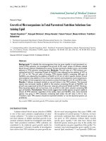
The four panels of Fig. 2 show the equilibrium deoxy and
8 ns CO photoproduct spectra for the m(Fe–L) band of
H93G(Im), H93G(1-MeIm), H93G(2-MeIm), and
H93G(4-MeIm) adducts in 90% glycerol/buffer solution.
The m(Fe–L) frequencies of both the equilibrium deoxy and
the 8 ns photoproduct species are significantly dependent
on the presence of glycerol. In the deoxy species no protein
relaxation occurs, suggesting that glycerol affects the
electrostatic environment of the heme. Increased osmotic
pressure due to glycerol removes a distal water molecule,
inducing a shift in m(Fe–L) typically toward lower fre-
quency. In contrast, the frequency shift in photoproduct
spectra is ascribed to a slowing of the protein relaxation
following photolysis, and thus the frequency is typically
higher in a viscous solvent, such as glycerol, compared with
buffer. A frequency shift of 2.6 cm
)1
in the m(Fe–His) band
of the CO photoproduct for wild-type sperm whale Mb has
been observed in 90% glycerol/buffer relative to buffer [37]
and is in agreement with the data in Table 1. The imidazole
and 4-methyl imidazole deoxy adducts show shifts of 3 and
4cm
)1
, respectively, to lower frequency in 90% glycerol/
buffer solution. The photoproduct spectra for the imidazole
and 4-methyl imidazole adducts show shifts of 0 and
+1 cm
)1
in 90% glycerol. These shifts result in a *CO-
deoxy difference frequency that is both positive in value and
larger for the H93G(Im) and H93G(4-MeIm) adducts in
90% glycerol/buffer solution than in buffer alone, and is
consistent with the data obtained for wild-type Mb.
The deoxy H93G(2-MeIm) species shows an increase of
2cm
)1
in the m(Fe–His) frequency in 90% glycerol, while
the photoproduct of this species shows a decrease of 1 cm
)1
.
Both of these values are in the opposite direction of what is
normally seen for wild-type Mb in 90% glycerol, and thus
the *CO-deoxy difference frequency is decreased to 8 cm
)1
,
a value significantly smaller value than the 11 cm
)1
shift
seen in buffer. The frequencies of both the H93G(2-MeIm)
and H93G(4-MeIm) differ from those reported in the
previous study that used continuous wave laser excitation
[36]. The origin of these differences is not known at present
and may result from laser excitation using 8 ns pulses.
Following photolysis of CO, some imidazole proximal
ligands in H93G dissociate on a time scale much longer than
8 ns. This is certainly the case for H93G-*CO shown in
Fig. 3, as it is known that a ligand switch occurs in H93G-
*CO [32]. Of the ligands used in this study, it is likely that
2-methyl imidazole dissociates the most rapidly given the
steric hindrance between the 2-methyl group of this ligand
and the heme.
Unlike the other ligands, H93G(1-MeIm)*CO shows no
shift in the photoproduct spectrum obtained in 90%
glycerol/buffer or in buffer alone. The relative intensities
of the two bands in the Fermi doublet change as the protein
relaxes from the photoproduct intermediate conformation
to the deoxy state in buffer while in 90% glycerol/buffer
solvent less difference in relaxation is seen. In 90% glycerol
the photoproduct peaks are essentially the same as in buffer,
while the deoxy peaks change intensity such that they more
closely resemble the photoproduct spectra.
Figure 3 shows the spectra obtained for the H93G
protein prepared without exogenous ligand, denoted the
Table 1. Frequencies of m(Fe–L) Raman modes for H93G(L) in buffer and 90% glycerol/buffer solutions. Frequencies of the Raman shift for the
equilibrium deoxy and 8 ns CO photoproduct spectra are given for each adduct. All values are presented in cm
)1
.
Species Buffer deoxy Buffer *CO
Difference
(*CO-deoxy)
Glycerol
deoxy Glycerol *CO
Difference
(*CO-deoxy)
H93G(Im) 225 224 )1 222 224 2
H93G(1-MeIm) 212 + 234 213 + 234 0 213 + 234 213 + 234 0
H93G(2-MeIm) 211 222 +11 213 221 +8
H93G(4-MeIm) 216 215 )1 212 216 +4
Wild type 218.4 219.7 1.3 216 220 +4
320280240200
Raman Shift (cm
-1
)
H93G(4-Me Im)
D
320280240200
Raman Shift (cm
-1
)
H93G(1-Me Im)
C
H93G(2-Me Im)
B
H93G(Im)
8 ns
Deoxy
A
Raman Intensity
Fig. 2. Equilibrium deoxy and 8 ns photo-
product resonance Raman spectra for H93G(L)
Mb with exogenous ligands in 90% glycerol/
buffer solution. (A) L ¼ imidazole; (B) L ¼
2-MeIm; (C) L ¼ 1-MeIm; and (D) L ¼
4-MeIm. The feature at 180 cm
)1
is artifact
from the hydrogen Raman shifter used in the
experiment. The mode at 300 cm
)1
is c
7
[35].
Ó FEBS 2002 Dynamic proximal ligand motions in H93G myoglobin (Eur. J. Biochem. 269) 4881
ligand-free form of H93G. Earlier work shows that the
ground state spectrum of the exogenous ligand-free form
with CO bound (H93G-CO) has Fe–C and CO stretch-
ing frequencies similar to wild-type Mb determined by
resonance Raman and FTIR spectroscopy, respectively [32].
The nomenclature H93G-CO reflects the fact that no
exogenous proximal ligand is added during sample prepar-
ation. However, the exogenous ligand-free adduct of deoxy
H93G has a five-coordinate heme high frequency Raman
spectrum (spin sensitive region, 1300–1650 cm
)1
), in spite of
the fact that there is no evidence of an axial ligand in the
spectral region from 200 to 250 cm
)1
.Them(Fe–L) axial
iron–ligand out-of-plane mode is absent in the H93G
Raman spectrum. The small bands observed at 240 cm
)1
in
the H93G spectrum shown in Fig. 3 are present in all
heme resonance Raman spectra of Mb and have been
assigned to the A
1g
mode m
9
[35]. The 8 ns photoproduct
spectrum shown in Fig. 3 reveals the appearance of a
m(Fe–L) band in H93G-*CO. The photoproduct spectra
obtained for H93G(Im), H93G(1-MeIm), H93G(2-MeIm)
and H93G(4-MeIm) serve as a reference for studies of the
ligand-free H93G protein. The appearance of a band at
220 cm
)1
in the photoproduct spectrum of H93G-*CO is
similar to the average frequency for the wild-type, H93G(4-
MeIm), H93G(2-MeIm) and H93G(Im) photoproduct
spectra in buffer indicating the presence of a nitrogenous
imidazole ligand in H93G-*CO.
Raman spectra for all samples in this study exhibit
essentially identical high frequency modes. For example, the
m
7
band at 672 cm
)1
in the H93G(Im) adducts studied in
buffer and 90% glycerol/buffer show shifts of less than
0.2 cm
)1
. Similar observations have been made for the
electron density marker and core size modes of deoxy and
photoproduct spectra in previous studies [32,36].
DISCUSSION
The photoproduct spectra indicate that there are significant
differences in the dynamics for heme iron ligands in the
proximal cavity during the first 8 ns following CO photo-
lysis. As the proximal ligands are not covalently bound to
the protein, the dynamics can arise from three effects. First,
steric interactions between the some of the ligands (e.g.
2-methyl imidazole) and the heme may occur during the
change of the iron spin state from low spin in H93G(L)CO
to high spin in deoxy H93G(L). Second, steric interactions
of the protein may cause ligands to reorient following
photolysis. For example, the methyl groups of ligands such
as 4-methyl imidazole and 1-methyl imidazole may inter-
act with protein side chains and the methyl group of the
2-methyl imidazole interacts strongly with the heme. Third,
changes in ligation or ligand switching can occur due to
proximal ligand lability, as has been proposed for the
H93G*-CO photoproduct [32]. We consider each of
these effects in photoproducts of H93G(L)*CO where L is
Im, 2-MeIm, 1-MeIm and 4-MeIm. The photoproduct data
on H93G*-CO provides further evidence for the model
presented in a previous study [32] in which a histidine from
the globin is bound in the six-coordinate form of the H93G
mutant when no exogenous proximal ligand is present.
The relative thermodynamic stability of the ligands in the
proximal pocket has been determined [29]. The increased
relative stability of imidazole and 4-methyl imidazole
ligands over the 1-methyl and 2-methyl imidazoles is likely
due to the fact that the first two species are closest in binding
geometry to the wild-type histidine side-chain. It is expected
that Im and 4-MeIm would fit the pocket well and are
stabilized by hydrogen bonds similar to those in wild-type
Mb. However, the X-ray crystal structure for H93G(4-
MeIm) indicates an Np–Fe–Ne–Cd dihedral angle of 49° for
H93G(4-MeIm) as opposed to 38° for H93G(Im) (wild type
has an 10° dihedral angle). Moreover, the imidazole plane
in H93G(4-MeIm) is tilted away from the heme normal by
more than 10°, creating a geometric distortion that leads to
a much shorter hydrogen bond to Ser92 [34]. These data are
significant because the stability of the ligand in the
photoproduct state is key to understanding the Raman
band shifts observed here.
Spectral changes reflect strain and coupling of ligand
and porphyrin modes: H93G(2-MeIm) models
conformational changes in Hb
The change in frequency for the photoproduct spectrum
relative to the equilibrium deoxy spectrum of H93G(2-
MeIm) is surprisingly similar to that for photolyzed
carbonmonoxy Hb (Hb*CO). In Hb*CO, there is good
evidence that strain introduced by changes in protein
conformation is communicated to the heme iron [33]. The
shift of the m(Fe–His) band from 230 cm
)1
in Hb*CO to
213 cm
)1
in deoxy Hb provides key evidence for a
mechanism that involves the communication of strain
introduced at protein subunits to the diatomic binding site
at the iron [33,38,39]. This 17 cm
)1
shift is comparable to
the shift observed in the photoproduct H93G(2-MeIm)
adduct in buffer solution. Adducts of heme model systems
with 2-methyl imidazole have been used a models of the
strain in the deoxy state of Hb (T-state) [40]. The data in
Fig. 1 suggest that H93G(2-MeIm) may be an excellent
example of the effect of strain on the heme iron in a protein
model system.
The origin of proximal histidine strain in Hb involves
more than simply an increase in the histidine–iron bond
Raman Intensity
800700600500400300200
Raman Shift (cm
-1
)
Ligand-free form
8 ns H93G-CO
deoxy H93G
Fig. 3. Equilibrium deoxy and 8 ns photoproduct resonance Raman
spectra for H93G-CO Mb with no exogenous ligand. Thesamplewas
prepared by heme reconstitution into apoMb in 10 m
M
phosphate
buffer [32]. The dashed line is the deoxy H93G sample and the solid
line is the 8 ns photoproduct spectrum.
4882 S. Franzen et al. (Eur. J. Biochem. 269) Ó FEBS 2002
length with a concomitant weakening of the bond. Models
of strain in human Hb also include the tilting of the
imidazole due to translation of the F-helix as it makes
contact with the CD loop region of a neighboring subunit
resulting in the formation of salt bridges and hydrophobic
contacts [41]. Changes in the iron–ligand bond tilt angle
may modify the m(Fe–L) stretching frequency through
anharmonic coupling to low frequency modes [42]. In
analogy with Hb, the large shift in photoproduct m(Fe–L)
frequencies for H93G(2-MeIm)*CO observed in both buffer
and 90% glycerol/buffer (Figs 1 and 2, respectively)
strongly suggests that proximal strain is present in
H93G(2-MeIm)*CO and that this strain arises from a
time-dependent change in Np–Fe–Ne tilt angle as the
2-MeIm ligand relaxes in the proximal pocket.
In wild-type Mb*CO the protein relaxations are smaller
than in Hb*CO. The frequency shift of the m(Fe–His) band
for the Mb photoproduct (1.5 cm
)1
) following photolysis is
very small compared to that for the Hb photoproduct (12–
17 cm
)1
). Figure 1 shows that the frequency shifts for
H93G(Im)*CO and H93G(4-MeIm)*CO photoproduct
spectra are similar in magnitude (i.e. very small) to those
observed in Mb*CO, however, their sign is reversed. The
changes in Mb structure upon photolysis can be divided
conceptually into a distal and proximal component [43]. On
the distal side, the protein structure must accommodate the
photolyzed ligand in a docking site parallel to the heme
plane. On the proximal side, the structural changes in both
Hb and Mb must also allow the heme iron to move out of
the heme plane as it changes from low spin to high spin
following photolysis and in Hb this movement is ultimately
followed by a shifting of the F-helix. These changes in
protein conformation in the proximal cavity of the H93G
mutants can have an effect on the stability and electronic
structure of the bound proximal ligand and thereby are also
reflected in spectroscopic changes in m(Fe–L) that are
sensitive to the heme pocket conformation.
The significance of Soret band shift, m(Fe–L) shifts
and geminate recombination rates
In an earlier study, time-dependent frequency shifts of the
heme deoxy Soret band following photolysis in 90%
glycerol/buffer solution were observed for the species
H93G(Im)CO, H93G(4-MeIm)CO, and H93G(1-MeIm)-
CO as well as wild-type Mb [44]. For these proximal ligands,
the time scale of the Soret band shift at room temperature
was nearly identical (< 10
)6
s), although there was a slight
increase in the rate in the order wild type < 1-methyl
imidazole @ 4-methyl imidazole < imidazole. For this same
set of proximal ligands, the geminate phase of the CO
rebinding was found to increase in the order wild type < 4-
methyl imidazole < imidazole < 1-methyl imidazole and
occurred on a time scale similar to the Soret band shift [30].
As shown in Table 1, in 90% glycerol the difference in the
photoproduct vs. deoxy m(Fe–L) Raman frequencies, CO–
deoxy, for these ligands decreases in the same order that the
previously reported geminate rate increased: wild type
(4 cm
)1
) ¼ 4-methyl imidazole (4 cm
)1
) > imidazole
(2 cm
)1
) > 1-methyl imidazole (0 cm
)1
). From these data
it would appear that the conformational coordinates that
control the Soret shift in H93G(L)*CO are not related to
those that govern the geminate rebinding rate and the shift
in the photoproduct m(Fe–L) frequency from its equilibrium
deoxy position but that the latter two observable pheno-
mena are correlated. These phenomena are consistent with a
separation of contributions from proximal and distal pocket
conformational relaxations that can be understood as
follows. The observed geminate phase decay rate can be
expressed as the sum of two rates for a three state model: the
rebinding rate for the CO from within the pocket and
the escape of the CO to the solvent, k
gem
¼ k
21
+ k
23
,where
the system can be portrayed as follows:
MbCO !
k
12
Mb:CO !
k
23
Mb þ CO
The rebinding rate, k
21
, is a function of the rebinding
barrier, and this is in part controlled by the strain on the
proximal ligand after photolysis as observed in the
photoproduct spectra. The difference in the photoproduct
m(Fe–L) frequency with respect to the deoxy value
correlates well with the amount of geminate rebinding
that occurs and this is interpreted to be due to the fact that
the m(Fe–L) frequency typically decreases due to an
increase in proximal strain in the Fe–L bond, as described
above. On the other hand, the escape rate, k
23
,islargely
controlled by distal pocket conformational changes. The
Soret band shift and the escape rate constant k
23
are not
highly dependent on the identity of the proximal ligand in
H93G(L)*CO [30,44]. However, mutations in the distal
heme pocket result in changes that are distinct from those
of H93G; the Soret band shift, k
23
and k
21
are all affected
concomitantly [16,47].
H93G(1-MeIm) probes hydrogen bonding
in the proximal pocket
The X-ray crystal structure of the metaquo form of
H93G(4-MeIm) and H93G(1-MeIm) show nearly identical
conformations for methyl imidazole in the proximal pocket
[34]. The X-ray structure for H93G(4-MeIm) is consistent
with hydrogen bonding for 4-methyl imidazole with S92
that is stronger than in wild-type Mb. However, no
hydrogen bonding is possible for 1-methyl imidazole
because the Nd position is bonded to a methyl group in
this ligand. Moreover, the pKa is similar for both H93G(4-
MeIm) and H93G(1-MeIm) indicating that differences in
frequency are not due to differences in ligand basicity.
Nonetheless, there is a substantial difference in the
proximal dynamics for these two ligands. It is reasonable
to suggest that the inability of 1-methyl imidazole to form
a hydrogen bond is responsible for the differences in
proximal dynamics [29]. Comparison with other proximal
mutations such as S92A and L89I indicates that hydrogen
bonding does not have a large effect when native histidine
is ligated to the heme iron [45]. However, proximal ligands
are destabilized by the H93G/S92A double mutant so that
no m(Fe–L) frequencies have been obtained for the latter
[29]. Thus, hydrogen bonding likely has a larger effect on
the non-native ligands to heme iron in H93G(L) and
H93G(L)*CO than observed in mutants such as S92A or
L89I that remove hydrogen bonds to Nd-H. For example,
neither viscosity/hydration effects from addition of glycerol
nor photoproduct spectra affect the frequency of the
nonhydrogen-bonding ligand H93G(1-MeIm) shown in
Figs 1 and 2.
Ó FEBS 2002 Dynamic proximal ligand motions in H93G myoglobin (Eur. J. Biochem. 269) 4883
Ligand-free H93G data indicate a ligand switching
mechanism
The covalent attachment of the ligand can be affected by the
change in hydrogen bonding suggesting that ligand lability
(i.e. a ligand switch) may also be a factor in differences in
m(Fe–L) frequency observed in Figs 1 and 2. Evidence for a
ligand switch can be obtained by comparison of the
photoproduct spectra with time-resolved FTIR and satura-
tion Raman experiments on H93G-CO. The ligand switch
does not appear to occur on an ultrafast time scale. In fact, it
appears to be quite slow (> 5 ls) [32]. The six-coordinate
form of H93G-CO appears to have a nitrogenous ligand
bound to the heme iron as shown in Fig. 3. The photo-
product data provide evidence that the ligand trans to CO in
H93G-CO is an endogenous histidine and likely is His97.
The ligand-free H93G*-CO photoproduct spectrum
shown in Fig. 3 has an interesting effect not observed for
the imidazole adducts. The deoxy spectrum shows no axial
m(Fe–L) mode. However, there must be an axial ligand
because the high frequency region of the deoxy spectrum is
consistent with a five-coordinate heme adduct. The buffer
contains only 10 m
M
phosphate and thus the axial ligand in
H93G must be H
2
O, phosphate or an amino acid side chain.
The photoproduct spectrum shows a distinct increase
in intensity at 220 cm
)1
consistent with a change in ligation.
In a previous study we proposed that the axial ligand is
H
2
O in H93G, i.e. H93G(H
2
O), and a histidine residue in
H93G*-CO [i.e. H93G(His)CO]. Most likely, the axial
ligand observed in the CO photoproduct spectrum is also
bound trans to CO in the equilibrium form of H93G-CO.
This ligand is hypothesized to be a histidine due to the
frequency of 220 cm
)1
, which is almost identical to that of
wild-type Mb. Although assignment of the histidine is not
certain, studies of the CO stretching frequency for double
mutants indicate that the distal histidine (His64) probably
does not give rise to the signal observed in Fig. 3 [46]. His97
is immediately adjacent to His93 on the proximal side and
we are currently investigating whether it is the side chain
that ligates the heme iron trans to CO in H93G-CO.
The data in Fig. 3 indicate that the axial ligand of the
heme iron is not the same in the H93G-CO and deoxy
H93G species. This is in agreement with step-scan FTIR
and saturation Raman data that indicate a dynamic ligand
switch in H93G Mb [32]. The histidine that appears bound
in the 8 ns photoproduct spectrum dissociates from the iron
on a time scale < 5 ls. CO recombines to give rise to a
transient H93G(H
2
O)CO species. This species then returns
to the equilibrium form, H93G(His)CO, on the millisecond
time scale. If His97 is ligated to the heme iron, it must be
sufficiently unstable that it is replaced by H
2
O in the deoxy
form. Substantial protein strain is required to permit His97
ligation. Furthermore, it is nearly impossible for His97 to
hydrogen bond in a manner analogous to His93. Thus, the
dynamic change in ligation is driven by conformational
strain the H93G(His)CO protein.
CONCLUSION
The 8 ns CO photoproduct spectra of the m(Fe–L) band in
H93G Mb reveal an important role for steric interactions
and hydrogen bonding in the proximal pocket. The shifts in
the m(Fe–L) photoproduct spectra of H93G(Im)*CO,
H93G(4-MeIm)*CO and H93G(1-MeIm)*CO are small
or absent, while the shift is relatively large for H93G(2-
MeIm). The Raman data further confirm the hypothesis
that the ligand-free form is a H93G(His)CO adduct that is
strained. In this adduct, the histidine dissociates within 5 ls
after CO photolysis but is clearly bound 8 ns after
photolysis as shown in Fig. 3. There is no evidence for a
four-coordinate intermediate in heme dissociations of this
type and it is likely that the off-rate of the ligand is slow
because of the requirement for water to enter the proximal
cavity to serve as a replacement for the nitrogenous ligand.
Thus, the proximal ligand in H93G(L)*CO is destabilized
leading to dissociation or altered frequencies due to ligand
strain. The stability of the proximal ligand is also modulated
by the strength of Nd-H hydrogen bond.
Although the relaxation in the proximal pocket shown
here does not affect the distal pocket relaxation probed by
the Soret band shift or band III shift, it may affect CO
rebinding kinetics [44]. For example, although H93G(1-
MeIm) and H93G(4-MeIm) are isostructural and have
similar basicity, their CO rebinding kinetics are quite
different [30]. H93G(1-MeIm) has a geminate recombina-
tion rate constant nearly one order of magnitude larger
than that of H93G(4-MeIm) in a 90% glycerol/buffer glass
[44]. Both ligand strain and proximal ligand dissociation
can lead to rapid CO rebinding kinetics. Thus, the ligands
that cannot hydrogen bond and fit poorly in the proximal
pocket are expected to have more rapid geminate CO
recombination rate constants. The data presented here
show the utility of the H93G mutant for separating
proximal and distal effects in Mb. This is a key step
toward making definitive assignments of spectroscopic
shifts in terms of globin structure. The m(Fe–L) band shifts
measured in this study further support the model advanced
earlier that protein relaxation monitored by band III and
the Soret band shifts represents motions of amino acid
residues in the distal pocket in response to CO photolysis.
The distal relaxation is distinct from the proximal effects
observed here that previously have been demonstrated to
have a large effect on the kinetics of geminate CO
recombination.
ACKNOWLEDGMENTS
SF acknowledges support by the NSF (MCB)9874895); JMF
acknowledges support from NIH (R01 HL58247, RO1 G58890) and
the W.M. Keck Foundation; and SGB acknowledges support from
NIH (GM27738).
REFERENCES
1. Rousseau, D.L. & Friedman, J.M. (1988) In: Biological Applica-
tions of Raman Spectroscopy, Vol. III (Spiro, T.G., ed), pp. 133–
215. Wiley & Sons, New York.
2. Kitagawa, T. (1988) Biological applications of raman spectros-
copy. In Biological Applications of Raman Spectroscopy,Vol.III
(Spiro, T.G., ed), pp. 97–131. Wiley & Sons, New York.
3. Jayaraman, V., Rodgers, K.R., Mukerji, I. & Spiro, T.G. (1995)
Hemoglobin allostery: resonance Raman spectroscopy of kinetic
intermediates. Science 269, 1843–1848.
4. Barrick, D. (2000) Trans-substitution of the proximal hydrogen
bond in myoglobin. II. Energetics, functional consequences, and
implications for hemoglobin allostery. Proteins Struct. Func.
Genet. 39, 291–308.
4884 S. Franzen et al. (Eur. J. Biochem. 269) Ó FEBS 2002
5. Scott, T.W. & Friedman, J.M. (1984) Tertiary-structure relaxation
in hemoglobin: a transient Raman study. J. Am. Chem. Soc. 106,
5677–5687.
6. Petersen,E.,Chien,E.,Sligar,S.&Friedman,J.(1998)Functional
implications of the proximal hydrogen-bonding network in
myoglobin. A resonance Raman and kinetic study of Leu89,
Ser92, His97 and F-helix swap mutants. Biochemistry 37, 12301–
12319.
7. Jackson, T.A., Lim, M. & Anfinrud, P.A. (1994) Complex non-
exponential relaxation in myoglobin after photodissociation of
MbCO: measurement and analysis from 2 ps to 56 ls. Chem. Phys.
180, 131–140.
8. Gilch, H., Schweitzer-Stenner, R., Dreybrodt, W., Leone, M.,
Cupane, A. & Cordone, L. (1996) Conformational substates of the
Fe
2+
–His F8 linkage in deoxymyoglobin and hemoglobin probed
in parallel by the Raman band of the Fe–His stretching vibration
and the near infrared absorption band III. Int. J. Quantum Chem.
59, 301–313.
9. Chavez, M.D., Courtney, S.H., Chance, M.R., Kuila, D., Nocek,
J., Hoffman, B.M., Friedman, J.M. & Ondrias, M.R. (1990)
Structural and functional significance of inhomogeneous line
broadening of band III in hemoglobin and Fe–Mn hybrid
hemoglobins. Biochemistry 29, 4844–4852.
10. Kiger, L., Stetzkowski-Marden, F., Poyart, C. & Marden, M.
(1995) Correlation of carbon monoxide association rates and
position of absorption band III in hemeproteins. Eur. J. Biochem.
228, 665–668.
11. Schlichting, I., Berendzen, J., Phillips, G.N. Jr & Sweet, R.M.
(1994) Crystal structure of an intermediate of CO binding to
myoglobin. Nature 371, 808–812.
12. Srajer, V., Teng, T.Y., Ursby, T., Pradervand, C., Ren, Z.,
Adachi, S., Schildkamp, W., Bourgeois, D., Wulff, M. & Moffat,
K. (1996) Photolysis of the carbon monoxide complex of myo-
globin: nanosecond time resolved crystallography. Science 274,
1726–1729.
13. Teng, T Y., Srajer, V. & Moffat, K. (1994) Photolysis-induced
structural changes in single crystals of carbonmonoxy myoglobin
at 40 K. Nat. Struct. Biol. 1, 701–705.
14. Hartmann, H., Zinser, S., Komninos, P., Schneider, R.T.,
Nienhaus, G.U. & Parak, F. (1996) X-ray structure determination
of a metastable state of carbonmonoxy myoglobin after photo-
dissociation. Proc.NatlAcad.Sci.U.S.A.93, 7013–7016.
15. Lim, M.H., Jackson, T.A. & Anfinrud, P.A. (1997) Ultrafast
rotation and trapping of carbon monoxide dissociated from
myoglobin. Nat. Struct. Biol. 4, 209–214.
16. Lambright, D.G., Balasubramanian, S. & Boxer, S.G. (1993)
Dynamics of protein relaxation in site-specific mutants of human
myoglobin. Biochemistry 32, 10116–10124.
17. Ansari, A., Jones, C.M., Henry, E.R., Hofrichter, J. & Eaton,
W.A. (1992) Conformational relaxation and ligand rebinding in
myoglobin. Science 256, 1796–1798.
18. Steinbach, P.J., Ansari, A., Berendzen, J., Braunstein, D., Chu,
K., Cowen, B.R., Ehrenstein, D., Frauenfelder, H., Johnson, J.B.,
Lamb, D.C., Luck, S., Mourant, J.R., Nienhaus, G.U., Ormos, P.,
Philipp, R., Xie, A. & Young, R.D. (1991) Ligand binding to heme
proteins: connection between dynamics and function. Biochem-
istry 30, 3988–4001.
19. Tian,W.D.,Sage,J.T.,Champion,P.M.,Chien,E.&Sligar,S.G.
(1996) Probing heme protein conformational equilibration rates
with kinetic selection. Biochemistry 35, 3487–3502.
20. Ostermann,A.,Waschipky,R.,Parak,F.G.&Nienhaus,G.U.
(2000) Ligand binding and conformational motions in myoglobin.
Nature 404, 205–208.
21. Goodin, D.B. & McRee, D.E. (1993) The Asp-His-Fe triad of
cytochrome c peroxidase controls the reduction potential, elec-
tronic structure, and coupling of the tryptophan free radical to the
heme. Biochemistry 32, 3313–3324.
22. Smulevich, G., Hu, S.Z., Rodgers, K.R., Goodin, D.B., Smith,
K.M. & Spiro, T.G. (1996) Heme-protein interactions in cyto-
chrome c peroxidase revealed by site-directed mutagenesis and
resonance Raman spectra of isotopically labeled hemes. Biospec-
troscopy 2, 365–376.
23. Sun, J., Fitzgerald, M.M., Goodin, D.B. & Loehr, T.M. (1997)
Solution and crystal structures of the H175G mutant of
cytochrome c peroxidase: a resonance Raman study. J. Am. Chem.
Soc. 119, 2064–2065.
24. Vogel, K.M., Spiro, T.G., Shelver, D., Thorsteinsson, M.V. &
Roberts, G.P. (1999) Resonance Raman evidence for a novel
charge relay activation mechanism of the CO-dependent heme
protein transcription factor CooA. Biochemistry 38, 2679–2687.
25. Callahan, P.M. & Babcock, G.T. (1981) Insights into heme
structure from soret excitation Raman spectroscopy. Biochemistry
20, 952–958.
26. Schelvis, J.P.M., Kim, S.Y., Zhao, Y.D., Marletta, M.A. &
Babcock, G.T. (1999) Structural dynamics in the guanylate cyclase
heme pocket after CO photolysis. J. Am. Chem. Soc. 121, 7397–
7400.
27. Frauenfelder, H., McMahon, B.H., Austin, R.H., Chu, K. &
Groves, J.T. (2001) The role of structure, energy landscape,
dynamics, and allostery in the enzymatic function of myoglobin.
Proc. Natl Acad. Sci. USA 98, 2370–2374.
28. Kuriyan, J., Wilz, S., Karplus, M. & Petsko, G.A. (1986) J. Mol.
Biol. 192, 133–154.
29. Decatur, S.M., Belcher, K.L., Rickert, P.K., Franzen, S. & Boxer,
S.G. (1999) Hydrogen bonding modulates binding of exogenous
ligands in a myoglobin proximal cavity mutant. Biochemistry 38,
11086–11092.
30. Franzen, S. (2002) Carbonmonoxy rebinding kinetics in h93g
myoglobin: separation of proximal and distal side effects. J. Phys.
Chem. 106, 4533–4542.
31. DePillis, G., Decatur, S.M., Barrick, D. & Boxer, S.G. (1994)
Functional cavities in proteins – a general method for proximal
ligand subsitution in myoglobin. J. Am. Chem. Soc. 116, 6981–
6982.
32. Franzen, S., Bailey, J., Dyer, R.B., Woodruff, W.H., Hu, R.B.,
Thomas, M.R. & Boxer, S.G. (2001) A photolysis-triggered heme
ligand switch in H93G myoglobin. Biochemistry 40, 5299–5305.
33. Petersen, E.S. & Friedman, J.M. (1998) A possible allosteric
communication pathway identified through a resonance Raman
study of four beta 37 mutants of human hemoglobin A. Bio-
chemistry 37, 4346–4357.
34. Barrick, D. & Dahlquist, F.W. (2000) Trans-substitution of the
proximal hydrogen bond in myoglobin. I. Structural consequences
of hydrogen bond deletion. Proteins Struct. Func. Genet. 39, 278–
290.
35. Hu, S., Smith, K.M. & Spiro, T.G. (1996) Assignment of proto-
heme resonance Raman spectrum by heme labeling in myoglobin.
J. Am. Chem. Soc. 118, 12638–12646.
37. Sage, J.T., Schomacker, K.T. & Champion, P.M. (1995) Solvent-
dependent structure and dynamics in myoglobin. J. Phys. Chem.
99, 3394–3405.
36. Franzen, S., Boxer, S.G., Dyer, R.B. & Woodruff, W.H. (2000)
Resonance Raman studies of heme-axial ligation in H93G myo-
globin. J. Phys. Chem. B 104, 10359–10367.
38. Matsukawa,S.,Mawatari,K.,Yoneyama,Y.&Kitagawa,T.
(1985) Correlation between the iron-histidine stretching fre-
quencies and oxygen affinity of hemoglobins. A continuous strain
model. J. Am. Chem. Soc. 107, 1108–1113.
39. Friedman, J.M., Scott, T.W., Stepnowski, R.A., Ikeda-Saito, M.
& Yonetani, T. (1983) The iron-proximal histidine linkage and
protein control of oxygen binding in hemoglobin. J. Biol. Chem.
258, 10564–10572.
40. Nagai, K., Kitagawa, T. & Morimoto, H. (1980) Quaternary
structures and low frequency molecular vibrations of haems of
Ó FEBS 2002 Dynamic proximal ligand motions in H93G myoglobin (Eur. J. Biochem. 269) 4885
deoxy and oxyhaemoglobin studied by resonance Raman scat-
tering. J. Mol. Biol. 136, 271–289.
41. Franzen, S., Lambry, J.C., Bohn, B., Poyart, C. & Martin, J.L.
(1994) Direct evidence for heme-iron doming as the primary event
in the quaternary structure change of hemoglobin. Nat. Struct.
Biol. 1, 230–233.
42. Rosenfeld, Y.B. & Stavrov, S.S. (1994) Anharmonic coupling of
soft modes and its influence on the shape of the iron-histidine
resonance Raman band of heme proteins. Chem. Phys. Lett. 229,
457–464.
43. Franzen, S., Bohn, B., Poyart, C. & Martin, J.L. (1995) Evidence
for sub-picosecond heme doming in hemoglobin and myoglobin.
A time-resolved resonance Raman comparison of carbonmonoxy
and deoxy species. Biochemistry 34, 1224–1237.
44. Franzen, S. & Boxer, S.G. (1997) On the origin of heme absorp-
tion band shifts and associated protein structural relaxation in
myoglobin following flash photolysis. J. Biol. Chem. 272, 9655–
9660.
45. Peterson, E., Chien, E., Sligar, S. & Friedman, J. (1998) Functional
implications of the proximal hydrogen-bonding network in myo-
globin. A resonance Raman and kinetic study of Leu89, Ser92,
His97 and F-helix swap mutants. Biochemistry 37, 12301–12319.
46. Hu, R.B. (1999) Structure-Function Relationship in Myoglobins
with Distal and Proximal Mutation,PhDThesis,Stanford
University, Stanford, CA, USA.
47. Nienhaus,K.,Lamb,D.C.,Deng,P.&Nienhaus,G.U.(2002)
The effect of ligand dynamics on heme electron transition band III
in myoglobin. Biophys. J. 82, 1059–1067.
4886 S. Franzen et al. (Eur. J. Biochem. 269) Ó FEBS 2002