Báo cáo Y học: Novel fish hypothalamic neuropeptide Cloning of a cDNA encoding the precursor polypeptide and identification and localization of the mature peptide pptx
Bạn đang xem bản rút gọn của tài liệu. Xem và tải ngay bản đầy đủ của tài liệu tại đây (364.63 KB, 9 trang )
Novel fish hypothalamic neuropeptide
Cloning of a cDNA encoding the precursor polypeptide and identification
and localization of the mature peptide
Kaori Sawada
1,2
, Kazuyoshi Ukena
1,2
, Honoo Satake
3
, Eiko Iwakoshi
3
, Hiroyuki Minakata
3
and Kazuyoshi Tsutsui
1,2
1
Laboratory of Brain Science, Faculty of Integrated Arts and Sciences, Hiroshima University, Higashi-Hiroshima, Japan,
2
Core Research of Evolutional Science and Technology (CREST), Japan Science and Technology Corporation, Tokyo, Japan and
3
Suntory Institute for Bioorganic Research, Wakayamadai 1-1-1, Shimamoto-cho, Mishima-gun, Osaka, Japan
Recently, we identified novel avian and amphibian
hypothalamic neuropeptides that inhibited gonadotropin
release and stimulated growth hormone release. They were
characterized by a similar structure including the C-terminal
LPLRF-NH
2
motif. To clarify that the expression of these
novel hypothalamic neuropeptides is a conserved property in
vertebrates, we characterized a cDNA encoding a similar
novel peptide, having LPLRF-NH
2
from the goldfish brain,
by a combination of 3¢ and 5¢ rapid amplification of cDNA
ends (RACE). The deduced peptide precursor consisted of
197 amino acid residues, encoding three putative peptide
sequences that included -LPXRF (where X is L or Q) at their
C-termini. Mass spectrometric analyses revealed that a tri-
decapeptide (SGTGLSATLPQRF-NH
2
) was derived from
the precursor in the brain as an endogenous ligand. Southern
blotting analysis of reverse-transcriptase-mediated PCR
products demonstrated a specific expression of the goldfish
peptide gene in the diencephalon. In situ hybridization
revealed the cellular localization of goldfish peptide mRNA
in the nucleus posterioris periventricularis in the hypotha-
lamus. Immunoreactive cell bodies were also restricted to the
the nucleus posterioris periventricularis and the nervus ter-
minalis and immunoreactive fibers were distributed in sev-
eral brain regions including the nucleus lateralis tuberis pars
posterioris and pituitary. Thus, the goldfish hypothalamus
expresses a novel neuropeptide containing the C-terminal
-LPQRF-NH
2
sequence, which may possess multiple regu-
latory functions and act, at least partly, on the pituitary to
regulate pituitary hormone release.
Keywords: cDNA cloning; goldfish; hypothalamic neuro-
peptide; in situ hybridization; mass spectrometry.
Since the molluscan neuropeptide Phe-Met-Arg-Phe-NH
2
(FMRFamide) was found in the ganglia of the venus clam
[1], immunohistochemical studies using the antiserum
against FMRFamide suggested that the vertebrate hypo-
thalamus possesses some unknown neuropeptide similar to
FMRFamide. In fact, neuropeptides having the RFamide
motif at their C-termini (RFamide peptides) have been
identified in the brains of several vertebrates. For the first
time Leu-Pro-Leu-Arg-Phe-NH
2
(LPLRFamide), a chicken
pentapeptide, has been purified from the vertebrate brain
[2]. Two pain modulatory neuropeptides, NPFF and NPAF
[3], prolactin-releasing peptide (PrRP) [4] and its fish
counterpart, Carassius RFamide, [5] are also RFamide
peptides. To date, these RFamide peptides have been shown
to have important physiological roles in neuroendocrine,
behavioral, sensory and autonomic functions [6–8].
We have also identified a novel hypothalamic RFamide
peptide (SIKPSAYLPLRF-NH
2
) inhibiting gonadotropin
release in the quail brain and termed this dodecapeptide
gonadotropin-inhibitory hormone (GnIH) [9]. Subse-
quently, we have cloned a cDNA encoding GnIH from
the quail brain [10]. Interestingly, the GnIH cDNA encoded
GnIH and its related peptides (GnIH-RP-1 and GnIH-
RP-2), which contained a C-terminal -LPXRF-NH
2
(X is L
or Q) sequence [10]. The chicken pentapeptide LPLRF-
amide [2] may be a fragment of GnIH [9]. Recently, we
have further isolated an amphibian dodecapeptide
(SLKPAANLPLRF-NH
2
) that is closely related to GnIH
from the bullfrog hypothalamus [11]. This peptide possessed
growth hormone (GH)-releasing activity and was designa-
ted as frog GH-releasing peptide (fGRP) [11]. In addition, a
gene database search [12] has determined cDNAs encoding
Correspondence to K. Tsutsui, Laboratory of Brain Science,
Faculty of Integrated Arts and Sciences, Hiroshima University,
Higashi-Hiroshima 739–8521, Japan,
Fax: +81 824-24-0759, Tel.: +81 824-24-6571,
E-mail:
Abbreviations: DIG, digoxigenin; fGRP, frog growth hormone-
releasing peptide; GnIH, gonadotropin-inhibitory hormone;
GnIH-RP, GnIH gene-related peptide; LPXRFamide, a peptide
containing a C-terminal -LPXRF-NH
2
(X is Leu or Gln); nano
ESI-TOF-MS, nanoflow electrospray ionization time-of-flight MS;
NLTp, nucleus lateralis tuberis pars posterioris; NPPv, nucleus pos-
terioris periventricularis; NT, nervus terminalis; PC, precursor con-
vertase; PrRP, prolactin-releasing peptide; PVO, paraventricular
organ; RACE, rapid amplification of cDNA ends; RFamide peptide, a
peptide containing a C-terminal Arg-Phe-NH
2
;RFRP,RFamide-
related peptide; RT-PCR, reverse-transcriptase-mediated PCR; VT,
ventral telencephalon; OTec, optic tectum.
Note: The nucleotide sequence data are available in the DDBJ, EMBL
and GenBank Nucleotide Sequence Databases under the accession
number AB078976.
(Received 13 May 2002, revised 16 July 2002, accepted 24 July 2002)
Eur. J. Biochem. 269, 6000–6008 (2002) Ó FEBS 2002 doi:10.1046/j.1432-1033.2002.03351.x
novel RFamide-related peptides (RFRPs) similar to GnIH
and fGRP in mammalian brains and the deduced RFRPs
have been suggested to participate in neuroendocrine [12]
and pain mechanisms [13] in the rat. Two peptides have
been predicted to be encoded in the cDNA of rodent
RFRPs. More recently, we have identified an octadecapep-
tide (ANMEAGTMSHFPSLPQRF-NH
2
) as one of rodent
RFRPs [14]. In addition, a pentatriacontapeptide (SLTFEE
VKDWAPKIKMNKPVVNKMPPSAANLPLRF-NH
2
)
has been isolated as one of the bovine RFRPs [15].
Collectively, these peptides identified from the brain of
several vertebrates are characterized by the LPXRF-NH
2
motif at their C-termini.
The presence of novel neuropeptides featuring the
C-terminal -LPXRF-NH
2
sequence (LPXRFamide pep-
tides) may be a conserved property of vertebrate brains, in
particular the hypothalamus. In view of the immunohisto-
chemical finding indicating that some of FMRFamide-like
immunoreactive neurons project to an area close to or
within the pituitary of fish [16–18], we looked for novel
LPXRFamide peptides from the goldfish brain. In the
present study, a cDNA encoding the LPXRFamide peptide
was characterized and subsequently a mature endogenous
peptide was identified in the goldfish brain. The localization
of its transcript in the goldfish brain was further investi-
gated.
MATERIALS AND METHODS
RNA preparation
Adults goldfish (Carassius auratus)werekeptinordinary
water aquariums at 20 ± 2 °C and used in the present
study. The experimental protocol was approved in accord-
ance with the Guide for the Care and Use of Laboratory
Animals prepared by Hiroshima University, Japan. Total
RNA of the diencephalon was extracted by the guanidium
thiocyanate/phenol/chloroform extraction method followed
by the isolation of poly(A)
+
RNA with Oligotex-(dT) 30
(Daiichikagaku, Tokyo, Japan) in accordance with the
manufacturer’s instructions.
Determination of the cDNA 3¢-end sequence
All PCR amplifications were carried out in a reaction
mixture containing Taq polymerase [Ex Taq polymerase
(Takara Shuzo, Kyoto, Japan) or gene Taq polymerase
(NIPPON GENE, Tokyo, Japan)] and 0.2 m
M
dNTP on a
thermal cycler (Program Temp Control System PC-700,
Astec, Fukuoka, Japan). First-strand cDNA was synthes-
ized with the oligo(dT)-anchor primer supplied in the 5¢/3¢
rapid amplification of cDNA ends (RACE) kit (Roche
Diagnostics, Basal, Switzerland) and amplified with the
anchor primer (Roche Diagnostics) and the first degenerate
primers 5¢-(T/C)TIAA(A/G)CCIGCIGCIAA(T/C)(T/C)
TICC-3¢ (I represents inosine), corresponding to the fGRP
sequence sequence, Leu2-Lys3-Pro4-Ala5-Ala6-Asn7-Leu8-
Pro9 [11]. First-round PCR products were reamplified with
the anchor primer and the second degenerate primers
5¢-GCIAA(T/C)(T/C)TICCI(T/C)TI(A/C)GITT(T/C)GG-3¢,
corresponding to the fGRP Ala6-Asn7-Leu8-Pro9-Leu10-
Arg11-Phe12-Gly13 [11]. Both first- and second-round
PCRs consisted of five cycles for 30 s at 94 °C, 30 s at
45 °C and 2.5 min at 72 °C, and of 30 cycles for 30 s at
94 °C, 30 s at 50 °C and 2.5 min at 72 °C(5.5minforlast
cycle). The second-round PCR products were subcloned
into a TA-cloning vector in accordance with the manufac-
turer’s instructions (Invitrogen, San Diego, CA, USA). The
DNA inserts of the positive clones were amplified by PCR
with universal M13 primers.
Determination of the cDNA 5¢-end sequence
Template cDNA was synthesized with an oligonucleotide
primer complementary to nucleotides 444–463 (5¢-GGTCT
AAAGGAAATATGTTC-3¢), followed by dA-tailing of
the cDNA with dATP and terminal transferase (Roche
Diagnostics). The tailed cDNA was amplified with the
oligo(dT)-anchor primer (Roche Diagnostics) and gene-
specific primer 1 (5¢-TATGTTCCTCCTCCCAAACC-3¢,
complementary to neucleotides 431–450); this was followed
by further amplification of the first-round PCR products
with the anchor primer and gene-specific primer 2 (5¢-AAA
CCTTTGCGGTAGGGTGG-3¢, complementary to nucle-
otides 416–435). Both first-round and second-round PCRs
were performed for 35 cycles for 30 s at 94 °C, 30 s at 55 °C
and 1 min at 72 °C (11 min for last cycle). The second-
round PCR products were subcloned and the inserts were
amplified as described above.
DNA sequencing
All nucleotide sequences were determined with an ABI
PRISM
TM
Dye terminator cycle sequencing ready reaction
kit (PE-Biosystems, Foster, CA, USA) and a model 373A
automated DNA sequencer (PE-Biosystems), then analysed
using
DNASIS
-
MAC
software (Hitachi Software Engineering,
Kanagawa, Japan). Universal M13 primers or gene-specific
primers were used to sequence both strands.
Northern blot hybridization
A digoxigenin (DIG)-labelled precursor polypeptide cDNA
(complementary to nucleotides 6–623, including all open
reading frames) was synthesized with a DNA-labelling kit
(Roche Diagnostics) and used as a probe for Northern blot
analysis. mRNA was separated on a denaturing 1% (w/v)
agarose/formaldehyde gel and fixed on a Hybond N
+
membrane (Amersham Pharmacia Life Science, Uppsala,
Sweden) by UV irradiation. Hybridization and detection
were performed in accordance with the manufacturer’s
standard procedure (Roche Diagnostics). RNA size was
estimated with the use of DIG-labelled RNA molecular
markers (Roche Diagnostics).
Immunoaffinity purification and mass spectrometry (MS)
To identify endogenous mature peptides in the brain, we
carried out affinity purification and immunoassay with the
antiserum raised against fGRP, which cross-reacted with
three putative peptides [goldfish LPXRFamide peptide-1,
-2 and -3 (see Results and Fig. 1)]. Brains (n ¼ 200) were
boiled for 7 min and homogenized in 5% acetic acid as
described previously [9,11,14]. The homogenate was cen-
trifuged at 15 000 g for 20 min at 4 °C and the superna-
tant was collected. After precipitation with 75% acetone,
Ó FEBS 2002 Novel fish hypothalamic neuropeptide (Eur. J. Biochem. 269) 6001
the supernatant was passed through a disposable C-18
cartridge column (Mega Bond-Elut; Varian, Harbor, CA,
USA) and the retained material eluted with 60% methanol
was loaded onto an immunoaffinity column. The affinity
chromatography was carried out as described elsewhere
[3,14]. The antibodies against fGRP were conjugated to
CNBr-activated Sepharose 4B as an affinity ligand. The
brain extract was applied to the immunoaffinity column at
4 °C and the adsorbed materials were eluted with 0.3
M
acetic acid containing 0.1% 2-mercaptoethanol. The eluted
fractions were concentrated and subjected to a reversed-
phase HPLC column (ODS-80TM; Tosoh, Tokyo, Japan)
with a linear gradient of 16–36% acetonitrile containing
0.1% trifluoroacetic acid for 100 min at a flow rate of
0.5 mLÆmin
)1
. The isolated immunoreactive substances
(1 mL each) were then subjected to MS analyses as
described below.
After evaporation of the isolated material, the residue was
dissolved in 50% methanol containing 0.1% formic acid
and the molecular mass was analysed by a nanoflow
electrospray ionization time-of-flight MS (nano ESI-TOF-
MS) (Q-TOF, Micromass, Wythenshawe, UK) as described
previously [10,14,19]. The expected mass value of each
deduced peptide was calculated using the protein prospector
program (
UCSF
) and a corresponding peak was further
examined in a tandem MS analysis. The needle voltage was
optimized at 1000 V and the cone voltage was set at 50 V.
Argon was used as the collision gas and the energy was set at
28 V.
Southern blot hybridization of RT-PCR products
The first-strand cDNA was synthesized from total RNA
(1 lg) prepared from each brain region with M-MLV
reverse transcriptase (Promega, Madison, USA) and an
oligo(dT) primer in accordance with the manufacturer’s
instruction (Promega). The oligonucleotide primer set used
for the amplification of peptide cDNA fragments was
5¢-CACCATCCTGCGACTTCAC-3¢ (identical with
nucleotides 234–252) and 5¢-GGTCTAAAGGAAATAT
GTTC-3¢ (complementary to nucleotides 444–463); primers
for the amplification of b-actin cDNA fragments were
5¢-CTACAACGAGCTGCGTGTTG-3¢ (identical with
nucleotides 296–315 in the goldfish b-actin gene, gb
AB039726) and 5¢-TGCCAATGGTGATGACCTGC-3¢
(complementary to nucleotides 761–780 in the goldfish
b-actin gene). PCR was performed for 30 cycles consisting
of 1 min at 94 °C, 1 min at 55 °C and 1 min at 72 °Cinthe
PCR reaction as described above. PCR products were
resolved on a 1.5% (w/v) agarose gel followed by transfer to
a Hybond N
+
membrane (Amersham Pharmacia Life
Science). The membrane was hybridized with DIG-labelled
oligonucleotide probe (5¢-AAACCTTTGCGGTAGGG
TGG-3¢, complementary to nucleotides 416–435). DIG-
DNA labelling and detection were performed in accordance
with the DIG system protocol (Roche Diagnostics).
In situ
hybridization
In the present study, the site of the peptide mRNA
expression in the brain was further localized by in situ
hybridization. Adult goldfish were killed by decapitation
and brains were immediately immersion-fixed in 4%
paraformaldehyde in phosphate-buffered saline (NaCl/P
i
;
pH 7.3) overnight at 4 °C. Subsequently, brain tissues were
placed in refrigerated 30% sucrose in NaCl/P
i
until they
settled. Sections (10-lm thick) of the brain were made using
acryostatat)20 °C and were placed onto 3-amino-
propyltriethoxysilane-coated slides. In situ hybridization
was carried out according to our previous method [20,21]
using the DIG-labelled antisense RNA probe. The DIG-
labelled antisense RNA probe was produced with RNA
labelling kit (Roche Diagnostics) from a part of the peptide
precursor cDNA (complementary to nucleotides 6–623,
including all open reading frames). Control for specificity of
the in situ hybridization of the peptide mRNA was
performed using the DIG-labelled sense RNA probe, which
is complementary to a sequence of antisense probe.
Competitive enzyme-linked immunosorbent assay (ELISA)
Because the deduced peptide precursor encoded three
putative LPXRFamide peptides [goldfish LPXRFamide
peptide-1, -2 and -3 (see Results and Fig. 1)], we synthesized
these three peptides. In this study, peptide levels in the brain
were quantified by a competitive ELISA using the anti-
fGRP serum [11]. This anti-fGRP serum was confirmed to
recognize specifically three putative goldfish LPXRFamide
peptides, as well as fGRP, by a competitive ELISA. The
IC
50
values (concentrations yielding 50% displacement) in
the competitive ELISA were estimated as follows;
0.46 pmol for goldfish LPXRFamide peptide-1, 3.43 pmol
for goldfish LPXRFamide peptide-2, 1.13 pmol for goldfish
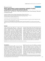
Fig. 1. Nucleotide sequence and deduced amino acid sequence of the
goldfish LPXRFamide peptide precursor cDNA. The sequences of
putative goldfish LPXRFamide peptides are boxed. Single or pairs of
basic amino acids as cleavage sites are shown in bold. The poly(A)
adenylation signal AGTAAA is underlined.
6002 K. Sawada et al. (Eur. J. Biochem. 269) Ó FEBS 2002
LPXRFamide peptide-3, 0.74 pmol for fGRP, 20.96 pmol
for chicken RFamide (LPLRFamide) and more than
1000 pmol for other RFamide peptides, e.g. Carassius
RFamide (SPEIDPFWYVGRGVRPIGRFamide) and
molluscan RFamide (FMRFamide).
Acetic acid extracts derived from different brain regions
of the adult goldfish were passed through disposable C-18
cartridges (Sep-pak; Waters, Milford, MA, USA) and the
retained material was subjected to the competitive ELISA as
described previously [9,22,23]. In brief, different concentra-
tions of the standard peptide, fGRP (0.01–100 pmolÆmL
)1
),
or adjusted tissue extracts were added together with the
antiserum (1 : 1000 dilution) to each well of a 96-well
microplate and incubated for 1 h at 37 °C. After the
reaction with alkaline phosphatase-labelled goat anti-rabbit
IgG, immunoreactive products were obtained in substrate
solution of p-nitrophenyl-phosphate and the absorbance
was measured at 415 nm on a microtiter plate reader (MTP-
120, CORONA, Ibaragi, Japan).
Immunohistochemistry
Immunohistochemical analysis was performed as described
previously [9,11,22,23]. In brief, adult goldfish were killed by
decapitation, and brains were fixed as described above.
Brains were transversely or sagittally frozen-sectioned at
10 lm thickness on a cryostat at )20 °C. After blocking
of nonspecific binding components, the sections were
immersed with the anti-fGRP serum at a dilution of
1 : 1000 overnight at 4 °C and subsequently with rhodam-
ine-conjugated goat antirabbit IgG. The specificity of the
staining was assessed by a substitution of the control serum
for the antiserum; in this control serum, the antiserum
(1 : 1000 dilution) was preabsorbed independently by
incubation overnight with the identified peptide, goldfish
LPXRFamide peptide-3, at a saturating concentration
(10 lgÆmL
)1
). Immunoreactive cell bodies and fibers in the
goldfish brain were studied using a Nikon fluorescence
microscope. The structures of goldfish brain were identified
according to the goldfish brain atlas [24].
RESULTS
Characterization of a cDNA encoding the novel
goldfish LPXRFamide peptide precursor
To obtain novel LPXRFamide peptide precursor cDNA
fragments from the goldfish diencephalon, we initially per-
formed an RT-PCR experiment with degenerate primers
corresponding to the partial fGRP sequence and the anchor
primer, followed by reamplification of the first-round PCR
products with degenerate primers corresponding to the other
partial fGRP sequence and the same anchor primer. Here,
the C-terminal amide group was thought to be derived from
a C-terminal Gly residue, which is known to be a typical
amidation signal [25,26]. Electrophoresis of the nested PCR
mixture revealed a major product of 0.5 kb (results not
shown). The predicted amino acid sequence included two
copies of the potential peptide sequence, LPQRFG, down-
stream of the partial fGRP sequence derived from the
second-round PCR primer, implying that this cDNA clone
encoded also a peptide including a C-terminal sequence
similartothatoffGRP(LPLRF-NH
2
). To determine the
5¢-end sequence, we performed 5¢ RACE with specific
primers for the clone. A single product of 0.45 kb (results
not shown) was obtained and sequenced, revealing that
these cDNA clones contained a LPLRFG sequence. The
entire novel goldfish LPXRFamide peptide precursor
cDNA was identified by combining nucleotide sequences
determined by these RACE experiments. As shown in
Fig. 1, the peptide precursor cDNA was composed of 742
nucleotides containing a short 5¢-untranslated sequence of
15 bp, a single open reading frame of 591 bp, and a
3¢-untranslated sequence of 136 bp with the addition of
various lengths of poly(A) tail. The open reading frame
region began with a start codon at position 16 and
terminated with a TAA stop codon at position 607. A
single polyadenylation signal (AGTAAA) was found in the
3¢-untranslated region at position 721. We predicted that the
goldfish LPXRFamide peptide transcript would be trans-
lated with Met1, because a hydropathy plot analysis of the
precursor demonstrated that the most hydrophobic moiety,
which is typical in a signal peptide region, followed Met1.
The cleavage site of the signal peptide was the Gly12-Thr13
bond, which is supported by the -3, -1 rule [27]. As shown in
Fig. 1, the deduced precursor polypeptide consisted of 197
amino acid residues, encoding three putative sequences that
included -LPXRF (X is L or Q) at their C-termini. As the
previous characterization of cDNAs encoding avian and
mammalian LPXRFamide peptides, i.e. GnIH [10], GnIH-
RP-2 [10], rat RFRP-2 [14] and bovine RFRP-1 [15], has
shown that N-terminal cleavage sites of these peptides are
between Arg and Ser/Ala, the sequences of mature goldfish
LPXRFamide peptides are predicted as follows: SLE
IEDFTLNVAPTSGRVSSPTILRLHPKITKPTHLHAN
LPLRF-NH
2
(goldfish LPXRFamide peptide-1), AKS
NINLPQRF-NH
2
(goldfish LPXRFamide peptide-2), and
SGTGLSATLPQRF-NH
2
(goldfish LPXRFamide pep-
tide-3). These predicted peptides are flanked on both ends
by single or pairs of endoproteolytic residues Arg (Fig. 1).
Glycine preceding the C-terminal cleavage site may serve as
a C-terminal amidation signal as described above [25,26].
Northern blot analysis of poly(A)
+
RNA detected a
single band of 0.75 kb (Fig. 2), suggesting that no
alternatively spliced forms were present. In addition, the
apparent migration of 0.75 kb was in good agreement
with the estimated length of the cDNA, 742 bp. This result
indicates that the cDNA clone includes a full-length
nucleotide sequence encoding the precursor of novel gold-
fish LPXRFamide peptides.
Detection of a novel goldfish LPXRFamide peptide
in the brain
As shown in Fig. 1, three LPXRFamide peptides (goldfish
LPXRFamide peptide-1, -2 and -3) were predicted to be
encoded in the cDNA. In the present study, we further
investigated naturally occurring LPXRFamide peptides in
the brain by immunoaffinity purification combined with
mass spectrometry. Acetic acid extracts of goldfish brains
were passed through a disposable C-18 reversed-phase
cartridge column. The retained material, eluted with 60%
methanol, was then subjected to an affinity chromatography
with the anti-fGRP serum which cross-reacted with three
deduced goldfish LPXRFamide peptides as well as fGRP
(see Materials and methods). The eluted fractions were
Ó FEBS 2002 Novel fish hypothalamic neuropeptide (Eur. J. Biochem. 269) 6003
subjected to the reversed-phase HPLC purification, and the
eluate was fractionated every 2 min.
Each isolated substance was then examined by mass
spectrometry. The mass values of predicted peptides were
calculated on the basis of the sequence of goldfish prepro-
protein. On the nano ESI-TOF-MS, a molecular ion peak in
the spectrum of the substance eluted at 36–38 min was
667.35 m/z ([M + 2H]
2+
). This value was identical to the
mass number calculated for goldfish LPXRFamide peptide-
3. Therefore, the sequence was determined by a tandem
mass spectrometric analysis (Fig. 3). Assignment of the
observed typical fragment ions, i.e. N-terminal (b) and
C-terminal (y) ions, indicated that the amino acid sequence
of this peak was compatible with the sequence SGT
GLSATLPQRF-NH
2
(Fig. 3). In contrast, mature forms
corresponding to goldfish LPXRFamide peptide-1 and
-2 were not detected by the mass spectrometry which was
conducted twice using different samples.
Expression of the novel goldfish LPXRFamide peptide
gene in different brain regions
The expression pattern of the novel goldfish LPXRFamide
peptide gene in four different regions of the brain was
determined by Southern blotting analysis of RT-PCR
products prepared from the telencephalon, diencephalon,
mesencephalon and rhombencephalon. As an internal
control, we detected the expression of the gene encoding
goldfish b-actin in each brain region. The goldfish b-actin
cDNA fragment with the size of 0.5 kb was amplified
with the primer set based on the goldfish b-actin gene
sequence in all brain tissues at a similar level (Fig. 4C). In
contrast, a single hybridized band for the 230 bp RT-PCR
product between nucleotides 234–463 was detected exclu-
sively in the diencephalon (Fig. 4A and B). We therefore
conclude that goldfish LPXRFamide peptide(s) is biosyn-
thesized exclusively in the diencephalon, which includes the
hypothalamus.
Cellular localization of novel goldfish LPXRFamide
peptide mRNA in the diencephalon
In situ hybridization of the goldfish LPXRFamide peptide
mRNA was examined in the brain using RNA probe with
sequences complementary to the precursor mRNA.
Expression was detected finally by enzyme immunohisto-
chemistry. An intense expression of goldfish LPXRFamide
peptide mRNA was detected only in the nucleus posterioris
periventricularis (NPPv) in the hypothalamus (Fig. 5A and
C). The control study using sense RNA probe resulted in a
complete absence of the expression of goldfish LPXRF-
amide peptide mRNA in the NPPv (Fig. 5B), suggesting
that the reaction was specific for goldfish LPXRFamide
peptide mRNA. Furthermore, in the serial section the NPPv
cells expressing goldfish LPXRFamide peptide mRNA
(Fig. 5C) were also stained by the anti-fGRP serum cross-
reacted with three deduced peptides including the identified
one, goldfish LPXRFamide peptide-3 (Fig. 5D).
Distribution of novel goldfish LPXRFamide peptide(s)
in the brain
In the present study, goldfish LPXRFamide peptide(s) was
further localized in the brain. As measured using ELISA,
the peptide concentration was greater in the diencephalon
and telencephalon than in other brain regions (Fig. 6A).
The peptide content per region was maximal in the
diencephalon and minimal in the rhombencephalon
Fig. 3. Detection of a goldfish LPXRFamide peptide in the goldfish
brain by tandem MS. (A) Fragmentation patterns of the purified
substance with the observed mass number of 667.35 m/z
([M + 2H]
2+
) by a tandem MS analysis. The spectrum shows typical
mass values of predicted tridecapeptide fragment ions. (B) Observed
N-terminal (b) and C-terminal (y) fragmentation ions are assigned in
the sequence of the tridecapeptide, goldfish LPXRFamide peptide-3.
Fig. 2. Transcript size of goldfish LPXRFamide peptide mRNA. Nor-
thern blot analysis of mRNA prepared from the goldfish diencepha-
lon. mRNA was purified from 50 lg total RNA of the goldfish
diencephalon and subjected to Northern blot hybridization with a
digoxigenin-labelled goldfish LPXRFamide peptide cDNA probe. The
single positive band is indicated by an arrow. The positions of RNA
molecular mass markers are shown on the left.
6004 K. Sawada et al. (Eur. J. Biochem. 269) Ó FEBS 2002
(Fig. 6B). To examine the precise peptide localization in the
brain, we conducted immunohistochemical analysis. In the
diencephalon, abundant immunoreactive cell bodies were
localized in the NPPv in the hypothalamus (Fig. 7C and D).
Interestingly, some of these immunoreactive cells may
project to the nucleus lateralis tuberis pars posterioris
(NLTp) in the diencephalon (Fig. 8C) and to the pituitary
(Fig. 8D). In addition to the NPPv, some immunoreactive
cell bodies were detected in the nervus terminalis (NT) of the
olfactory bulb, which was characterized morphologically in
the previous study [17] (Fig. 7A). Immunoreactive fibers
were present in the ventral telencephalon (VT) (Fig. 8A)
Fig. 6. Quantitation of goldfish LPXRFamide peptide(s) in different
brain regions by ELISA. (A) Peptide concentration per unit weight
tissue. (B) Peptide content per each brain tissue. Each column and
vertical line represent the mean ± SEM (n ¼ 6 samples, one sample
from 10 fish). *P <0.05, **P < 0.01 (vs. rhombencephalon) by
Duncan’s multiple range test.
Fig. 5. Cellular localization of goldfish LPXRFamide peptide mRNA in
the goldfish brain. The expression of goldfish LPXRFamide peptide
mRNA was localized by in situ hybridization. Cellular localization of
goldfish LPXRFamide peptide mRNA expression in the NPPv on
transverse (A) or sagittal (C) hypothalamic sections of goldfish.
Hybridization of goldfish LPXRFamide peptide mRNA by the sense
probe (control) on transverse (B) brain sections. Immunohistochemical
staining on sagittal brain sections (D) of goldfish with the antifGRP
serum cross-reacted with deduced goldfish LPXRFamide peptides.
Scale bars (A–D), 50 lm.
Fig. 4. Localized expression of goldfish LPXRFamide peptide mRNA.
RT-PCR analysis together with Southern hybridization of goldfish
LPXRFamide peptide mRNA in different brain regions of the gold-
fish. (A) Gel electrophoresis of RT-PCR products for goldfish
LPXRFamide peptide mRNA. (B) Identification of the band by
Southern hybridization using digoxigenin-labelled oligonucleotide
probe for goldfish LPXRFamide peptide cDNA corresponding to
1 lg total RNA extracted from the brain was used for a PCR reaction.
(C) RT-PCR for goldfish b-actin as the internal control, in which
cDNA corresponding to 10 ng total RNA was used as template.
Ó FEBS 2002 Novel fish hypothalamic neuropeptide (Eur. J. Biochem. 269) 6005
and the optic tectum (OTec) in the mesencephalon (Fig. 8B)
as well as the NLTp (Fig. 8C) and the pituitary (Fig. 8D). A
few immunoreactive fibers were also scattered in other brain
regions. As shown in the olfactory bulb (Fig. 7B), a
complete absence of such an immunoreaction in all of the
positively stained cell bodies and fibers was observed by
preabsorbing the antiserum with an excess of synthetic
goldfish LPXRFamide peptide-3.
DISCUSSION
As summarized in Table 1, all of the identified novel
peptides in the brains of mammalian, avian and amphibian
species include a -LPXRF-NH
2
sequence (X is L or Q) at
their C-termini (LPXRFamide peptides) [9–11,14,15]. To
determinewhetherthepresenceofLPXRFamidepeptides
in the brain is a conserved property in vertebrates, we
looked for novel fish LPXRFamide peptides having a
similar C-terminal structure. In the present study we first
identified a cDNA encoding the novel fish LPXRFamide
peptide from the goldfish diencephalon by a combination of
3¢ and 5¢ RACE. We found that the precursor polypeptide
encodes three putative LPXRFamide peptide sequences
that are flanked on both ends by monobasic or dibasic
endoproteolytic residues, Arg (Fig. 1). Moreover, a series of
mass spectrometric analyses verified the expression of
goldfish LPXRFamide peptide-3 (SGTGLSATLPQRF-
NH
2
) as a mature endogenous ligand. From the previous
[9–11,14,15] and present findings (see Table 1), it may be
stated that the presence of LPXRFamide peptides is a
generally conserved property in vertebrate brains. On the
other hand, the mature peptides corresponding to goldfish
LPXRFamide peptide-1 and -2 were not detected in this
study. Because both putative goldfish LPXRFamide pep-
tide-1 and -2 lack dibasic C-terminal cleavage sequences in
contrast with goldfish LPXRFamide peptide-3, it is unlikely
that these peptides are generated as mature forms. However,
we cannot rule out the possibility that premature goldfish
LPXRFamide peptide-1 and -2 are subjected to further
processing and modification or that these two predicted
peptides are present below the detectable levels for the
present mass spectrometric analysis.
The present RT-PCR analysis together with Southern
hybridization indicated a specific expression of the goldfish
LPXRFamide peptide gene in the diencephalon, suggesting
a regional difference in the expression. Identification of the
cells expressing goldfish LPXRFamide peptide mRNA in
the brain must be taken into account when studying the
neuropeptide action. We therefore characterized the site
showing the expression of goldfish LPXRFamide peptide
mRNA by in situ hybridization. The expression was
Fig. 8. Immunohistochemically labelled fibers in the goldfish brain.
Immunohistochemical staining of transverse telencephalic (A),
mesencephalic (B) and diencephalic (C) or sagittal pituitary (D) sec-
tions of goldfish with the antifGRP serum cross-reacted with deduced
goldfish LPXRFamide peptides. VT, ventral telencephalon; Otec,
optic tectum; NLTp, nucleus lateralis tuberis pars posterioris; C,
cerebellum. Scale bars (A–D), 200 lm. Arrows show immunoreactive
fibers.
Table 1. Novel neuropeptides including the C-terminal LPXRF-NH
2
motif in vertebrate brains.
Sequence Animal Name Reference
SLTFEEVKDWAPKIKMNKPVVNKMPPSAANLPLRF-NH
2
Bovine RFRP-1 [15]
ANMEAGTMSHFPSLPQRF-NH
2
Rat RFRP-2 [14]
SIKPSAYLPLRF-NH
2
Quail GnIH [9]
SSIQSLLNLPQRF-NH
2
Quail GnIH-RP-2 [10]
SLKPAANLPLRF-NH
2
Bullfrog fGRP [11]
SGTGLSATLPQRF-NH
2
Goldfish Goldfish LPXRFa peptide-3 This study.
Fig. 7. Immunohistochemically labelled cell bodies in the goldfish brain.
Immunohistochemical staining of sagittal olfactory bulb (A and B) or
transverse diencephalic (C and D) sections of goldfish with the anti-
fGRP serum cross-reacted with deduced goldfish LPXRFamide pep-
tides (A, C and D) or with the antiserum preabsorbed with a saturating
concentration of the identified goldfish LPXRFamide peptide-3 (B).
The inset in (C) is shown magnified in (D). NPPv, nucleus posterioris
periventricularis; NT, nervus terminalis. Scale bars (A–D), 200 lm.
6006 K. Sawada et al. (Eur. J. Biochem. 269) Ó FEBS 2002
localized in the NPPv in the hypothalamus. The control
study using sense RNA probe resulted in a complete
absence of the expression of goldfish LPXRFamide peptide
mRNA, suggesting the validity of the in situ hybridization
technique. Interestingly, the NPPv cells expressing goldfish
LPXRFamide peptide mRNA stained specifically by the
antiserum that cross-reacted with the goldfish LPXRF-
amide peptide. Because preadsorption of the antiserum with
the synthetic goldfish LPXRFamide peptide-3, which was
identified by the mass spectrometric analysis, resulted in a
complete disappearance of the reaction product, the immu-
nohistochemical staining was considered to be specific for
the peptide. A striking observation in the immunohisto-
chemical experiment was the distribution of stained cell
bodies and fibers in the diencephalic region. Immunoreac-
tive cell bodies and fibers were localized in the NPPv and the
NLTp, respectively. In addition, some of immunoreactive
fibers projected to the pituitary gland. These immunohisto-
chemical findings are in good agreement with the previous
findings, indicating that FMRFamide-like immunoreactive
cells project to an area close to or within the pituitary of fish
[16–18]. It has been demonstrated that the paraventricular
organ (PVO) including the NPPv is a source of pituitary
afferents in the goldfish [28]. The NLTp is known to be
involved in the control of pituitary functions in the teleost
[29]. Taken together, these results suggest that goldfish
LPXRFamide peptide-3 identified here acts at least partly
on the pituitary to regulate pituitary hormone secretion, like
GnIH [9], fGRP [11] and RFRP [12].
In addition to the NPPv, we found immunoreactive cell
bodies in the NT. However, the goldfish LPXRFamide
peptide mRNA signal was detected only in the NPPv. The
present in situ hybridization did not detect the signal in the
NT, which may be due to the low expression of goldfish
LPXRFamide peptide mRNA. Otherwise, the localization
of immunoreactive cell bodies in the NT may suggest the
presence of other undiscovered peptide(s) which cross-react
with the antiserum used in this study. Immunoreactive fibers
were also distributed in other brain regions, such as the VT
and OTec. These findings are in harmony with the ELISA
data indicating that the peptide content was maximal in the
diencephalon and high in the telencephalon and mesen-
cephalon. Although the telencephalon included many
immunoreactive fibers, the peptide content in the telen-
cephalon was lower than that in the diencephalon, due to
small tissue mass. Judging from such a distribution pattern,
the goldfish LPXRFamide peptide may be multifunctional
as with other LPXRFamide peptides, e.g. GnIH [9], fGRP
[11] and RFRPs [12–15,23] and other RFamide peptides,
e.g. PrRP [8] and neuropeptide FF [7]. Further experiments
are needed to understand the possible multiple regulatory
functions of the goldfish LPXRFamide peptide that was
identified in this study.
ACKNOWLEDGEMENTS
This work was supported in part by Grants-in-Aid for Scientific
Research from the Ministry of Education, Science and Culture, Japan
(12440233, 12894021, 13210101 to K. T. and 12640669 to H. M.) and
the SUNBOR Grant from Suntory Institute for Bioorganic Research,
Osaka, Japan (to K. U.). We are grateful to Dr Miki Hisada (Suntory
Institute for Bioorganic Research, Osaka, Japan) for her valuable
discussion.
REFERENCES
1. Price, D.A. & Greenberg, M.J. (1977) Structure of a molluscan
cardioexcitatory neuropeptide. Science 197, 670–671.
2. Dockray, G.J., Reeve, J.R. Jr, Shively, J., Gayton, R.J. & Bar-
nard, C.S. (1983) A novel active pentapeptide from chicken brain
identified by antibodies to FMRFamide. Nature 305, 328–330.
3. Yang, H Y.T., Fratta, W., Majane, E.A. & Costa, E. (1985)
Isolation, sequencing, synthesis, and pharmacological character-
ization of two brain neuropeptides that modulate the action of
morphine. Proc. Natl Acad. Sci. USA. 82, 7757–7761.
4. Hinuma, S., Habata, Y., Fujii, R., Kawamata, Y., Hosoya, M.,
Fukusumi, S., Kitada, C., Masuo, Y., Asano, T., Matsumoto, H.,
Sekiguchi, M., Kurokawa, T., Nishimura, O., Onda, H. & Fujino,
M. (1998) A prolactin-releasing peptide in the brain. Nature. 393,
272–276.
5. Fujimoto, M., Takeshita, K., Wang, X., Takabatake, I., Fujisawa,
Y., Teranishi, H., Ohtani, M., Muneoka, Y. & Ohta, S. (1998)
Isolation and characterization of a novel bioactive peptide,
Carassius RFamide (C-RFa), from the brain of the Japanese
crusian carp. Biochem. Biophys. Res. Commun. 242, 436–440.
6. Panula, P., Aarnisalo, A.A. & Wasowicz, K. (1996) Neuropeptide
FF, a mammalian neuropeptide with multiple functions. Prog.
Neurobiol. 48, 461–487.
7. Panula, P., Kalso, E., Nieminen, M., Kontinen, V.K., Brandt, A.
& Pertovaara, A. (1999) Neuropeptide FF and modulation of
pain. Brain Res. 848, 191–196.
8. Ibata, Y., Iijima, N., Kataoka, Y., Kakihara, K., Tanaka, M.,
Hosoya, M. & Hinuma, S. (2000) Morphological survey of pro-
lactin-releasing peptide and its receptor with special reference to
their functional roles in the brain. Neurosci. Res. 38, 223–230.
9. Tsutsui, K., Saigoh, E., Ukena, K., Teranishi, H., Fujisawa, Y.,
Kikuchi, M., Ishii, S. & Sharp, P.J. (2000) A novel avian
hypothalamic peptide inhibiting gonadotropin release. Biochem.
Biophys. Res. Commun. 275, 661–667.
10. Satake, H., Hisada, M., Kawada, T., Minakata, H., Ukena, K. &
Tsutsui, K. (2001) Characterization of a cDNA encoding a novel
avian hypothalamic neuropeptide exerting an inhibitory effect on
gonadotropin release. Biochem. J. 354, 379–385.
11. Koda, A., Ukena, K., Teranishi, H., Ohta, S., Yamamoto, K.,
Kikuyama, S. & Tsutsui, K. (2002) A novel amphibian hypotha-
lamic neuropeptide: isolation, localization and biological activity.
Endocrinology. 143, 411–419.
12. Hinuma, S., Shintani, Y., Fukusumi, S., Iijima, N., Matsumoto,
Y., Hosoya, M., Fujii, R., Watanabe, T., Kikuchi, K., Terao, Y.,
Yano, T., Yamamoto, T., Kawamata, Y., Habata, Y., Asada,
M., Kitada, C., Kurokawa, T., Onda, H., Nishimura, O., Tanaka,
M., Ibata, Y. & Fujino, M. (2000) New neuropeptides containing
carboxy-terminal RFamide and their receptor in mammals.
Nature Cell Biol. 2, 703–708.
13. Liu, Q., Guan, X.M., Martin, W.J., McDonald, T.P., Clements,
M.K., Jiang, Q., Zeng, Z., Jacobson, M. & Williams, D.L. Jr,
YuH., Bomford, D., Figueroa, D., Mallee, J., Wang, R., Evans, J.,
Gould, R. & Austin, C.P. (2001) Identification and characteriza-
tion of novel mammalian neuropeptide FF-like peptides that
attenuate morphine-induced antinociception. J. Biol. Chem. 276,
36961–36969.
14. Ukena, K., Iwakoshi, E., Minakata, H. & Tsutsui, K. (2002) A
novel rat hypothalamic RFamide-related peptide identified by
immunoaffinity chromatography and mass spectrometry. FEBS
Lett. 512, 255–258.
15. Fukusumi, S., Habata, Y., Yoshida, H., Iijima, N., Kawamata,
Y., Hosoya, M., Fujii, R., Hinuma, S., Kitada, C., Shintani, Y.,
Suenaga, M., Onda, H., Nishimura, O., Tanaka, M., Ibata, Y. &
Fujino, M. (2001) Characteristics and distribution of endogenous
RFamide-related peptide-1. Biochim. Biophys. Acta. 1540, 221–
232.
Ó FEBS 2002 Novel fish hypothalamic neuropeptide (Eur. J. Biochem. 269) 6007
16. Vallarino, M., Salsotto-Cattaneo, M.T., Feuilloley, M. & Vaudry,
H. (1991) Distribution of FMRFamide-like immunoreactivity in
the brain of the elasmobranch fish Scyliorhinus canicula. Peptides.
12, 1321–1328.
17. Bonn, U. & Konig, B. (1989) FMRFamide immunoreactivity in
the brain and pituitary of Carassius auratus (Cyprinidae, Tele-
ostei). J. Hirnforsch. 30, 361–370.
18. Fujii, K. & Kobayashi, H. (1992) FMRFamide-like immuno-
reactivity in the brain and pituitary of the goldfish, Carassius
auratus. Ann. Anat. 174, 217–222.
19. Iwakoshi, E., Hisada, M. & Minakata, H. (2000) Cardioactive
peptides isolated from the brain of a Japanese octopus, Octopus
minor. Peptides 21, 623–630.
20. Ukena, K., Kohchi, C. & Tsutsui, K. (1999) Expression and
activity of 3b-hydroxysteroid dehydrogenase/D
5
-D
4
-isomerase in
the rat Purkinje neuron during neonatal life. Endocrinology. 140,
805–813.
21. Matsunaga, M., Ukena, K. & Tsutsui, K. (2001) Expression and
localization of cytochrome P450, 17a- hydroxylase/c17,20-lyase in
the avian brain. Brain Res. 899, 112–122.
22.Sakamoto,H.,Ubuka,T.,Kohchi,C.,Li,D.,Ukena,K.&
Tsutsui, K. (2000) Existence of galanin in lumbosacral
sympathetic ganglionic neurons that project to the quail uterine
oviduct. Endocrinology. 141, 4402–4412.
23. Ukena, K. & Tsutsui, K. (2001) Distribution of novel RFamide-
related peptide-like immunoreactivity in the mouse central ner-
vous system. Neurosci. Lett. 300, 153–156.
24. Peter, R.E. & Gill, V.E. (1975) A stereotaxic atlas and technique
for forebrain nuclei of the goldfish, Carassius auratus. J. Comp.
Neurol. 159, 69–102.
25. Bradbury, A.F., Finnie, M.D. & Smyth, D.G. (1982) Mechanism
of C-terminal amide formation by pituitary enzymes. Nature. 298,
686–688.
26. Eipper, B.A., Perkins, S.N., Husten, E.J., Johnson, R.C.,
Keutmann, H.T. & Mains, R.E. (1991) Peptidyl a-hydroxyglycine
a-amidating lyase. J. Biol. Chem. 266, 7827–7833.
27. von Heijne, G. (1986) A new method for predicting signal
sequence cleavage sites. Nucleic Acids Res. 14, 4683–4690.
28. Fryer, J.N., Boudreault-Chateauvert, C. & Kirby, R.P. (1985)
Pituitary afferents originating in the paraventricular organ (PVO)
of the goldfish hypothalamus. J. Comp. Neurol. 242, 475–484.
29. Ball, J.N. (1981) Hypothalamic control of the pars distalis in
fishes, amphibians, and reptiles. General Comp. Endocrinol. 44,
135–170.
6008 K. Sawada et al. (Eur. J. Biochem. 269) Ó FEBS 2002