Báo cáo Y học: Characteristics of binding of insulin-like growth factor (IGF)-I and IGF-II analogues to the type 1 IGF receptor determined by BIAcore analysis pptx
Bạn đang xem bản rút gọn của tài liệu. Xem và tải ngay bản đầy đủ của tài liệu tại đây (371.47 KB, 8 trang )
Characteristics of binding of insulin-like growth factor (IGF)-I
and IGF-II analogues to the type 1 IGF receptor determined
by BIAcore analysis
Correlation of binding af®nity with ability to prevent apoptosis
Briony E. Forbes
1,
*, Perry J. Hart®eld
2,
*, Kerrie A. McNeil
1
, Kathy H. Surinya
1,
², Steven J. Milner
3
,
Leah J. Cosgrove
4
and John C. Wallace
1
1
Department of Molecular Biosciences, Adelaide University, SA Australia;
2
School of Biomedical Sciences, Faculty of Medical
and Health Sciences, University of Newcastle, Callaghan, NSW, Australia;
3
GroPep Ltd, Thebarton, SA, Australia;
4
CSIRO Health Sciences and Nutrition, Adelaide, SA, Australia
Insulin-like growth factor ( IGF) binding to the type 1 IGF
receptor (IGF1R) e licits mitogenic eects, promotion of
dierentiation and protection f rom apoptosis. This study
has systematically measured IGF1R binding anities of
IGF-I, IGF-II and 14 IGF analogues to a recombinant
high-anity form of the IGF1R using BIAcore technology.
The analogues assessed could be divided into two groups:
(a) those d esigned t o i nvestigate b inding of I GF-binding
protein, which exhibited IGF1R-binding anities similar to
those of IGF-I or IGF-II; (b) those generated to probe
IGF1R i nteractions with greatly redu ced IGF1R-binding
anities. The relative binding anities of IGF-I analogues
and IGF-I for the IGF1R d etermined by B IAcore analysis
agreed closely with existing data from receptor-binding
assays using cells or tissue membranes, demonstrating that
BIAcore technology is a powerful tool for measuring
anities of IGFs for IGF1R. In parallel studies, IGF1R-
binding anities were related to ability to protect against
serum withdrawal-induced apoptosis in three dierent
assays including Hoechst 33258 staining, cell survival, and
DNA fragmentation assays using the rat pheochromo-
cytoma cel l line, PC12. In this model s ystem, IGF-I a nd
IGF-II at low nanomolar concentrations are able t o p revent
apoptosis completely. We conclude that ability to protect
against apoptosis is directly related t o ability t o bind t he
IGF1R.
Keywords: apoptosis; BIAcore analysis; insulin-like growth
factor (IGF); type 1 IGF receptor.
Insulin-like growth factors-I and -II ( IGF-I and IGF-II) are
small peptides, which are able to promote cell proliferation,
differentiation a nd survival resulting predominantly from
interactions with the type 1 IGF receptor (IGF1R).
Prevention of apoptosis by an activated IGF1R plays a
major role in the survival of many cell types, including
neurons and haemopoietic cells after interleukin-3 with-
drawal (reviewed by B aserga et al. [1]). Signi®cantly, t he
ability of IGFs to protect against apoptosis has been
implicated in potentiation of aberrant growth in disease
situations such as cancer, where abnormally high levels of
circulating IGFs are evident [2].
The IGF1R is a ubiquitously expressed transmembrane
homodimeric tyrosine kinase receptor [3,4]. It consists of two
extracellular ligand-binding a domains and t wo transmem-
brane b domains. The tyrosine kinase domain i s l ocated
within the cytoplasmic region and is responsible for s ignal-
ling events initiated by ligand activation via the extracellular
domain. Upon receptor a utop hosphorylation, two m ajor
downstream pathways are activated, namely the Ras/Raf/
mitogen-activated protein kinase and phosphatidylinositol
3-kinase/Akt pathways. In s ome situations, a third pathway
involving 14.3.3 protein modulation of Raf activation is also
involved in IGF1R signalling [5,6]. Anti-apoptotic activity
of the IGF1R requires activation of at least two of these
pathways [5], with the phosphatidylinositol 3-kinase/Akt
pathway being perhaps the most important [1,7].
The IGF1R and insulin receptor (IR) are structurally
related [3], as are the IGFs and insulin [8±10]. I t is not
surprising therefore that IGF-I binds with high af®nity to
the IGF1R and also binds the IR, but with 100-fold lower
af®nity. IGF-I also binds the structurally unrelated type 2
IGF receptor (IGF2R) with low a f®nity [11]. IGF-II binds
IGF2R and an isoform of the IR with h igh af®nity and also
binds the IGF1R, albeit with twofold to threefold lower
af®nity th an IGF-I [11]. In ad dition, both IGFs bind t o six
Correspondence to B. E. Forbes, Department of Molecular
Biosciences, Adelaide University, SA 5005, Australia.
Fax: + 61 8 8303 4348, Tel.: + 61 8 8303 5581,
E-mail:
Abbreviations: IGF, insulin-like growth factor; IGF1R, type 1 I GF
receptor; rhIGF1R, recombinant human high-anity IGF1R;
IR, insulin receptor; IGF2R, type 2 IGF recept or; IGFBP,
IGF-binding protein; NGF, nerve growth factor; HBS,
Hepes-buered saline; DMEM, Dulbecco's modi®ed Eagle's medium;
MTT, 3-(4,5-dimethylthiazol-2-yl)-5-(3-carboxymethoxyphenyl)-
2-(4-sulfophenyl)-2H-tetrazolium.
*Note: these authors contributed equally to this work.
Present address: Department of Clinical Biochemistry,
Addenbrooke's Hospital, University of Cambridge, UK.
(Received 3 August 2001, revised 7 December 2001, accepted 11
December 2001)
Eur. J. Biochem. 269, 961±968 (2002) Ó FEBS 2002
high-af®nity binding proteins (IGFBPs) with 10-fold
higher af®nity than their binding to IGF receptors. IGFBPs
thereby in¯uence t he availability o f IGF to bind to IGF
receptors [1 2].
Over the past 14 years, a large number of IGF analogues
have been designed (initially by comparing IGF-I and
insulin sequences) a nd recombinantly expressed as tools to
investigate which residues are important for interactions
with IGF1R, IGF2R and the IGFBP family. Initially, key
residues involved in IGF1R binding were identi®ed in the
IGF-I B-domain (Tyr24) [13,14] and C-domain ( Tyr31) [14],
whereas residues important for b inding of IGF2R (Phe49,
Arg50, Ser51 [15]) and IGFBP (Glu3, Thr4, Gln15, Phe16
[16]) were located in the A and B domains, respectively.
Further mutations have led to the general consensus that the
B and C domains contain residues most involved in IGF1R
binding, t he A domain represents the IGF2R-binding site,
while the B domain and the ® rst A domain helix contain the
major determinants for IGFBP binding.
In this study we show, using IGF-I, IGF-II and 14 IGF
analogues, that potency in an tiapoptotic activity is directly
related to ability to b ind to the IGF1R. Previously we have
expressed a recombinant f orm o f the human IGF1R by
fusing the cDNA encoding the receptor ectodomain (resi-
dues 1±944) to the cDNA of the mouse Fc domain o f
immunoglobulin (encoding residues of the CH2 and CH3
domains) [17]. Expression in mammalian cells y ielded a
homodimer, with the Fc domain replacing the transmem-
brane domain of the high-af®nity IGF receptor. We
determined that the af®nity of the r ecombinant receptor
for IGF was comparable to that of native type 1 IGF
receptors as measured using conventional ELISA-based and
whole cell binding studies. With this valuable tool we have
now assessed IGF±receptor interaction s using the BIAcore.
With parallel studies, we h ave determined the a bilities of
IGF and IGF analogue to prevent serum withdrawal-
induced apoptosis in the rat pheochromocytoma PC12 cell
line, a w ell-established model of neuronal apoptosis [18±20].
This study clearly i ndicates that the binding af®nity of IGF
analogues to IGF1R correlates directly with their ability to
prevent apoptosis.
MATERIALS AND METHODS
Materials
IGF-I, IGF-II and IGF analogues (Table 1 ) were either
obtained from GroPep Pty. L td. (Adelaide, South Austra-
lia) or p rovided by S . J. Milner as a member o f the CRC for
Tissue Growth and Repair (includes L ong IGF-I a nd Gly3-
IGF-I). Nerve growth factor (NGF) was acquired from
Alomone (Jerusalem, Israel). BIAcore C M5 sensor chips,
amine coupling kits and surfactant A were from BIAcore
Inc. (Melbourne, A ustralia). Hepes-buffered s aline (HBS)
for BIAcore analysis contained 10 m
M
Hepes, 150 m
M
NaCl, 3.4 m
M
EDTA, pH 7.4, and 0.005% surfactant A.
Hoechst 33258 was from Calbiochem.
Preparation of recombinant high-af®nity
type 1 IGF receptor (rhIGF1R)
rhIGF1R was expressed in BHK-21 neonatal hamster
kidney ®broblasts from a cDNA clone encoding the
ectodomain of t he IGF1R fused to the mouse Fc domain
of immunoglobulin (K. H. Surinya, B. E. Forbes,
F. Occhidoro, K. A. McNeil, J. C. Wallace & L. J.
Cosgrove, unpublished r esults). rhIGF1R was puri®ed
from cell culture medium using an a nti-mouse IgG af®nity
column and was dialysed against HBS before use i n further
experiments.
Table 1. Kinetic constants obtained from B IAcore analysis of binding o f IGF-I, IGF-II and mutant IGF analogue to rhIGF1R. Data wer e analy sed
using BIAevaluation software 3.0 and ®tted to a Langmuir 1 : 1 binding model as outlined in Materials and methods. T he dissociation constant
(K
d
) was determined from th e calculation of k
d
/k
a
,wherek
a
is t he association rate and k
d
is the dissociation rate. Relative K
d
is equal to K
d
of
IGF-I/K
d
of IGF analogu e. Dashes ind icate d ata inap propriate f or assessing association and dissociation rates. N o d etectable bind ing (ND) was
seen with Ala31-IGF-I (800 n
M
), Leu60-IGF-I (800 n
M
), Leu27-IGF-II (1 l
M
), or des-(1±6,10)-Leu27-IGF-II (1 l
M
).
k
a
´ 10
5
(1/
M
ás)
k
d
´ 10
)3
(1/s)
K
d
´ 10
)9
(
M
)
Relative
K
d
IGF-I and analogues
IGF-I 2.70 1.16 4.29 1.00
Long 2.52 1.07 4.25 1.01
Arg3 3.60 0.88 2.44 1.76
Gly3 3.65 1.90 5.20 0.83
des-(1±3) 2.89 0.94 3.25 1.32
Leu24 ± ± 88.6 0.05
des-(2,3)-Leu24 ± ± 161.0 0.03
Ala31 ± ± 53.4 0.08
des-(2,3)-Ala31 ± ± 48.2 0.09
Leu60 ± ± 945.0 0.005
Ala31Leu60 ± ± ND ND
IGF-II and analogues
IGF-II 1.00 2.35 23.50 1.000
Des-(1±6) 0.51 2.90 56.86 0.41
Arg6 0.97 2.25 23.20 1.01
Leu27 ± ± ND ND
Des-(1±6,10)-Leu27 ± ± ND ND
962 B. E. Forbes et al.(Eur. J. Biochem. 269) Ó FEBS 2002
BIAcore analysis
BIAcore sensor chip preparation. Coupling of rhIGF1R
to CM5 BIAsensor chip via amine group linkage was
achieved using standard coupling procedures [21]. Brie¯y,
CM5 sensor chips were activated by injecting 35 lL N-ethyl-
N¢-[(dimethylamino)propyl]carbodiimide/ N-hydroxysucci-
nimide a t 5 lLámin
)1
. Subsequently, rhIGF1R was coupled
to the CM5 se nsor chip by injecting 3 5 lLrhIGF1R(4lg)
in 10 m
M
sodium acetate, pH 4.5, at 5 lLámin
)1
. Unreacted
groups were inactivated w ith 35 lL1
M
ethanolamine/HCl,
pH 8.5. A sensor surface with 8±10 000 response units of
coupled rhIGF1R would routinely result in a response of
100 response units with 200 n
M
IGF-I.
Generation of kinetic binding data. Kinetic studies with a
range of analyte concentrations were determined at a ¯ow
rate of 30 lLámin
)1
to minimize mass transfer effects, and
by allowing 300 s for association and 900 s for dissociation.
In the case of IGF-I and related analogues, the concentra-
tions used were 12.5, 25, 50, 100, and 200 n
M
,andfor
IGF-II and r elated analogues c oncentrations of 25, 50, 100,
200, and 400 n
M
were utilized. The rhIGF1R-coated
biosensor surface was regenerated with 0.3
M
sodium
citrate/0.4
M
NaCl, pH 4.5. Kinetic data were analysed
with BIAevaluation 3.0 software. For each binding curve,
the response obtained u sing control surfaces (n o protein
coupled) was subtracted. Both IGF-I and IGF-II binding
®tted a 1 : 1 Langmuir binding model using global ®tting.
This model describes a simple reversible interaction of two
molecules in a 1 : 1 complex. Goodness of ® t measured as a
v
2
value w as not greater than 5 for rhIGF1R b inding. All
binding experiments were repeated at least in duplicate and
biosensor chips coupled at different times yielded surfaces
with identical binding af®nities. T he binding af®nities of
IGF-I and IGF-II to rhIGF1R (K
d
4.45 and 2 3 n
M
,
respectively) were comparable to the binding af®nities
(K
d
3.5 a nd 20 n
M
, respectively) repor ted i n t he study
by Jansson et al. [ 22], who used a homodimer of the IGF1R
ectodomain fused to the IgG-binding Z domain in BIAcore
experiments.
Apoptosis assays
Cell culture. The rat PC12 pheochromocytoma cell line
was generously provided by R. Rush (Flinders Medical
Centre, Ad elaide, South Australia). Stock cultures were
maintained in Dulbecco's modi®ed Eagle's medium
(DMEM; high glucose) supplemented with 10% horse
serum, 5% fetal bovine serum, 100 UámL
)1
penicillin and
100 lgámL
)1
streptomycin. PC12 cells were detached by
trituration, resuspended in complete DMEM, and plated on
poly(
L
-ornithine)-coated plastic culture dishes for 18±24 h
before further treatments.
Determination of levels of apoptosis
by ¯uorescence microscopy
Levels of apoptosis were quantitated after labelling the cells
with the nuclear stain Hoechst 33258 and visualization by
¯uorescence microscopy as described previously [23]. Brie¯y,
PC12 cells were plated in six-well plastic culture dishes
(4 ´ 10
4
cells per well), washed in seru m-free DMEM and
resuspended in DMEM in either the presence or absence
(control) of IGF-I, IGF-II or IGF a nalogues at various
concentrations. Treatments were for 24 h and the cells were
subsequently stained with Hoechst 33258 (5 lgámL
)1
).
Nuclei that were condensed or fragmented were scored as
apoptotic.
Cell survival assays. PC12 cells were plated in 96-well
tissue culture plates (1 ´ 10
4
cells per well) in serum -free
DMEM either in the absence or the presence of IGF-I, I GF-
II or IGF analogues at the concentrations indicated. Cell
survival was determined after 24 h using a commercial
[3-(4,5-dimethylthiazol-2-yl)-5-(3-carboxymethoxyphenyl)-
2-(4-sulfophenyl)-2H-tetrazolium (MTT)-based] assay (Cell
Titer, 96Ò, Aqueous One Solution Assay; Promega).
Absorbances were measured at 490 nm using a microtitre
plate reader (Titertek Multiskan MCC).
DNA fragmentation ELISA. PC12 ce lls (1 ´ 10
5
)were
treated for 24 h in serum-free DMEM e ither in the absence
or the presence of IGF-I, IGF-II, or IGF analogues at the
concentrations indicated, scraped from the dish, centrifuged
(400 g, 10 min), washed once, lysed (30 min, 4 °C). Cyto-
plasmic f ractions were prepared from lysates by centrifuga-
tion (20 000 g, 1 0 min). The cell death detection ELISA
(Roche Molecular Biochemicals), which quantitatively
determines levels of cytoplasmic histon e-associated D NA
fragments generated during apoptotic DNA fragmentation,
was performed according to the manufacturer's i nstruc-
tions, and absorbances were measured at 405 nm.
RESULTS
BIAcore analysis of rhIGF1R binding by IGF analogues
High af®nity binding of IGF-I, IGF-II and their analogues
to rhIGF1R was measured by BIAcore analysis (Figs 1
and 2), and relative binding af®nities were determined
(Table 1). Arg3-IGF-I, des-(1±3)-IGF-I, Gly3-IGF-I and
Long IGF-I (with an N-terminal fusion partner consisting
of 13 additional residues of porcine growth hormone [24])
exhibited rhIGF1R binding af®nities similar to IGF-I
(no more than 1.5-fold difference in K
d
, see Fig. 1A±E
and Table 1). These analogues were d esigned speci®cally to
disrupt IGFBP binding and were therefore expected to
maintain rhIGF1R-binding af®nities similar to IGF-I.
Long Gly3-IGF-I and Long Arg3-IGF-I also had similar
rhIGF1R-binding af®nities to IGF-I (data not shown).
The a nalogues Leu24-IGF-I, des-(2,3)-Leu24-IGF-I,
Ala31-IGF-I, des-(2,3)-Ala31-IGF-I and Leu60IGF-I
were made to probe interactions with the IGF1R. These
analogues bound with much lower af®nities to the
rhIGF1R than IGF-I (Fig. 1F±H and Table 1). The
association and dissociation rates of these analogues
were too rapid to be ac curately measured, a nd therefore
their s teady-state a f®nities were determined. T here was a
200-fold difference in af®nity between Leu60-IGF-I and
IGF-I for binding to the rhIGF1R. In addition, the
effect of the double substitution of Ala31Leu60-IGF-I
resulted in no binding being detected, which s uggests
its af®nity for t he rhIGF1R i s below the d etection limit
of the BIAcore ( 10
)5
M
). This analogue has previously
been shown to have a very low af®nity for IGF1R on
Ó FEBS 2002 IGF analogue binding to IGF1R and cell survival (Eur. J. Biochem. 269) 963
human placental membranes ( >500-fold less than IGF-I
[14]).
There w as an approximately f ourfold difference in the
af®nities of IGF-I (4.45 n
M
) and IGF-II (17.8 n
M
)forthe
rhIGF1R (Figs 1A and 2A, Table 1 ). Substitu tion of Glu
at position 6 to A rg (Arg6-IGF-II) or deletion of the ®rst
six amino acids of IGF-II (des-(1±6)-IGF-II), which are
equivalent analogues to Arg3-IGF-I and des-(1±3)-IGF-I,
had minimal effect on rhIGF1R-binding af®nity compared
with native IGF-II (Fig. 2B,C and Table 1). However,
replacement of Tyr27 of IGF-II with Leu results in
comprehensive loss of af®nity for IGF1R [25]. The af®ni-
ties of Leu27-IGF-II (equivalent analogue to Leu24-IGF-I)
and des-(1±6,10)-Leu27-IGF-II we re too low to be detected
in this study using BIAcore analysis (Table 1).
Prevention of PC12 cell apoptosis by IGF analogues
The effects of IGF analogues on the prevention of PC12 cell
apoptosis were determined using three separate assays:
¯uorescent staining (Hoechst 33258) of nuclei, MTT-based
cell survival assays and an apoptotic DNA fragmentation
ELISA. PC12 cells cultured in complete serum are fully
viable and actively proliferate. Deprivation of serum (24 h)
induces 47.5% apoptosis (Fig. 3 C), and serum deprivation-
induced apoptosis is completely prevented by NGF
(100 ngámL
)1
,Fig.3C).IGF-I(1n
M
) essentially prevented
apoptosis induced by serum deprivation (Fig. 3A) and
reduced the levels of apoptosis to an equivalent degree as
NGF (100 ngámL
)1
, < 5% apoptotic cells). IGF-II also
strongly prevented apoptosis, reducing the levels of apop-
tosisto8%with1n
M
IGF-II and completely preventing
20
100
180
50 450
850
A
Response (RU)
20
100
180
50 450
850
B
20
100
180
50 450 850
20
100
180
50 450
850
D
Time (s)
C
Time (s)
20
100
180
50 450
850
E
10
70
130
50 450 850
F
10
30
50
50 450 850
G
10
30
50
50 450
850
H
20
100
180
50 450
850
A
Response (RU)
20
100
180
50 450
850
B
20
100
180
50 450 850
20
100
180
50 450 850
20
100
180
50 450
850
D
Time (s)
C
Time (s)
20
100
180
50 450
850
E
10
70
130
50 450 850
10
70
130
50 450 850
F
10
30
50
50 450 850
G
10
30
50
50 450
850
H
Fig. 1. BIAcore analysis of IGF-I and IGF-I analogue binding to high-
anity recombinant IGF1R. Binding of IGF-I (A), des-(1±3)-IGF-I
(B), Arg3-IGF -I (C), Gly3-IGF-I (D), Lon g IGF-I (E), Ala31-IGF-I
(F), Leu 24-IGF-I (G) and L eu60-IGF-I (H) to rhIGF1R was mea-
sured by using concentrations of 12.5, 25, 50, 100 and 200 n
M
.Asso-
ciation w as measured for 300 s (starting after 100 s) and dissociation
was m easured for 900 s in the presen ce o f HBS alon e. Receptor
binding is expressed in response units (RU). These results are r epre-
sentative of at l east du plicate experiments performed on dierent
sensor chip surfaces. Binding of des-(2,3)-Leu24 and des-(2,3)-Ala31-
IGF-I is not shown but kinetic analysis i s summarized in Table 1.
Time (s)
0
40
80
0 400 800 1200
Response (RU)
0
40
80
120
400 800
12000
A
0
40
80
0 400 800 1200
B
C
Time (s)
0
40
80
0 400 800 1200
Response (RU)
0
40
80
120
400 800
12000
A
0
40
80
0 400 800 1200
B
C
Fig. 2. BIAcore analysis of IGF-II and IGF-II analogue binding to high-
anity recombinant IGF1R. Bin ding of IGF-II (A), d es-(1±6)-IGF-II
(B) and Arg6-IGF-II (C) to rhIGF1R measured by BIAcore analysis
using concentrations of 25, 50, 100, 200 and 400 n
M
. Association was
measured for 300 s (starting after 100 s) and dissociation was mea-
sured for 900 s in the presence of HBS alone. Receptor binding
is expressed in response units (RU). These results are representative
of at le ast duplicate experiments performed on dierent sen sor chip
surfaces .
964 B. E. Forbes et al.(Eur. J. Biochem. 269) Ó FEBS 2002
apoptosis at 10 n
M
IGF-II(Fig. 3C).LongIGF-I,des-(1±3)-
IGF-I, Arg3-IGF-I and Gly3-IGF-I e ssentially prevented
serum deprivation-induced PC12 cell a poptosis at concen-
trations of 1 n
M
(Fig. 3A). In addition, the combination of
the presence of the 13-amino acid N-terminal fusion p artner
with charge reversal/neutralization (Long Arg3 and Long
Gly3) did not alter the ability of the analogues to prevent
apoptosis (data not shown).
The second group of analogues were designed to probe
for the IGF1R-binding site. Leu24-IGF-I and Ala31-IGF-I
were not able to prevent apoptosis at concentrations of
1n
M
, where levels of apoptosis between 25 and 30% were
measured, but results indicated that these analogues were
able to fully protect against apoptosis at concentrations of
10 n
M
(Fig. 3B). The truncated forms, des-(2,3)-Leu24-
IGF-I and des-(2,3)-Ala31-IGF-I, behaved in a similar
manner to their full-length counterparts (Fig. 3). Interest-
ingly, Leu60-IGF-I only prevented apoptosis at 100 n
M
,
and high levels of apoptosis were measured at concentra-
tions of 1 n
M
and 10 n
M
. Ala31Leu60-IGF-I failed to
prevent serum deprivation-induced apoptosis at 1 n
M
and
10 n
M
and only reduced the levels o f apoptosis by < 50% at
100 n
M
(Fig. 3B).
The IGF-II analogues Arg6-IGF-II a nd des-(1±6)-IGF-II
were essentially as effective as native IGF-II in preventing
serum deprivation-induced apoptosis with complete protec-
tion at 10 n
M
(Fig. 3C). In contrast, Leu27-IGF-II only
provided full protection at 100 n
M
. Interestingly, we cannot
detect binding of Leu27-IGF-II to the I GF1R (Table 1).
It is possible that there is a very w eak a f®nity for the IGF1R
that results in a poor ability t o protect against apoptosis
although binding was not detected using BIAcore or
conventional cell-based assays. Des-(1±6,10)-Leu27-IGF-II
was unable t o signi®cantly protect at all the concentrations
tested and only reduced the levels of apoptosis to 30% at
100 n
M
(Fig. 3C). D es-(1±6,10)-Leu27-IGF-II does not
bind IGFBPs but binds the IGF2R with equal af®nity to
Leu27 (our unpublished data). We can conclud e that
deletion of Gly at position 10 must result in an even poorer
interaction with t he IGF1R leading to the inability of des-
(1±6,10)-Leu27-IGF-II to protect PC12 cells from serum
starvation-induced apoptosis.
The present results con®rmed previous investigations
[18,19] showing that PC12 cell apoptosis (induced by 24 h
serum-free conditions) was completely prevented and that
cell viability was fully maintained by IGF-I and IGF-II at
concentrations of 1 n
M
and 10 n
M
, respectively (Figs 3 a nd
4). Indeed, I GF-I wa s a ble t o promote survival a t levels
> 100%, indicating that IGF-I not only promotes cell
survival but also stimulates a d egree o f cell proliferation
even in serum-free conditions. Long I GF-I, des-(1±3)-IGF-I,
Arg3-IGF-I and Gly3-IGF-I (all at 1 n
M
)promotedPC12
cellsurvivaltothesamedegreeasnativeIGF-I(Fig. 4A).In
contrast, Leu24-IGF-I, d es-(2,3)-Leu24-IGF-I, Leu60-IGF-I
and Ala31Leu60-IGF-I (all at 1 n
M
) failed to prevent serum
deprivation-induced cell death, and Ala31-IGF-I and des-
(2,3)-Ala31-IGF-I e xhibited a s mall survival-promoting
effect (Fig. 4C). IGF-II (10 n
M
) completely prevented
serum deprivation-induced cell death (Fig. 4E) and Arg6-
IGF-II and des(1±6)-IGF-II were essentially equipotent to
IGF-II. Leu27-IGF-II and d es-(1±6,10)-Leu27-IGF-II did
not exhibit any survival-promoting effects at 10 n
M
(Fig. 4E).
Finally, l evels o f PC12 cell a pop tosis w ere quantitated
using a DNA fragmentation ELISA. I n the absence of
serum, high levels of DNA fragmentation were detected,
and IGF-I (1 n
M
) prevented apoptotic DNA fragmentation
(Fig. 4B). Long IGF-I, des-(1±3)-IGF-I, Arg3-IGF-I
and Gly3-IGF-I ( all at 1 n
M
) similarly prevented DNA
fragmentation (Fig. 4B), whereas Ala31-IGF-I and des-
(2,3)-Ala31-IGF-I slightly reduced the levels of DNA
fragmentation (Fig. 4D). Leu24-IGF-I, des-(2,3)-Leu24-
IGF-I, Leu60-IGF-I and Ala31Leu60-IGF-I (all at 1 n
M
)
had no effect and failed to reduce the levels of DNA
fragmentation induced by serum deprivation (Fig. 4D).
IGF-II (10 n
M
) prevented DNA fragmentation and Arg6-
IGF-II and des(1±6)-IGF-II showed comparable protective
Fig. 3. Concentration-dependent eects of IGF-I, IGF-II and mutant
IGF analogues on the prevention of serum deprivation-induced PC12 cell
apoptosis measured by ¯uorescence microscopy analysis of nuclear
morphology. PC12 cells were incubated in serum-free (SF) me dium for
24 h in either the presence or absence of IGF-I, IGF-II or mutant IGF
analogues at the indicated c oncentrations. (A) E ects of IGF-I ana-
logues that bind IGF-binding proteins with low ani ty; (B) eects of
IGF-I analogues with substitutions that alter binding to IGF1R;
(C) eects of IGF-II analogues t hat either b ind with low anity to
IGF-binding pro teins [ Arg6 and d es-(1±6)] a nd/or bin d with low
anity to IGF1R [Leu27 and d es-(1±6,10)-Leu27]. In addition, th e
levels of apoptosis induced by serum deprivation (24 h) an d its pre-
vention by NGF (100 ngámL
)1
) are shown in (C). PC12 cells were
stained with Hoechst 33258 (5 lgámL
)1
) and le vels of apoptosis were
quantitatively determined by ¯uorescence microscopy a s described in
Materials and methods. The number of apoptotic cells is expressed as a
percentage of all cells counted (% apoptosis), and data are presented as
means SEM from at least four independent experiments.
Ó FEBS 2002 IGF analogue binding to IGF1R and cell survival (Eur. J. Biochem. 269) 965
effects, but Leu27-IGF-II and des-(1±6,10)-Leu27-IGF-II
were completely unable to p revent apoptotic DNA frag-
mentation (Fig. 4F).
DISCUSSION
We have made a direct comparison of rhIGF1R binding by
IGF-I, IG F-II and 1 4 analogues using BIAcore analysis.
IGF1R binding by all of these analogues has not previously
been compared in a single binding assay. Analogues included
in this study were originally designed to analyse e ither
IGF1R [14,22] or IGFBP binding [24,26,27], a nd mutations
consist of amino-acid substitutions (e.g. Tyr24 to Leu24
IGF-I), charge reversals (Glu3 to Arg3 I GF-I) or a mino-acid
deletions (e.g. des-(1±3)-IGF-I). We have con®rmed that
Tyr24, Tyr31 and Tyr60 o f IGF-I play a s igni®cant role in t he
interaction with IGF1R, whereas the ®rst three amino acids
of IGF-I, and in p articular Glu3, ar e not critical residues for
binding to IGF1R. Corresponding residues of IGF-II
(namely Tyr27 and residues 1±6 or Glu6) h ave similar
importance in the interaction between IGF-II and IGF1R.
When comparing the receptor's binding af®nities for the
IGF analogues with its af®nity for IGF-I or IGF-II,
respectively, our analyses generally con®rm t he r esults of
conventional r eceptor binding assays. For example , there
was a reported 1.36-fo ld difference in IGF1R binding
between des-(1±3)-IGF-I and IGF-I as measured in L6
myoblast competition binding assays [24,26,28] and a 1.48-
fold higher af®nity for r hIGF1R determined by BIAcore
analysis in this study. Similarities were also seen between our
present ®ndings and reported IGF1R-binding af®nities for
Gly3-IGF-I, Arg3-IGF-I [24] and the ÔLongÕ forms of these
analogues [26]. In addition, human placental membrane
IGF1Rs had an 18-fold lower af®nity for Leu24-IGF-I than
native IGF-I [14], and a similar reduction (20-fold) in
rhIGF1R-binding af®nity was measured in our experi-
ments. Therefore, this study demonstrates that BIAcore
analysis is an excellent technique for the assessment of
relative binding af®nities of IGF analogues and IGF-I for
the IGF1R.
The IGF1R-binding af®nities measured in the present
study are comparable to the af®nities reported by J ansson
et al . [22] using an immobilized recombinant IGF1R IgG-
binding Z domain f usion protein in BIAcore experiments.
The af®nities are, however, 10-fold lower than af®nities
calculated from binding to soluble IGF1R preparations [29].
Coupling via amine g roups (predominantly through the
N-terminus of the F c domain under these conditions) may
be limiting interactions between rhIGF1R and IGF or
hindering the ¯exibility o f r hIGF1R. Differences in relative
binding af®nities between our BIAcore analyses a nd cell-
based or membrane-based receptor-binding assays for
Ala31-IGF-I and Leu60-IGF [14] may also re¯ect the
Fig. 4. Comparative eects of IGF-I, I GF-II and mutant IGF analogues
on PC12 measured by MTT-based cell survival assay and apoptotic
DNA fragmentation. PC12 cells were incubated in serum-free medium
for 24 h in either the presence or absence of IGF-I (1 n
M
), IGF-II
(10 n
M
), IGF-I analogues (1 n
M
)orIGF-IIanalogues(10n
M
). The
eects of IGF-I analogues that b ind IGF-bindin g proteins with low
anity (A, B), IGF a nalogues with substitution s that alter b inding to
IGF1R (C, D) and IGF-II analogues (E, F) were determine d on cell
survival (A, C, E) an d apoptotic DNA fragmentation (B, D, F). I GF-I
(1 n
M
) and IGF-II (10 n
M
) complet ely prevented serum d ep rivation -
induced PC12 cell death and DNA fragmentation. Survival assays
(MTT-based assay) and DNA fragmentation ELISAs were performed
as described in Materials and methods, and the d ata are presented as
means SD from at least three independent experiments.
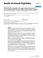
Fig. 5. Correlation b etween bind ing anities of IGF-I anal ogues t o
recombinant human IGF1R and prevention of apoptosis by IGF-I ana-
logues. The dissociation constants (K
d
) for binding of IGF-I and IGF-I
analogues to rhIGF1R were plotted against levels of apoptosis (%)
determined by ¯u orescence m icroscopy (Ho echst 33258 staining) in the
presence of 1 n
M
IGF-I a nd IGF-I analogues. Symbol representations
are I GF-I (d), des-(1±3) (s), Arg3 (j), Gly3 (h), des-(2,3)-Ala31(m),
Long (n), Leu24 (.), Ala31(,), Leu60 (r), des-(2,3)-Leu24 (e).
966 B. E. Forbes et al.(Eur. J. Biochem. 269) Ó FEBS 2002
differences in the assay systems used (i.e. immobilized
rhIGF1R vs. cell membrane-bound receptor), with the
greatest effect being on interactions involving residue 60 of
IGF-I. We are currently introducing a biotinylated spacer
arm at the C-terminus of rhIGF1R, which will allow a
guaranteed homogeneously coupled chip and may perhaps
improve ¯exibility and access to the receptor.
Having established the re lative binding af®nities of IGF-I,
IGF-II and the 14 analogues for rhIGF1R, we related these
to their abilities to prevent apoptosis. IGFs have powerful
antiapoptotic effects and promote survival in a d iverse array
of cell types throu gh activation o f IGF1R signalling (for
reviews, see [1] and [ 7]). This study investigated the survival-
promoting effects of IGF analogues on serum deprivation-
induced apoptosis in the rat PC12 cell line, which has been
used extensively as a model system to study the mechanism
of neuronal cell s urvival and apoptosis [18±20,23]. I GF-I
and IGF-II c ompletely prevented serum deprivation-
induced PC12 cell apoptosis at con centrations of 1 n
M
and 1 0 n
M
, r espectively, which are similar levels t o those
reported previously [18±20]. Importantly, PC12 cells have a
limited capacity to produce and secrete IGFBPs, and
existing evidence indicates that PC12 cells only synthesize
very low levels of IGFBP-6 [30]. This fact further suggests
that PC12 cells constitute a useful cell model for investiga-
ting direct biological actions of IGFs through IGF1R
signalling, as IGFBPs will not regulate or negatively i mpact
IGF±IGF1R interactions. We have made a direct correla-
tion between r eceptor binding af®nity (K
d
) and ability to
prevent apoptosis for IGF-I and the IGF-I analogues
(Fig. 5). Essentially, the ability of IGF-I o r IGF-I analogues
to prevent apoptosis correlated directly with their IGF1R-
binding af®nities, and a strong correlation coef®cient
(r
2
0.97) was obtained for this relationship (Fig. 5).
Thus, those IGF-I mutants designed to investigate IGFBP
binding [Long IGF-I, des-(1±3)-IGF-I, Arg3-IGF-I, Gly3-
IGF-I, Arg6-IGF-II and des(1±6)IGF-II] have basically
equal abilities to prevent apoptosis. Conversely, IGF-I
analogues with d isrupted IGF1R binding have a corre-
sponding loss in their ability to p revent apoptosis.
Despite the signi®cant role of IGFs in antiapoptotic
actions, assays measuring apoptosis have not traditionally
been used to analyse biological activity of analogues.
However, comparisons of receptor binding and biological
activities, such as protein and DNA synthesis and protein
degradation, have been made in the past and they generally
support our conclusions. For example, analogues designed
to probe for interactions between IGFs and I GFBPs
[des-(1±3)-IGF-I, Arg3-IGF-I a nd Long Arg3-IGF-I,
des-(1±6)-IGF-II, Arg6-IGF-II] were analysed in L6 myo-
blast receptor-binding assays and inhibition of protein
breakdown assays [26,27]. In these studies the ability to
inhibit protein breakdown was directly related to receptor-
binding af®nity. A lso, IGF analogues with perturbed
receptor interactions (Leu24-IGF-I, Ala31-IGF-I, Leu60-
IGF-I and combinations of these) exhibited r educed ability
to stimulate DNA synthesis in L7 murine ®broblasts
compared with IGF-I [14]. Interestingly, strong correlations
between insulin or insulin mutant binding to the insulin
receptor and metabolic potencies (glucose transport and
lipogenesis) have also been made [31]. However, mitogenic
potency of insulin analogues does not correlate as tightly
with overall receptor af®nity but rather with the dissociation
rate or occupancy time of receptor [31,32]. In the present
study, the analysis of association and dissociation rates of
the IGF analogues did not reveal distinct mechanisms of
IGF1R activation l eading to different b iological activities
(for example apoptosis vs. protein degradation).
The mechanism of signalling resulting from IGF1R
activation by the analogues tested above was not investi-
gated. However, both the Akt (via I RS-1) and the MAP
kinase (via Shc) pathways are involved in IGF-induced
antiapoptotic signalling via the IGF1R in PC12 cells [20].
It would be interesting in the future to determine whether
analogues differ in their abilities to activate these pathways.
In summary, we have clearly demonstrated a direct
correlation b etween the IGF1R-binding af®nity of IGF
and IGF analogues and the prevention of ap optosis
mediated through IGF1R signalling, suggesting that
IGF1R-binding af®nity is the primary determinant when
assessing the antiapoptotic potential of IGF analogues.
ACKNOWLEDGEMENTS
This work was supported by an Australian government Cooperative
Research Centre grant. We thank Mr A dam Denley and Ms. Filomena
Occhiodoro for technical assistance. In addition, we gratefully
acknowledge Mr Geo Francis (GroPep Ltd) for his critical
comments.
REFERENCES
1. Baserga, R., R esnico, M., D'Ambrosio, C. & Valentinis, B.
(1997) The role of the IGF-I receptor in apoptosis. Vitam. Horm.
53, 65±98.
2. Chan, J.M., Stampfer, M.J., Giovanucci, E., Gann, P.H., Ma, J.,
Wilkinson, P., Hennekens, C.H. & Pollak, M. (1998) Plasma
insulin-like g rowth factor-I and prostate cancer risk: a prospective
study. Science 279, 563±566.
3. Adams, T.E., Epa, V .C., Garrett, T.P. & W ard, C.W. (2000 )
Structure and function of the type 1 insulin-like growth factor
receptor. Cell. Mol. Life Sci. 57, 1050±1093.
4. Rubin, R. & Baserga, R. (1995) Insulin-like growth factor-I
receptor. Its role in cell proliferation, apoptosis, and tumori-
genicity. Lab. Invest. 73, 311±331.
5. Navarro, M. & Baserga, R. (2001) Limited redundancy of survival
signals from t he type 1 insulin-like g rowth factor receptor. Endo-
crinology 142, 1073±1081.
6. Peruzzi, F., Prisco, M., Dews, M., Salomoni, P., Grassilli, E.,
Romano, G., Calabretta, B. & Baserga, R. (1999) Multiple sig-
naling pathways of the insulin-like growth factor 1 receptor in
protection from apoptosis. Mol. Cell. Biol. 19, 7203±7215.
7. Butt,A.J.,Firth,S.M.&Baxter,R.C.(1999)TheIGFaxisand
programmed cell death. Immunol. Cell Biol. 77 , 256±262.
8. Cooke, R.M., Harvey, T.S. & Campbell, I.D. (1 991) Solution
structure of h uman insulin-like growth factor 1: a nuclear m ag-
netic resonance and restrained molecular dynamics study. Bio-
chemistry 30, 5484±5491.
9. Torres, A.M., Forbes, B.E., Aplin, S.E., Wallace, J.C., Francis,
G.L. & Norton, R.S. (1995) Solution s tructure of hu man insulin-
like g rowth factor. II. Relationship to receptor and binding pro-
tein interactions. J. Mol. Biol. 248, 385±401.
10. Sato, A ., K oyama, S., Yamada, H., Suzuki, S., T amura, K.,
Kobayashi, M., Niwa, M., Yasuda, T., Kyogoku, Y. & Kobay-
ashi, Y. (2000) Three-dimensional solution structure of a disul®de
bond isomer of the human insulin-like growth factor-I. J. Peptide
Res. 56, 218±230.
11. Braulke, T. (1999) Type-2 IGF receptor: a multi-ligand binding
protein. Horm. Metab. Res. 31, 242±246.
Ó FEBS 2002 IGF analogue binding to IGF1R and cell survival (Eur. J. Biochem. 269) 967
12. Baxter, R .C. (2000) Insulin-like growth factor (IGF)-binding
proteins: inte ractions with IGFs and intrinsic bioactivities. Am. J.
Physiol. 278, E967±E976.
13. Cascieri, M.A., Chicchi, G.G., Applebaum, J., Hayes, N.S.,
Green, B.G. & Bayne, M.L. (1 988) Mutants o f human insulin-like
growth factor I with red uced a nity for the type 1 insulin-like
growth factor receptor. Biochemistry 27, 3229±3233.
14. Bayne, M.L., Applebaum, J., Chicchi, G.G., Miller, R.E. &
Cascieri, M.A. (1990) The roles of tyrosines 24, 31, a nd 60 in the
high anity binding of insulin-like growth f actor-I to the typ e 1
insulin-like growth factor receptor. J. Biol. Chem. 265, 15648±
15652.
15. Cascieri, M.A., Chicchi, G.G., Applebaum, J ., Green, B.G.,
Hayes, N.S. & Bayne, M.L. (1989) Structural analogs of h uman
insulin-like growth factor (IGF) I with altered anity for type 2
IGF receptors. J. Biol. Chem. 264, 2199±2202.
16. Bayne, M.L., Applebaum, J., Chicchi, G.G., Hayes, N.S., Gre en,
B.G. & Cascieri, M.A. (1988) Structural analogs of hum an insulin-
like growth factor I with reduced anity for s erum binding pro-
teins and the type 2 insulin-like growth factor receptor. J. Biol.
Chem. 263, 6233±6239.
17. Surinya, K.H., Forbes, B.E., Occhiodoro, F., Brown , A., M urray,
A., Francis, G.L., Cosgrove, L.J. & Wallace, J.C. (1999) Pro-
duction and functional analysis o f a soluble high a nity insulin-
like growth factor type 1 receptor. Growth Horm. IGF Res. 9, 389.
18. Hale, K., Murray, A.W., Cosgrove, L.J., Bach, L.A. & Hart®eld,
P.J. (2000) P reventio n of apoptosis by insulin-lik e g rowth fact or
(IGF)-I and IGF-II is dierentially atten uated by IGF-bind ing
proteins in PC12 cells. Neurosci. Res. C ommun. 27, 7 5±83.
19. Rukenstein, A., Rydel, R.E. & G reene, L.A. (1991) Multiple
agents rescue PC12 cells from serum-free cell death by t ranslation-
and transcription-independent mechanisms. J. Neurosci. 11, 2552±
2563.
20. Parrizas, M., Saltiel, A.R. & LeRoith, D. (1997) Insulin-like
growth factor 1 inhibits apoptos is using the phosphatidylinositol
3¢-kinase and mitogen-activated protein kinase pathways. J. Biol.
Chem. 272, 154±161.
21. Lofas, S. & Johnsson, B. (1990) A novel hydrogel matrix on gold
surfaces in surface plasmon resonance sensors for f ast and ecient
covalent immobilization of ligands. J. Chem. Soc. Chem. Commun.
21, 1526±1528.
22. Jansson, M., Hallen, D., Koho, H., A ndersso n, G., Berghard, L.,
Heidrich, J., Nyberg, E., Uhlen, M., Kordel, J. & Nilsson, B.
(1997) Characterization of ligand binding of a soluble human
insulin-like growth factor I receptor variant suggests a ligand-
induced conformational change. J. Biol. Chem. 272, 8189±8197.
23. Hart®eld, P.J., Mayne, G.C. & Murray, A.W. (1997) Ceramide
induces apoptosis in PC12 cells. FEBS Lett. 401, 148±152.
24. King, R., Wells, J.R., Krieg, P., Snoswell, M., Brazier, J., Bagley,
C.J., Wallace, J.C., Ballard, F.J., Ross, M . & F rancis, G.L. ( 1992)
Production and characterization of recombinant insulin-like
growth factor-I (IGF-I) and potent analogues of IGF-I, with Gly
or Arg substituted for Glu3, following their expression in
Escherichia coli as fusion proteins. J. Mol. Endocrinol. 8, 29±41.
25. Beukers, M.W., Oh, Y., Zhang, H., Ling, N. & Rosenfeld, R.G.
(1991) [Leu27] insulin-like growth factor II is highly selective for
the t ype-II IGF recepto r in binding, cross-linking and thymidine
incorporation experiments. Endocrinology 128, 1201±1203.
26. Francis, G.L. , Ross, M., B allard, F.J., Milner, S .J., Senn, C.,
McNeil, K.A., Wallace, J.C., King, R. & Wells, J.R. (1992) Novel
recombinant fusion protein analogues o f i nsulin-like growth fact or
(IGF)-I indicate the relative importance of IG F-binding protein
and receptor binding for enhanced biological potency. J. Mol.
Endocrinol. 8, 213±223.
27. Francis, G.L., Aplin, S.E., Milner, S.J., McNeil, K.A., B allard,
F.J. & Wallace, J.C. (1993) Insulin-like growth factor (IGF)-I
binding to IGF-binding p roteins an d IGF receptors i s modi®ed by
deletion of the N-terminal hexapeptide or substitution of arginine
for glutamate-6 in IGF-II. Biochem. J. 293, 713±719.
28. Ross, M., Francis, G.L., Szabo, L ., Wallace, J.C. & Ballard, F.J.
(1989) Insulin-like growth factor (IGF)-binding proteins inhibit
the biological activities of I GF-1 and IGF-2 but not des-(1±3)-
IGF-1. Biochem. J. 258, 267±272.
29. Schumacher, R., Mosthaf, L., Schlessinger, J., Brandenburg, D. &
Ullrich, A. (1991) Insulin and insulin-like growth factor-1 binding
speci®city is determined by distinct regions of their cognate
receptors. J. Biol. Chem. 266, 19288±19295.
30. Bach, L.A., Tseng, L.Y., Swartz, J.E. & Rechler, M.M. (1993) Rat
PC12 pheochromocytoma cells synthesize insulin-like growth
factor-binding protein-6. Endocrinology 133, 990±995.
31. Kurtzhals, P., Schaer, L., Sorensen, A., Kristensen, C., Jonassen,
I., Schmid, C. & Trub, T. (2000) Correlations of r eceptor binding
and metabolic and mitogenic potencies of insulin analogs designed
for clinical use. Diabetes 49, 999±1005.
32. Fawcett, J., Hamel, F.G., Bennett, R.G ., Vajo, Z. & D uckwo rth,
W.C. (2001) Insulin and analogue eects on protein degradation i n
dierent cell types. Dissociation between binding and activity.
J. Biol. C hem. 276, 11552±11558.
968 B. E. Forbes et al.(Eur. J. Biochem. 269) Ó FEBS 2002