Báo cáo y học: "Significance of Conversation between Mast Cells and Nerves" pdf
Bạn đang xem bản rút gọn của tài liệu. Xem và tải ngay bản đầy đủ của tài liệu tại đây (253.81 KB, 16 trang )
65
Functional communication between mast cells
and nerves has been shown to occur in a variety
of both physiologic and pathologic situations.
1,2
Neuronal mechanisms are involved in mast cell
activation, and mast cells act as principle trans-
ducers of information between peripheral nerves
and local inflammatory events. Neuropeptides,
released from autonomic or nonadrenergic non-
cholinergic nerves, may influence the recruitment,
proliferation, and activation of leukocytes. On the
other hand, inflammatory cells may modulate the
neuronal phenotype and function.
Association of Mast Cells and Nerves
It is well established that there is an anatomic
association between mast cells and nerves in most
tissues.
3–6
In various studies, tissue mast cells
invariably showed ultrastructural evidence of acti-
vation even in normal healthy conditions, sug-
gesting that these cells are constantly providing
information to the nervous system. Mutual asso-
ciations between nerves and mast cells have been
observed in normal conditions and in pathologic
ones such as human irritable bowel syndrome,
atopic dermatitis, and interstitial cystitis.
7
Amor-
phometric study in both infected and healthy rat
intestine showed that mast cells and nerves were
closely and invariably approximated in rat intesti-
nal villi.
8
Electron microscopy showed evident
membrane-membrane association between
mucosal mast cells and nerves with dense core
vesicles at the points of contact. Other than in the
intestine, nerve and mast cell associations are
found in rat trachea and peripheral lung tissue,
9
skin,
10
urinary bladder,
11
brain,
12
and several other
tissues.
13,14
Besides an anatomic association, there is a
functional bidirectional communication pathway
in vivo. For example, psychological stress in rats
causes increased chloride ion secretion by the
intestinal epithelium, increased colonic mucin
Review Article
Significance of Conversation
between Mast Cells and Nerves
Hanneke P. M. van der Kleij, MD;
John Bienenstock, CM, MD (Hon), FRCP, FRCP(C), FRSC
Abstract
More and more studies are demonstrating interactions between the nervous system and the immune
system. However, the functional relevance of this interaction still remains to be elucidated. Such asso-
ciations have been found in the intestine between nerves and mast cells as well as between eosinophils
and plasma cells. Similar morphologic associations have been demonstrated in the liver, mesentery, uri-
nary bladder, and skin. Unmyelinated axons especially were found to associate with mast cells as well
as Langerhans’ cells in primate as well as murine skin. Although there are several pathways by which
immune cells interact with the nervous system, the focus in this review will be on the interaction between
mast cells and nerves.
H. P. M. van der Kleij, J. Bienenstock—Brain-Body
Institute and Department of Pathology and Molecular
Medicine, St. Joseph’s Healthcare, Hamilton, Ontario, and
McMaster University, Hamilton, Ontario
Correspondence to: John Bienenstock, Department of
Pathology and Molecular Medicine, McMaster University,
1200 Main Street West, Hamilton, Ontario, L8N 3Z5 Canada
66 Allergy, Asthma, and Clinical Immunology / Volume 1, Number 2, Spring 2005
secretion, and increased intestinal permeability,
mediated in part by both mast cells and substance
P.
15–17
Furthermore, mast cells and substance
P–containing nerves are also obligatory
components in a hapten-induced model of lung
inflammation.
18
Rozniecki and colleagues
provided evidence for morphologic, anatomic,
and functional interactions of dura mast cells with
cholinergic and peptidergic neurons containing
substance Pand calcitonin gene-related peptide.
19
Mast Cells
Mast cells are widely distributed throughout the
body in both connective tissue and at mucosal sur-
faces. They form a heterogeneous population of
cells with differences in their development, medi-
ator content, and their ability to interact with the
local environment.
20
Therefore, it seems likely
that mast cells have many diverse functions.
They are thought to play a major role in resistance
to infection and are extensively involved in
inflammation and subsequent tissue repair.
21
Moreover, there is evidence to support the con-
cept that mast cells are functionally important
modulators of hair follicle cycling, specifically
during anagen development.
22
This invites the
exploration of the murine hair cycle as a model
for dissecting the physiologic growth modulatory
functions of mast cells.
23
Furthermore, mast cells
are known to have a significant variety of actions
and interactions with other cells and physiologic
systems.
Mast cells can be divided into various sub-
populations with distinct phenotypes. Mast cell
secretory granules contain unique tryptic and chy-
motryptic serine proteases that differ between
species and tissues. The heterogeneity can express
itself as differences in histochemical, biochemi-
cal, and functional characteristics. The growth fac-
tors required for human mast cell differentiation have
been shown to be somewhat different than those for
such differentiation in rodents.
24
Although tryptase(s)
is found in most or every human mast cell, just a
single chymase has been defined. Human mast
cells are classified by the presence or relative
absence of this chymase.
25
In contrast, rodent mast
cell subsets store different chymase isoforms. Two
main subsets, connective tissue–type mast cells
(CTMCs) and mucosal mast cells (MMCs), are
recognized as distinct mast cell populations with dif-
ferent phenotypic and functional characteristics.
26,27
Another commonly used classification uses the
terms “MCt” and “MCtc”; the MCt phenotype con-
tains tryptase alone whereas the MCtc phenotype
contains chymase and tryptase.
28
In spite of their variation, the different mast-
cell subsets are derived from a common precur-
sor in the bone marrow. Mast cell progenitor cells
translocate from bone marrow to mucosal and
connective tissues to locally undergo differentia-
tion into mature forms. They possess a remarkable
degree of plasticity, so that even apparently fully
differentiated CTMCs will transform their phe-
notype to that of MMCs if transplanted into a
mucosal environment.
29
Mast Cell Mediators
Mast cells are capable of the synthesis of a large
number of pro- and anti-inflammatory mediators,
including cytokines, growth factors and products
of arachidonic acid metabolism. Pre-stored medi-
ators, such as histamine, serine proteases, pro-
teoglycans, sulphatases, and tumour necrosis
factor (TNF), are released within minutes after
degranulation of the cell.
30
After this primary
response, a second wave of newly synthesized
mediators are released, including prostaglandins
and leukotrienes. In the late-phase allergic
response, cytokines such as interleukin (IL)-4,
IL-5, IL-6, IL-8, IL-13, and TNF are induced and
secreted.
30
Expression of this host of cytokines
has led to the assumption of a role for mast cells
in host defense, for example, in immunoglobu-
lin E (IgE)–dependent immune responses to cer-
tain parasites, in natural immunity to bacterial
infections, and in inflammatory and allergic
diseases.
The communication between mast cells and
nerves via cytokines has not received much atten-
tion. TNF, which is pre-stored and is released
rapidly on degranulation, has an important func-
tional effect. Mast cells also secrete newly
Significance of Conversation between Mast Cells and Nerves — van der Kleij and Bienenstock 67
synthesized TNF within 30 minutes following cer-
tain stimuli.
31
Furthermore, TNF is able itself to
induce mast cell degranulation. TNF is involved
in changing neuronal cell function because it
can modulate the susceptibility of neurons to
electrical stimuli. The sensitizing effect of TNF
seems to primarily target C fibres.
32
In vitro incu-
bation of rat sensory nerves with TNF enhanced
the response of C fibres to capsaicin.
33
It is known
that TNF can activate nerve endings, causing a
lowering of the threshold to stimulation. Astudy
by Aranguez and colleagues indicated that mouse
astrocytes express TNF receptor 1 (TNFR1).
34
Furthermore, rat microglia transcribe messen-
ger ribonucleic acid (mRNA) for both TNFR1 and
TNFR2.
35
These results indicate that neuronal
tissue probably expresses both TNF receptors
and implies that communication between mast
cells and nerves may be mediated, at least in
part, by TNF.
Another major mast cell mediator is tryptase,
known to be present in all mast cell subtypes.
Although proteases (tryptase, chymase) are not
classified as cytokines, they have many cytokine-
like effects. These cytokine-like activities often
activate cells via protease-activated receptors
(PARs), cleavage of which results in signal trans-
duction.
36
Proteases regulate neurons and glia in
the central nervous system by cleaving PAR.
Myenteric neuron protease-activated receptor 2
(PAR2) expression has been detected by reverse
transcription polymerase chain reaction. Tryptase
has recently been shown to cleave PAR2 on pri-
mary spinal afferent neurons, which causes the
release of substance P, activation of the neu-
rokinin 1 receptor, and amplification of inflam-
mation and thermal and mechanical hyperalgesia.
37
Corvera and colleagues showed that purified
tryptase stimulates calcium mobilization in myen-
teric neurons.
38
They hypothesized that tryptase
excites neurons through PAR2 because activation
of PAR2 with trypsin or peptide agonists strongly
desensitizes the response to tryptase. In addition,
a tryptase inhibitor suppressed calcium mobi-
lization in response to degranulated mast cells.
This indicates that tryptase is a major mast cell
mediator with the capacity of activating myenteric
neurons through PAR2.
Growth Factors
The classic mediators of inflammation are not
alone in their ability to influence the interaction
between mast cells and nerves. Nerve and mast cell
growth factors are thought to play prominent reg-
ulatory roles as well. One such factor, nerve growth
factor (NGF), acts as a chemoattractant, thereby
causing an increase in the number of mast cells as
well as their degranulation.
39–41
NGF receptors on
mast cells act as autoreceptors, regulating mast cell
NGF synthesis and release while at the same time
being sensitive to NGF from the environment.
Inflammation can lead to an enhanced produc-
tion and release of NGF. In turn, NGF induces the
expression of neuropeptides and lowers the thresh-
old of neurones for firing.
41
In vivo administration of NGF in neonatal
rats caused a great increase in the size and num-
ber of mast cells in the peripheral tissues.
42
Furthermore, NGF has been shown to induce
degranulation and histamine release from mast
cells.
43,44
To complete the circle, mast cells are
capable of producing NGF.
45
Therefore, it is not
surprising that injection of NGF causes mast cell
proliferation, in part by mast cell degranulation.
46
NGF can have proinflammatory as well as
anti-inflammatory effects, depending on the sit-
uation and on the concentration of the compound.
Braun and colleagues recently showed that nasal
treatment of mice with NGF induced airway
hyperresponsiveness as measured by electrical
field stimulation.
47
Another study by Braun and
colleagues showed that nasal treatment of mice
with anti-NGF prevented the development of air-
way hyperresponsiveness.
48
On the other hand, the
expression of NGF is increased after brain injury.
There is evidence that the increased production
of NGF in the central nervous system during
brain disease such as multiple sclerosis can sup-
press inflammation by switching the immune
response to an anti-inflammatory suppressive
model.
49
In a compelling study, the injection of
CD4
+
lymphocytes transfected with the NGF
gene, either before or after the induction of aller-
gic encephalomyelitis, inhibited the onset of
demyelination.
50
This powerful inhibition of an
autoimmune process showed that local expression
of NGF prevented the migration of inflamma-
tory cells across the epithelium.
Mast Cell Activation by Tachykinins:
Expression of the Neurokinin 1 Receptor
In addition to the classic neurotransmitters acetyl-
choline and noradrenaline, a wide number of pep-
tides with neurotransmitter activity have been
identified in the past few decades. Among them,
the tachykinins substance P, neurokinin A, and
neurokinin B appear to act as mediators of non-
adrenergic noncholinergic excitatory neuro-
transmission.
The tachykinin substance Pcan activate mast
cells via distinct mechanisms. First, substance P
can activate mast cells without an intermediary
receptor through direct combination with G pro-
teins on the cell surface.
51,52
Second, tachykinins
interact with specific membrane proteins belong-
ing to the family of G protein–coupling cell
membrane receptors. Three distinct tachykinin
receptor subtypes have been identified and are
denoted as neurokinin 1 (NK1), neurokinin 2
(NK2), and neurokinin 3 (NK3); these receptors
have the highest affinity for substance P,
neurokinin A, and neurokinin B, respectively.
53–55
Several investigators have discussed the increased
in vivo expression of NK1 receptor in inflamed tis-
sue.
56,57
Therefore, it can be proposed that NK1
receptor expression on immune cells such as mast
cells is influenced by environmental inflammatory
factors such as cytokines. In previous work, Karimi
and colleagues demonstrated the increased
sensitivity of bone marrow–derived mast cells
(BMMCs) to substance P after a short coculture
with the cytokines IL-4 and stem cell factor.
58
The NK1 receptor appears to be present on the
basophil leukemia cell line (RBL).
59
Similar find-
ings were made in rat peritoneal mast cells, which
also express NK1 receptors.
60
In an in vitro cocul-
ture model, the activation of nerves with scor-
pion venom elicited the degranulation of RBL
cells via substance P.
61
It was shown that this sub-
stance-Pactivation is initiated only at the point of
contact between nerve fibres and associated RBL
cells through NK1 receptors.
62
Recently, it has been shown that functional
expression of NK1 receptors on BMMCs (which
are phenotypically immature mast cells) varies
according to culture conditions. The extent of
degranulation of BMMCs depends directly on
both the concentration of substance Pused and the
amount of NK1 receptor expression.
63
Similarly,
in an in vitro coculture model of BMMCs and neu-
rites, we showed that expression of NK1 by mast
cells lowers the threshold of activation induced by
nerve stimulation.
64
Furthermore, the response in
coculture was inhibited by pretreatment with
SR140333, an NK1-specific receptor antagonist
strongly pointing to an NK1 receptor–dependent
mechanism.
Very recently, Bischoff and colleagues exam-
ined the expression of tachykinin receptors on
human mast cells and found that human mast cells
derived from intestinal mucosa do not constitu-
tively express NK1, NK2, or NK3 receptors.
65
However, when stimulated by IgE receptor cross-
linking, these mast cells started to express NK1
receptors but not NK2 or NK3 receptors, again sug-
gesting that specific tissue conditions such as
allergic inflammation may lead to mast cell expres-
sion of NK1 receptors.
Interaction of Mast Cells and Nerves
Mast cells and nerves are in constant contact with
each other in both physiologic and pathologic sit-
uations. Many arguments suggest that mast cells
and nerves may be seen as a functional unit. They
share a number of activating signals, for some of
which both cells express receptors (such as vanil-
loids).
66
Furthermore, both mast cells and nerves
respond to stimulation by degranulating preformed
mediators, many of which are produced by both
cells (NGF, neuropeptides, and endothelin-1).
Mast cells can be activated by neuropeptides such
as substance P, and many mast cell mediators,
including serotonin and tryptase, can cause the
release of tachykinins from sensory nerve end-
ings.
3,67–69
Moreover, mast cells and nerves coop-
erate in a number of pathologic and physiologic
processes such as the regulation of hair follicle
cycling and development and such as wound
68 Allergy, Asthma, and Clinical Immunology / Volume 1, Number 2, Spring 2005
Significance of Conversation between Mast Cells and Nerves — van der Kleij and Bienenstock 69
healing.
70,71
Also, stress has been shown to trigger
skin mast cell degranulation, an action not only
dependent on corticotropin-releasing hormone but
apparently also involving substance P.
72
Stimula-
tion of the enteric nervous system by mast cell acti-
vation is likely to play an important role in mast
cell–mediated host defense in infections, espe-
cially infections induced by bacteria.
21,73
Interac-
tions between mast cells and nerves have also
been interpreted as important neuronal tissue repair
mechanisms following injury.
71,74
An enhanced interaction between mast cells and
nerves can lead to neurogenic inflammation. Inflam-
matory models have shown a significant increase
in the number of mast cells, resulting in the increased
release of inflammatory mediators on degranulation.
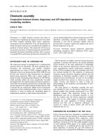
Inflammatory mast cell mediators may modulate
sensory nerves through the activation of receptors
on nerve terminals (Figure 1). Nonadrenergic non-
cholinergic (NANC) nerve endings express recep-
tors for histamine (H1 and H3) and serotonin
(5HT2A).
75–77
Under inflammatory-like conditions,
primary NANC nerves show an up-regulation of at
least histamine H1receptor expression.
78
A recent
report by Shubayev and Myers provides evidence
of expression of TNFR1 and TNFR2 in dorsal root
ganglia (DRG) neurons in adult rats.
79
Both recep-
tor subtypes were up-regulated in DRG neurons dur-
ing inflammation. Capsaicin-sensitive nerves can
be altered in this way and could result in an increased
release of neuropeptides. Allergen/hapten chal-
lenge can also lead to production of substance Pin
a subset of sensory nerve fibres that are typically
devoid of neuropeptides. In other words, aller-
gen/hapten challenge leads to a phenotypic switch
in the sensory neuropeptide innervation in the air-
ways, probably via mast cell activation, again
increasing the interaction between mast cells and
substance P–immunoreactive nerves.
80,81
Thus, mast
cell activation can result in an increase in the
excitability of sensory nerves and the production and
secretion of neuropeptides.
Neurogenic Inflammation
Neurogenic inflammation involves a change in
function of sensory neurons owing to inflamma-
tory mediators, inducing an enhanced release of
neuropeptides from the sensory nerve endings.
82
Neurogenic inflammation has been shown to occur
in different tissues, including the skin, urinary
tract, digestive system, and airways.
83–86
Given
the close proximity of mast cells and nerves to
blood vessels in most tissues, they may be con-
sidered an important functional unit in neurogenic
inflammation.
3
It is becoming apparent that by affecting neu-
ronal functioning, the mast cell and its mediators
play an important role in neurogenic inflamma-
tion.
3,87
Mast cells pass information on through
afferent nerves to local tissues by axon reflexes and
to the spinal cord and thence the brain. Stimula-
tion of C fibres by a range of chemical and phys-
ical factors results in afferent neuronal conduction
that elicits parasympathetic reflexes and antidromic
impulses travelling to the peripheral nerve termi-
nal. Axon reflexes account for many of the local
physiologic responses to antigen (for instance, in
sensitized lung and gut tissues) and have long
Figure 1 Mast cell–nerve interactions. Inflammatory
mediators may modulate sensory nerve endings through
the activation of receptors on nerve terminals. Neu-
ropeptides can stimulate mast cells via a receptor-
dependent and a receptor-independent mechanism.
Under inflammatory-like conditions, receptor expres-
sion on nerve endings and mast cells can be up-regu-
lated. CGRP= calcitonin gene–related peptide; H = his-
tamine; 5HT2A = serotonin 2a; NGF = nerve growth
factor; NK-1 = neurokinin 1; PAR = protease-acti-
vated receptor; TNFR = tumour necrosis factor recep-
tor; trk = neurotrophin tyrosine kinase receptor.
been recognized to be involved in local vasodi-
latation in the skin.
88–91
Antidromic stimulation
of guinea pig vagal sensory fibres results in con-
tractions of the isolated airway smooth muscle,
mediated by tachykinins.
92
Further studies indicate
that neuropeptide release can also be induced via
direct depolarization of the nerve terminal.
93
Priming
It is widely accepted that the effect of substance
P as a mast cell secretagogue is found only at
high concentrations. However, exposure of mast
cells to very small amounts of this neuropeptide
may be expected to reduce the threshold of acti-
vation of the cells for subsequent challenge with
antigen or neuropeptides. Therefore, mast cells can
be primed when exposed to physiologically rele-
vant low concentrations of substances, which low-
ers their thresholds to subsequent activation.
Priming appears to be a broadly based biologic
process and has been reported in several cell types.
Mast cells have been reported to be primed by dif-
ferent cytokine growth factors for activation by dif-
ferent agonists.
94
Stem cell factor (SCF), for
instance, can act as a priming agent in some cir-
cumstances.
95
We have shown that SCF and IL-4
prime BMMCs to induce increased responsiveness
to substance P.
63
Mast cells can also be primed by
substance P itself because repeated doses of very
low concentrations (picomolars) of substance Pcan
induce mast cell degranulation and can lower the
threshold for degranulation via subsequent cross-
linking of IgE receptors by anti-IgE.
96
The concept
of priming also applies to neurons. TNF may exert
a priming effect (rather than a direct stimulatory
effect) on sensory activity.
33,97
Mast Cell Activation versus
Mast Cell Degranulation
Exocytosis is the most obvious event associated
with secretion of the mediator molecules con-
tained in granules. It used to be believed that mast
cell activation was “all or nothing” and that IgE
cross-linking induces the functional consequences
of allergic reactions and anaphylaxis. However, the
activity of mast cells in health and disease is
clearly much more complicated. Secretion can
occur without evidence of degranulation, and even
molecules stored within the same granules can
be released and secreted in a discriminatory pat-
tern.
98
Mast cells have been increasingly implicated
in inflammatory processes in which explosive
degranulation is not commonly observed. Astudy
by Ratliff and colleagues ultrastructurally showed
mast cells in close proximity to unmyelinated
nerve fibres.
99
These mast cells contained granules
showing ultrastructural features of activation or
piecemeal degranulation, which have been asso-
ciated with differential secretion. Furthermore,
Gottwald and colleagues found increases in the his-
tamine content of intestinal tissues after electrical
vagal stimulation without degranulation of mast
cells.
100
These data support the potential for intesti-
nal mucosal mast cell regulation by the central ner-
vous system and suggest modulation of mast cells
without degranulation. Furthermore, IL-1 stimu-
lates secretion of IL-6 without release of the
granule-associated protease tryptase.
101
Selective
secretion of IL-6 from mast cells appears to be dis-
tinct from degranulation and may contribute to the
development of inflammation, in which the impor-
tance of IL-6 has been recognized. Serotonin can
be released separately from histamine, and dif-
ferential synthesis and release of arachidonic acid
metabolites, prostaglandins, and leukotrienes have
been reported.
102,103
Interaction of Mast Cells
and Nerves in Tissues
Brain and Immune System
The brain and the nervous and immune systems
are the major adaptive systems of the body.
104
Several pathways have been shown to link the
brain and the immune system, such as (1) the
autonomic nervous system via direct neural influ-
ences and (2) the neuroendocrine humoral outflow
via the pituitary. Corticotropin-releasing hormone
(CRH), secreted by the pituitary gland, is a major
regulator of the hypothalamic-pituitary-adrenal
70 Allergy, Asthma, and Clinical Immunology / Volume 1, Number 2, Spring 2005
(HPA) axis and cortisone synthesis and acts as a
coordinator of the stress response.
105
CRH is also
thought to be involved peripherally in tissue
responses to stress in the skin, respiratory tract, and
intestine.
Mast cells are resident in the brain of many
species.
106
They appear to enter the brain via pen-
etrating blood vessels. Brain mast cells are asso-
ciated with blood vessels throughout the brain
and especially in the meninges.
107
They seem to
be involved in behavioural activity, such as the
courting behaviour of doves.
108
Large numbers of
tryptase-containing mast cells have been described
as surrounding the pituitary gland and are thought
to act as an immune gate for HPAaxis activity.
109
These mast cells can respond to antigens and reg-
ulate CRH secretion via histamine effects.
105
The physiologic significance of mast cells in
brain function and/or metabolism is unclear. How-
ever, they can modulate neuroendocrine control
systems,
2
and they could play a role in the regu-
lation of meningeal blood flow and vessel per-
meability.
110
Pavlovian conditioning has also been
shown to be able to promote mast cell degranu-
lation through as yet unknown mechanisms.
111
Apart from their being resident cells, mast
cells can move through the brain in the absence
of inflammation. Mast cells in the central ner-
vous system may participate in the regulation of
inflammatory responses through interactions with
the HPAaxis. Matsumoto and colleagues showed
that in the dog, degranulation of mast cells evoked
HPAactivation in response to histamine release.
109
The physiologic effects of psychological stress
are often largely mediated by CRH, released either
centrally or peripherally, and mast cell–nerve
interactions are important components of this
response.
112
In response to psychological stress or
certain physical stressors, an inflammatory process
may occur through the release of neuropeptides
(especially substance P) from sensory nerves and
the activation of mast cells or other inflammatory
cells. Central neuropeptides initiate a systemic
stress response by activation of neuroendocrine
pathways (such as the sympathetic nervous
system, the hypothalamic-pituitary axis, and the
renin-angiotensin system) with the release of stress
hormones (ie, catecholamines, corticosteroids,
growth hormone, glucagons, and renin).
113
These
effects have been found in a variety of stress mod-
els, including cold, restraint stress, and water
avoidance stress.
15,114,115
The Skin
The dermis is richly innervated by primary effer-
ent sensory nerves, postganglionic cholinergic
parasympathetic nerves, and postganglionic adren-
ergic and cholinergic sympathetic nerves.
116
Neu-
ropeptides, released by cutaneous nerves, have
been shown to activate a number of target cells,
including Langerhans’cells, endothelial cells, and
mast cells.
117
In the skin, neuropeptides are released
in response to nociceptive stimulation by pain
and by mechanical and chemical irritants, to medi-
ate skin responses to infection, injury, and wound
healing.
118,119
Substance P is one of the main neu-
ropeptides responsible for the skin reaction char-
acterized by erythema, pain, and swelling.
119
In
addition, substance Pcan cause the release of his-
tamine
120
and TNF
121
from skin mast cells, which
in turn leads to vasodilation.
Interestingly, capsaicin (which releases neu-
ropeptides from nerves) applied to human skin
induces the release of chymase within 6 hours
and the induction of E-selectin in adjacent
microvascular endothelium, events consistent with
release of substance Pfrom axons and subsequent
stimulation of cytokine-mediated mast cell inter-
action with endothelial cells. However, an iden-
tical application of capsaicin to human skin grafted
onto immunodeficient mice (and thus experi-
mentally lacking in unmyelinated axons) failed to
yield similar findings.
5
These results indicate that
unmyelinated axons connect Langerhans’ cells
and dermal mast cells.
Recent studies have suggested that mast cells
play a crucial role in the down-regulation of
immune responses and the induction of tolerance
after exposure of skin to ultraviolet B radiation
(UVB). Hart and colleagues reported the involve-
ment of histamine in UVB-induced suppression in
mice, and mast cells have been shown to be the
source of UVB-induced histamine.
122,123
Further-
more, interactions between mast cells and the
nervous system appear to be involved in UVB-
mediated immune suppression. TNF, reported to
Significance of Conversation between Mast Cells and Nerves — van der Kleij and Bienenstock 71
be derived from mast cells, is a major cytokine
implicated in signalling the immunosuppressive
effects of UVB.
124
Evidence indicates that mast
cells are triggered to release TNF in response to
the neuropeptide calcitonin gene–related peptide
(CGRP), which is released from UVB-damaged
cutaneous nerve endings.
125
Airways
Efferent and afferent autonomic nerves regulate
many aspects of human and animal airway func-
tion. In addition to cholinergic and adrenergic
innervation, the NANC nervous system is an
important third neural network in the lung.
Inhibitory NANC nerves contain vasoactive intesti-
nal peptide (VIP) and nitric oxide, which are
potent relaxants of the airways and which coun-
teract bronchoconstriction.
Excitatory NANC nerves or so-called sen-
sory nerves are mainly localized in and beneath
the airway epithelium. Tachykinins and CGRP
are the predominant excitatory NANC neuropep-
tides in the airways.
126
Mast cells lining the mucosal layer of the res-
piratory tract have been found in close proximity
to substance P-immunoreactive and CGRP-
immunoreactive nerves of rat trachea and periph-
eral lung tissue.
10
Immunohistochemical studies of
neuronal tachykinins in the airways of asthmatic
patients have yielded conflicting results. Whereas
an increase in both the number and length of
tachykinin-immunoreactive nerve fibres in the
airways was found in some studies, other studies
detected significantly less substance P–like
immunoreactivity in lung tissue from asthmatic
patients as compared to nonasthmatic patients.
127–130
However, this latter finding may reflect an
augmented release of substance P followed by
degradation. Studies on autopsy tissue,
130
plasma
levels,
131
lung lavage fluid,
128
and sputum
132
sug-
gest that tachykinins are present in increased
amounts in asthmatic airways.
Neuropeptides influence the recruitment, pro-
liferation, and activation of inflammatory cells
such as mast cells. There is growing evidence that
tachykinins and CGRPare involved in neurogenic
inflammation of the airways. Structural studies
show that mast cells associate with nerves in the
lung. Furthermore, Forsythe and colleagues have
demonstrated that substance P and neurokinin A
induce histamine release from human airway mast
cells.
133
Moreover, antigen causes a secretory
response in the rat trachea via an interaction depen-
dent on mast cells and nerves.
89
Gastrointestinal Tract
The gastrointestinal tract is characterized by a
unique accumulation of immune and inflammatory
cells. The mechanism of interaction between nerve
and inflammatory cells in the intestine is, however,
very unclear. Intestinal mast cells have been repeat-
edly reported to communicate with the enteric
nervous system. Furthermore, Stead and
colleagues, on the basis of electron microscopy
studies, reported an anatomic association between
mast cells and nerves in the human intestinal
mucosa.
134
Nerve stimulation has been reported to cause
mast cell degranulation in the intestine. First,
Shanahan and colleagues showed that substance
Pcaused mediator release from intestinal mucosal
mast cells.
135
Subsequently, substance Pand CGRP
fibres have been reported to activate peptidergic
mast cells in the intestinal mucosa of healthy and
infected rats as well as in patients with inflam-
matory bowel disease.
1
Mast cell mediators also appear to have an
effect on the nerves in the intestine. Intestinal
mast cell infiltration may perturb nerve function,
leading to abdominal pain perception in patients
with irritable bowel syndrome (IBS).
136
Recent
evidence for activated mast cells associated with
enteric nerves in IBS strongly implies that mast
cells are involved in this symptom complex.
136
A
study by Jiang and colleagues using an intestinal
model for anaphylaxis showed that serotonin and
histamine, released from the mast cells after
intestinal anaphylaxis, stimulate mesenteric affer-
ents via 5-HT3 and histamine H1 receptors.
137
Mesenteric afferent-nerve discharge increased
approximately 1 minute after luminal antigen
challenge and was attenuated by serotonin and his-
tamine receptor antagonists. Mast cell–nerve
association appears to function as a homeostatic
unit in the regulation of gut physiology and in
response to antigens.
138
72 Allergy, Asthma, and Clinical Immunology / Volume 1, Number 2, Spring 2005
Perdue and colleagues determined the exis-
tence of an integral nerve-to-mast cell and mast
cell-to-nerve connection during intestinal ana-
phylaxis.
139
Arole for the mast cell-to-nerve con-
nection was established by increases in the short-
circuit current after antigen challenge. The response
to antigenic stimulation was reduced in mast
cell–deficient W/Wv mice as compared to their +/+
litter mates and was inhibited by different mast cell
antagonists in +/+ mice but not in W/Wv mice,
pointing to a mast cell-to-nerve connection.
Furthermore, reconstitution of the mast cell defi-
ciency was followed by a restoration of the neural
response. In sensitized guinea pig intestine, the
short-circuit-current secretory response to anti-
gen occurred simultaneously with acetylcholine
release and could be blocked by atropine.
140
This
showed conclusively that nerve excitation and the
secretion of the main cholinergic neurotransmit-
ter could be induced by antigen via mast cells
through an immune-mediated response. The effects
of Clostridium difficile toxin on intestinal seg-
ments has also been shown to be dependent on
intact mast cells and substance P–containing
nerves.
141,142
It can be reasonably concluded that nerves and
mast cells form a physiologic unit that presumably
maintains and regulates homeostasis of the mucosal
epithelial secretory response. This unit is involved
in health, in response to stress, and also in response
to injuries and environmental pathogens.
Therapy
In different tissues and species, there is constant
communication between mast cells and the nervous
system. This functional communication has been
shown to occur in a variety of both physiologic and
pathologic situations.
6
The concept of these inter-
actions is very interesting and may bring about new
therapeutic and diagnostic approaches.
In humans, an inhaled long-acting 
2
agonist
inhibits mast cell mediator release and plasma
exudation and may reduce sensory nerve activa-
tion. In combination with a corticosteroid, the
low systemic effect of these drugs does not result
in any significant adverse effects, and there is a
strong scientific rationale for long-term asthma
therapy.
143
In the skin, cyclosporin A has power-
ful therapeutic effects on severe therapy-resistant
atopic dermatitis.
144
Treating the skin with
cyclosporin A increases the stable granule popu-
lation and results in the disappearance of the close
interrelation of mast cells and cutaneous nerves.
These findings suggest that cyclosporin A may
exert its therapeutic effect by inhibiting mast cell
activation and by affecting the interaction between
mast cells and nerves.
Exogenous administration of neuropeptides
to maintain normal immune defences represents
a new field of pharmacotherapeutics against bac-
terial invasion. But besides this positive health
effect of neuropeptides, there is the negative fact
that neuropeptides can activate mast cells and
result in an enhanced communication between
mast cells and nerves, causing an inflammatory
response. Mast cell mediators can sensitize sen-
sory neurons, which further activate the mast
cells by releasing neurotransmitters or neu-
ropeptides (eg, neurotensin, somatostatin, sub-
stance P, and acetylcholine). It has been shown
that in the gastrointestinal tract, CGRP, substance
P, and VIP-immunoreactive nerve fibres are
involved in protection of the tissue.
145,146
In a rat
colitis model, an early decrease in these neu-
ropeptides may be an essential condition for the
development of colitis. That the intensity and
density of substance P and VIP-IR nerve fibres
increased after the induction of colitis suggests
their possible involvement in tissue repair.
147
Again, on the other hand, these neuropeptides can
activate mast cells that play a pivotal role in
inflammation. An enhanced interaction between
mast cells and nerves can also lead to neuro-
genic inflammation.
From everything we know so far of the asso-
ciation between mast cells and nerves, it is becom-
ing clearer that the interaction is involved in the
regulation of physiologic processes as well as in
disease mechanisms. First, therapeutic targets
have to be very selective. Because these associa-
tions of mast cells and nerves seem to appear
throughout the body, it may be very difficult to find
a drug that is selectively effective at a particular
site in the body. Second, if a selective drug that
Significance of Conversation between Mast Cells and Nerves — van der Kleij and Bienenstock 73
74 Allergy, Asthma, and Clinical Immunology / Volume 1, Number 2, Spring 2005
provides protection against disease is found, inter-
ference in the cross-communication between mast
cells and nerves also increases the risk of chang-
ing the healthy balance that is essential for main-
taining tissue homeostasis.
More physiologic studies are needed for a
better understanding of how the activation of mast
cells and nerves is modulated, how sensory nerves
control mast cell functions, how mast cells use sen-
sory nerves in inducing inflammation, and the
role of nerve fibres and their mediators. New find-
ings will continue to increase our understanding
of mast cell–nerve associations and their func-
tion in health and disease and will be followed by
new therapeutic and diagnostic approaches.
Conclusions
Extensive crosstalk exists between nerves and
mast cells. Although differences in species have
been reported, morphologic as well as functional
associations are found in most tissues in humans
and in rodents. Many of these associations have
been shown to occur between substance P- and
CGRP-containing neurons and mast cells of all
subtypes.
The role of this bidirectional communication
between mast cells and nerves appears to be mul-
tifactorial. Mast cells are thought to play a major
role in resistance to infection and are extensively
involved in inflammation and subsequent tissue
repair. The communication with the nervous sys-
tem allows the peripheral and central nervous sys-
tems to be involved in the regulation of defence
mechanisms, inflammation, and response to infec-
tion. The involvement of mast cell–nerve com-
munication in the response to stress, for instance,
points to an extensive communication between the
nervous and immune systems.
However, the complexity of the picture has
increased further as it has become clear that clas-
sic neurotransmitters such as acetylcholine and
neuropeptides are produced by nonneuronal cells.
Nonneuronal cells of the immune system, such as
monocytes, macrophages, T lymphocytes, and
eosinophils, have been shown to produce endoge-
nous substance P.
148,149
This alternative source of
immune cells could represent an additional source
of tachykinins in inflamed tissues, providing a
nonneurogenic tachykininergic contribution to
the local inflammatory process.
150
References
1. Marshall JS, Waserman S. Mast cells and the
nerves—potential interactions in the context of
chronic disease. Clin Exp Allergy 1995;25:
102–10.
2. van der Kleij HPM, Blennerhassett M,
Bienenstock J. Nerve-mast cell interactions—
partnership in health and disease. In:
Bienenstock J, Blennerhassett M, Goetzl E, edi-
tors. Autonomic neuroimmunology. Autonomic
Neuroscience Series. Vol. 15. London, (UK):
Taylor&Francis group; 2003 p. 139–170.
3. Purcell WM, Atterwill CK. Mast cells in neu-
roimmune function: neurotoxicological and
neuropharmacological perspectives. Neurochem
Res 1995;20:521–32.
4. Arizono N, Matsuda S, Hattori T, et al.
Anatomical variation in mast cell nerve asso-
ciations in the rat small intestine, heart, lung,
and skin: similarities of distances between neural
processes and mast cells, eosinophils, or plasma
cells in the jejunal lamina propria. Lab Invest
1990;62:626–34.
5. Bienenstock J, MacQueen G, Sestini P, et al.
Mast cell/nerve interactions in vitro and in vivo.
Am Rev Respir Dis 1991;143:S55–8.
6. Pang X, Boucher W, Triadafilopoulos G, et al.
Mast cell and substance P-positive nerve
involvement in a patient with both irritable
bowel syndrome and interstitial cystitis. Urology
1996;47:436–8.
7. Bauer O, Razin E. Mast cell-nerve interactions.
New Physiol Sci 2000;15:213–8.
8. Stead RH, Tomioka M, Quinonez G, et al.
Intestinal mucosal mast cells in normal and
nematode-infected rat intestines are in intimate
contact with peptidergic nerves. Proc Natl Acad
Sci U S A 1987;84:2975–9.
9. Undem BJ, Riccio MM, Weinreich D, et al.
Neurophysiology of mast cell-nerve interac-
tions in the airways. Int Arch Allergy Immunol
1995;107:199–201.
10. Egan CL, Viglione-Schneck MJ, Walsh LJ, et
al. Characterization of unmyelinated axons unit-
ing epidermal and dermal immune cells in
Significance of Conversation between Mast Cells and Nerves — van der Kleij and Bienenstock 75
primate and murine skin. J Cutan Pathol
1998;25:20–9.
11. Letourneau R, Pang X, Sant GR, Theoharides
TC. Intragranular activation of bladder mast
cells and their association with nerve processes
in interstitial cystitis. Br J Urol 1996;77:41–54.
12. Keller JT, Marfurt CF. Peptidergic and sero-
toninergic innervation of the rat dura mater. J
Comp Neurol 1991;309:115–34.
13. Olsson Y. Mast cells in the nervous system. Int
Rev Cytol 1968;24:27–70.
14. Newson B, Dahlstrom A, Enerback L, Ahlman
H. Suggestive evidence for a direct innervation
of mucosal mast cells. Neuroscience 1983;
10:565–70.
15. Pothoulakis C, Castagliuolo I, Leeman SE.
Neuroimmune mechanisms of intestinal
responses to stress. Role of corticotropin-
releasing factor and neurotensin. Ann N Y
Acad Sci 1998;840:635–48.
16. Santos J, Saunders PR, Hanssen NP, et al.
Corticotropin-releasing hormone mimics stress-
induced colonic epithelial pathophysiology in
the rat. Am J Physiol 1999;277:G391–9.
17. Santos J, Benjamin M, Yang PC, et al. Chronic
stress impairs rat growth and jejunal epithelial
barrier function: role of mast cells. Am J Physiol
Gastrointest Liver Physiol 2000;278:G847–54.
18. Kraneveld AD, van der Kleij HP, Kool M, et al.
Key role for mast cells in nonatopic asthma. J
Immunol 2002;169:2044–53.
19. Rozniecki JJ, Dimitriadou V, Lambracht-Hall
M, et al. Morphological and functional demon-
stration of rat dura mater mast cell-neuron
interactions in vitro and in vivo. Brain Res
1999;849:1–15.
20. Bradding P, Holgate ST. Immunopathology and
human mast cell cytokines. Crit Rev Oncol
Hematol 1999;31:119–31.
21. Malaviya R, Ikeda T, Ross E, Abraham SN.
Mast cell modulation of neutrophil influx and
bacterial clearance at sites of infection through
TNF-alpha. Nature 1996;381:77–80.
22. Maurer M, Fischer E, Handjiski B, et al.
Activated skin mast cells are involved in murine
hair follicle regression (catagen). Lab Invest
1997;77:319–32.
23. Maurer M, Paus R, Czarnetzki BM. Mast cells
as modulators of hair follicle cycling. Exp
Dermatol 1995;4:266–71.
24. Welle M. Development, significance, and het-
erogeneity of mast cells with particular regard
to the mast cell-specific proteases chymase and
tryptase. J Leukoc Biol 1997;61:233–45.
25. Beil WJ, Schulz M, Wefelmeyer U. Mast cell
granule composition and tissue location—a close
correlation. Histol Histopathol 2000;15:937–46.
26. Befus AD, Dyck N, Goodacre R, Bienenstock
J. Mast cells from the human intestinal lamina
propria. Isolation, histochemical subtypes, and
functional characterization. J Immunol
1987;138:2604–10.
27. Galli SJ. New insights into “the riddle of the
mast cells”: microenvironmental regulation of
mast cell development and phenotypic hetero-
geneity. Lab Invest 1990;62:5–33.
28. Church MK, Clough GF. Human skin mast cells:
in vitro and in vivo studies. Ann Allergy Asthma
Immunol 1999;83:471–5.
29. Kitamura Y, Kanakura Y, Sonoda S, et al. Mutual
phenotypic changes between connective tissue
type and mucosal mast cells. Int Arch Allergy
Appl Immunol 1987;82:244–8.
30. Church MK, Levi-Schaffer F. The human mast
cell. J Allergy Clin Immunol 1997:99;155–60.
31. Gordon JR, Galli SJ. Mast cells as a source of
both preformed and immunologically inducible
TNF-alpha/cachectin. Nature 1990:346;274–6.
32. Junger H, Sorkin LS. Nociceptive and inflam-
matory effects of subcutaneous TNF alpha. Pain
2000;85:145–51.
33. Nicol GD, Lopshire JC, Pafford CM. Tumor
necrosis factor enhances the capsaicin sensi-
tivity of rat sensory neurons. J Neurosci
1997;17:975–82.
34. Aranguez I, Torres C, Rubio N. The receptor
for tumor necrosis factor on murine astrocytes:
characterization, intracellular degradation, and
regulation by cytokines and Theiler’s murine
encephalomyelitis virus. Glia 1995;13:185–94.
35. Dopp JM, Mackenzie-Graham A, Otero GC,
Merrill JE. Differential expression, cytokine
modulation, and specific functions of type-1
and type-2 tumor necrosis factor receptors in
rat glia. J Neuroimmunol 1997;75:104–12.
36. Mirza H, Schmidt VA, Derian CK, et al.
Mitogenic responses mediated through the pro-
teinase-activated receptor-2 are induced by
expressed forms of mast cell alpha- or beta-
tryptases. Blood 1997;90:3914–22.
76 Allergy, Asthma, and Clinical Immunology / Volume 1, Number 2, Spring 2005
37. Defea K, Schmidlin F, Dery O, et al.
Mechanisms of initiation and termination of
signalling by neuropeptide receptors: a com-
parison with the proteinase-activated receptors.
Biochem Soc Trans 2000;28:419–26.
38. Corvera CU, Dery O, McConalogue K, et al.
Thrombin and mast cell tryptase regulate guinea-
pig myenteric neurons through proteinase-
activated receptors-1 and -2. J Physiol 1999;
517:741–56.
39. Marshall JS, Gomi K, Blennerhassett MG,
Bienenstock J. Nerve growth factor modifies
the expression of inflammatory cytokines by
mast cells via a prostanoid-dependent mecha-
nism. J Immunol 1999;162:4271–6.
40. Horigome K, Pryor JC, Bullock ED, Johnson
EM. Mediator release from mast cells by nerve
growth factor. Neurotrophin specificity and
receptor mediation. J Biol Chem 1993;
268:14881–7.
41. Lindsay RM, Harmar AJ. Nerve growth factor
regulates expression of neuropeptide genes in
adult sensory neurons. Nature 1989;337:362–4.
42. Aloe L, Levi-Montalcini R. Nerve growth factor
induced overgrowth of axotomized superior cer-
vical ganglia in neonatal rats. Similarities and
differences with NGF effects in chemically axo-
tomized sympathetic ganglia. Arch Ital Biol
1979;117:287–307.
43. Pearce FL, Thompson HL. Some characteris-
tics of histamine secretion from rat peritoneal
mast cells stimulated with nerve growth factor.
J Physiol 1986;372:379–93.
44. Aloe L. The effect of nerve growth factor and
its antibody on mast cells in vivo. J
Neuroimmunol 1988;18:1–12.
45. Leon A, Buriani A, Dal Toso R, et al. Mast cells
synthesize, store, and release nerve growth
factor. Proc Natl Acad Sci USA 1994;91:
3739–43.
46. Marshall JS, Stead RH, McSharry C, et al. The
role of mast cell degranulation products in mast
cell hyperplasia. I. Mechanism of action of nerve
growth factor. J Immunol 1990;144:1886–92.
47. Braun A, Quarcoo D, Schulte-Herbruggen O,
et al. Nerve growth factor induces airway hyper-
responsiveness in mice. Int Arch Allergy
Immunol 2001;124:205–7.
48. Braun A, Lommatzsch M, Lewin GR, et al.
Neurotrophins: a link between airway inflam-
mation and airway smooth muscle contractil-
ity in asthma? Int Arch Allergy Immunol
1999;118:163–5.
49. Villoslada P, Hauser SL, Bartke I, et al. Human
nerve growth factor protects common mar-
mosets against autoimmune encephalomyelitis
by switching the balance of T helper cell type
1 and 2 cytokines within the central nervous
system. J Exp Med 2000;191:1799–806.
50. Flugel A, Matsumuro K, Neumann H, et al.
Anti-inflammatory activity of nerve growth
factor in experimental autoimmune
encephalomyelitis: inhibition of monocyte
transendothelial migration. Eur J Immunol
2001;31:11–22.
51. Mousli M, Bronner C, Bockaert J, et al.
Interaction of substance P, compound 48/80, and
mastoparan with the alpha-subunit C-terminus
of G protein. Immunol Lett 1990;25:355–7.
52. Mousli M, Hugli TE, Landry Y, Bronner C.
Peptidergic pathway in human skin and rat
peritoneal mast cell activation. Immuno-
pharmacology 1994;27:1–11.
53. Severini C, Improta G, Falconieri-Erspamer G,
et al. The tachykinin peptide family. Pharmacol
Rev 2002;54:285–322.
54. Nakanishi S. Mammalian tachykinin receptors.
Annu Rev Neurosci 1991;14:123–36.
55. Regoli D, Boudon A, Fauchere JL. Receptors
and antagonists for substance Pand related pep-
tides. Pharmacol Rev 1994;46:551–99.
56. Kaltreider HB, Ichikawa S, Byrd PK, et al.
Upregulation of neuropeptides and neuropep-
tide receptors in a murine model of immune
inflammation in lung parenchyma. Am J Respir
Cell Mol Biol 1997;16:133–44.
57. Mantyh CR, Vigna SR, Bollinger RR, et al.
Differential expression of substance P recep-
tors in patients with Crohn’s disease and
ulcerative colitis. Gastroenterology 1995;
109:850–60.
58. Karimi K, Redegeld FA, Blom R, Nijkamp FP.
Stem cell factor and interleukin-4 increase
responsiveness of mast cells to substance P. Exp
Hematol 2000;28:626–34.
59. Cooke HJ, Fox P, Alferes L, et al. Presence of
NK1 receptors on a mucosal-like mast cell line,
RBL-2H3 cells. Can J Physiol Pharmacol
1998;76:188–93.
Significance of Conversation between Mast Cells and Nerves — van der Kleij and Bienenstock 77
60. Okada T, Hirayama Y, Kishi S, et al. Functional
neurokinin NK-1 receptor expression in rat peri-
toneal mast cells. Inflamm Res 1999;48:274–9.
61. Suzuki R, Furuno T, McKay DM, et al. Direct
neurite-mast cell communication in vitro occurs
via the neuropeptide substance P. J Immunol
1999;163:2410–5.
62. Mori N, Suzuki R, Furuno T, et al. Nerve-mast
cell (RBL) interaction: RBL membrane ruffling
occurs at the contact site with an activated neu-
rite. Am J Physiol Cell Physiol 2002;283:
C1738–44.
63. van der Kleij HP, Ma D, Redegeld FA, et al.
Functional expression of neurokinin 1 recep-
tors on mast cells induced by IL-4 and stem cell
factor. J Immunol 2003;171:2074–9.
64. Furuno T, Ma D, van der Kleij HP, et al. Bone
marrow-derived mast cells in mice respond in
co-culture to scorpion venom activation of supe-
rior cervical ganglion neuritis according to level
of expression of NK-1 receptors. Neurosci Lett
2004;372:185–9.
65. Bischoff SC, Schwengberg S, Lorentz A, et al.
Substance P and other neuropeptides do not
induce mediator release in isolated human
intestinal mast cells. Neurogastroenterol Motil
2004;16:185–93.
66. Biro T, Maurer M, Modarres S, et al.
Characterization of functional vanilloid recep-
tors expressed by mast cells. Blood
1998;91:1332–40.
67. Paus R, Heinzelmann T, Robicsek S, et al.
Substance P stimulates murine epidermal
keratinocyte proliferation and dermal mast cell
degranulation in situ. Arch Dermatol Res
1995;287:500–2.
68. Barnes PJ. Neurogenic inflammation and
asthma. J Asthma 1992;29:165–80.
69. Holzer P. Local effector functions of capsaicin-
sensitive sensory nerve endings: involvement
of tachykinins, calcitonin gene-related peptide
and other neuropeptides. Neuroscience 1988;24:
739–68.
70. Paus R, Peters EM, Eichmuller S, Botchkarev
VA. Neural mechanisms of hair growth control.
J Investig Dermatol Symp Proc 1997;2:61–8.
71. Gottwald T, Coerper S, Schaffer M, et al. The
mast cell-nerve axis in wound healing: a hypoth-
esis. Wound Repair Regen 1998;6:8–20.
72. Singh LK, Pang X, Alexacos N, et al. Acute
immobilization stress triggers skin mast cell
degranulation via corticotropin-releasing hor-
mone, neurotensin, and substance P: a link to
neurogenic skin disorders. Brain Behav Immun
1999;13:225–39.
73. Echtenacher B, Mannel DN, Hultner L. Critical
protective role of mast cells in a model of acute
septic peritonitis. Nature 1996;381:75–7.
74. Murphy PG, Borthwick LS, Johnston RS, et al.
Nature of the retrograde signal from injured
nerves that induces interleukin-6 mRNAin neu-
rons. J Neurosci 1999:19;3791–800.
75. Nemmar A, Delaunois A, Beckers JF, et al.
Modulatory effect of imetit, a histamine H3
receptor agonist, on C-fibers, cholinergic fibers,
and mast cells in rabbit lungs in vitro. Eur J
Pharmacol 1999;371:23–30.
76. Sekizawa S, Tsubone H, Kuwahara M, Sugano
S. Does histamine stimulate trigeminal nasal
afferents? Respir Physiol 1998;112:13–22.
77. Imamura M, Smith NC, Garbarg M, Levi R.
Histamine H3-receptor-mediated inhibition of
calcitonin gene-related peptide release from car-
diac C fibers. A regulatory negative-feedback
loop. Circ Res 1996;78:863–9.
78. Kashiba H, Fukui H, Morikawa Y, Senba E.
Gene expression of histamine H1 receptor in
guinea pig primary sensory neurons: a rela-
tionship between H1 receptor mRNA-expressing
neurons and peptidergic neurons. Brain Res Mol
Brain Res 1999;66:24–34.
79. Shubayev VI, Myers RR. Axonal transport of
TNF-alpha in painful neuropathy: distribution
of ligand tracer and TNF receptors. J
Neuroimmunol 2001;114:48–56.
80. Fischer A, McGregor GP, Saria A, et al.
Induction of tachykinin gene and peptide expres-
sion in guinea pig nodose primary afferent
neurons by allergic airway inflammation. J Clin
Invest 1996;98:2284–91.
81. Undem BJ, Hubbard W, Weinreich D.
Immunologically induced neuromodulation of
guinea pig nodose ganglion neurons. J Auton
Nerv Syst 1993;44:35–44.
82. Barnes PJ. Neurogenic inflammation in airways.
Int Arch Allergy Appl Immunol 1991;94:303–9.
83. Lundberg JM, Brodin E, Hua X, Saria A.
Vascular permeability changes and smooth
muscle contraction in relation to capsaicin-sen-
sitive substance P afferents in the guinea pig.
Acta Physiol Scand 1984;120:217–27.
78 Allergy, Asthma, and Clinical Immunology / Volume 1, Number 2, Spring 2005
84. Baluk P. Neurogenic inflammation in skin and
airways. J Investig Dermatol Symp Proc
1997;2:76–81.
85. Maggi CA, Giachetti A, Dey RD, Said SI.
Neuropeptides as regulators of airway function:
vasoactive intestinal peptide and the tachykinins.
Physiol Rev 1995;75:277–322.
86. Sann H, Dux M, Schemann M, Jancso G. Neu-
rogenic inflammation in the gastrointestinal
tract of the rat. Neurosci Lett 1996;219:
147–50.
87. Baraniuk JN, Kowalski ML, Kaliner MA.
Relationships between permeable vessels,
nerves, and mast cells in rat cutaneous neuro-
genic inflammation. J Appl Physiol
1990;68:2305–11.
88. Lundberg JM, Martling CR, Saria A. Substance
P and capsaicin-induced contraction of human
bronchi. Acta Physiol Scand 1983;119:49–53.
89. Sestini P, Dolovich M, Vancheri C, et al.
Antigen-induced lung solute clearance in rats
is dependent on capsaicin-sensitive nerves. Am
Rev Respir Dis 1989;139:401–6.
90. Baird AW, Cuthbert AW. Neuronal involvement
in type 1 hypersensitivity reactions in gut epithe-
lia. Br J Pharmacol 1987;92:647–55.
91. Westerman RA, Low A, Pratt A, et al.
Electrically evoked skin vasodilatation: a quan-
titative test of nociceptor function in man. Clin
Exp Neurol 1987;23:81–9.
92. Undem BJ, Myers AC, Barthlow H, Weinreich
D. Vagal innervation of guinea pig bronchial
smooth muscle. J Appl Physiol 1990;69:
1336–46.
93. White DM. Release of substance Pfrom periph-
eral sensory nerve terminals. J Peripher Nerv
Syst 1997;2:191–201.
94. Bischoff SC, Baggiolini M, de Weck AL,
Dahinden CA. Interleukin 8-inhibitor and
inducer of histamine and leukotriene release in
human basophils. Biochem Biophys Res
Commun 1991;179:628–33.
95. Coleman JW, Holliday MR, Kimber I, et al.
Regulation of mouse peritoneal mast cell secre-
tory function by stem cell factor, IL-3 or IL-4.
J Immunol 1993;150:556–62.
96. Janiszewski J, Bienenstock J, Blennerhassett
MG. Picomolar doses of substance P trigger
electrical responses in mast cells without degran-
ulation. Am J Physiol 1994;267:C138–45.
97. van Houwelingen AH, Kool M, de Jager SC, et
al. Mast cell-derived TNF-alpha primes sen-
sory nerve endings in a pulmonary
hypersensitivity reaction. J Immunol
2002;168:5297–302.
98. Theoharides TC, Kops SK, Bondy PK, Askenase
PW. Differential release of serotonin without
comparable histamine under diverse conditions
in the rat mast cell. Biochem Pharmacol
1985;34:1389–98.
99. Ratliff TL, Klutke CG, Hofmeister M, et al. Role
of the immune response in interstitial cystitis.
Clin Immunol Immunopathol 1995;74:209–16.
100. Gottwald TP, Hewlett BR, Lhotak S, Stead RH.
Electrical stimulation of the vagus nerve mod-
ulates the histamine content of mast cells in the
rat jejunal mucosa. Neuroreport 1995;7:313–7.
101. Kandere-Grzybowska K, Letourneau R,
Kempuraj D, et al. IL-1 induces vesicular secre-
tion of IL-6 without degranulation from human
mast cells. J Immunol 2003;171:4830–6.
102. Kraeuter Kops S, Theoharides TC, Cronin CT,
et al. Ultrastructural characteristics of rat peri-
toneal mast cells undergoing differential release
of serotonin without histamine and without
degranulation. Cell Tissue Res 1990;
262:415–24.
103. Payan DG, Levine JD, Goetzl EJ. Modulation
of immunity and hypersensitivity by sensory
neuropeptides. J Immunol 1984;132:1601–4.
104. Elenkov IJ, Wilder RL, Chrousos GP, Vizi ES.
The sympathetic nerve—an integrative inter-
face between two supersystems: the brain and
the immune system. Pharmacol Rev 2000:52;
595–638.
105. Cromlish JA, Seidah NG, Marcinkiewicz M, et
al. Human pituitary tryptase: molecular forms,
NH2-terminal sequence, immunocytochemical
localization, and specificity with prohormone
and fluorogenic substrates. J Biol Chem
1987;262:1363–73.
106. Silver R, Silverman AJ, Vitkovic L,
Lederhendler II. Mast cells in the brain: evi-
dence and functional significance. Trends
Neurosci 1996;19:25–31.
107. Persinger MA. Brain mast cell numbers in the
albino rat: sources variability. Behav Neural
Biol 1979;25:380–6.
108. Silverman AJ, Millar RP, King JA, et al. Mast
cells with gonadotropin-releasing hormone-like
Significance of Conversation between Mast Cells and Nerves — van der Kleij and Bienenstock 79
immunoreactivity in the brain of doves. Proc
Natl Acad Sci USA 1994;91:3695–9.
109. Matsumoto I, Inoue Y, Shimada T, Aikawa T.
Brain mast cells act as an immune gate to the
hypothalamic-pituitary-adrenal axis in dogs. J
Exp Med 2001;194:71–8.
110. Mares V, Bruckner G, Biesold D. Mast cells in
the rat brain and changes in their number under
different light regimens. Exp Neurol
1979;65:278–83.
111. MacQueen G, Marshall J, Perdue M, et al.
Pavlovian conditioning of rat mucosal mast cells
to secrete rat mast cell protease II. Science
1989;243:83–5.
112. Theoharides TC, Singh LK, Boucher W, et al.
Corticotropin-releasing hormone induces skin
mast cell degranulation and increased vascular
permeability, a possible explanation for its
proinflammatory effects. Endocrinology 1998;
139:403–13.
113. Black PH. Stress and the inflammatory response:
a review of neurogenic inflammation. Brain
Behav Immun 2002;16:622–53.
114. Castagliuolo I, Wershil BK, Karalis K, et al.
Colonic mucin release in response to immobi-
lization stress is mast cell dependent. Am J
Physiol 1998;274:G1094–100.
115. Santos J, Saperas E, Nogueiras C, et al. Release
of mast cell mediators into the jejunum by cold
pain stress in humans. Gastroenterology
1998;114:640–8.
116. Rossi R, Johansson O. Cutaneous innervation
and the role of neuronal peptides in cutaneous
inflammation: a minireview. Eur J Dermatol
1998;8:299–306.
117. Ansel JC, Armstrong CA, Song I, et al.
Interactions of the skin and nervous system. J
Investig Dermatol Symp Proc 1997;2:23–6.
118. McDonald DM, Bowden JJ, Baluk P, Bunnett
NW. Neurogenic inflammation. A model for
studying efferent actions of sensory nerves. Adv
Exp Med Biol 1996;410:453–62.
119. Scholzen T, Armstrong CA, Bunnett NW, et al.
Neuropeptides in the skin: interactions between
the neuroendocrine and the skin immune
systems. Exp Dermatol 1998;7:81–96.
120. Church MK, Okayama Y, el-Lati S. Mediator
secretion from human skin mast cells provoked
by immunological and non-immunological stim-
ulation. Skin Pharmacol 1991;4:15–24.
121. Ansel JC, Brown JR, Payan DG, Brown MA.
Substance P selectively activates TNF-alpha
gene expression in murine mast cells. J Immunol
1993;150:4478–85.
122. Hart PH, Jaksic A, Swift G, et al. Histamine
involvement in UVB- and cis-urocanic acid-
induced systemic suppression of contact
hypersensitivity responses. Immunology
1997;91:601–8.
123. Hart PH, Grimbaldeston MA, Swift GJ, et al.
Dermal mast cells determine susceptibility to
ultraviolet B-induced systemic suppression of
contact hypersensitivity responses in mice. J
Exp Med 1998;187:2045–53.
124. Alard P, Niizeki H, Hanninen L, Streilein
JW. Local ultraviolet B irradiation impairs
contact hypersensitivity induction by trig-
gering release of tumor necrosis factor-alpha
from mast cells: involvement of mast cells
and Langerhans cells in susceptibility to
ultraviolet B. J Invest Dermatol 1999;113:
983–90.
125. Yoshikawa T, Streilein JW. Tumor necrosis
factor-alpha and ultraviolet B light have simi-
lar effects on contact hypersensitivity in mice.
Reg Immunol 1990–91;3:139–44.
126. Solway J, Leff AR. Sensory neuropeptides and
airway function. J Appl Physiol 1991;71:
2077–87.
127. Ollerenshaw SL, Jarvis D, Sullivan CE,
Woolcock AJ. Substance P immunoreactive
nerves in airways from asthmatics and nonasth-
matics. Eur Respir J 1991;4:673–82.
128. Nieber K, Baumgarten CR, Rathsack R, et al.
Substance Pand beta-endorphin-like immunore-
activity in lavage fluids of subjects with and
without allergic asthma. J Allergy Clin Immunol
1992;90:646–52.
129. Lilly CM, Hall AE, Rodger IW, et al. Substance
P-induced histamine release in tracheally per-
fused guinea pig lungs. J Appl Physiol
1995;78:1234–41.
130. Howarth PH, Djukanovic R, Wilson JW, et al.
Mucosal nerves in endobronchial biopsies in
asthma and non-asthma. Int Arch Allergy Appl
Immunol 1991;94:330–3.
131. Cardell LO, Uddman R, Edvinsson L. Low
plasma concentrations of VIPand elevated levels
of other neuropeptides during exacerbations of
asthma. Eur Respir J 1994;7:2169–73.
80 Allergy, Asthma, and Clinical Immunology / Volume 1, Number 2, Spring 2005
132. Tomaki M, Ichinose M, Miura M. Elevated
substance P content in induced sputum from
patients with asthma and patients with chronic
bronchitis. Am J Respir Crit Care Med
1995;151:613–7.
133. Forsythe P, McGarvey LP, Heaney LG, et al.
Sensory neuropeptides induce histamine release
from bronchoalveolar lavage cells in both
nonasthmatic coughers and cough variant asth-
matics. Clin Exp Allergy 2000;30:225–32.
134. Stead RH, Dixon MF, Bramwell NH, et al. Mast
cells are closely apposed to nerves in the human
gastrointestinal mucosa. Gastroenterology
1989;97:575–85.
135. Shanahan F, Denburg JA, Fox J, et al. Mast cell
heterogeneity: effects of neuroenteric peptides
on histamine release. J Immunol 1985;135:
1331–7.
136. Barbara G, Stanghellini V, De Giorgio R, et al.
Activated mast cells in proximity to colonic
nerves correlate with abdominal pain in irrita-
ble bowel syndrome. Gastroenterology
2004;126:693–702.
137. Jiang W, Kreis ME, Eastwood C, et al. 5-HT3
and histamine H1 receptors mediate afferent
nerve sensitivity to intestinal anaphylaxis in
rats. Gastroenterology 2000;119:1267–75.
138. McKay DM, Bienenstock J. The interaction
between mast cells and nerves in the gastroin-
testinal tract. Immunol Today 1994;15:533–8.
139. Perdue MH, Masson S, Wershil BK, Galli SJ.
Role of mast cells in ion transport abnormali-
ties associated with intestinal anaphylaxis:
correction of the diminished secretory response
in genetically mast cell-deficient W/Wv mice
by bone marrow transplantation. J Clin Invest
1991;87:687–93.
140. Javed NH, Wang YZ, Cooke HJ. Neuroimmune
interactions: role for cholinergic neurons in
intestinal anaphylaxis. Am J Physiol 1992;263:
G847–52.
141. Wershil BK, Castagliuolo I, Pothoulakis C.
Direct evidence of mast cell involvement in
Clostridium difficile toxin A-induced enteritis
in mice. Gastroenterology 1998;114:956–64.
142. Castagliuolo I, LaMont JT, Letourneau R, et al.
Neuronal involvement in the intestinal effects
of Clostridium difficile toxin A and Vibrio
cholerae enterotoxin in rat ileum.
Gastroenterology 1994;107:657–65.
143. Barnes PJ. Cytokine modulators as novel ther-
apies for airway disease. Eur Respir J Suppl
2001;34:67s–77s.
144. Toyoda M, Morohashi M. Morphological assess-
ment of the effects of cyclosporin A on mast
cell–nerve relationship in atopic dermatitis. Acta
Derm Venereol 1998;78:321–5.
145. Mazelin L, Theodorou V, Fioramonti J, Bueno
L. Vagally dependent protective action of cal-
citonin gene-related peptide on colitis. Peptides
1999;20:1367–74.
146. Reinshagen M, Patel A, Sottili M, et al. Action
of sensory neurons in an experimental rat
colitis model of injury and repair. Am J Physiol
1996;270(1 Pt 1):G79–86.
147. Miampamba M, Sharkey KA. Distribution of
calcitonin gene-related peptide, somatostatin,
substance P, and vasoactive intestinal polypep-
tide in experimental colitis in rats.
Neurogastroenterol Motil 1998;10:315–29.
148. Lambrecht BN, Germonpre PR, Everaert EG,
et al. Endogenously produced substance Pcon-
tributes to lymphocyte proliferation induced by
dendritic cells and direct TCR ligation. Eur J
Immunol 1999;29:3815–25.
149. Weinstock JV, Blum A, Walder J, Walder R.
Eosinophils from granulomas in murine schis-
tosomiasis mansoni produce substance P. J
Immunol 1988;141:961–6.
150. Maggi CA. The effects of tachykinins on inflam-
matory and immune cells. Regul Pept
1997;70:75–90.