Báo cáo Y học: Protein methylation as a marker of aspartate damage in glucose-6-phosphate dehydrogenase-deficient erythrocytes docx
Bạn đang xem bản rút gọn của tài liệu. Xem và tải ngay bản đầy đủ của tài liệu tại đây (227.7 KB, 8 trang )
Protein methylation as a marker of aspartate damage
in glucose-6-phosphate dehydrogenase-deficient erythrocytes
Role of oxidative stress
Diego Ingrosso
1,2
, Amelia Cimmino
1
, Stefania D’Angelo
1
, Fiorella Alfinito
3
, Vincenzo Zappia
1,2
and Patrizia Galletti
1,2
1
Department of Biochemistry and Biophysics, School of Medicine, Second University of Naples, Italy;
2
Cardiovascular Research
Centre, School of Medicine, Second University of Naples, Italy;
3
Department of Hematology, School of Medicine,
University of Naples Federico II, Italy
The ÔMediterraneanÕ variant of glucose-6-phosphate dehy-
drogenase (G6PD) deficiency is due to the C563CT point
mutation, leading to replacement of Ser with Phe at position
188, resulting in acute haemolysis triggered by oxidants.
Previous work has shown increased formation of altered
aspartate residues in membrane proteins during cell ageing
and in response to oxidative stress in normal erythrocytes.
These abnormal residues are specifically recognized by
the repair enzyme
L
-isoaspartate (
D
-aspartate) protein
O-methyltransferase (PCMT; EC 2.1.1.77).
The aim of this work was to study the possible involve-
ment of protein aspartate damage in the mechanism linking
the G6PD defect and erythrocyte injury, through oxidative
stress. Patients affected by G6PD deficiency (Mediterranean
variant) were selected. In situ methylation assays were per-
formed by incubating intact erythrocytes in the presence of
methyl-labelled methionine. Altered aspartate residues were
detected in membrane proteins by methyl ester quantifica-
tion.
We present here evidence that, in G6PD-deficient ery-
throcytes, damaged residues are significantly increased in
membrane proteins, in parallel with the decay of pyruvate
kinase activity, used as a cell age marker. Erythrocytes from
patients were subjected to oxidative stress in vitro,bytreat-
ment with t-butylhydroperoxide, monitored by a rise in
concentration of both methaemoglobin and thiobarbituric
acid-reactive substances.
L
-Isoaspartate residues increased
dramatically in G6PD-deficient erythrocytes in response to
such treatment, compared with baseline conditions.
The increased susceptibility of G6PD-deficient erythro-
cytes to membrane protein aspartate damage in response
to oxidative stress suggests the involvement of protein
deamidation/isomerization in the mechanisms of cell injury
and haemolysis.
Keywords: erythrocyte membrane; glucose-6-phosphate
(G6PD) deficiency;
L
-isoaspartate residues; oxidative stress;
protein methylation.
Several biochemical variants of glucose-6-phosphate dehy-
drogenase (G6PD), corresponding to about 100 different
point mutations of the gene encoding this protein, have been
described [1]. Many of these are associated with chronic or
acute haemolysis. The ÔMediterraneanÕ clinical variant,
resulting from the C563CT change in the gene sequence,
results in the replacement of serine with phenylalanine at
position 188. Clinical outcome is characterized by neonatal
jaundice and acute haemolysis and haemoglobinuria.
Haemolysis is triggered by exposure to oxidants, e.g. fava
beans(thediseaseisoftenreferredtoasÔfavismÕ, and acute
haemolysis is called Ôfavic crisisÕ) or administration of drugs
such as primaquine, nitrofurantoin, and sulfamethoxazole,
or infection [1]. G6PD activity is almost undetectable in
most patients [2,3]. However, despite the vast amount of
data on the characterization of different G6PD variants, the
pathophysiological link between the enzyme defect and
haemolysis has not been unequivocally elucidated. Among
G6PD-deficient erythrocytes, aged cells are the most
sensitive to haemolysis, because of age-dependent decay of
the activity of several enzymes, including G6PD, and
antioxidant systems. There is further evidence that altera-
tions in the plasma membrane are central to the mechanism
of cell destruction [4,5]. Under normal conditions, G6PD-
deficient erythrocytes are removed, mainly by phagocytosis,
upon opsonization by autologous immunoglobulins and
complement [5]. Haemolysis is in most cases autocompen-
sated, as it appears to be limited to the oldest erythrocyte
population [5]. These observations indicate that biochemical
alterations that occur naturally during erythrocyte ageing
are part of the mechanism of haemolysis induced by
oxidative stress in these patients.
Spontaneous post-biosynthetic modifications of mem-
brane proteins have been shown to occur during erythrocyte
ageing, in particular deamidation of asparagine residues and
Correspondence to D. Ingrosso, Department of Biochemistry and
Biophysics, Via Costantinopoli 16, 80138 Napoli, Italy.
Fax: + 39 081441688, Tel.: + 39 0815667522,
E-mail:
Abbreviations: G6PD, glucose-6-phosphate dehydrogenase; PCMT,
L
-isoaspartyl-protein O-methyltransferase; PK, pyruvate kinase;
RFLP, restriction fragment length polymorphism; TBARS, thio-
barbituric acid-reactive substances; t-BHP, t-butylhydroperoxide;
AdoMet, S-adenosylmethionine; AdoHcy, S-adenosylcysteine.
Enzyme:
L
-isoaspartate (
D
-aspartate) protein O-methyltransferase
(PCMT; EC 2.1.1.77).
(Received 24 October 2001, revised 8 February 2002, accepted 15
February 2002)
Eur. J. Biochem. 269, 2032–2039 (2002) Ó FEBS 2002 doi:10.1046/j.1432-1033.2002.02838.x
isomerization of aspartate residues [6]. Major targets of
these alterations, also called Ôprotein molecular fatigue
damageÕ [7], are cytoskeletal components, such as ankyrin
and bands 4.1 and 4.2, as well as the integral membrane
protein band 3 (AE1; the anion transporter) [6]. In this
respect, asparaginyl deamidation has been shown to play a
role in the shift of band 4.1b to 4.1a [8] during erythrocyte
ageing. Increased molecular fatigue damage of several
cytoskeletal proteins has been found associated not only
with erythrocyte ageing, but also with intrinsic defects of
erythrocytes [7]. In fact,
L
-isoaspartate, a major protein
fatigue degradation product, is increased in hereditary
spherocytosis, and its increase correlates positively with the
degree of spectrin deficiency [9]. Erythrocyte passage
through the spleen microcirculation has also been found
to be a key determinant of this type of protein alteration in
spherocytosis [10]. Therefore, deamidation and isomeriza-
tion of membrane proteins have been proposed to play a
role in spleen conditioning [10].
Major byproducts of protein fatigue at the Asn/Asp
(Asx) level are
L
-isoaspartate residues. These residues are
selectively recognized by a specific S-adenosylmethionine
(AdoMet)-dependent enzyme,
L
-isoaspartate (
D
-aspartate)
protein O-methyltransferase (PCMT; EC 2.1.1.77) [11,12].
Enzymatic methylation of abnormal aspartate residues is
physiologically involved in the repair and/or disposal of
fatigue damaged proteins [13]. Because of its unusual
substrate specificity, methylation can be used to monitor the
occurrence of these protein alterations as the erythrocyte
ages [7,8].
A number of erythrocyte stress conditions, including
oxidation, have been shown to significantly increase isoas-
partate content in normal erythrocyte membrane proteins,
detected by measuring methylation levels [14,15]. The
rationale of this work was to detect the occurrence of such
alterations in G6PD deficiency and assess their extent in an
oxidative microenvironment. Data presented here show that
the effect of oxidative stress on membrane protein deam-
idation/isomerization is significantly higher in G6PD-defi-
cient erythrocytes (Mediterranean variant) than normal.
The implications of these findings in the pathophysiology of
haemolysis are discussed.
MATERIALS AND METHODS
Materials
S-Adenosyl[methyl-
14
C]methionine (58 mCiÆmmol
)1
)and
[methyl-
3
H]methionine (55 CiÆmmol
)1
) were from Amer-
sham International. Ready Gel liquid-scintillation cocktail
was from Beckman Inc. (Cuppertino, CA, USA). Percoll
was purchased from Pharmacia (Uppsala, Sweden). Selec-
tographin was obtained from Schering (Berlin, Bergkamen,
Germany). Thiobarbituric acid and t-butylhydroperoxide
(t-BHP) (70% aqueous solution) were from Sigma Co. (St
Louis, MO, USA).
Patient enrolment and sample processing
All subjects to be enrolled were assessed by a standard
screening panel, evaluating blood G6PD activity and the
presence of the C563T point mutation. A group of patients
and age/sex-matched normal controls were selected.
Patients gave informed consent and were made aware of
the outcomes of the study. All procedures and manipula-
tions, including blood sampling and genetic diagnostics,
were subject to authorization by the patients. Experimental
design was subject to approval by the bioethics committee,
as required. At the time of the study, patients were free of
haemolysis and in good clinical condition. Routine bio-
chemical blood tests (Hitachi 911 Automatic Analyzer) and
a standard haematological screening test for anaemia were
performed. For all erythrocyte testing, blood samples were
withdrawninEDTA(1mgÆmL
)1
blood) and further
processed for determination of G6PD activity [16]. Control
G6PD activity was 8.34 ± 1.59 UÆg
)1
haemoglobin, as
defined in the literature [16]. Normal controls showed
G6PD activity within this reference range, whereas patients
had almost undetectable levels.
When indicated, erythrocytes were separated according
to density as previously described [17]. The two most
abundant age-density fractions were used for the enzyme
assays, and referred to as Ôbuoyant fractionÕ and Ôdense
fractionÕ. Pyruvate kinase (PK) activity was measured, as a
cell-age marker, in individual erythrocyte populations.
Diagnostic evaluation and identification of the G6PD
gene defect
The study investigated 15 unrelated, G6PD-deficient men.
The molecular defect was assessed as described previously [1].
Briefly, lymphocytes from peripheral blood were isolated on
a Ficoll-Telepaque gradient; DNA was extracted [18], and
G6PD exon 1 was amplified by PCR and isolated by agarose
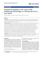
gel electrophoresis. As shown in Fig. 1, the C563T point
mutation, responsible for the Mediterranean clinical variant,
was then identified by digestion of PCR-amplified product
with MboII [19]. A complete haematological screening of
these subjects revealed no other alteration. For control
purposes we also investigated 14 healthy men of the same age.
Erythrocyte oxidative treatment
Erythrocytes were subjected to oxidative stress as described
previously [15], with modifications. Cells, prepared as
described above, were filtered through nylon net and
thoroughly washed with phosphate buffered saline,
spH 7.4, before the oxidative treatment. This was per-
formed by incubating the cells at 37 °C in a shaking water
bath, in the presence of t-BHP at the indicated concentra-
tion, in 25-mL flasks (final haematocrit 10%). After
incubation, supernatants were used to determine levels
of thiobarbituric acid-reactive substances (TBARS) as
described below. Erythrocytes were washed seven times
with isotonic buffer to remove t-BHP.
Evaluation of oxidation markers
Determination of methaemoglobin and oxyhaemoglo-
bin. Methaemoglobin and oxyhaemoglobin contents were
determined by a spectrophotometric method [20]. Briefly,
5 lL packed oxidized erythrocytes were mixed with 995 lL
stabilizing solution (2.7 m
M
EDTA, pH 7.0, and 0.7 m
M
2-mercaptoethanol). After shaking, oxyhaemoglobin and
methaemoglobin concentrations were measured spectro-
photometrically.
Ó FEBS 2002 Protein methylation in G6PD-deficient erythrocytes (Eur. J. Biochem. 269) 2033
Evaluation of lipid peroxidation. Lipid peroxidation was
evaluated by detecting the amount of TBARS, mainly
malondialdehyde, as described previously [21]. Briefly, 2 mL
of the supernatant of oxidized erythrocyte pellet was mixed
with 1 mL 30% (w/v) trichloroacetic acid and centrifuged at
5000 g for 15 min. A 2-mL aliquot of supernatant was added
to 0.5 mL 1% (w/v) thiobarbituric acid in 0.05
M
NaOH and
heated in a boiling-water bath for 10 min. The absorbance of
the developing pink chromophore was measured at 532 nm.
Methyl esterification of membrane proteins in intact
erythrocytes (
in situ
assay)
Every day at least one patient and one control sample was
processed in parallel. When oxidative stress was applied,
control and patient samples were treated at the same time.
Intact erythrocytes were incubated with methyl-labelled
methionine, the in vivo precursor of AdoMet [6]. First,
250 lL packed erythrocytes were resuspended in an equal
volume of 5 m
M
Tris/HCl buffer (pH 7.4), containing
160 m
M
NaCl, 0.96 m
M
MgCl
2
,and2.8m
M
glucose. Then
0.93 nmol
L
-[methyl-
3
H]methionine (15 lCi) was added and
the mixture incubated at 37 °C for 60 min. Cells were then
haemolysed in hypotonic buffer (5 m
M
sodium phosphate,
pH 8.0, containing 25 m
M
phenylmethanesulfonyl fluoride).
Membranes were then washed twice with the same hypotonic
solution at decreasing pH (7.2 and 6.2) to preserve methyl
ester stability. Radioactivity incorporated as protein methyl
esters was determined after solubilizationof10 lLmembrane
preparation in 125 lL10 m
M
acetic acid/2.5% SDS. Protein
concentration was determined as described previously [6].
Electrophoretic analysis of membrane proteins
SDS/PAGE of membrane erythrocytes was performed by
method of Fairbanks et al.[22] with modifications [9]. The
gels were 1.5 mm thick and contained acrylamide mix 5.6%
(mass/vol), in the presence of 1% SDS, at pH 7.4. All
samples were run in duplicate so that one control and one
treated (and/or patient) samples were analysed in parallel on
each gel half of the same gel. At the end of the run, gels were
cut into half, and one half was stained with Coomassie
Brilliant Blue to visualize protein bands and densitometri-
cally scanned for area quantification [9]. The other half was
used for methyl ester quantification. For this, lanes were
sliced into 2-mm fractions and the incorporated radioacti-
vity was determined after elution of proteins from each slice
[6,9]. Radioactivity was expressed as d.p.m./band area.
Determination of AdoMet and
S
-adenosylhomocysteine
intracellular content
Intracellular concentrations of AdoMet and S-adenosyl-
homocysteine were determined by HPLC in a perchloric
acid-soluble fraction of erythrocyte cytosol [23]. All samples
were filtered through a 0.2-lm pore filter before injection on
to a Zorbax C8 reverse-phase column (25 cm · 4 mm; Du
Pont-New England Nuclear, Boston, MA, USA), equili-
brated with buffer A (50 m
M
NaH
2
PO
4
/10 m
M
heptanesulf-
onic acid buffer, pH 3.2), containing 4% (w/v) acetonitrile.
Nucleosides were eluted with a 15-min linear gradient of
4–20% acetonitrile, followed by a 10-min linear gradient of
20–25% acetonitrile, at a flow rate of 1 mLÆmin
)1
.
Enzyme assays
PCMT specific activity was determined, in vitro, in the
cytosol of erythrocytes subjected to oxidative stress, as
previously described [9,24]. Erythrocyte lysates were
obtained by rapid freeze–thawing after 10-fold dilution of
oxidized erythrocytes with a stabilizing solution (2.7 m
M
EDTA, pH 7.0, and 0.7 m
M
2-mercaptoethanol) [16].
Wild type
AB
Mediterranean
417 120
120100317
1a 1b 2
417
1 2 3 4 5 6 7 8 9 10 11 St
317
120
100
Fig. 1. Diagnostic assessment of molecular defect in G6P-deficient patients. Patient selection, sampling and DNA extraction were as described in
Materials and Methods. The C563T mutation, associated with the Mediterranean variant was identified after PCR amplification of exon 5 and 6,
followed by digestion with MboII restriction enzyme. (A) Schematic representation of the expected restriction fragment length polymorphism
(RFLP) in wild-type and Mediterranean mutants, where the latter show an additional MboII site. (B) RFLP analysis of some patients and controls.
1, 3, 10, Mediterranean variant male patients; 4, 5, heterozygous females; 2, 7, 9, 11, normal controls. Mutants are characterized by sensitivity to
MboII digestion of PCR amplified fragments, yielding additional bands of 317 and 100 bp, respectively.
2034 D. Ingrosso et al.(Eur. J. Biochem. 269) Ó FEBS 2002
Membranes were removed by centrifugation at 10 000 g for
20 min. The assay mixture contained, in a final volume of
40 lL, 1.6 mg ovalbumin as the methyl acceptor, 2.8 mg
cytosolic proteins, 0.1
M
sodium citrate buffer, pH 6.0, and
30 l
M
(final concentration) S-adenosyl-
L
-[methyl-
14
C]-
methionine as the methyl donor. After incubation at
37 °C for 10 min, the reaction was quenched by adding
an equal volume (40 lL) of 0.2
M
NaOH/1% (w/v) SDS.
Radioactivity due to methyl incorporation was determined
as previously described [9]. Results are expressed as enzyme
units (pmol methyl ester formedÆmin
)1
) per mg haemoglo-
bin. Haemoglobin concentration was determined spectro-
photometrically [24].
PK activity was evaluated as a cell-age marker by the
method of Beutler et al.[16].
Statistical analysis
Statistical analysis was performed by Student’s paired or
unpaired t-test. Results are presented as the mean ± SE.
Differences were considered significant at P < 0.05.
RESULTS AND DISCUSSION
Methyl esterification of membrane proteins
is increased particularly in the G6PD-deficient
‘dense’ erythrocyte fraction
There is evidence from at least three independent labora-
tories that methyl esterification of membrane proteins,
catalysed by PCMT, is increased as erythrocytes age in the
circulation [6,25,26]. This has been related to an increased
number of abnormal aspartate residues, spontaneously
arising from
L
-asparaginyl deamidation and/or
L
-aspartyl
isomerization reactions [7]. In addition it has been reported
that isoaspartate residues, detected by the PCMT in situ
assay, increase in membrane proteins of normal erythro-
cytes subjected to oxidative stress [14], suggesting that
susceptibility to oxidative damage may render these mem-
brane protein components more prone to deamidation/
isomerization.
We measured methyl esterification of membrane proteins
in G6PD-deficient erythrocytes, to establish if abnormal
isoaspartate residues occur, in this condition, at a higher
rate than normal, while in the circulation. To this end, cells
were fractionated according to density, the two most
abundant, intermediate fractions being used in the subse-
quent procedure. Cell recovery in these fractions, with
respect to the total amount of cells loaded on to the
gradient, was 70.3 ± 5.1% (control) vs. 80.9 ± 3.8%
(G6PD). Cell percentages were 46.6 ± 4.1% (buoyant
fraction) vs. 23.7 ± 1.9 (dense fraction) for the control
samples, and 61.0 ± 3.2% (buoyant fraction) vs.
19.9 ± 2.3 (dense fraction) for the G6PD samples. An
equal number of cells from each fraction was incubated with
methyl-labelled methionine, the in vivo AdoMet precursor.
PK activity was measured in parallel, as a cell age marker, in
cytosolic extracts of the same erythrocyte fractions. PK
activities in the dense cell fraction, in both normal and
G6PD-deficient cells, were always significantly lower than in
the corresponding buoyant cell fraction (Fig. 2A), confirm-
ing that PK is a suitable cell age marker in the G6PD-
deficient as well the normal erythrocyte. Moreover PK
activity in the G6PD-deficient erythrocytes was significantly
higher than in the corresponding control cell populations
(Fig. 2A). This is consistent with G6PD-deficient erythro-
cytes having a reduced half-life in the circulation.
Each cell fraction from individual patient populations
was then subjected to the in situ methylation assay. A
general increase in methyl ester formation with cell age was
found in membrane proteins of both pathological and
normal erythrocytes. However, this age-dependent increase
in methylation (i.e. protein damage) was significantly more
marked in the membrane of G6PD-deficient erythrocytes
than controls (Fig. 2B), despite the fact that the control cells
were older (i.e. their life span was prolonged) according to
PK activity.
As a whole, the results show that, in G6PD deficiency,
erythrocyte membrane proteins have an increased tendency
to isoaspartate formation, in spite of the reduced half-life of
the circulating erythrocyte population. In other words, the
increase in altered residues resulting from protein deamida-
tion/isomerization reactions show a premature onset with
erythrocyte ageing, in the G6PD deficiency Mediterranean
variant.
Oxidative stress increases membrane protein ‘fatigue’
damage in G6PD-deficient erythrocytes
It has been reported that oxidation, induced by cell
treatment with t-BHP, leads to significant membrane
alterations, including the occurrence of deamidated/isomer-
ized Asx residues of membrane-cytoskeletal proteins [15].
Fig. 2. Membrane protein methylation levels and PK activity of density-
fractionated G6PD-deficient erythrocytes. (A) PK activity, as a cell-age
marker, was determined in erythrocyte cytosol, as detailed in Materials
and methods. (B) Membrane protein methylation levels were deter-
mined in two different erythrocyte age/density fractions obtained by
isopycnic centrifugation on a Percoll gradient. Methyl esterification in
intact erythrocytes was assayed by incubating them in the presence of
[
3
H]methionine according to the in situ procedure (see Materials and
methods).
Ó FEBS 2002 Protein methylation in G6PD-deficient erythrocytes (Eur. J. Biochem. 269) 2035
We were therefore intrigued to investigate whether the
abnormal susceptibility of G6PD-deficient erythrocytes to
oxidative stress could be responsible for their increased
tendency to isoaspartate formation in membrane proteins.
Therefore we monitored development of this alteration to
evaluate its pathophysiological meaning in the mechanism
of cell damage, using the in situ methylation assay, in
isolated G6PD-deficient erythrocytes subjected to oxidative
stress.
Erythrocytes from both normal control and G6PD-
deficient patients were subjected to oxidant treatment, with
t-BHP, before the in situ methylation assay. To limit
possible interference of cell manipulation with the oxidative
stress conditions, cells were not fractionated according to
density. The effects of reactive oxygen species on erythrocyte
membranes were assessed by measuring lipid peroxidation
products (Fig. 3A), which showed a dramatic rise. Proteins
were also generally affected by the oxidative treatment, as
demonstrated by the increase in methaemoglobin concen-
tration (Fig. 3B). As expected, the effects of the oxidant
treatment were much more dramatic on G6PD-deficient
erythrocytes than on normal cells.
We next evaluated the effects of such treatment on
isoaspartate formation, by measuring the levels of mem-
brane protein methyl esters by the in situ methylation assay.
PK activity was also measured in parallel, to assess the loss
of the older cell fractions as the result of possible haemol-
ysis. Figure 4A shows that erythrocyte exposure to oxida-
tive conditions resulted in higher intracellular PK activity,
probably due to haemolysis, which affected the oldest,
intrinsically less resistant cells, so that the remaining
erythrocyte population was significantly younger. This
effect was clearly more pronounced in G6PD-deficient than
normal erythrocytes.
Methyl esterification of membrane proteins was mea-
sured in parallel, in order to monitor isoaspartate forma-
tion. Figure 4B shows that such abnormal residues
increased in membrane proteins in response to oxidants in
both normal and pathological erythrocytes, but to different
extents. This effect was in fact significantly more marked in
G6PD deficiency, particularly when we consider that, in this
disease, the erythrocyte population that survived the in vitro
oxidative stress was younger than the controls (compare
Figs 4A and 4B). No significant differences were noted in
the AdoMet and AdoHcy concentrations, as well as in
PCMT specific activity, after oxidative treatment, confirm-
ing our previous findings [15].
As a whole, the results indicate that the lack of
reducing power is a crucial element in conditioning
erythrocyte susceptibility to undergo membrane protein
damage in the form of Asx deamidation/isomerization.
Isoaspartate formation may also be one of the ultimate
events in cell destruction. Evidence shows that erythrocyte
removal during cell ageing or after oxidative damage is
mediated by binding of band 3 antibodies to band 3
antigenic sites [5]. Therefore, the occurrence of altered
aspartate residues in band 3 of normal and abnormal
erythrocytes during ageing [6,7] or oxidative stress [15]
may be relevant to the fact that the same protein becomes
a major site of new antigen generation under the same
conditions.
It should be pointed out, in this respect, that erythrocyte
ageing was initially believed to be the main determinant of
isoaspartate formation in membrane proteins [6]. Our
results are in line with a different interpretation, which
underscores the equally important role played by cell stress
in the occurrence of such protein damage. This may be
particularly relevant to pathological conditions, such as
Fig. 3. Evaluation of oxidation markers in G6PD-deficient erythrocytes
subjected to oxidative stress. Measurements were performed on both
G6PD-deficient and normal control erythrocytes after exposure to
oxidative stress with t-BHP. (A) TBARS evaluation of incubation
medium; (B) methaemoglobin content in erythrocyte cytosol.
Fig. 4. Membrane protein methylation levels and PK activity of G6PD-
deficient erythrocytes subjected to oxidative stress. (A) PK activity, as a
cell age marker, was measured in parallel in the cytosolic fraction of the
same cell preparations. (B) Membrane protein methylation levels were
evaluated by the in situ methylation assay.
2036 D. Ingrosso et al.(Eur. J. Biochem. 269) Ó FEBS 2002
spherocytosis or G6PD deficiency, in which erythrocytes are
intrinsically altered, so that they are more prone to this type
of protein alteration than normal.
Protein methyl esterification as an adaptive response
to cell exposure to damaging conditions
Different spontaneous post-biosynthetic modifications have
been shown to occur during erythrocyte senescence. For
example nonenzymatic glycosylation of haemoglobin (gly-
cation) has been shown to increase in the course of
mismanaged hyperglycaemia in diabetes [27]. Haemoglobin
is a useful protein marker of this kind of damage, although a
number of other protein molecules are known to be affected
by this alteration, with unpredictable functional conse-
quences.
Deamidation/isomerization of Asx residues has been
shown to occur in haemoglobin a chain [28]. Haemoglobin
mutations have been also shown to increase its susceptibility
to deamidation, such as in the case of haemoglobin
ÔProvidenceÕ [29]. Nevertheless the major targets of Asx
deamidation/isomerization during cell ageing are several
constituents of the membrane-cytoskeletal network. The
electrophoretic shift of protein 4.1 has been shown to occur
in aged erythrocytes, and it is due to deamidation of
sensitive asparagine residues [8]. This protein is involved in
the maintenance of erythrocyte shape and deformability, by
stabilizing interactions between the spectrin–actin network
and integral membrane proteins glycophorin C and band 3
(AE1) [30].
The functional consequences of deamidation/isomeriza-
tion have often been investigated under near-pathological
conditions. Homozygous knockout mice for PCMT are
affected by growth retardation, and die prematurely with
tonic-clonic seizures [31,32]. In these animals, isomerized
proteins accumulate in all organs and tissues, indicating lack
of PCMT-driven repair activity [31,32]. However, the
functional outcome of such alterations on individual
proteins is still uncertain, although the biological activity
of different proteins appears to be compromised in vitro by
deamidation and isomerization. Previous experience with
several cell models has shown that the isoaspartate content
of intracellular proteins is increased as the result of heat
shock [33] as well as of UVA irradiation [34]. As far as the
Fig. 5. SDS/PAGE profile of membrane proteins from G6PD-deficient
and normal erythrocytes subjected to oxidative stress. Oxidative stress
was induced, where indicated, by t-BHP treatment. Lane 1, nonoxi-
dizednormalerythrocyte;lane2,oxidizednormalerythrocyte;lane3,
nonoxidized G6PD-deficient erythrocyte; lane 4, oxidized G6PD-
deficient erythrocyte.
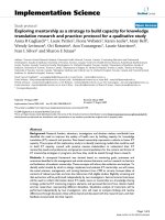
Fig. 6. Schematic representation of the overall hypothesis on the relationships between oxidative stress and isoaspartate formation in G6PD deficiency.
G6PD-deficient erythrocytes are intrinsically less resistant to subliminal oxidant levels, so that protein deamidation/isomerization products (i.e.
isoaspartate residues) tend to accumulate despite the fact that the life span of these cells is, on average, shorter than normal. In other words, they
reach levels of aspartate damage that are typical of a much older normal erythrocyte population. Exposure to certain foods or drugs (fava beans,
nonsteroidal anti-inflammatory drugs, antimalaria drugs, chemotherapeutics, etc.) trigger the haemolytic crisis, which is also associated with a
further increase in the levels of deamidated/isomerized proteins. The mechanism linking oxidation to haemolysis involves membrane alterations.
Ó FEBS 2002 Protein methylation in G6PD-deficient erythrocytes (Eur. J. Biochem. 269) 2037
erythrocyte is concerned, there is evidence that deamida-
tion/isomerization of Asx residues, monitored by methyla-
tion, is significantly increased under cell stress conditions.
This occurs when erythrocytes are subjected to haemody-
namic shear forces in a metabolically hostile microenviron-
ment, such as the spleen microcirculation [35]. These results
support the role of protein fatigue damage in the mechanism
of spleen conditioning, in haemocatheresis [35,36]. It has
also been shown that membrane–cytoskeletal proteins of
resealed/engineered erythrocytes display increased suscepti-
bility to Ômolecular fatigueÕ, detected by methyl esterifica-
tion, after repeated osmotic stress [14].
In a previous report on the effects of oxidative stress on
normal erythrocytes, we found that treatment with t-BHP
increased isoaspartate occurrence in membrane proteins
[15]. Conversely, we did not observe under such conditions
any of the extensive membrane alterations described by
others, including formation of protein aggregates with
haemoglobin [15].
The electrophoretic pattern of membrane proteins from
G6PD-deficient cells, both treated and untreated with
oxidants, was similar to that of controls (Fig. 5). This
allows us to conclude that molecular alterations, in the form
of isoaspartate residues, take place on t-BHP treatment,
before and not as a consequence of massive alterations of
membrane protein composition. The results indicate that
protein damage at the aspartate level is a sensitive and early
marker of erythrocyte exposure to oxidants, before the
appearance of more extensive damage of morphological
relevance [15].
In conclusion, the data presented here show that G6PD
deficiency, which renders erythrocyte adaptation to an
oxidative microenvironment more difficult, makes mem-
brane proteins more prone to isoaspartate formation, both
during cell ageing and, even more so, under stress conditions
(see scheme in Fig. 6). Taken as a whole, the results support
the role of this post-biosynthetic protein modification in the
mechanism of haemolysis in G6PD deficiency.
ACKNOWLEDGEMENTS
Genetic testing of patients was accomplished at the International
Institute of Genetic and Biophysics (I.I.G.B.) of the National Research
Council, Naples, Italy, under the supervision of Dr Giuseppe Martini
and Stefania Filosa. The work was supported in part by research grants
from Ministero dell’Istruzione, dell’Universita
`
e della Ricerca, Progetti
di Rilevante Interesse Nazionale (M.I.U.R. P.R.I.N., 1999): ÔExtra and
intracellular nucleotide and nucleoside: chemical signals, metabolic
regulators and potential drugsÕ and ÔHyperhomocysteinemia as a
cardiovascular risk factor: biochemical mechanism(s)Õ.
REFERENCES
1. Luzzatto, L., Mehta, A. & Vulliamy, T. (2001) Glucose 6-phos-
phate dehydrogenase deficiency. In The Metabolic and Molecular
Bases of Inherited Disease (Scriver,C.R.,Beaudet,A.L.,Sly,W.S.
& Valle, D., eds), vol. 3, pp. 4517–4553. McGraw-Hill, New York.
2. Mason, J.P. (1996) New insights into G6PD deficiency. Br.
J. Haematol. 94, 585–591.
3. Beutler, E., Vulliamy, T. & Luzzatto, L. (1996) Hematological
important mutation: glucose-6-phosphate dehydrogenase. Blood
Cell Mol. Dis. 22, 49–56.
4. Pandolfi, P.P., Sonati, F., Rivi, R., Mason, P., Grosveld, F. &
Luzzatto, L. (1995) Target disruption of the housekeeping gene
encoding glucose 6-phosphate dehydrogenase (G6PD): G6PD is
dispensable for defense against oxidative stress. EMBO J. 14,
5209–5215.
5. Arese, P. & De Flora, A. (1990) Pathophysiology of hemolysis in
glucose-6-phosphate dehydrogenase deficiency. Semin. Hematol.
27, 1–40.
6. Galletti, P., Ingrosso, D., Nappi, A., Gragnaniello, V., Iolascon,
A.&Pinto,L.(1983)Increasedmethylesterificationofmembrane
proteins in aged red-blood cells. Preferential esterification of
ankyrin and band 4.1 cytoskeletal proteins. Eur. J. Biochem. 13,
25–31.
7. Galletti, P., Ingrosso, D., Manna, C., Clemente, G. & Zappia, V.
(1995) Protein damage and methylation-mediated repair in the
erythrocyte. Biochem. J. 306, 313–325.
8. Inaba, M., Gupta, K.C., Kuwabara, M., Takahashi, T., Benz, E.J.
Jr & Maede, Y. (1992) Deamidation of human erythrocyte protein
4.1: possible role in ageing. Blood 79, 3355–3361.
9. Ingrosso, D., D’Angelo, S., Perna, A.F., Iolascon, A., Miraglia del
Giudice, E., Perrotta, S., Zappia, V. & Galletti, P. (1995) Increased
membrane-protein methylation in hereditary spherocytosis. A
marker of cytoskeletal disarray. Eur. J. Biochem. 228, 894–898.
10. Ingrosso, D., D’Angelo, S., Perrotta, S., D’Urzo, G., Iolascon, A.,
Perna, A.F., Galletti, P., Zappia, V. & Miraglia del Giudice, E.
(1996) Cytoskeletal behaviour in spectrin and in band 3 deficient
spherocytic red cells: evidence for a differentiated splenic con-
ditioning role. Br. J. Haematol. 93, 38–41.
11. Johnson, B.A., Langmack, E.L. & Aswad, D.W. (1987) Partial
repair of deamidation-damaged calmodulin by protein carboxyl
methyltransferase. J. Biol. Chem. 262, 12283–12287.
12. Ryttersgaard, C., Griffith, S.C., Sawaya M.R., MacLaren D.C.,
Clarke, S. & Yeates, T.O. (2002) Crystal structure of human
L
-isoaspartyl methyltransferase. J. Biol. Chem., Manuscript
M200229200.
13. Ota, I.M. & Clarke, S. (1990) The function and enzymology of
protein
D
-aspartyl/
L
-isoaspartyl methyltransferase in eukaryotic
and prokaryotic cells. In Protein Methylation (Paik, W.K. & Kim,
S., eds), pp. 179–194. CRC Press, Boca Raton, FL.
14. Ingrosso, D., Cotticelli, M.G., D’Angelo, S., Buro, M.D., Zappia,
V. & Galletti, P. (1997) Influence of osmotic stress on protein
methylation in reasealed erythrocytes. Eur. J. Biochem. 244,918–
922.
15. Ingrosso, D., D’Angelo, S., di Carlo, E., Perna, A.F., Zappia, V. &
Galletti, P. (2000) Increased methyl esterification of altered
aspartyl residues in erythrocyte membrane proteins in response to
oxidative stress. Eur. J. Biochem. 267, 4397–4405.
16. Beutler, E., Blume, K.G., Lohr, G.W., Ramot, B. & Valentine,
W.N. (1977) International committee for standardization in hae-
matology: recommended methods for red-cell enzyme analysis.
Br. J. Haematol. 35, 331–341.
17. Vettore, L., De Matteis, M.C. & Zampini, P. (1980) A new density
gradient system for the separation of human red blood cells.
Am. J. Hematol. 8, 291–297.
18. Sykes, B.C. (1983) DNA in heritable disease. Lancet 2, 787–788.
19. Alfinito, F., Cimmino, A., Ferraro, F., Cubellis, M.V., Vitagliano,
L.,Francese,M.,Zagari,A.,Rotoli,B.,Filosa,F.&Martini,G.
(1997) Molecular characterization of G6PD deficiency in Southern
Italy: heterogeneity, correlation genotype-phenotype and descrip-
tion of a new variant (G6PD Neapolis). Br. J. Haematol. 98, 41–46.
20. Winterbourn, C.C. (1990) Oxidative reactions of hemoglobin.
Methods Enzymol. 186, 265–272.
21. Caprari, P., Bozzi, A., Malorni, W., Bottini, A., Iosi, F., Santini,
M.T. & Salvati, A.M. (1995) Junctional sites of erythrocyte ske-
letal proteins are specific targets of tert-butylhydroperoxide oxi-
dative damage. Chem-Biol Interact. 94, 243–258.
22. Fairbanks, G., Steck, T.L. & Wallach, D.F.H. (1971) Electro-
phoretic analysis of the major polypeptides of the human ery-
throcyte membrane. Biochemistry 10, 2606–2616.
2038 D. Ingrosso et al.(Eur. J. Biochem. 269) Ó FEBS 2002
23. Cools, M., Hasobe, M., De Clerq, E. & Borchardt, R.T. (1990)
Mechanism of the synergistic antiviral and cytostatic activity of
(RS)-3-(adenin-9-yl)-2-hydroxypropanoic acid isobutyl ester and
D
,
L
-homocysteine. Biochem. Pharmacol. 39, 195–202.
24. Gilbert, J.M., Fowler, A.V., Bleibaum, J. & Clarke, S. (1988)
Purification of homologous protein carboxyl methyltransferase
isozymes from human and bovine erythrocytes. Biochemistry 27,
5227–5233.
25. Barber, J.R. & Clarke, S. (1983) Membrane protein carboxyl
methylation increases with human erythrocyte age. J. Biol. Chem.
258, 1189–1196.
26. O’Connor, M.C. & Yutzey, K.E. (1998) Enhanced carboxyl
methylation of membrane-associated hemoglobin in human ery-
throcytes. J. Biol. Chem. 263, 1386–1130.
27. Vlassara, H. (1990) Advanced non-enzymatic tissue glycosylation:
mechanism implicated in the complications associated with aging.
In Molecular Biology of Aging (Finch, C.E. & Johnson, T.E., eds),
pp. 171–185. Wiley-Liss, New York.
28. Ladino, C.A. & O’Connor, C.M. (1991) Identification of a site for
carboxyl methylation in human a-globin. Biochem. Biophys. Res.
Commun. 180, 742–747.
29. Bonaventura, J., Bonaventura, C., Sullivan, B., Ferruzzi, G.,
McCurdy, P.R., Fox, J. & Moo-Penn, W.F. (1976) Hemoglobin
providence. Functional consequences of two alterations of the
2,3,diphosphoglycerate binding site at position b82. J. Biol. Chem.
251, 7563–7571.
30. Cohen, C.M. & Gastard, P. (1992) Regulation and post-trasnla-
tional modofocation of erythrocyte membrane protein and
membrane skeletal proteins. Semin. Hematol. 29, 244–292.
31. Kim, E., Lowenson, J.D., MacLaren, D.C., Clarke, S. & Young,
S.G. (1997) Deficiency of a protein-repair enzyme results in
the accumulation of altered proteins, retardation of growth,
and fatal seizures in mice. Proc. Natl. Acad. Sci. USA 94,
6132–6137.
32. Kim, E., Lowenson, J., Clarke, S. & Young, S.G. (1999) Pheno-
typic analysis of seizure-prone mice lacking
L
-isoaspartate
(
D
-aspartate) O-methyltransferase. J. Biol. Chem. 274, 20671–
20678.
33. Ladino, C.A. & O’Connor, C.M. (1992) Methylation of atypical
protein aspartyl residues during the stress response of the HeLa
cells. J. Cell. Physiol. 153, 297–304.
34. D’Angelo, S., Ingrosso, D., Perfetto, B., Baroni, A., Zappia, M.,
Lobianco Lubrano, L., Tufano, M.A. & Galletti, P. (2001) UVA-
irradiation induces
L
-isoaspartyl formation in melanoma cell
proteins. Free Radic. Biol. Med. 31,1–9.
35. Gallagher, P.G., Forget, B.G. & Lux, S.E. (1998) Disorders of the
erythrocyte membrane. In Hematology of Infancy and Childhood
(Nathan. D.G. & Oski, F.A., eds), pp. 544–664. Saunders, Phila-
delphia.
36. Bartosz, G. (1990) Erythrocyte membrane changes during ageing
in vivo.InBlood Cell Biochemistry (Harris, J.R., ed.), pp. 45–81.
Kluwer Academic/Plenum Publishers, New York.
Ó FEBS 2002 Protein methylation in G6PD-deficient erythrocytes (Eur. J. Biochem. 269) 2039