Báo cáo Y học: Binding of Thermomyces (Humicola) lanuginosa lipase to the mixed micelles of cis-parinaric acid/NaTDC Fluorescence resonance energy transfer and crystallographic study ppt
Bạn đang xem bản rút gọn của tài liệu. Xem và tải ngay bản đầy đủ của tài liệu tại đây (327.54 KB, 9 trang )
Binding of
Thermomyces
(
Humicola
)
lanuginosa
lipase to the mixed
micelles of
cis
-parinaric acid/NaTDC
Fluorescence resonance energy transfer and crystallographic study
Ste
´
phane Yapoudjian
1
, Margarita G. Ivanova
1
, A. Marek Brzozowski
2
, Shamkant A. Patkar
3
, Jesper Vind
3
,
Allan Svendsen
3
and Robert Verger
1
1
Laboratoire de Lipolyse Enzymatique CNRS-IFR1, Marseille, France;
2
Structural Biology Laboratory, Chemistry Department,
University of York, UK;
3
Enzyme Research, Novozymes A/S, Bagsvaerd, Denmark
The binding of Thermomyces lanuginosa lipase and its
mutants [TLL(S146A), TLL(W89L), TLL(W117F,
W221H, W260H)] to the mixed micelles of cis-parinaric acid/
sodium taurodeoxycholate a t pH 5.0 led t o the quenching of
the intrinsic tryptophan fluorescence emission (300–380 nm)
and to a simultaneous increase in the cis-parinaric acid
fluorescence emission (380–500 nm). These findings were
used to characterize the Thermomyces lanuginosa lipase/cis-
parinaric acid interactions occurring in the presence of
sodium taurodeoxycholate.The fluorescence resonance
energy transfer and Stern–Volmer quenching constant
values obtained were correlated with the accessibility of the
tryptophan r esidues to the cis-parinaric acid and with the lid
opening ability of Thermomyces lanuginosa lipase (and its
mutants). TLL(S146A) was found to have the highest
fluorescence resonance energy t ransfer. In ad dition, a
TLL(S146A)/oleic acid complex was crystallised and its
three-dimensional structure was solved. Surprisingly, two
possible binding modes (sn-1 and antisn1) were found to exist
between oleic acid and the catalytic cleft of the open con-
formation of TLL(S146A). Both binding modes involved an
interaction with tryptophan 89 of the lipase lid, in agreement
with fluorescence resonance energy transfer experiments.As
a consequence, we concluded that TLL(S146A) mutant is
not an appropriate substitute for the wild-type Thermomyces
lanuginosa lipase for mimicking the interaction between the
wild-type enzyme and lipids.
Keywords: lipase; X-ray crystallography; cis-parinaric acid;
fluorescence resonance energy transfer.
Lipases (EC 3.1.1.3) can be defined as enzymes that catalyze
the hydrolysis of long-chain acyl-glycerols [1]. In addition to
playing an important role in fat catabolism, they have
numerous applications in the food, cosmetics, detergent and
pharmaceutical industries [2–5].
In recent years, the three-dimensional structures of lipases
and lipase–inhibitor complexes have been determined using
X-ray crystallographic methods [6–11]. All lipases show a
common a–b hydrolase fold [12] and a catalytic triad
composed of a nuc leophilic serine, which is ac tivated via
hydrogen bond s as part of a charge relay system, along with
the histidine and the aspartate or glutamate residues [6,7].
The crystal structures of some lipases have shown that t he
active site is covered by a helical surface loop or ÔlidÕ that
renders the active site inaccessible to substrate. This is
referred to as the closed conformation of the lipase. On the
other hand, the three-dimensional structures of lipases
complexed with inhibitors shows a rearrangement of the lid,
allowing free access to the active site in the so-called op en
conformation, in which a large hydrophobic surface around
the catalytic triad is exposed.
Thermomyces lanuginosa lipase (TLL) has four trypto-
phan residues located in positions 89, 117, 221 and 260. The
side chains of W117, W221 and W260 are buried into the
protein core, whereas the W89 residue is located in the
central part of the helical lid [13]. In the crystal structures of
the open forms of TLL, W89 is in close van der Waals
contact with the acyl moiety of an in hibitor mimicking the
transition state [14,15].
The fluorescence technique was used previously to study
the binding of TLL to small or large unilamellar vesicles of
1-palmitoyl-2-oleoylglycero-sn-3-phosphoglycerol (POPG)
and to vesicles of zwitterionic phospholipids such as
1-palmitoyl-2-oleoylglycero-sn-3-phosphocholine [16]. The
authors concluded that TLL may bind with a similar a ffinity
to all t ypes of pho spholipid vesicles and may adopt a
catalytically active conformation and be involved in inter-
facial activation processes only when small unilamellar
vesicles of POPG are used. Furthermore, molecular
Correspondence to R. Verger, Laboratoire de Lipolyse Enzymatique,
CNRS-IFR1, 31 chemin Joseph Aiguier, 13402 Marseille cedex 20,
France.Fax:+3391715857,Tel.:+3391164093,
E-mail:
Abbreviations: cis-PnA, cis-parinaric acid; CMC, critical micellar
concentration; FRET, fluorescence resonance energy transfer; K
SV
,
Stern–Volmer quenching constant; NaTDC, sodium taurodeoxycho-
late; OA, oleic acid; POPG, 1-palmitoyl-2-oleoylglycero-sn-3-phos-
phoglycerol; RFI, relative fluorescence intensity; TLL, Thermomyces
lanuginosa lipase; TLL(S146A), inactive mutant with S146 mutated to
A; TLL(W89L), mutant with W89 mutated to L; TLL(W117F,
W221H, W260H), mutant with only the W89; W 117 mutated to F,
W221 mutated to H and W260 mutated to H.
Note: t he atomic co -ordinat es have been deposited in the Brookhaven
ProteinDataBankwiththeaccessioncode1gt6.
(Received 20 August 2001, revised 5 December 2001, accepted 14
January 2002)
Eur. J. Biochem. 269, 1613–1621 (2002) Ó FEBS 2002
dynamics simulations [17] indicated that the replacement of
a single amino acid at the active site (S146A) may lead to
conformational alterations in TLL.
The aim of the present study was to investigate the TLL/
fatty acid interactions using the fluorescence resonance
energy transfer (FRET) technique.
One of the prerequisites to be able to observe the FRET
between a donor (TLL tryptophans) and an acceptor
(fattyacid)isthattheremustexistaspectraloverlap
between the donor emission and the accep tor absorption
spectra, and the donor and acceptor groups must be
the right distance apart a nd properly orien ted [18].
Therefore 9,11,13,15-cis,trans,trans,cis-oct adecatetraenoic
acid (cis-PnA), a naturally fluorescent f atty acid with
thoroughly characterized spectroscopic properties [19], was
chosen for use as a probe. It h as been previously
established that cis-PnA can act as an acceptor for the
tryptophan fluorescence emission [20], and its spectroscop-
ic properties have been used in studies on fatty acid
binding to various proteins [20,21].
Bile salts are the main detergent-like molecules respon-
sible for the solubilization of lipolytic products (monoglyc-
erides and free fatty acids) during the digestion of dietary
fats. Sodium taurodeoxycholate (NaTDC) is a conjugated
bile acid, which forms very small micelles in an aqueous
solution [22]. The mixe d micelles of cis-PnA/NaTDC turned
out to be a convenient model system for studying the
interactions between a water soluble protein (TLL) and a
fattyacidintheformofmixedmicelles.
First, we studied the lipase free cis-PnA/NaTDC system
in order to characterize the cis-PnA/NaTDC mixed micellar
system. The binding behavior of TLL (and its mutants) to
pure cis-PnA and to mixed micelles of cis-PnA/NaTDC was
then studied using the FRET technique. In addition, X-ray
crystallogaphy studies were performed on the S146A
mutant in order to elucidate the particular properties of its
complexes with f atty acids.
MATERIALS AND METHODS
Materials
NaTDC was from Sigma and cis-PnA was from Molecu-
lar Probes. A stock solution of 3.2 m
M
of cis-PnA in
ethanol containing 0 .001% (w/v) but ylhydroxytoluene
(BHT) as an antioxydant was stored in the dark at
)20 °C under a n argon atmosphere. T hese precautions
were taken to ensure that no polyene decomposition
would occur [20].
The TLL wild-type, its single mutants: TLL(S146A),
TLL(W89L), and triple mutant TLL(W117F, W221H,
W260H) were used. All enzymes were kindly provided by
A. Svendsen and S. A. Patkar from Novo Nordisk,
Denmark and prepared as described previously [23,24].
The buffers used were 10 m
M
Tris/HCl pH 8.0, 150 m
M
NaCl, 21 m
M
CaCl
2
,1m
M
EDTA and 10 m
M
acetate
pH 5.0, 100 m
M
NaCl, 20 m
M
CaCl
2
.
UV absorption spectroscopy
Differential absorption spectra were recorded on a Uvikon
860 spectrophotometer from Kontron Instruments. All
assays were carried out using two quartz cuvettes (optical
path length 1 cm) of 3.5 mL each: one for the assay and one
for t he control assay. The contents of each cuvette were
mixed 5–10 times by gentle inversion of the cuvette capped
with Teflon stopper, and were then left unstirred during the
measurement procedure. Measurements were performed at
room temperature. Two types of experiments were per-
formed. (a) Titration of cis-PnA was carried out by the
increasing amounts of NaTDC at pH 5.0, in the absence of
TLL. The assay and control cuvettes were both filled with
buffer and NaTDC at the various concentrations tested. cis-
PnA w as subsequently added to the assay c uvette from an
ethanolic stock solution and differential absorption spectra
were recorded between 200 and 450 mn. (b) Absorption
spectra of cis-PnA in the presence of TLL at pH 5.0 or
pH 8.0. The assay and control cuvettes were both filled with
buffer, NaTDC and TLL. cis-PnA was added afterwards
into the assay cuvette and the differential absorption spectra
were recorded.
Fluorescence spectroscopy
Fluorescence measurements were carried out at 29 °C
under con stant stirring using a SFM 25 spectrofluorimeter
from Kontron Instruments and a 3.5-mL quartz cuvette
(optical path length 1 cm). During all the fluorescence
measurements, the optical density was < 0.1 in the spectral
range between 280 nm and 500 nm to avoid inner filter
effect. T wo types of fluorescence experiments were
performed.
Titration of cis-PnA at various NaTDC concentrations at
pH 5.0. The cuvette was filled with buffer containing
NaTDC at a given concentration. cis-PnA was then added
to the cuvette and a fluorescence emission spectrum was
recorded at an excitation wavelength of 320 nm by
scanning at an emission wavelength ranging from 350 nm
to 5 00 nm.
FRET experiments. TLL (w ild-type or mutant) was titrated
at pH 5.0 or pH 8 .0 by adding increasing amounts of cis-
PnA in the presence and absence of NaTDC. The excitation
wavelength was set to 280 nm and the emission wavelength
ranged from 300 nm to 500 nm.
The accessibility of tryptophan to cis-PnA was estimated
by measuring the quenching of the TLL flu orescence
effected by cis-PnA, according to the Stern–Volmer equa-
tion [25]:
F
0
F
¼ 1 þ K
sv
½Qð1Þ
where F
0
and F are the fluorescence emission intensities in
the absence and in the presence of a quencher, respectively,
[Q] is the molar quencher concentration and K
SV
is the
Stern–Volmer quenching constant. K
SV
is appropriate for
collisional quenching i n which binding is not involved.
However, the Stern–Volmer equation fits well our experi-
mental results, even though binding is clearly involved.
Consequently, K
SV
will be replaced by ÔK
SV
Õ.
Protein crystallization and crystallography
TLL(S146A) solution was washed s everal times in 10K
Centricon in 10 m
M
Tris/HCl pH 8.0 buffer and concen-
1614 S. Yapoudjian et al. (Eur. J. Biochem. 269) Ó FEBS 2002
trated up to 20 mgÆmL
)1
. Crystallization trials were
performed u sing the hanging drop technique at 291 K.
Screening for the crystallization conditions was performed
simultaneously at pH 8.0 (0.1
M
Tris/HCl buffer) and
pH 5.0 (0.1
M
acetate buffer). OA was used instead of cis-
PnA for crystallization experiments to avoid oxidation
during the crystallization. OA was dissolved in iso-propanol
and mixed in this form with a protein sample at a 5 : 1
molar ratio (OA/lipase). After a 1-h incubation, the
resulting precipitate was removed by centrifugation in a
Sigma Eppendorf centrifuge (5 min, 18 000 g)andthe
remaining protein was used in the crystallization experi-
ments. NaTDC was added to the crystallization trials
separately at a concentration of 10 m
M
. Crystals were flash
frozen in the liquid n itrogen and characterized in-house o n
a Rigaku RU200 rotating anode source (k ¼ 1.5418 A
˚
),
MAR Research 345 imaging plate scanner, Osmic focusing
mirrors and Oxford Cryosystem set at 120 K. The X-ray
data were subsequently collected at the ESRF in Grenob le
on the MAR Research C CD detecto r at 100 K, proc essed
with
DENZO
and scaled and merged with
SCALEPACK
[26].
The s tructure was d etermined using th e Molecular
Replacement method. The lid was removed by molecular
modelling in
QUANTA
to get a model for molecular
replacement (lipase m inus lid). The
AMORE
software
program [27] was used and the wild-type TLL structure
[14] (minus the lid) was used as a model. The structure was
refined using maximum likelihood techniques with
REFMAC
[28]; other calculations were carried out using the
CCP
4
suite of programs (Collaborative Computational Project,
Number 4, 1994).
Electron d ensity map inspection, m odel building and
analysis were carried out with the
X
-
FIT
options of the
QUANTA
software program (Molecular Simulations Inc.).
RESULTS
Absorption spectroscopy
The UV absorption spectrum of cis-PnA was determined in
an ethanolic solution (95%) and found to be identical to
that obtained by Sklar et al.[19].Ascis-PnA is prone to
oxidation, the absorption spectrum of the stock solution
was checked regularly and no changes in the cis-PnA
absorption spectra were observed in t he ethanolic solution
upon storage.
The UV absorption spectrum of cis-PnA at pH 5.0 and
pH 8.0 as well as the fluorescence emission spectrum of
TLL (excited at 280 nm, at pH 5.0) in the presence of 1 m
M
NaTDC are shown in Fig. 1. At pH 5.0, the cis-PnA UV
absorption spectrum overlapped the TLL emission spec-
trum in the 290–380 nm wavelength range, whereas no
overlap can be observed at pH 8.0. No significant changes
in the TLL emission spectrum were detected between
pH 5.0 and pH 8.0 (data not shown).
The effects o f NaTDC on the UV absorption spectrum of
cis-PnA at pH 5.0 are shown in Fig. 2. In the absence of
NaTDC, the cis-PnA solution was slightly turbid. As soon
as the NaTDC concentration reached at least 1 m
M
,the
solution became optically clear changing simultaneously the
absorption spectrum of cis-PnA. Three main absorption
peaks appeared at 298 nm, 304 nm and 326 nm and
increased in proportion to the NaTDC concentration. This
increase in the attenuence of cis-PnA leveled off at NaTDC
concentrations above 4 m
M
.
Fluorescence spectroscopy
No significant NaTDC fluorescence was recorded under our
experimental conditions. The excitation and emission spec-
tra of cis-PnA were recorded at various NaTDC concen-
trations at pH 5.0. The maximum of the excitation and the
emission spectra were found to occur at 320 nm and
410 nm, respectively. In order to estimate the critical
micellar concentration (CMC) of NaTDC, the relative
fluorescence intensity (RFI) of cis-PnA at 410 nm (excita-
tion wavelength at 320 nm) was measured as a function of
the NaTDC c oncentration at pH 5.0. At NaTDC concen-
trations lower than 1 m
M
, t he fluorescence of cis-PnA was
Fig. 1. Fluorescence Emission spectra of TLL (––) and UV absorption
spectra of cis-PnA (- - -). TLL and cis-PnA concentrations were 0.8 l
M
and 10 l
M
, respectively. The buffer used was 10 m
M
acetate (pH 5.0)
100 m
M
NaCl, 20 m
M
CaCl
2
or 10 m
M
Tris (pH 8.0) 150 m
M
NaCl,
21 m
M
CaCl
2
,1m
M
EDTA. NaTDC concentration was 1 m
M
.The
excitation wavelength used to obtain the fluorescence emission spectra
was 280 nm.
Fig. 2. Effects of various NaTDC concentrations on the UV absorption
spectra of a solution of cis-PnA. The cis-PnA concentration was kept
constant at 10 l
M
.Bufferwas10m
M
acetate (pH 5.0) 100 m
M
NaCl,
20 m
M
CaCl
2
.( )0m
M
NaTDC, (– - –) 1 m
M
NaTDC, (– –)
2m
M
NaTDC, (––) 4 m
M
NaTDC. The s chematic diagram on the
right illustrates the experimental protocol u sed.
Ó FEBS 2002 Lipase binding to lipid, FRET and structural study (Eur. J. Biochem. 269) 1615
negligible. The RFI increased in parallel with the rise in the
NaTDC concentration above 1 m
M
. This increase leveled
off at NaTDC concentrations higher than 4 m
M
(data not
shown) .
The results of the FRET recordings obtained between
TLL and cis-PnA, at wavelengths r anging from 300 t o
500 nm in the presence of NaTDC at pH 5.0, a re presented
in Fig. 3. As the molar ratio (R) between cis-PnA and TLL
increased, the R FI decreased at wavelengths ranging from
300 to 380 nm and increased simultaneously at wavelengths
ranging from 380 to 500 nm.
From the data presented in Fig. 3, the decrease in RFI
(%), measured at the maximal emission wavelength (k
max
),
as well as the increase of R FI (%), measured at 410 nm, as a
function of cis-PnA concentration are presented in Fig. 4 . A
good quantitative correlation between increase and decrease
of RFI as a function of increasing concentration of cis-PnA
can be seen. Furthermore, a plateau value is reached when
one molecule of TLL is added to one molecule of cis-PnA
(R ¼ 1). Similar curves as those presented in Fig. 4 were
also obtained for TLL(S146A), TLL(W117F, W221H,
W260H) and TLL(W89L) (data not shown).
Similar FRET experiments were also pe rformed with
TLL, TLL(S146A), TLL(W117F, W221H, W260H),
TLL(W89L) and cis-PnA i n the presence and absence of
NaTDC (Fig. 5). In t he presence of NaTDC, the FRET was
observed between TLL, TLL(S146A), TLL(W117F,
W221H, W260H), TLL(W89L) and cis-PnA. In the absence
of NaTDC, the FRET was negligible. Surprisingly, in the
absence of NaTDC, a clear-cu t quenching process was
observed only with TLL(S146A) and TLL(W89L). Similar
experiments were performed at pH 8.0, in the presence of
Fig. 3. FRET between TLL and cis-PnA. The numbers refer to the
values of the molar ratio R of cis-PnAtoTLL.Inalltheassays,the
excitation wavelength was 280 nm. The dotted line corresponds to
the fluorescence emission spectra of cis-PnA (1 l
M
) recorded in the
absence of TLL under the same experimental conditions. The dashed
line corresponds to the arithmetic sum of the TLL and cis-PnA spectra
recorded separately. The correlation between quenched tryptophan
RFI (325 nm) and sensitized RFI of cis-PnA (410 nm) is p resented in
Fig. 4. TLL concentration was 0.8 l
M
and cis-PnA concentration
varied from 0 to 1 l
M
. For the sake of clarity, only spectrum corres-
ponding to three cis-PnA concentrations (0, 0.4 and 0.8 l
M
)areshown
(plain lines). The NaTDC concentr atio n was 1 m
M
.Bufferwas10m
M
acetate (pH 5.0) 100 m
M
NaCl, 20 m
M
CaCl
2
.
Fig. 4. RFI decrease ( d)atk
max
as well as RFI increas e (s)atk
410 nm
as a funct io n of cis-PnA concentration. Data from Fig. 3.
Fig. 5. FRET between TLL (and its mutants) and cis-PnA in the
presence and absence of NaTDC. The protein concentration was 0.8 l
M
and the cis-PnA concentration was varied stepwise from 0 to 1 l
M
(0,0.2,0.4,0.8,1l
M
). Excitation wavelength: 280 nm. Buffer pH 5.0
as in Fig. 2. The data in the graph at the uppermost left hand corner
are identical to those shown in Fig. 3.
1616 S. Yapoudjian et al. (Eur. J. Biochem. 269) Ó FEBS 2002
NaTDC (data not shown). Quenching was observed only
between TLL(S146A), TLL(W 89L) and cis-PnA.
The maximum fluorescence emission wavelengths (k
max
)
of TLL, TLL(S146A), TLL(W117F, W221H, W260H), and
TLL(W89L) (excitation at 280 nm, pH 5.0) with or without
NaTDC, in the presence or absence of cis-PnA are
summarized i n Table 1. In the absence of cis-PnA, th e
addition of NaTDC led to a blue shift of the k
max
of all the
lipases tested, except for TLL(W89L). Furthermore, in
contrast to what occurred with TLL(W89L), the addition of
cis-PnA in the presence of 1 m
M
NaTDC also led to a
significant blue shift in the case of TLL, TLL(S146A) and
TLL(W117F, W221H, W260H). It is worth noting that
TLL(W89L) displayed no significant blue shift under any of
the experimental c onditions tested.
Stern–Volmer plots for the fluorescence quenching of
TLL (mutants) by cis-PnA were calculated from the data
presented in Fig. 5, in the presence of 1 m
M
NaTDC (data
not shown). The ÔK
SV
Õ constants calculated for TLL,
TLL(S146A), TLL(W117F, W221H, W260H) and
TLL(W89L) were 3.2.10
6
, 4.6.10
6
, 3.4.10
6
and 1.5.10
6
M
)1
,
respectively.
X-ray crystallography
Good X-ray quality crystals of TLL(S146A) were obtained
in the presence o f O A in 0.1
M
Tris/HCl pH 8.0 buffer,
10 m
M
NaTDC, 25% w/v poly(ethylene glycol) 5K MME,
25 m
M
MgCl
2
. Crystallization at pH 5.0 and control setups
were unsuccessful under similar conditions with wild-type
TLL. Crystals of the TLL(S146A) mutant were found to
belong to the P2
1
2
1
2 space group and to have two
molecules in the asymmetric unit, a packing density of
2.64 A
˚
3
ÆDa
)1
and a solvent content of 53%. The final X-ray
data are 97.8% complete up to 2.20 A
˚
resolution (96.2% in
the 2.28–2.20 resolution shell) with an overall R
merge
of
0.075 (0.44), I/r(I) of 12.2, and a mean multiplicity of 3.2
observations per reflection. The final model has a crystal-
lographic factor of 21.4 and a R
free
of 23.9 against all
reflections in the resolution range of 20–2.20 A
˚
. The overall
root mean square deviations (rmsd) from geometrical
ideality are 0.009 A
˚
in bond lengths, 1.3° in bond angles,
and 1.2 A
˚
2
for the DB between bonded atoms. This model
is composed of all the atoms of all the residues between E1
and L269 in the case of molecule A (chain A) and molecule
B (chain B), w ith a rmsd of 0.17 A
˚
between the corres-
ponding Ca atoms of molecule A a nd B. However, due to
the high mobility and the resulting lack of clarity of the
electron density maps, occupancies of only a few residues
were set at zero during the refinement procedure and
consequently in the final model as well. This was the case in
particular with loop 24–44 in molecule B and few residues
of this loop in molecule A. 262 w ater molecules were
identified and refined. B oth molecules have open ( Ôfully
activeÕ) conformations, as discussed previously [15]. After
satisfactory convergence of the refinement of the protein
and water molecules, the remaining positive electron density
in the surroundings of t he active site cavities was a nalyzed.
This made it possible to model and refine the full-length OA
molecule in the active site of m olecule B. Due to the residual
electron density present in the active site of molecule A, the
modeling of the ligand was restricted to its carboxylic group
and the alkyl chain between C2 and C9. The remaining
atoms of the OA in molecule A, C10–C18, were included in
the final protein model for the sake of overall clarity but
their occupancies were set to zero, as the electron density of
these atoms was negligible. The OA compound was trapped
in the active site of molecule B in an unexpected manner
(Fig. 6A); it was rotated by 180° with respect to the main
sn-1 TLL alkyl chain binding site [14,15], which is r eferred
to here as antisn1. Structural studies of fatty acids bound to
human serum a lbumin also revealed this unexpected
behaviour [29]. The OA carboxylic group o f lipid molecule
is thrust deeply into this alternative binding cleft of
molecule B, where it is anchored via hydrogen bonds
between its carboxylic group and the carbonyl oxygen of
N92 (2.7 A
˚
) and NE2 of H110 (Fig. 6A). The C9–C10
cis double bond lies near the b carbon of A146 ( 3.0 A
˚
),
causing this a lkyl chain to bend and become wrapped
around the W89 residue of the lid, and the C18 carbon is
finally wedged between CD1 of I255 and CH2 of W89.
The partially defined electron density map of the OA in
molecule A b inding site cavity was used to model the
carboxylic group lying on the top of A146, which is
stabilized by a hydrogen bond with the carbonyl oxygen of
this residue (2.8 A
˚
) and the NE2 atom of H256 (2.8 A
˚
). The
fact that C2-C9 carbon atoms of the lipid occupy the sn-1
position in the active site indicates that the lip id binds
according to the Ôconventional modeÕ in this molecule. The
location o f the remaining OA atoms is not very clear, due to
the very weak electron density but the position of the first
atom of the cis C9–C10 bond was used to model the
remaining part of thsis moiety in a similar manner to the
model of one of the alkyl chains in the TLL complex with
di-dodecyl phosphatidylcholine [15] (not presented in
Fig. 6B for the sake of clarity), and with dodecyl phospho-
nate inhibitor [14], which occupies the main sn-1 binding site
as depicted in Fig. 6B.
Table 1. Effects of NaT DC (1 m
M
) and/or cis-PnA (1 l
M
)onk
max
(nm) of the RFI of TLL and its mutants. Data from Fig. 5. Buffer was 10 m
M
acetate (pH 5.0) 100 m
M
NaCl, 20 m
M
CaCl
2
. The protein conce ntration was 0.8 l
M
and the excitation wavelength was 280 nm.
Protein (0.8 l
M
)
[NaTDC] ¼ 0m
M
[NaTDC] ¼ 1m
M
[PnA] ¼ 0 l
M
[PnA] ¼ 1 l
M
Dk
max
[PnA] ¼ 0 l
M
[PnA] ¼ 1 l
M
Dk
max
TLL 326 326 0 322 315 )7
TLL(S146A) 329 328 )1 324 310 )14
TLL(W117F, W221H, W260H) 335 335 0 324 304 )20
TLL(W89L) 313 312 )1 313 311 )2
Ó FEBS 2002 Lipase binding to lipid, FRET and structural study (Eur. J. Biochem. 269) 1617
DISCUSSION
Mixed micelles of
cis-
PnA/NaTDC
Adding NaTDC to an aqueous solution of cis-PnA resulted
in a drastic change in the UV absorption spectra of this fatty
acid (Fig. 2). This spectroscopic property of cis-PnA is
probably associated with the transformation of cis-PnA
aggregates into mixed micelles of cis-PnA/NaTDC. The
presence of mixed micelles of cis-PnA/NaTDC was also
suggested by the increase in the fluorescence emission
intensity of cis-PnA recorded with increasing amounts of
NaTDC (data not shown). The results obtained using
cis-PnA as a fluorescent reporter, indicate that the increase in
the cis-PnA fluorescence intensity was due to its incorpor-
ation i nto the NaTDC micelles, resulting in a drastic change
in the mic roenvironment t o which ci s-PnA was exposed. T his
phenomenon has already been used as the b asis of a s ensitive,
continuous and specific lipase assay involving the use of the
naturally fluorescent oil from Parinari glaberrimum [30].
The CMC ( 1m
M
)ofNaTDCmeasuredusingcis-PnA
as reporter is in good agreement w ith the previously
published values [22]. However, it is worth noting that cis-
PnA c annot be used as a general fluorescent probe to
evaluate the CMC of synthetic detergents such as Tween 20,
Chaps, and Nansa (alkyl benzene sulfonate). In contrast to
what occurred with NaTDC, no sharp increase was
observed in the RFI of cis-PnA at the respective CMCs of
the above mentioned detergents. Therefore, NaTDC
appeared to be the most suitable d etergent for the present
studies, as FRET measurements can be performed after the
incorporation of cis-PnA into mixed micelles.
We have observed by direct excitation at 280 nm an
increase of the RFI of cis-PnA with increasing concentra-
tions of NaTDC (data not shown). Consequently, we have
selected a NaTDC concentration of 1 m
M
to perform the
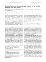
Fig. 6. Oleic acid b inding modes revealed by TLL-OA complex crystal structure. (A) Anti sn-1 position of the OA in the hydrophobic catalytic cleft
of the open conformation of TLL(S146A). The REFMAC 2Fo-Fc electron density map of the active site surroundings in molecule B (contoured at
1 r level), with the ligand – oleic acid (OA) – omitted f rom this calculation; dashed lines in dicate hydrogen bond s between OA and the protein.
(B) Comparisons between several ligand binding modes in the catalytic c left of TLL. The stereoview of OA binding modes in molecule A and B
(OA_A and OA_B, respectively, thick lines); C9 and C10 correspond to the position of these atoms in the OA_A ligand, where the electron density
was visible from its carboxylic group up to the C9-C10 carbon atoms: the alkyl chain of OA_A is included in the model beyond this double bond for
the sake of clarity. The corresponding part of the covalent complex of the C12 posphonate inhibitor–TLL [14], in which the C12 alkyl moiety
occupies sn -1 sit e of the active c enter, is also shown (protein shown by a thin line, ligand marked C12), for comparisons.
1618 S. Yapoudjian et al. (Eur. J. Biochem. 269) Ó FEBS 2002
FRET experiments in order to optimize the signal to noise
ratio. Under these conditions (NaTDC concentration of
1m
M
), some cis-PnA molecules are not incorporated into
the mixed micelles of cis-PnA/NaTDC, as already indicated
from the data presented in Fig. 2. It is known that the
micelle formation process of bile salts is a complex process.
Pre-micellar aggregates of various sizes have been described
previously [22,31]. This may explain why at 1 m
M
NaTDC,
we observed a clear and characteristic UV absorption
spectrum of cis-PnA, different from the one recorded in the
absence o f NaTDC (see Fig. 2). Despite this limitation
(a high RFI background resulting from a direct cis-PnA
excitation at 280 nm), we observed a significant FRET at a
NaTDC concentration of 4 m
M
(data not shown). Further-
more, the addition of aliquots of pure ethanol (up to a
concentration of 10%, v/v) to a mixed solution of cis-PnA/
NaTDC (1 l
M
/1 m
M
) does not significantly change the R FI
of the cis-PnA (data not shown).
Binding of TLL (and mutants) to
cis-
PnA and NaTDC
In the presence of NaTDC, without cis-PnA. The presence
of NaTDC at a concentration of 1 m
M
resulted in a blue
shiftofthek
max
of the RFI of TLL (4 nm), TLL(S146A)
(5 nm) and TLL(W117F, W221H, W260H) (11 nm) (see
Table 1), which was probably d ue to the decreasing polarity
of the local environment of W89 in these three molecules.
The lack of any wavelength shift observed for TLL(W89L)
and the highest blue shift observed for TLL(W117F,
W221H, W260H) indicate that the lid’s W89 was involved
in the i nteraction with NaTDC micelles.
These findings are in an agreement with the data obtained
from other studies [16,32], showing that W89 is the only
accessible tryptophan of the TLL and that the lid region is
directly involved in the binding of TLL to micelles of the
pentaoxyethylene octyl e ther (C
8
E
5
)detergent[32].
In the absence of NaTDC, with cis-PnA. The addition of
cis-PnA to TLL (or mutants) did not lead to any significant
changes in the k
max
of the RFI of the proteins (see Table 1).
Moreover, no FRET (Fig. 5) was observed for TLL or for
its mutants upon addition of cis-PnA, probably due to the
physico-chemical state of cis-PnA in the absence of NaTDC
micelles. However, the presence of cis-PnA led to a
quenching of the fluorescence emission only with
TLL(S146A) and TLL(W89L). The S146A and W89L
mutations are located either in the catalytic triad or in the
lid, respectively, and favor the open structure of the lipase in
solution, enhancing its interaction with cis-PnA. Hence, the
mutant TLL(S146A) is not an appropriate substitute for the
wild-type TLL for mimicking the interaction between lipase
and lipids.
In the presence of NaTDC and cis-PnA. The absence of
significant FRET at pH 8.0 with an excitation wavelength
ranging from 250 to 290 nm (data not shown), may be
attributed to the lack of spectral overlap between the UV
absorption spectrum of cis-PnA and the fluorescence
emission spectrum of TLL (Fig. 1).
The FRET between TLL and mixed micelles of cis-PnA/
NaTDC at pH 5.0 can be illustrated by comparing the
separately taken spectra of TLL and cis-PnA (dashed curve
from Fig. 3 corresponding to their arithmetic sum) with
spectra obtained after mixing TLL and mixed micelles of
cis-PnA/NaTDC. Moreover, as the molar ratio (R)between
cis-PnA and TLL increased, the RFI recorded at wave-
lengths ranging from 300 to 380 nm decreased, while
increasing simultaneously at wavelength ranging from 380
to 500 nm (Fig. 4). This behavior indicates that a FRET
occurred between TLL and cis-PnA. An isobestic point was
observed at 380 nm. The distance between the donor and
the acceptor can be estimated to be around 25 A
˚
[20].
Similar experiments were performed with a TLL mutant in
which all four tryptophan residues were mutated to
nonfluorescent a mino acids (data not shown). In this case,
the RFI decreased by 10-fold and no significant FRET was
observed. This indicates that any contributions of the
tyrosine residues to the FRET were negligible under our
experimental conditions.
When mixed micelles of cis-PnA/NaTDC were added, a
blue shift of the k
max
of the RFI of TLL (7 nm),
TLL(S146A) (14 nm) or TLL(W117F, W221H, W260H)
(20 nm) was observed, which indicates that the environment
of their tryptophan residues became less polar (see Table 1).
The greatest blue shift which occurred with TLL(W117F,
W221H, W260H) and the smallest one with TLL(W89L)
(2 nm) suggest that the lid is involved in the binding of the
mixed micelles of cis-PnA/NaTDC. On the other hand, these
shifts might result from the quenching of the tryptophan
fluorescence in t he presence of the mixed micelles, which
simultaneously reveals the fluorescence of the tyrosine
residues. However, this explanation can be ruled out, as no
significant shift was observed with the TLL(W89L) mutant
in the presence of the mixed micelles. Similar experiments
were performed w ith BSA in a control experiment and no
blue shift was observed, although FRET showed a
characteristic decrease in the RFI at 330 nm and a
simultaneous increase at 410 nm (data not shown). If the
quenching of the tryptophan e mission had revealed t he
tyrosine emission, then we would also h ave observed a
spectral shift in the experiments performed with BSA and
TLL(W89L), which was not the case. We can therefore
attribute t he spectral b lue shifts observed with m utants
TLL(S146A) and TLL(W117F, W221H, W260H) to a
change in the local surroundings of their W89 residues
towards a less polar environment.
The accessibility of the tryptophan residues to the cis-
PnA quencher was used to estimate the changes taking place
in the lid region of TLL and its mutants. As the values of
ÔK
sv
Õ calculated with TLL and TLL(W117F, W221H,
W260H) were the same, we can assume that W89 is the
only tryptophan side chain accessible to cis-PnA. This
conclusion was a lso confirmed by the lowest ÔK
sv
Õ obtained
for TLL(W89L).
The b lue s hift observed in the cas e of TLL(W117F,
W221H, W260H) confirms that the lid is involved in the
interaction between TLL and the mixed micelles of cis-PnA/
NaTDC. The h ighest accessibility of W89, assessed by ÔK
SV
Õ
values, observed w ith TLL(S146A) i ndicates that this mutant
has a higher binding affinity towards mixed micelles of cis-
PnA/NaTDC than the wild-type TLL. The S146A mutation
presumably destabilizes the closed conformation of the
lipase, exposing a cluster of hydrophobic amino acids,
including L206, F95, F113, F211, Y21, A146, L147 and
A146, and consequently enhances the interaction between
the lipase and the lipid aggregates, resulting in an efficient
Ó FEBS 2002 Lipase binding to lipid, FRET and structural study (Eur. J. Biochem. 269) 1619
FRET. This is in agreement with the molecular dynamics
simulations data obtained by Peters et al.[17],which
indicated that the mobility of the lid was enhanced in the
TLL(S146A) mutant. Furthermore, the results of independ-
ent direct binding measurements carried out on TLL and
TLL(S146A) with monomolecular films of substrate ana-
logues have shown that TLL(S146A) has the highest affinity
for these substrates (S. Yapoudjian, M. Ivanova, I. Douchet,
A. Ze
´
nath, M. Sentis, W.Marine, A. Svendsenand R. Verger,
unpublished data). This confirms that TLL(S146A) is not an
appropriate substitute for the wild-type TLL for mimicking
the interaction between lipase an d lipids.
Crystal structure of TLL(S146A) complexed with OA
The crystal structure of TLL(S146A) in a complex with OA
is virtually identical to other complexed structures of this
enzyme [14,15], and can be classified as Ôfully activeÕ
conformation of TLL according to Brzozowski et al.[15].
The lids in both molecules (A and B) are well defined in the
electron density maps and are in the same fully opened
conformations. They are not involved in the interactions
between molecule A and B, and a re almost free from
intermolecular contacts. For example, the W89 in molecule
Ais 8A
˚
from its nearest crystal lattice neighbour (F211)
and 16 A
˚
from W89 of symmetry related molecule. The
nearest ( 4A
˚
) and only intermolecular contact of the lid in
molecule A is between its E87 and symmetry linked D111.
There is only one (nondirect) hydrogen bond via water
molecules between E87 and N95–D96 o f the symmetry
related molecule. The lid in molecule B is also exposed to a
large crystal cavity and is free from strong intermolecular
interactions. However, the proximity ( 4A
˚
)ofL90from
L90 and D94 of symmetry related molecule might have
some stabilizing effect on the lid in this molecule. This
would explain much better definition of the OA electron
density in the molecule B in comparison with higher
disorder of the ligand in the molecule A. However, despite
of the lack of strong lattice contacts that may affect the
conformation of the lid, its high mobility (described in other
TLL crystal structure with unrestricted lid positioning [9]) is
not observed i n the reported structure.
The OA has been found in the exposed, catalytic cleft o f
the enzyme in two completely different orientations: Ôclas-
sicalÕ sn-1 position and, unexpectedly, in a antisn1 binding
mode. These two different OA binding modes may shed
some light on the unusual spectroscopic properties of the
Ser146fiAla mutant d escribed and d iscussed above. Firstly,
the Ser146fiAla mutation abolishes OG Ser146–NE2
His258 hydrogen bond (2.7 A
˚
), which stabilizes 144–148
loop with the active serine at its apex [6,14,15]. As the
Ala146 r esidue is released from this interaction it collapses
slightly deeper ( 0.62 A
˚
S146Ca–A146Ca distance) into
the protein core creating more space and flexibility f or the
putative ligand in the active site region. This relaxation,
together with the lack of steric h indrance created usually by
the hydroxyl g roup of Ser146 in the w ild-type enzyme,
results in a larger binding cavity, capable of accommodation
of lipid analogues normally not acceptable by the wild-type
active site. Secondly, the local perturbation caused by
Ser146fiAla mutation may affect the stability of its
neighbouring residue: L147. This can lead to a disruption
of the weak ( 3.8 A
˚
), but likely stabilizing, van der Waals
contact of L147 with W89 of the lid that is present in all
closed wild-type TLL structures, resulting in a higher
mobility of the lid in the S146A mutant. Moreover, the
removal of the potential structural strain of Ser146–His258
hydrogen bond may be propagated through freed His258
into the C-terminal (262–269) region of Tl lipase, which is
thought to be one of the crucial structural element of this
enzyme involved in the first steps of interfacial activation
[15,16]. Any small changes in this C-terminus/Arg84 switch
area may therefore contribute substantially to the destabil-
ization of the closed (low activity or activated form [15])
forms of Tl lipase, facilitating faster opening of the lid and its
transformation into the fully active form. It is also likely,
that the lack of a hydroxyl group of Ser146 diminishes the
role of this residue in the stabilization of the substrate–
ligand molecule, leading to the ÔconfusionÕ of the enzyme in
the process of the ligand recognition. This may result in the
two alternative OA binding modes observed here. The better
defined electron density for OA in m olecule B may than in
molecule A suggests that the antisn1 binding mode of this
lipid is favored b y the mutant. This is p robably due to the
stabilizing effect of the hydrogen bonds of D92 and H110
with carboxyl group of the OA molecule, specific for this
particular ligand conformation. Whether this form of OA
binding is enhanced further by the more stable conforma-
tion of the lid in molecule B is difficult to assess as generally
very weak crystal contacts of the lid in molecule B are only
marginally stronger than in molecule A, and the tempera-
ture factors of these regions are similar in both molecules.
The control crystallizations of the wild-type of TLL
under the conditions used for the S 146A mutant, have been
unsuccessful. This deficiency of crystals of the wild-type
TLL–OA complex may result from the lack of interactions
between TLL and OA in the crystallization conditions that
is in an agreement with the FRET data observed here.
Hence the physiological relevance of the two binding modes
of OA observed in S146A mutant should be interpreted with
caution, as they may result from the small changes in the
ligand binding cavity caused by the S146A mutation.
Micellar or molecular binding?
It is worth noticing that the FRET technique used in the
present study cannot, in principle, distinguish a micellar from
a molecular binding mode. In other words, one could expect
to observe comparable fluorescence signals whether a lipase
molecule binds to the mixed micelle of cis-PnA/NaTDC or
toa single molecule of cis-PnA incorporated into the mixed
micelles. These two alternative models are reminiscent to a
long standing discussion about the surface dilution pheno-
menon concerning the interaction of phospholipase A
2
with mixed micelles of Triton X-100/phospholipid [33].
As seen in Fig. 4, the FRET signal reaches a plateau
value when a molecule of TLL is added per molecule of cis-
PnA. This sto echiometry is more in f avour of a molecular
recognition rather than a micellar binding mode. More-
over, we determined the three-dimensional structure of the
open TLL(S146A), co-crystallised with mixed OA/NaTDC
micelles. We identified one OA molecule per TLL mono-
mer lying in the cat alytic site, in contact with W 89,
according to two binding mo des (see F ig. 6). These
structural considerations suggest that the FRET was
probably due to a molecular binding of TLL to cis-PnA.
1620 S. Yapoudjian et al. (Eur. J. Biochem. 269) Ó FEBS 2002
Thus FRET can be taken as an index of the open
conformation of TLL.
ACKNOWLEDGEMENTS
This research was carried out with financial suppo rt of the BIOTECH
program of the European Union under contract no. BIO4-CT97-2365.
We would like to thank the staff and beamline managers at the
European Synchrotron Radiation Facility (ESRF) (Grenoble) and SRS
Daresbury for their assistance with the data collection. The infrastruc-
ture of the Structural Biology Laboratory in York is supported by the
Biology and Biotechnology Science Research Council (BBSRC). We
would like to thank Dr Antonie J. VISSER (Wageningen Agricultural
University, MicroSpectroscopy Centre, Laboratory of Biochemistry,
the Netherlands) and Prof. J. Sturg is (LISM, CNRS, Mar seille, Fra nce)
for fruitful discussions. The assistance of Dr Jessica Blanc is
acknowledged for revising the English manuscript.
REFERENCES
1. Verger, R. (1997) Interfacial activation of lipases: facts and arti-
facts. Trends Biotech. 15, 32–38.
2. Schmid, R.D. & Verger, R. (1998) Lipases: interfacial enzymes
with at tractive applications. Angew. Che m. International
1608–1633.
3. Faber, K. (1992) Bio-Transformations in Organic Chemistry.
Springer-Verlag, Berlin.
4. Alberghina, L. (2000) Protein Engineering in Industrial Biotech-
nology, Harwood Academic Publishers, Australia, Canada, France.
5. Bornscheuer, U.T. & Kazlauskas, R.J . (1999) H ydrolases in
Organic Synthesis: Regio- and Stereo-selective Biotransformations.
Wiley-VCH, Weinheim, New York.
6. Brady, L., Brzozowski, A.M., Derewenda, Z.S., Dodson, E.,
Dodson, G., Tolley, S., Turk enburg, J.P., Christiansen, L.,
Huge-Jensen,B.,Norskov,L.,Thim,L.&Menge,U.(1990)A
serine protease triad forms the catalytic centre of a triacylglycerol
lipase. Nature 343, 767–770.
7. Winkler, F.K., d’Arcy, A. & Hunziker, W. (1990) Structure of
human pancreatic lipase. Nature 343, 771–774.
8. van Tilbeurgh, H., Egloff, M P., Martinez, C., Rugani, N.,
Verger, R. & Cambillau, C. (1993) Interfacial activation of the
lipase–procolipase complex by mixed micelles r evealed by X-ray
crystallography. Nature 362, 814–820.
9. Derewenda, U., Swenson, L., Wei, Y.Y., Green, R., Kobos, P.M.,
Joerger,R.,Haas,M.J.&Derewenda,Z.S.(1994)Conforma-
tional lability of lipases observed in the absence of an oil–water
interface. Crystallographic studies of enzymes from the fungi
Humicola lanuginosa and Rhizopus delemar. J. Lipid Res. 35,
524–534.
10. Brzozowski, A.M., Derewenda, U., Derewenda, Z.S., Dodson,
G.G., Lawson, D.M., Turkenburg, J.P., Bjorkling, F., Huge-
Jensen, B., Patkar, S.A. & Thim, L. (1991) A model for interfacial
activation in lipases from the structure of a fungal lipase–inhibitor
complex. Nature 351, 491–494.
11. Grochulski, P., Li, Y., Schrag, J.D. & Cygler, M. (1994) Two
conformational states of Candida rugosa lipase. Protein Sci. 3,
82–91.
12. Ollis, D.L., Cheah, E., Cygler, M., Dijkstra, B., Frolow, F.,
Franken, S.M., Harel, M., Remington, S.J., Silman, I., Schrag, J.,
Sussman, J.L., Verschueren, K.H.G. & Goldman, A. (1992) The
a/b hydro lase fold. Protein Eng. 5, 197–211.
13. Derewenda, U., Swenson, L., Green, R ., Wei, Y., Yamaguchi, S.,
Joerger, R., Haas, M.J. & Derewenda, Z.S. (1994) Current pro-
gress in crystallographic studies of new lipases from filamentous
fungi. Protein Eng. 7, 551–557.
14.Lawson,D.M.,Brzozowski,A.M.,Rety,S.,Verma,C.&
Dodson, G.G. (1994) Probing the nature of substrate binding in
Humicola lanuginosa lipase through X-ray crystallography and
intuitive modelling. Protein Eng. 7, 543–550.
15. Brzozowski, A.M., Savage, H., Verma, C.S., Turkenburg, J.P.,
Lawson,D.M.,Svendsen,A.&Patkar,S.A.(2000)Structural
origins of the interfacial activation in Thermomyces (Humicola)
lanuginosa lipase. Biochemistry 39 , 15071–15082.
16. Cajal,Y.,Svendsen,A.,Girona,V.,Patkar,S.A.&Alsina,M.A.
(2000) Interfacial control of lid opening in Thermomyces lanugi-
nosa lipase. Biochemistry 39, 413–423.
17. Peters, G.H., Svendsen, A., Langberg, H., Vind, J., Patkar, S.A.,
Toxavaerd, S. & Kinnunen, P.K.J. (1998) Active serine involved in
the stabilization of th e active site lo op in the Humicola lanuginosa
lipase. Biochemistry 37, 12375–12383.
18. Cantor, C.R. & Schimmel, P.R. (1980) Fluorescence Spectroscopy
in Biophysical C hemistry, pp. 433–465. W.H. Free man, San
Francisco.
19. Sklar, L.A., Hudson, B.S., Petersen, M. & Diamond, J. (1977)
Conjugated polyene fatty acids as fluorescent probes: spectro-
scopic characterization. Biochemistry 16, 813–818.
20. Sklar, L.A., Hudsen, B.S. & Simoni, R.D. (1977) Conjugated
polyene fatty acids as fluorescent probes: binding to bovine serum
albumin. Biochemistry 16, 5100–5108.
21. Dansen, T.B., Westerman, J., Wouters, F.S., Wanders, R.J.A.,
van Hoek, A., Gadella, T.W.J. & Wirtz, K.W.A. (1999) High-
affinity binding of very-long-chain fatty acyl-CoA esters to the
peroxisomal non-specific lipid-transfer protein (sterol carrier
protein-2). Biochem. J. 339, 193–199.
22. Small, D.M. (1971) The physical chemistry of cholanic acids. In:
The Bile Acids (Nair, P.P. & Kritchevski, D ., eds), pp. 249–356.
Plenum Press, N.Y.
23. Svendsen, A., Clausen, I.G., Patkar, S.A., Borch, T. & Thellersen,
M. (1997) Protein engineering of microbial lipases of industrial
interest. Methods Enzymol. (Dennis, E . & Rub in, B., ed s),
pp. 317–340.
24. Holmquist, M., Martinelle, M., Clausen, I.G., Patkar, S.,
Svendsen, A. & Hult, K. (1994) Trp89 in the lid of Humicola
lanuginosa lipase is important for efficient hydrolysis of tributyrin.
Lipids 29, 599–603.
25. Lehrer, S.S. (1971) Solute per turbation of protein fluorescence.
The quenching of the tryptophyl fluorescence of model compounds
and of lysozyme by iodide ion. Biochemistry 10, 3254–3263.
26. Otwinowski, Z. & Minor, W. (1997) Processing of X-ray diffrac-
tion data collection in oscillation mode. Methods Enzymol. 276,
307–326.
27. Navaza, J. (1994) AMORE: an automated package for molecular
replacemen t. Acta Crystallogr. A50, 157–163.
28. Murshudov, G.N., Vagin, A.A. & Dodson, E.J. (1997) Refine-
ment of macromolecular structures by the maximum-likelihood
method. Acta Crystallogr. D53, 240–255.
29. Bhattacharya, A.A., Gru
¨
ne,T.&Curry,S.(2000)Crystal-
lographic analysis reveals common modes of binding of medium
and long-chain fatty acids to human serum albumin. J. Mo l. Biol.
303, 721–732.
30. Beisson, F., Ferte
´
,N.,Nari,J.,Noat,G.,Arondel,V.&Verger,R.
(1999) Use of naturally fluorescent triacylglycerols from Parinari
glaberrimum to detect low lipase activities from Ar abid opsis
thaliana seedlings. J. Lipid Res. 40, 2313–2321.
31. Small, D.M. (1968) Size and structure of bile salt micelles. Advan.
Chem. Series 84, 31–52.
32. Jutila, A., Zhu, K., Patkar, S.A., Vind, J., Svendsen, A. &
Kinnunen, P.K.J. (2000) Detergent-induced conformational
changes of Humicola lanuginosa lipase studied by fluorescence
spectroscopy. Biophys. J. 78, 1634–1642.
33. Deems, R.A., Eaton, B.R. & Dennis, E.A. (1975) Kinetic analysis
of phospholipase A 2 activity toward mixed micelles and its
implications for the study of lipolytic enzymes. J. Biol. Chem. 250,
9013–9020.
Ó FEBS 2002 Lipase binding to lipid, FRET and structural study (Eur. J. Biochem. 269) 1621