u thực quản trang
Bạn đang xem bản rút gọn của tài liệu. Xem và tải ngay bản đầy đủ của tài liệu tại đây (4.89 MB, 26 trang )
ESOPHAGEAL CANCER
Nguyễn Thị Thu Trang
CK1-K25
Epidemiology
Esophageal cancer is responsible for <1% of all cancers and 4-10% of all gastrointestinal malignancies
M:F = 4:1
Risk factors:
- alcohol
- smoking
- Asbestosis
- ionizing radiation
- obesity
- HPV
Pathology
Squamous cell carcinoma
Adenocarcinoma
other types: Mucoepidermoid carcinoma,
Rhabdomyosarcoma, Fibrosarcoma…
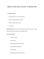
Esophageal Divisions
anatomy
Lymph node stations: 1 = supraclavicular, 2L = left
paratracheal, 2R = right paratracheal, 3P = posterior
mediastinal, 4L = left tracheobronchial angle, 4R = right
tracheobronchial angle, 5 = aortopulmonary, = anterior
mediastinal, 7 = subcarinal, 8L = lower
paraesophageal, 8M = middle paraesophageal, 9 =
inferior pulmonary ligament, 10L = left hilar, 10R = right
hilar, 6 15 = diaphragmatic, 16 = paracardial, 17 = left
gastric, 18 = common hepatic, 19 = splenic, 20 = celiac
M Stage
M1a -Metastases to cervical or celiac nodes
M1b - metastases to distant sites
Staging: TNM system- AJJC
In patients with a tumor in the upper thoracic esophagus, cervical lymph node metastases are
designated as M1a disease
In patients with a tumor in the midthoracic esophagus, cervical or celiac axis lymph node
metastases are considered to be M1b disease
In patients with a tumor in the distal thoracic esophagus, celiac axis lymph node metastases are
also classified as M1a disease
Radiographic features
esophageal air-fluid level
widened azygo-esophageal recess with convexity toward right lung.
tracheal deviation
retrocardiac or posterior mediastinal mass
repeated aspiration pneumonia (with tracheo-esophageal fistula)
Fluoroscopy/Barium Swallow
There is a 4.2 centimeter non-circumferential mass
in the mid esophagus, with an irregular surface
and shouldering demonstrated. This lesion is
suspicious for malignancy.
Endoscopic US
Endoscopic US is considered to be the most
accurate imaging modality currently available for
primary tumor staging (T staging). It defines the
layers of the esophageal wall hence can
differentiate T1, T2, and T3 tumors.
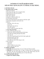
T1 N0 M0 (stage I) SCC of the midesophagus in a 52-year-old
man.
Endoscopic US image clearly depicts a polypoid lesion
(arrow) with extension into the second (hypoechoic) deep
mucosal layer
CT
eccentric or circumferential wall thickening >5 mm
peri-esophageal soft tissue and fat stranding
dilated fluid- and debris-filled esophageal lumen is proximal to an obstructing lesion
tracheobronchial invasion appears as a displacement of the airway (usually the trachea or left
mainstem bronchus) as a result of mass effect by the esophageal tumor
aortic invasion
CT: Local invasion
loss of fat planes between the tumor and adjacent structures
displacement or indentation of other mediastinal structures
Aortic invasion is suggested if 90° or more of the aorta is in contact with the tumor
obliteration of the triangular fat space between the esophagus, aorta, and spine adjacent to
the primary tumor
Displacement of the trachea or bronchus, or indentation of the posterior wall of the trachea
or bronchus by the tumor
Pericardial invasion is suspected if pericardial thickening, pericardial effusion, or indentation
of the heart with loss of the pericardial fat plane is seen
T1 N0 M0 (stage I) SCC of the midesophagus in a 52-year-old man. (a) Contrast material– enhanced CT scan obtained at the level
of the left superior pulmonary vein shows a small, nodular protruding lesion (arrow). (b) Endoscopic US image clearly depicts a
polypoid lesion (arrow) with extension into the second (hypoechoic) deep mucosal layer. Note the normal alternating hyper- and
hypoechoic architecture of the esophageal wall (arrowheads). The first layer is hyperechoic and represents the interface between
balloon and superficial mucosa, the second layer (hypoechoic) represents the lamina propria and muscularis mucosae, the third
layer (hyperechoic) represents the submucosa, the fourth layer (hypoechoic) represents the muscularis propria, and the fifth
layer (hyperechoic) represents the interface between the serosa and surrounding tissues.
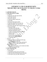
T4 N1 M0 (stage III) SCC of the midesophagus in a 61-year-old man. Contrast-enhanced CT scan obtained at
the level of the mainstem bronchi shows marked esophageal wall thickening with tumor extension into the
periesophageal fat. Note the diffuse wall thickening and narrowing of the left main bronchus (arrowheads).
There is loss of the normal fat plane (arrows) between the esophagus and the thoracic aorta, a finding that is
suggestive of aortic invasion. Sagittal reformatted CT image shows a broad interface (arrowheads) between
the esophageal mass and the thoracic aorta.
PET/CT
PET/CT is useful for detecting esophageal primary tumors.
PET/CT is also superior to CT for detecting lymph node metastases and can depict metastases in
normal-sized lymph nodes.
The most common sites of distant metastases detected at PET (but frequently missed at CT) are
the bones and liver.
PET/CT
Contrast-enhanced CT scan shows circumferential wall thickening in the lower esophagus (arrowhead), a
finding that is consistent with esophageal cancer. There is also a suspect low-attenuation lesion in the posterior
wall of the left ventricle (arrow), a finding that was missed at initial interpretation.
Fused PET/CT image shows intense FDG uptake by the primary tumor (arrowhead) and an unexpected
additional focus of FDG uptake in the left ventricle (arrow), a finding that is consistent with metastasis.
PET/CT
Pitfalls in the determination of N stage with FDG PET. (a) CT scan obtained at the level of the right inferior pulmonary
vein shows esophageal wall thickening (arrow), a finding that corresponds to esophageal cancer. Note also the
enlarged periesophageal lymph node (arrowhead), a finding that is suggestive of N1 disease. (b) Coronal PET scan
shows intense FDG uptake by the primary tumor (arrowhead). However, this uptake is difficult to differentiate from
the FDG uptake in the periesophageal lymph node seen in a due to the limited spatial resolution of PET. The
periesophageal lymph node was confirmed to be malignant (metastatic) at subsequent surgery.