Báo cáo khoa học: Do bacterial genotoxins contribute to chronic inflammation, genomic instability and tumor progression? pot
Bạn đang xem bản rút gọn của tài liệu. Xem và tải ngay bản đầy đủ của tài liệu tại đây (343.25 KB, 12 trang )
REVIEW ARTICLE
Do bacterial genotoxins contribute to chronic
inflammation, genomic instability and tumor progression?
Lina Guerra, Riccardo Guidi and Teresa Frisan
Department of Cell and Molecular Biology, Karolinska Institutet, Stockholm, Sweden
Introduction
Epidemiological evidence has linked chronic bacterial
infections with an increased risk of tumor development.
Heleicobacter pylori is associated with gastric cancers
and has been classified as a type I carcinogen by the
World Health Organization [1]. Other known associa-
tions between bacterial infection and human cancers are
Salmonella enterica serovar Typhi and carcinoma of the
gallbladder in chronic carriers; Streptococcus bovis and
colon cancer; persistent Chlamydia pneumoniae and
lung cancer; as well as Bartonella species and vascular
tumor formation [2].
The acquisition of genomic instability is a crucial
feature in tumor initiation and progression. Because
the baseline mutation rate is insufficient to account for
the multiple genetic changes required for cancer pro-
gression, tumor cells must acquire a ‘mutator pheno-
type’ that enhances the mutation frequency, and
allows the evolution from a pre-malignant to an inva-
sive cancer cell [3]. This phenotype can be caused by a
failure to repair damaged DNA and ⁄ or altered activa-
tion of the DNA damage-induced checkpoint
responses that selectively eliminate irreversibly dam-
aged cells. In the case of bacterial infection, several
events (e.g. the establishment of chronic inflammation,
as well as the production of genotoxins or bacterial
products that interfere with regulation of cell cycle
Keywords
bacterial genotoxin; chronic inflammation;
colibactin, cytolethal distending toxin; DNA
damage; DNA damage response; genomic
instability; tumor induction ⁄ progression
Correspondence
T. Frisan, Department of Cell and Molecular
Biology, Karolinska Institutet, Box 285,
S-171 77, Stockholm, Sweden
Fax: +46 8 337412
Tel: +46 8 52486385
E-mail:
(Received 1 March 2011, revised 4 April
2011, accepted 13 April 2011)
doi:10.1111/j.1742-4658.2011.08125.x
Cytolethal distending toxin, produced by several Gram-negative bacteria,
and colibactin, secreted by several commensal and extraintestinal patho-
genic Escherichia coli strains, are the first bacterial genotoxins to be
described to date. Exposure to cytolethal distending toxin and colibactin
induces DNA damage, and consequently activates the DNA damage
response, resulting in cell cycle arrest of the intoxicated cells and DNA
repair. Irreversible DNA damage will lead to cell death by apoptosis or to
senescence. It is well established that chronic exposure to DNA damaging
agents, either endogenous (reactive oxygen species) or exogenous (ionizing
radiation), may cause genomic instability as a result of the alteration of
genes coordinating the DNA damage response, thus favoring tumor initia-
tion and progression. In this review, we summarize the state of the art of
the biology of cytolethal distending toxin and colibactin, focusing on the
activation of the DNA damage response and repair pathways, and discuss
the cellular responses induced in intoxicated cells, as well as how prolonged
intoxication may lead to chronic inflammation, the accumulation of geno-
mic instability, and tumor progression in both in vitro and in vivo models.
Abbreviations
AaCDT, Aggregatobacter actinomycetemcomitans CDT; CDT, cytolethal distending toxin; DDR, DNA damage response; DSB, double strand
break; EcCDT, Escherichia coli CDT; ER, endoplasmic reticulum; H2AX, histone 2AX; HdCDT, Haemophilus ducreyi CDT; HR, homologous
recombination; MRN, Mre11-Rad50-Nbs1; NF, nuclear factor; NRPS, nonribosomal peptide megasynthetase; PARP, poly(ADP-ribose)
polymerase; PKS, polyketide megasynthetase; ROS, reactive oxygen species; StCDT, serovar Typhi CDT; Th, T helper.
FEBS Journal 278 (2011) 4577–4588 ª 2011 The Authors Journal compilation ª 2011 FEBS 4577
progression and apoptosis), in association with host
genetic factors, may contribute to the acquisition of
the mutator phenotype.
In this review, we focus on the two bacterial prod-
ucts that act as genotoxins and can directly damage
the host DNA: cytolethal distending toxin (CDT) and
colibactin. We discuss the biology of these two geno-
toxins, as well as their contribution to induction of
chronic infection ⁄ inflammation, genomic instability
and tumor progression.
CDT
CDT is the first bacterial genotoxin to be described and
is produced by a variety of Gram-negative bacteria, such
as Escherichia coli, Aggregatobacter actinomycetemcom-
itans, Haemophilus ducreyi, Shigella dysenteriae, Cam-
pylobacter sp. and Helicobacter sp, and S. enterica [4].
CDT is generally a exotoxin, and the active holotox-
in is a tripartite complex encoded by a single operon
[5,6], formed by the CdtA, CdtB and CdtC subunits
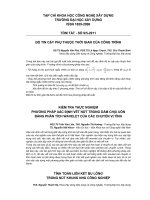
(Fig. 1A). Nesic et al. [7] solved the crystal structure of
the H. ducreyi CDT (HdCDT) and demonstrated that
the CdtA and CdtC subunits are lectin-like molecules,
sharing structural homology with the B-chain repeats
of the plant toxin ricin [7] (Fig. 1B). CDT is an
exotoxin secreted by the pathogen at the infection site.
Functional studies have identified CdtA and CdtC as
comprising essential proteins for mediating toxin bind-
ing to the membrane and internalization into target
cells [8–10].
The CdtB subunit adopts the canonical four-layered
fold of the DNase I family: a central 12-stranded b-
sandwich packed between outer a-helices and loops on
each side of the sandwich [7]. The crystal structure
confirms previous data demonstrating that the CdtB
subunit is functionally homologous to the mammalian
DNase I, and also possesses DNase capacity both
in vitro and when ectopically expressed or microinjected
in eukaryotic cells. Mutation in any conserved residue
important for the catalytic activity or the Mg
2+
bind-
ing abolishes the ability of CdtB to cleave DNA in vitro
and to induce DNA damage responses (DDRs) in
model cell lines [11–14].
CDT is therefore defined as an A-B
2
toxin, where
CdtA and CdtC are required for binding the holotoxin
to the plasma membrane of target cells, allowing entry
of the active CdtB, which can translocate to the
nucleus and induce DNA lesions.
In addition to the well-characterized DNA damaging
activity of the CdtB subunit, Shenker et al. [15]
reported that the active subunit from A. actinomyce-
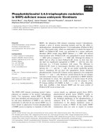
Fig. 1. Structure of CDT holotoxin and the psk genomic island. (A) Schematic representation of the CDT genes from H. ducreyi and S. enter-
ica, serovar Typhi. In all CDT-producing bacteria identified to date, excluding S. enterica, the three cdt genes are organized in an operon and
are transcribed as monocistrons. Conversely, in S. enterica, the three genes required for an active holotoxin are present as two separate
units: one unit containing the cdtB gene, encoding the active subunit, and the pltB ⁄ pltA unit, encoding proteins possibly required for the
proper traffic of CdtB to the nucleus of target cells. No homologous genes for the cdtA and cdtC subunits have been identified in this bacte-
rium. (B) Crystal structure of the HdCDT, adapted from Nesic et al. [7]. Protein data bank code: 1SR4. (C) Schematic representation of the
pks genomic island that encodes the enzymes and accessory proteins required for synthesis of an active colibactin in the E. coli strain Nissle
1917 [30].
Bacterial genotoxins and genomic instability L. Guerra et al.
4578 FEBS Journal 278 (2011) 4577–4588 ª 2011 The Authors Journal compilation ª 2011 FEBS
temcomitans exhibits PI-3,4,5-triphosphate phosphatase
activity [15]. However, there is evidence suggesting that
the DNA damaging activity alone is sufficient to con-
fer CdtB toxicity. Indeed, the G2 arrest and cell death
induced by conditional expression of CdtB in Saccha-
romyces cerevisiae depend exclusively on its DNase-
catalytic residue because yeast does not harbor the
substrate for CdtB phosphatidylinositol-3,4,5-triphos-
phate phosphatase activity [16] and specific CdtB
mutations that inhibit the phosphatase activity (but
retain DNase activity) are sufficient to induce the cell
death in proliferating U937 monocytes [17].
The discrepancy between the requirements of the dif-
ferent enzymatic activities of CDT may depend on the
cell type used as model. It is conceivable that T lym-
phocytes are more susceptible to the phosphatase
activity of CDT compared to all of the other cell lines
tested.
The only exception to the general structure of the
CDT family described so far is represented by the
S. enterica, serovar Typhi CDT (StCDT). In this intra-
cellular pathogen, the three genes required for an
active cytotoxin are not part of a single operon
because the gene for cdtB is not associated with genes
encoding for the CdtA and CdtC subunits. No homo-
logs for cdtA and cdtC genes have been found within
the complete Salmonella typhi genome [18]. However,
the toxicity of the StCDT on target cells requires the
transcription of two other genes: pltB (pertussis-like
toxin B) and pltA (pertussis-like toxin A) (Fig. 1A).
In vitro reconstitution experiments have shown that the
products of these three genes form an tripartite com-
plex inducing DNA damage in intoxicated eukaryotic
cells [19].
Interestingly, expression of the cdtB, pltB and pltA
genes is switched on upon bacterial uptake by the host
cells, and it is conceivable that the PltB and PltA sub-
units are required to transport CdtB from its site of
production within the cells to the extracellular med-
ium, from where StCDT can also intoxicate cells that
have not been infected, in a paracrine manner [19].
Several details of CDT binding to the plasma mem-
brane of target cells and its intracellular trafficking to
the nucleus have been elucidated (Fig. 2).
Interaction of the A. actinomycetemcomitans CDT
(AaCDT) occurs within GM1-enriched regions of the
plasma membrane, which are characteristic of mem-
brane rafts [14,20], and cholesterol depletion by
methyl-b-cyclodextrin reduces the ability of both
AaCDT and HdCDT to bind to Jurkat and HeLa cell
lines, respectively, and prevents intoxication [14,20].
Furthermore, inactivation of mutations within the
SGMS1 gene that reduce the levels of sphingomyelin
(a key component of lipid rafts) confers resistance to
the E. coli CDT (EcCDT) [21].
The identity of the toxin receptor is still unknown.
Fucose may represent the binding determinant for the
EcCDT-II [10], whereas another study indicated that
the AaCDT holotoxin binds to surface glycosphingoli-
pids and that inhibitors of glycosphingolipid synthesis
can prevent intoxication of the human monocytic
U937 cell line [22]. Site-directed mutagenesis of a
human cell line haploid for all chromosomes except
chromosome 8 identified mutants for the membrane-
expressed protein TMEM181 that were resistant to
EcCDT [21].
On the basis of such evidence, it is conceivable that
each individual CDT exhibits different receptor speci-
ficity. In line with this evidence, Eshraghi et al. [23]
reported that CDTs from H. ducreyi, A. actinomyce-
temcomitans, E. coli and Campylobacter jejuni differ in
their abilities to intoxicate host cells. The binding of
Aa, Hd and EcCDT-III, but not CjCDT, is dependent
on the presence of cholesterol. Unexpectedly, mutant
Chinese hamster ovary cells that lack N-linked com-
plex and hybrid carbohydrates, as well as cells that
lack glycosphingolipids or are deficient in fucose bio-
synthesis, are as similarly sensitive as the wild-type to
intoxication by all four CDTs tested, indicating that
N- and O-glycan, or fucosylated structures are dispens-
able for mediating toxin binding [23].
Upon binding to the plasma membrane, the HdCDT
is internalized in HeLa cells by dynamin-dependent
endocytosis, and it further transits to the endosomal
compartment [24]. Biochemical and imaging experi-
ments have demonstrated that the toxin is then retro-
gradely transported via the Golgi complex to the
endoplasmic reticulum (ER) [14]. Other bacterial and
plant toxins have been described to traffic from the
plasma membrane to the ER (Shiga, cholera and
ricin), and they subsequently reach their targets in the
cytosol by retrograde transport via the ER degradation
pathway (Fig. 2). However, HdCDT could not be
detected by biochemical assays in the cytosol of intoxi-
cated cells [25]: this opens the possibility that the CDT
active subunit may translocate directly from the ER to
the nucleus. Two studies have identified specific
nuclear localization signals (NLS) within AaCdtB and
EcCdtB-II. In AaCdtB, a nonconventional NLS is
localized at the N-terminus of the protein, and the
deletion of 11 amino acids within the this sequence
abolishes intoxication [26]. Conversely, two NLS
sequences, designated as NLS1 and NLS2, have been
identified in the carboxy-terminal region of the
EcCdtB-II [8]. Interestingly, the deletion of each of
these sequences produces a differential localization of
L. Guerra et al. Bacterial genotoxins and genomic instability
FEBS Journal 278 (2011) 4577–4588 ª 2011 The Authors Journal compilation ª 2011 FEBS 4579
the active toxin subunit, suggesting that they play dif-
ferent roles in the intracellular trafficking of EcCDT-
II. Cells intoxicated with a holotoxin containing the
EcCdtB-II-DNLS1 display a perinuclear distribution,
which is consistent with trapping of the active toxin
component in the late endosome and ⁄ or ER com-
partment, whereas a diffuse cytoplasmic staining is
observed in cells exposed to the EcCdtB-II-DNLS2-
containing toxin. It is conceivable that the NLS1
promotes the ER-to-nucleus translocation, whereas
NLS2 may function as an ER compartimentalization
signal preventing the escape of the CdtB molecules to
the cytosol, allowing their transit to the nucleus. It is
still not known why the presence of the putative NLS
domains are so divergent in AaCDT and EcCDT-II
because the structure of CdtB subunits presents a high
degree of conservation among different bacteria [27].
On the basis of such evidence, it is clear that the
nuclear translocation of the CdtB subunit into the
nucleus remains an open issue. Another interesting
question is whether the CdtA and CdtC subunits assist
the active component in its trafficking within the host
cells.
Colibactin
This toxin has been recently characterized and very
little information is available regarding its biology.
Colibactin is a putative hybrid peptide–polyketide
genotoxin found in both commensal and pathogenic
bacteria. Colibactin has been mostly characterized in
extraintestinal pathogenic E. coli strains of the phylo-
genetic group B2 [28]. The enzymes required for the
synthesis of colibactin are present in a 54 kb genomic
island, referred to as the pks island, located in the
asnW tRNA locus. This region contains 23 putative
ORFs, including three nonribosomal peptide megasyn-
thetases (NRPS), three polyketide megasynthetases
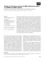
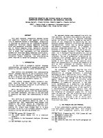
Fig. 2. Summary of the CDT internalization pathway and cellular responses induced by bacterial genotoxins. Binding of CDT is dependent on
the presence of intact lipid rafts, and the toxin is internalized via dynamin-dependent endocytosis into early and late endosomes. At this
stage, it is not known whether the CdtA (violet) and ⁄ or the CdtC (pink) subunits are internalized and follow the active CdtB subunit (green)
into the nucleus. The CdtB subunit further transits to the Golgi complex, and is then retrogradely transported to the ER. The mechanisms of
nuclear translocation have not yet been fully elucidated. Once in the nucleus, the CdtB subunit causes DNA damage and activates the ATM-
dependent DNA damage response, characterized by recruitment of the MRN complex, and full activation of ATM at the site of the damage,
which also requires a functional c-MYC. Activation of ATM promotes phosphorylation of histone H2AX and activation of the DNA damage
checkpoint responses via activation of: (a) the tumor suppressor p53 and its downstream effector p21, which results in G1 arrest, and (b)
the kinase CHK2 that blocks cell proliferation in the G2 phase of the cell cycle by inactivating the CDC25 phosphatase, resulting in hyper-
phosphorylation and inactivation of the cyclin-dependent kinase CDK1 (pCDK1). CDT intoxication also activates RhoA-dependent survival sig-
nals. This effect requires a functional ATM and is dependent on dephosphorylation of the guanine nucleotide exchange factor Net1.
Activation of RhoA regulates two distinct pathways: (a) induction of actin stress fibers, which requires the RhoA kinases ROCKI and ROCKII,
and (b) activation of p38 mitogen-activated protein kinase, associated with delayed cell death.
Bacterial genotoxins and genomic instability L. Guerra et al.
4580 FEBS Journal 278 (2011) 4577–4588 ª 2011 The Authors Journal compilation ª 2011 FEBS
(PKS), two hybrid NRPS ⁄ PKS megasynthetases, and
ten accessory, tailoring and editing enzymes (Fig. 1C).
Mutation analysis demonstrated that all the PKS and
NRPS and eight of the accessory genes are required
for production of an active genotoxin [28].
Screening studies have demonstrated that the pks
island is also present in other members of the Entero-
bacteriaceae family, such as Klebsiella pneumoniae,
Enterobacter aerogenes and Citrobacter koseri isolates
[29].
Gene expression of the ORFs required for the colib-
actin synthesis has been studied, using the nonpatho-
genic E. coli strain Nissle 1917 as a model, by
Homburg et al. [30], who identified seven transcripts,
four of which are polycistrons. The polycistronic tran-
scripts comprise the genes: (a) clbC to clbG; (b) clbI to
clbN; (c) clbO to clbP; and (d) clbR to clbA, whereas
the other ORFs are transcribed as monocistrons [30].
Luciferase reporter assays performed on the clbA,
clbB, clbQ and clbR genes demonstrated that their
expression increased during late logarithmic and early
stationary phase of the bacteria growth course. The
levels and the duration of expression depend on the
culture medium used, with DMEM being the best con-
dition compared to LB or minimal medium supple-
mented with 0.2% glucose [30]. Interestingly, the
transcription of these ORFs was not induced by direct
contact with the mammalian cell line HeLa [30]. It
remains to be determined whether the expression of
the other clb genes is dependent on interaction with
eukaryotic cells.
DDRs and genomic instability
CDT possesses DNase activity in vitro, and both CDT
and colibactin cause DNA damage in intoxicated cells
[11–14,28]. Thus, before discussing the cellular
responses to these genotoxins, we briefly review the
state of the art of the DNA damage sensing and repair
pathways in mammalian cells, as well as the conse-
quences of an altered DDR in the promotion of geno-
mic instability. On the basis of the type of DNA
damage induced by CDT and colibactin, we focus
mainly on the cellular responses to DNA double
strand breaks (DSB).
DDRs are essential for preserving the genetic infor-
mation and maintain genomic integrity in cells exposed
to the damaging activity of endogenous [e.g. reactive
oxygen species (ROS) produced by the cellular metab-
olism] and environmental agents (ionizing radiation
and UV radiation). Sensing and activation of the
DDR is coordinated by proteins of the phosphatidyl-
inositol 3-kinase-like protein kinase family: ATM,
ATR and DNA-PK [31]. The outcome is a block of
cell cycle progression and activation of the DNA dam-
age repair pathways. Successful repair will allow the
cell to resume the normal cell cycle progression,
whereas damage beyond the possibility of repair will
promote either apoptosis or senescence, precluding the
survival and replication of cells that can accumulate
genomic instability [32]. A key effector protein that
regulates activation of cell death or senescence in the
presence of chronic and unrepaired DNA damage is
the tumor suppressor gene p53, which acts as a barrier
for cancer initiation ⁄ progression [33].
Many DNA repair pathways have been evolved to
cope with all the possible insults to which the cellular
DNA is exposed. A mismatch DNA base is replaced
with the correct one by the mismatch repair [34],
whereas small base alterations, such as alkylation, are
repaired by the base excision repair, which removes
the altered base [35]. More complex lesions, such the
UV-induced pyrimidine dimers, require a longer exci-
sion (approximately 30 bp) and are repaired by the
nucleotide excision repair pathway [36]. DSB will be
processed by nonhomologous end joining or homolo-
gous recombination (HR) [37]. Nonhomologous end
joining occurs throughout the cell cycle and is based
on the identification of the break and subsequent
rejoining of the two ends. Consequently, there is a loss
or addition of nucleotides at the site of the lesion, and
the repair mechanism per se can contribute to a certain
degree of genomic alteration. Conversely, HR per-
forms an error-free repair of the lesion because the sis-
ter chromatid is used as template, restricting this type
of repair mechanism to the late S and G2 phases of
the cell cycle.
Recognition of DSB is mediated by several sensor
complexes: members of the poly(ADP-ribose) polymer-
ase (PARP) family, specifically PARP1 and PARP2,
the Mre11-Rad50-Nbs1 (MRN) complex and the
Ku70 ⁄ Ku80 hetorodimer [31].
PARP1 and 2 are activated by DNA DSB and catalyze
the addition of poly(ADP-ribose) chains on histones
and nuclear proteins. This step is essential for the
recruitment of the MRN complex, which initiates a
resection of the DNA ends to produce a 3¢ tail, and
promotes the accumulation and full activation of the
ATM kinase at the site of the damage [38]. In turn,
ATM coordinates the full DNA resection to promote
HR, and activates the checkpoint responses to block
cell cycle progression, allowing repair and preservation
of the genomic integrity. This is achieved by: (a)
recruitment and phosphorylation of c-histone 2AX
(H2AX) important to sustain the DDR; (b) activation
of effectors such as CtlP, BRCA1, ARTEMIS
L. Guerra et al. Bacterial genotoxins and genomic instability
FEBS Journal 278 (2011) 4577–4588 ª 2011 The Authors Journal compilation ª 2011 FEBS 4581
(DNA resection); and (c) phosphorylation ⁄ activation
of CHK2 and p53 that lead to cell cycle arrest and, in
ultimate instances, to apoptosis or senescence
[31,32,39].
Upon extensive DNA resection, the RPA complex
binds to the 3¢ ssDNA ends and modulates the activity
of effectors, such as BRCA2 and Rad51, which execute
the recombination process by performing strand inva-
sion and Holliday junction formation and resolution
to produce two completed undamaged sister chromat-
ids that preserve the original genetic information [31].
Figure 3 summarizes the main events in the ATM-
dependent activation of the DDR.
In nonhomologous end joining, the end of the
breaks are recognized and bound by the Ku79 ⁄ Ku80
heterodimer, which in turn recruits the DNA-PK and
initiates the DNA resection process. After binding to
the DNA, the DNA-PK is autophosphorylated and
provides access to the site of the resecting protein
ARTEMIS, and also the ligase complex XRCC4 ⁄ Lig4,
which promotes re-ligation of the broken ends [40].
Genomic instability is a characteristic of almost all
human tumors. The major genomic alterations
described in cancer cells include chromosomal instabil-
ity and microsatellite instability [41]. Chromosomal
instability is characterized by losses of entire or large
portions of chromosomes, resulting in aneuploidy,
translocation and loss of heterozygosity, whereas
microsatellite instability is defined as an expansion or
contraction of the number of oligonucleotide repeats
present in microsatellite sequences apart from the
nucleus.
The importance of the DDR in the maintenance of
genomic integrity is highlighted by the demonstration
that the acquisition of genomic instability is linked to
mutations in genes controlling DNA repair and mitotic
checkpoint pathways in hereditary cancers [42].
Conversely, high throughput analysis demonstrated
that, in sporadic cancers, the most frequently altered
genes are the TP53 tumor suppressor and genes that
regulate cell growth either positively (e.g. the oncoge-
nes EGFR and RAS) or negatively (e.g. PTEN and
CDKN2A) [42]. This pattern is not unexpeceted
because deregulation ⁄ overexpression of oncogenes will
lead to DNA replication stress and stalled replication,
resulting in the formation of DNA damage and activa-
tion of DDR. One of the consequences of this will be
the activation of p53 and the promotion of cell death
or senescence, a response that will pose a barrier to
tumor initiation ⁄ progression. Therefore, only cells in
which the deregulation of oncogenes is accompanied
by alteration of the p53 tumor suppression pathway
will have the possibility of overcoming this barrier.
Cellular responses to colibactin and
CDT
We now discuss the key cellular responses activated in
cells exposed to CDT or colibactin-producing bacteria,
focusing on the DDR and cell survival, which are both
relevant in the context of maintaining genome integ-
rity. For a more detailed analysis of the CDT-induced
cellular responses, several comprehensive reviews on
CDT biology are available [4,43,44].
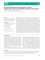
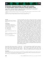
Fig. 3. Summary of the ATM-dependent
DNA damage responses to DNA DSBs.
Induction of DNA DSBs activates PARP1,
which mediates the initial recruitment of the
MRN complex and promotes the full activa-
tion of ATM. In turn, ATM coordinates the
DNA damage repair resulting in: (1) sus-
tained DDR by phosphorylating histone
H2AX; (2) resection of the damaged DNA to
allow activation of the HR process; and (3)
activation of the checkpoint responses that
block cell proliferation in distinct phases of
the cell cycle (G1 or G2) to allow repair. If
the damage is beyond repair, this response
will result in elimination of the altered cell
by apoptosis or the induction of cellular
senescence, a p53-dependent process
defined as the tumorigenesis barrier.
Bacterial genotoxins and genomic instability L. Guerra et al.
4582 FEBS Journal 278 (2011) 4577–4588 ª 2011 The Authors Journal compilation ª 2011 FEBS
It is now well established that CDT acts as a nucle-
ase cleaving DNA substrates in vitro, inducing nuclear
fragmentation and chromatin disruption when trans-
fected in cultured mammalian cells or Saccharomy-
ces cereviase, and promoting DNA fragmentation in
cells exposed to purified soluble toxin [11–13,45]. Simi-
larly exposure of mammalian cells to E. coli strains
expressing the pks island promotes DNA fragmenta-
tion, as detected by the comet assay [28].
As a result of the DNA damaging activity, intoxi-
cated cells activate the classical DDR, which includes
recruitment of the DNA damage sensor complex
MRN and the ATM kinase, phosphorylation of his-
tone H2AX, activation of p53 and its transcriptional
target, the cyclin-dependent kinase inhibitor p21, and
phosphorylation of checkpoint kinase CHK2. Tran-
scriptional upregulation of p21 leads G1 arrest,
whereas CHK2-dependent inactivation of the CDC25
phosphatase leads to an accumulation of the hyper-
phosphorylated form of cyclin-dependent kinase 1
(CDK1, also known as CDC2), and consequent induc-
tion of G2 arrest [9,46–51].
The prompt activation of the ATM-dependent
response to CDT or ionizing radiation-induced DNA
damage also requires a functional proto-oncogene
MYC [52].
As a consequence of the activation of ATM-depen-
dent checkpoint responses, cells exposed to CDT or
colibactin stop proliferating [28,46,51]. Furthermore,
CDT-intoxicated normal or tumor cells express the
hallmarks of cellular senescence, such as persistently-
activated DNA damage signaling (detected as
53BP1 ⁄ cH2AX-positive foci), enhanced senescence-
associated b-galactosidase activity, and expansion of
promyelocytic nuclear compartments [53]. In support
of the DNA damaging activity of CDT, a genome
wide analysis performed in S. cerevisae identified HR
and activation of the DNA damage checkpoints as
comprising essential mechanisms for the response to
damage induced by the conditional expression of the
active CdtB subunit from C. jejuni [54].
Another important aspect in the context of bacterial
genotoxins and carcinogenesis is the activation of sur-
vival signaling pathways because the survival of cells
with damaged DNA enhances the risk of acquiring
genomic instability and favors tumor initiation and ⁄ or
progression [55,56] (Fig. 4). The survival of CDT
intoxicated cells is dependent on the activation of the
small GTPase RhoA [45], which induces actin stress
fiber formation via the RhoA kinases, ROCKI and
ROCKII, and prevents cell death via activation of the
mitogen-activated protein kinase p38 and its down-
stream target mitogen-activated protein kinase-acti-
vated protein kinase 2 [57] (Fig. 2). The activation of
RhoA is dependent on the dephosphorylation on ser-
ine152 of the RhoA-specific guanine nucleotide
exchange factor Net1, and it appears to be part of the
DDR response because it requires a functional ATM
[57].
The cellular responses to the two bacterial toxins are
summarized in Fig. 2.
Infection with CDT-producing bacteria:
chronic inflammation and tumor
progression
Over the past 10 years, chronic inflammation has been
shown to be associated with an enhanced risk of tumor
development. How can inflammation favor the acquisi-
tion of the mutator phenotype? The inflammatory
environment is characterized by the production of
ROS and reactive nitrogen intermediates. These com-
pounds are potent genotoxic agents that may increase
the mutation rate and promote the accumulation of
genomic instability, thus altering the crucial biological
processes such as the regulation of DNA repair and
DDRs, allowing tumor initiation ⁄ progression [58,59].
Chronic inflammation is also associated with constit-
utive activation of pleiotropic nuclear factor (NF)-jB,
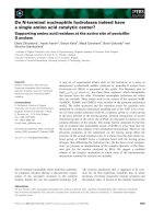
Fig. 4. Possible role of bacterial genotoxins in cancer development.
Chronic infection with CDT or colibactin-producing bacteria can pro-
mote the induction of genomic instability by direct secretion of bac-
terial genotoxins and activation of a chronic inflammation, which is
associated with the production of endogenous DNA damaging
agents, such as ROS. Persistent activation of the transcription fac-
tor NF-jB, via the pro-inflammatory cytokine TNF-a or sustained
triggering of pathogen recognition receptors (e.g. Toll-like recep-
tors), in combination with survival signals induced by cellular intoxi-
cation with genotoxins, may allow cells carrying genomic instability
to break through the tumorigenesis barrier, resulting in an increased
risk of tumor development.
L. Guerra et al. Bacterial genotoxins and genomic instability
FEBS Journal 278 (2011) 4577–4588 ª 2011 The Authors Journal compilation ª 2011 FEBS 4583
which promotes cell survival, and has been demon-
strated to contribute to tumor formation in models of
colitis-associated cancer and hepatocellular carcinoma
[60,61].
Bacteria that cause persistent infections associated
with chronic inflammation have a higher risk of pro-
moting carcinogenesis [58]. The best-studied example is
H. pylori and its association with gastric carcinoma
and mucosa-associated lymphoid tissue lymphoma
[62]. Chronic infection with this bacterium is associ-
ated with the downregulation of mismatch repair and
base excision repair proteins both in vitro and in vivo
[63], and these effects correlate with a five-fold
increased mutation frequency and also the induction of
microsatellite instabilities in the gastric epithelium of
mice 6 months after infection [64]. Epidemiological evi-
dence also demonstrate an increased risk of carcinoma
of the gallbladder in chronic carriers of S. enterica, ser-
ovar Typhi [2] and colon cancer in individuals colo-
nized by Bacteroides fragilis [65]. Bacterial products
can contribute to a sustained and deregulated inflam-
matory microenvironment by stimulation of the host
pathogen recognition receptors, leading to a constant
supply of ROS, reactive nitrogen intermediates and
cytokines. The most well-characterized family of PPRs
is the Toll-like receptor family. The majority of Toll-
like receptor signaling converges to the adaptor
molecule MyD88 and activates the transcription factor
NF-jB. MyD88 knockdown was shown to strongly
reduce the development of spontaneous colorectal
carcinoma in mice carrying heterozygous mutation of
the tumor suppressor genes APC (Apc
Min ⁄ +
mouse
model) [66]. In addition to the contribution of the
innate immune responses in inflammation-induced car-
cinogenesis via stimulation of PPR or pro-inflamma-
tory cytokine production, there is now evidence linking
T cell-mediated immune responses in infection-induced
carcinogenesis. The production and secretion of the
B. fragilis toxin induces colitis, which further develops
into colon cancer in the Apc
Min ⁄ +
mouse model, and
the carcinogenic capacity of B. fragilis toxin-producing
strains is directly associated with the recruitment of
the highly pro-inflammatory subset of T helper (Th) 17
lymphocytes [67].
Figure 4 summarizes the effectors that may contrib-
ute to carcinogenesis in chronic bacterial infections.
Considering the importance of establishing a chronic
infection as risk for tumor development, several studies
have assessed whether CDT may contribute to persis-
tent bacterial colonization of the gastrointestinal tract.
Fox et al. [68] demonstrated that a functional toxin
favors colonization of the stomach and lower bowel
of C57BL ⁄ 129 mice infected with C. jejuni. The
C57BL ⁄ 129 mouse model was not suitable for studying
the effect of a persistent infection with CDT-producing
bacteria in the induction of inflammation because bac-
teria were detected only in a proportion of the mice.
Conversely, a persistent colonization of the stomach
and the lower bowel for 100% of animals was achieved
in C57BL ⁄ 129 mice carrying a homozygous deletion of
p50 and heterozygous deletion of the p65 subunits of
the transcription factor NF-jB (p50
) ⁄ )
p65
+ ⁄ )
). In
this model of chronic infection, the presence of CDT-
producing bacteria was associated with significantly
enhanced severity of the gastritis and a greater induc-
tion of gastric hyperplasia and dysplasia, which is an
indication of an early neoplastic process [68].
Colonization of the Swiss Webster mice with Heli-
cobacter hepaticus was also dependent on expression of
a wild-type CDT [69]. This persistent infection was
associated with a highly inflammatory response, char-
acterized by the production of Th1-associated IgG2a,
Th2-associated IgG1 and mucosal IgA [69].
Similarly, a strong inflammatory response was
described in the liver of male A ⁄ JCr mice infected with
H. hepaticus carrying a wild-type CDT 10 months after
infection compared to mice infected with an isogenic
strain carrying a mutant toxin, where the cdtB gene
was inactivated by transposon mutagenesis. This
response was characterized by an increase in transcrip-
tion levels of pro-inflammatory (TNF-a, IFN-c and
Cox-2, IL-6 and TGF-a) and anti-apoptotic (Bcl-2 and
Bcl-X
L
) genes, as well as upregulation of hepatic
mRNA levels of components of the NF-jB pathway
(p65 and p50) [70]. The presence of CDT was further
associated with a progression of inflammation to dys-
plasia. The dysplastic lesions were characterized by the
presence of hepatocytes with a marked variation in cell
and nuclear size and shape, as well as a loss of hepatic
architecture [70].
An interesting notion, providing fuel for future stud-
ies, is to assess whether these conditions of dysplasia
are associated with the CDT-dependent induction of
DNA damage, chronic activation of the DDRs, acqui-
sition of genomic instability and alteration of cellular
pathways that regulate senescence, allowing cells to
break through the tumorigenesis barrier. Indeed, very
little is known about the ability of CDT to induce
genomic instability.
Effects of colibactin on genomic
instability
The effects of colibactin in induction of DNA damage
in vivo and genomic instability in vitro were studied by
Cuevas-Ramos et al. [50]. These authors reported that
Bacterial genotoxins and genomic instability L. Guerra et al.
4584 FEBS Journal 278 (2011) 4577–4588 ª 2011 The Authors Journal compilation ª 2011 FEBS
the expression of the clbA gene, a key gene for the syn-
thesis of this genotoxin, was detected in a mouse intes-
tinal loop model and in the colons of antibiotic-treated
BALB ⁄ cJ mice 6 h and 5 days, respectively, after infec-
tion with an E. coli strain harboring the psk genetic
island. In each case, an isogenic strain carrying a clbA
mutant gene (and therefore unable to produce colibac-
tin) was used as negative control. The expression of
the clbA gene was further associated with the induction
of DNA damage, as assessed by increased phosphory-
lation of the histone H2AX [50].
Short-term exposure of the Chinese hamster ovary
cell line to psk+ E. coli at low multiplicity of infection
(in the range five to 20 bacteria per cell) induced DNA
damage that could still be observed, although at a very
low level, up to 24 h post-infection in actively cycling
cells, indicating that the DNA repair process was not
completed. As a consequence of the partial DNA
repair, the infected cells presented anaphase bridges,
accumulated chromosomal aberrations in approxi-
mately 7% of the chromosomes, with ring chromo-
somes and translocations being the most common
alterations, and aneuploidy (a loss or gain of chromo-
somes) 72 h post-infection. Such aberrations were
maintained in a proportion of cells up to 21 days post-
infection. The chromosomal instability induced by
infection with colibactin-producing E. coli was further
associated with an enhanced rate of mutation fre-
quency and an increased ability of the cells to grow in
soft agar, which is a feature of anchorage-independent
growth [50].
Future perspectives
Our knowledge on how bacterial infections can con-
tribute to carcinogenesis has begun to be unraveled.
The journey started from epidemiological data and
several molecular mechanisms have been identified to
date, with special focus on the oncogenic role of
H. pylori infection. The identification of bacterial
genotoxins opens yet another new avenue.
We have come a long way in our understanding of
the biology of CDT, although many questions still
remain. We do not know: (a) when and under what
conditions the toxin is produced in vivo; (b) what is the
extent of the DNA damage and genomic instability in
in vivo models; and (c) whether there is a correlation
between chronic infection of CDT-producing bacteria
and an increased risk of cancer development. A retro-
spective study demonstrated that infection with the
enteropathogenic C. jejuni, where 99% of the strains
harbour cdt genes, did not correlate with an increased
risk of developing a tumor in the gastrointestinal tract
at least during the first 10 years after the detection of
infection [71]. However, this bacterium is rarely associ-
ated with the establishment of a chronic infection and
therefore may not represent a suitable model, despite
the fact that C. jejuni was found in tissue specimens
derived from intestinal mucosa-associated lymphoid
tissue lymphoma patients [72].
The biology of colibactin is still at its infancy
because this toxin was only characterized recently
and cannot yet be produced as a synthetic molecule
in vitro. Several interesting questions remain: (a) how
does it induce DNA damage; (b) how does it enter and
traffic within the host cells to reach the nuclear
compartment; and (c) does it contribute to long-term
bacterial colonization in vivo.
As a more general evolutionary aspect, we still do
not know how bacteria benefit from producing such
genotoxins.
The experimental work in the field of bacteria and
cancer, and specifically on bacterial genotoxins, has
been hampered by the complexity of the host–bacteria
interaction, although the development of suitable ani-
mal models and the implementation of high through-
put screenings will provide conditions that allow the
pursuit of this exciting issue in the field of medical
science.
Acknowledgements
This work was supported by the Swedish Research
Council, the Swedish Cancer Society, the A
˚
ke-Wiberg
Foundation, the Magnus Bergvall Foundation, the
Karolinska Institutet to T.F., and the Robert Lund-
berg Memorial Foundation to L.G. T.F. is supported
by the Swedish Cancer Society.
References
1 Humans IWGotEoCRt (1994) Schistosomes, Liver
Flukes and Helicobacter pylori. Lyon, France.
2 Vogelmann R & Amieva MR (2007) The role of bacte-
rial pathogens in cancer. Curr Opin Microbiol 10,
76–81.
3 Raptis S & Bapat B (2006) Genetic instability in human
tumors. EXS 96, 303–320.
4 Smith JL & Bayles DO (2006) The contribution of cyto-
lethal distending toxin to bacterial pathogenesis. Crit
Rev Microbiol 32, 227–248.
5 Scott DA & Kaper JB (1994) Cloning and sequencing
of the genes encoding Escherichia coli cytolethal dis-
tending toxins. Infect Immun 62, 244–251.
6 Lara-Tejero M & Galan JE (2001) CdtA, CdtB and
CdtC form a tripartite complex that is required for
L. Guerra et al. Bacterial genotoxins and genomic instability
FEBS Journal 278 (2011) 4577–4588 ª 2011 The Authors Journal compilation ª 2011 FEBS 4585
cytolethal distending toxin activity. Infect Immun 69,
4358–4365.
7 Nesic D, Hsu Y & Stebbins CE (2004) Assembly and
function of a bacterial genotoxin. Nature 429, 429–433.
8 McSweeney LA & Dreyfus LA (2004) Nuclear localiza-
tion of the Escherichia coli cytolethal distending toxin
CdtB subunit. Cell Microbiol 6, 447–458.
9 Hassane DC, Lee RB & Pickett CL (2003) Campylobac-
ter jejuni cytolethal distending toxin promotes DNA
repair responses in normal human cells. Infect Immun
71, 541–545.
10 McSweeney LA & Dreyfus LA (2005) Carbohydrate-
binding specificity of the Escherichia coli cytolethal
distending toxin CdtA-II and CdtC-II subunits. Infect
Immun 73, 2051–2060.
11 Elwell CA & Dreyfus LA (2000) DNAase I homologous
residues in CdtB are critical for cytolethal distending
toxin-mediated cell cycle arrest. Mol Microbiol 37,
952–963.
12 Lara-Tejero M & Galan JE (2000) A bacterial toxin
that controls cell cycle progression as a deoxyribonucle-
ase I-like protein. Science 290, 354–357.
13 Hassane DC, Lee RB, Mendenhall MD & Pickett CL
(2001) Cytolethal distending toxin demonstrates geno-
toxic activity in a yeast model. Infect Immun 69,
5752–5759.
14 Guerra L, Teter K, Lilley BN, Stenerlow B, Holmes
RK, Ploegh HL, Sandvig K, Thelestam M & Frisan T
(2005) Cellular internalization of cytolethal distending
toxin: a new end to a known pathway. Cell Microbiol 7,
921–934.
15 Shenker BJ, Dlakic M, Walker LP, Besack D, Jaffe E,
LaBelle E & Boesze-Battaglia K (2007) A novel mode
of action for a microbial-derived immunotoxin: the
cytolethal distending toxin subunit B exhibits phospha-
tidylinositol 3,4,5-triphosphate phosphatase activity.
J Immunol 178, 5099–5108.
16 Matangkasombut O, Wattanawaraporn R, Tsuruda K,
Ohara M, Sugai M & Mongkolsuk S (2010) Cytolethal
distending toxin from Aggregatibacter actinomycetem-
comitans induces DNA damage, S ⁄ G2 cell cycle arrest,
and caspase- independent death in a Saccharomyces
cerevisiae model. Infect Immun 78, 783–792.
17 Rabin SD, Flitton JG & Demuth DR (2009) Aggrega-
tibacter actinomycetemcomitans cytolethal distending
toxin induces apoptosis in nonproliferating macrophag-
es by a phosphatase-independent mechanism. Infect
Immun 77, 3161–3169.
18 Haghjoo E & Galan JE (2004) Salmonella typhi encodes
a functional cytolethal distending toxin that is delivered
into host cells by a bacterial-internalization pathway.
Proc Natl Acad Sci USA 101, 4614–4619.
19 Spano S, Ugalde JE & Galan JE (2008) Delivery of a
Salmonella typhi exotoxin from a host intracellular com-
partment.
Cell Host Microbe 3, 30–38.
20 Boesze-Battaglia K, Besack D, McKay T, Zekavat A,
Otis L, Jordan-Sciutto K & Shenker BJ (2006) Cho-
lesterol-rich membrane microdomains mediate cell
cycle arrest induced by Actinobacillus actinomycetem-
comitans cytolethal-distending toxin. Cell Microbiol 8,
823–836.
21 Carette JE, Guimaraes CP, Varadarajan M, Park AS,
Wuethrich I, Godarova A, Kotecki M, Cochran BH,
Spooner E, Ploegh HL et al. (2009) Haploid genetic
screens in human cells identify host factors used by
pathogens. Science 326, 1231–1235.
22 Mise K, Akifusa S, Watarai S, Ansai T, Nishihara T &
Takehara T (2005) Involvement of ganglioside GM3 in
G(2) ⁄ M cell cycle arrest of human monocytic cells
induced by Actinobacillus actinomycetemcomitans cytole-
thal distending toxin. Infect Immun 73, 4846–4852.
23 Eshraghi A, Maldonado-Arocho FJ, Gargi A, Cardwell
MM, Prouty MG, Blanke SR & Bradley KA (2010) Cy-
tolethal distending toxin family members are differen-
tially affected by alterations in host glycans and
membrane cholesterol. J Biol Chem 285, 18199–18207.
24 Cortes-Bratti X, Chaves-Olarte E, Lagerga
˚
rd T &
Thelestam M (2000) Cellular internalization of cytole-
thal distending toxin from Haemophilus ducreyi. Infect
Immun 68, 6903–6911.
25 Guerra L, Nemec KN, Massey S, Tatulian SA, Thele-
stam M, Frisan T & Teter K (2009) A novel mode of
translocation for cytolethal distending toxin. Biochim
Biophys Acta 1793, 489–495.
26 Nishikubo S, Ohara M, Ueno Y, Ikura M, Kurihara H,
Komatsuzawa H, Oswald E & Sugai M (2003) An
N-terminal segment of the active component of the
bacterial genotoxin cytolethal distending toxin B
(CDTB) directs CDTB into the nucleus. J Biol Chem
278, 50671–50681.
27 Pickett CL & Whitehouse CA (1999) The cytolethal dis-
tending toxin family. Trends Microbiol 7, 292–297.
28 Nougayrede JP, Homburg S, Taieb F, Boury M,
Brzuszkiewicz E, Gottschalk G, Buchrieser C, Hacker J,
Dobrindt U & Oswald E (2006) Escherichia coli induces
DNA double-strand breaks in eukaryotic cells. Science
313, 848–851.
29 Putze J, Hennequin C, Nougayrede JP, Zhang W,
Homburg S, Karch H, Bringer MA, Fayolle C, Carniel
E, Rabsch W et al. (2009) Genetic structure and distri-
bution of the colibactin genomic island among members
of the family Enterobacteriaceae. Infect Immun 77,
4696–4703.
30 Homburg S, Oswald E, Hacker J & Dobrindt U (2007)
Expression analysis of the colibactin gene cluster coding
for a novel polyketide in Escherichia coli. FEMS
Microbiol Lett 275, 255–262.
31 Ciccia A & Elledge SJ (2010) The DNA damage
response: making it safe to play with knives. Mol Cell
40, 179–204.
Bacterial genotoxins and genomic instability L. Guerra et al.
4586 FEBS Journal 278 (2011) 4577–4588 ª 2011 The Authors Journal compilation ª 2011 FEBS
32 Smith J, Tho LM, Xu N & Gillespie DA (2010) The
ATM-Chk2 and ATR-Chk1 pathways in DNA damage
signaling and cancer. Adv Cancer Res 108, 73–112.
33 Meek DW (2009) Tumour suppression by p53: a role
for the DNA damage response? Nat Rev Cancer 9,
714–723.
34 Jiricny J (2006) The multifaceted mismatch-repair sys-
tem. Nat Rev Mol Cell Biol 7, 335–346.
35 van Loon B, Markkanen E & Hubscher U (2010) Oxy-
gen as a friend and enemy: how to combat the muta-
tional potential of 8-oxo-guanine. DNA Repair (Amst)
9, 604–616.
36 Nouspikel T (2009) DNA repair in mammalian cells:
nucleotide excision repair: variations on versatility. Cell
Mol Life Sci 66, 994–1009.
37 Kass EM & Jasin M (2010) Collaboration and competi-
tion between DNA double-strand break repair path-
ways. FEBS Lett 584, 3703–3708.
38 Stracker TH & Petrini JH (2011) The MRE11 complex:
starting from the ends. Nat Rev Mol Cell Biol 12,
90–103.
39 You Z & Bailis JM (2010) DNA damage and decisions:
CtIP coordinates DNA repair and cell cycle check-
points. Trends Cell Biol 20, 402–409.
40 Lieber MR (2010) The mechanism of double-strand
DNA break repair by the nonhomologous DNA end-
joining pathway. Annu Rev Biochem 79, 181–211.
41 Negrini S, Gorgoulis VG & Halazonetis TD (2010)
Genomic instability – an evolving hallmark of cancer.
Nat Rev Mol Cell Biol 11, 220–228.
42 Pino MS & Chung DC (2010) The chromosomal insta-
bility pathway in colon cancer. Gastroenterology 138,
2059–2072.
43 Thelestam M & Frisan T (2006) Cytolethal distending
toxins. In The Comprehensive Sourcebook of Bacterial
Protein Toxins (Alouf J & Popoff M eds), pp. 448–467.
Elsevier, Academic Press, San Diego.
44 Guerra L, Cortes-Bratti X, Guidi R & Frisan T (2011)
The biology of the cytolethal distending toxin. Toxins
3, 172–190.
45 Frisan T, Cortes-Bratti X, Chaves-Olarte E, Stenerlo
¨
w
B & Thelestam M (2003) The Haemophilus ducreyi cyto-
lethal distending toxin induces DNA double strand
breaks and promotes ATM-dependent activation of
RhoA. Cell Microbiol 5, 695–707.
46 Cortes-Bratti X, Karlsson C, Lagergard T, Thelestam
M & Frisan T (2001) The Haemophilus ducreyi cytole-
thal distending toxin induces cell cycle arrest and apop-
tosis via the DNA damage checkpoint pathways. J Biol
Chem 276, 5296–5302.
47 Li L, Sharipo A, Chaves-Olarte E, Masucci MG, Levit-
sky V, Thelestam M & Frisan T (2002) The Haemophi-
lus ducreyi cytolethal distending toxin activates sensors
of DNA damage and repair complexes in proliferating
and non-proliferating cells. Cell Microbiol 4, 87–99.
48 Yamamoto K, Tominaga K, Sukedai M, Okinaga T,
Iwanaga K, Nishihara T & Fukuda J (2004) Delivery of
cytolethal distending toxin B induces cell cycle arrest
and apoptosis in gingival squamous cell carcinoma in
vitro. Eur J Oral Sci 112, 445–451.
49 Sato T, Koseki T, Yamato K, Saiki K, Konishi K,
Yoshikawa M, Ishikawa I & Nishihara T (2002) p53-
independent expression of p21(CIP1 ⁄ WAF1) in plasma-
cytic cells during G(2) cell cycle arrest induced by
Actinobacillus actinomycetemcomitans cytolethal
distending toxin. Infect Immun 70, 528–534.
50 Cuevas-Ramos G, Petit CR, Marcq I, Boury M,
Oswald E & Nougayrede JP (2010) Escherichia coli
induces DNA damage in vivo and triggers genomic
instability in mammalian cells. Proc Natl Acad Sci USA
107, 11537–11542.
51 Sert V, Cans C, Tasca C, Bret-Bennis L, Oswald E, Du-
commun B & De Rycke J (1999) The bacterial cytole-
thal distending toxin (CDT) triggers a G2 cell cycle
checkpoint in mammalian cells without preliminary
induction of DNA strand breaks. Oncogene 18, 6296–
6304.
52 Guerra L, Albihn A, Tronnersjo
¨
S, Yan Q, Guidi R,
Stenerlo
¨
w B, Sterzenbach T, Josenhans C, Fox JG,
Schauer DB et al. (2010) Myc is required for activation
of the ATM-dependent checkpoints in response to
DNA damage. PLoS ONE 5, e892.
53 Blazkova H, Krejcikova K, Moudry P, Frisan T,
Hodny Z & Bartek J (2010) Bacterial intoxication
evokes cellular senescence with persistent DNA damage
and cytokine signaling. J Cell Mol Med 14, 357–367.
54 Kitagawa T, Hoshida H & Akada R (2007) Genome-
wide analysis of cellular response to bacterial genotoxin
CdtB in yeast. Infect Immun 75, 1393–1402.
55 Kastan MB & Bartek J (2004) Cell-cycle checkpoints
and cancer. Nature 432, 316–323.
56 Shiloh Y (2003) ATM and related protein kinases:
safeguarding genome integrity. Nat Rev Cancer 3, 155–
168.
57 Guerra L, Carr HS, Richter-Dahlfors A, Masucci MG,
Thelestam M, Frost JA & Frisan T (2008) A bacterial
cytotoxin identifies the RhoA exchange factor Net1 as a
key effector in the response to DNA damage. PLoS
ONE 3, e2254.
58 Grivennikov SI, Greten FR & Karin M (2010) Immu-
nity, inflammation, and cancer. Cell 140, 883–899.
59 Karin M, Lawrence T & Nizet V (2006) Innate immu-
nity gone awry: linking microbial infections to chronic
inflammation and cancer. Cell 124, 823–835.
60 Greten FR, Eckmann L, Greten TF, Park JM, Li ZW,
Egan LJ, Kagnoff MF & Karin M (2004) IKKbeta
links inflammation and tumorigenesis in a mouse model
of colitis-associated cancer. Cell 118, 285–296.
61 Pikarsky E, Porat RM, Stein I, Abramovitch R,
Amit S, Kasem S, Gutkovich-Pyest E, Urieli-Shoval S,
L. Guerra et al. Bacterial genotoxins and genomic instability
FEBS Journal 278 (2011) 4577–4588 ª 2011 The Authors Journal compilation ª 2011 FEBS 4587
Galun E & Ben-Neriah Y (2004) NF-kappaB functions
as a tumour promoter in inflammation-associated can-
cer. Nature 431, 461–466.
62 Polk DB & Peek RM Jr (2010) Helicobacter pylori:
gastric cancer and beyond. Nat Rev Cancer 10, 403–414.
63 Machado AM, Figueiredo C, Seruca R & Rasmussen
LJ (2010) Helicobacter pylori infection generates genetic
instability in gastric cells. Biochim Biophys Acta 1806,
58–65.
64 Touati E, Michel V, Thiberge JM, Wuscher N, Huerre
M & Labigne A (2003) Chronic Helicobacter pylori
infections induce gastric mutations in mice. Gastroenter-
ology 124, 1408–1419.
65 Toprak NU, Yagci A, Gulluoglu BM, Akin ML, Dem-
irkalem P, Celenk T & Soyletir G (2006) A possible role
of Bacteroides fragilis enterotoxin in the aetiology of
colorectal cancer. Clin Microbiol Infect 12, 782–786.
66 Rakoff-Nahoum S & Medzhitov R (2007) Regulation
of spontaneous intestinal tumorigenesis through the
adaptor protein MyD88. Science 317, 124–127.
67 Wu S, Rhee KJ, Albesiano E, Rabizadeh S, Wu X,
Yen HR, Huso DL, Brancati FL, Wick E, McAllister F
et al. (2009) A human colonic commensal promotes
colon tumorigenesis via activation of T helper type 17 T
cell responses. Nat Med 15, 1016–1022.
68 Fox JG, Rogers AB, Whary MT, Ge Z, Taylor NS, Xu
S, Horwitz BH & Erdman SE (2004) Gastroenteritis in
NF-kappaB-deficient mice is produced with wild-type
Camplyobacter jejuni but not with C. jejuni lacking cyto-
lethal distending toxin despite persistent colonization
with both strains. Infect Immun 72 , 1116–1125.
69 Ge Z, Feng Y, Whary MT, Nambiar PR, Xu S, Ng V,
Taylor NS & Fox JG (2005) Cytolethal distending toxin
is essential for Helicobacter hepaticus colonization in
outbred Swiss Webster mice. Infect Immun 73, 3559–
3567.
70 Ge Z, Rogers AB, Feng Y, Lee A, Xu S, Taylor NS &
Fox JG (2007) Bacterial cytolethal distending toxin pro-
motes the development of dysplasia in a model of mi-
crobially induced hepatocarcinogenesis. Cell Microbiol
9, 2070–2080.
71 Brauner A, Brandt L, Frisan T, Thelestam M & Ekbom
A (2010) Is there a risk of cancer development after
Campylobacter infection? Scand J Gastroenterol 45, 893–
897.
72 Lecuit M, Abachin E, Martin A, Poyart C, Pochart P,
Suarez F, Bengoufa D, Feuillard J, Lavergne A, Gor-
don JI et al. (2004) Immunoproliferative small intestinal
disease associated with Campylobacter jejuni. N Engl J
Med 350, 239–248.
Bacterial genotoxins and genomic instability L. Guerra et al.
4588 FEBS Journal 278 (2011) 4577–4588 ª 2011 The Authors Journal compilation ª 2011 FEBS