Báo cáo khoa học: Enzymatic characterization and molecular modeling of an evolutionarily interesting fungal b-N-acetylhexosaminidase pot
Bạn đang xem bản rút gọn của tài liệu. Xem và tải ngay bản đầy đủ của tài liệu tại đây (1.27 MB, 16 trang )
Enzymatic characterization and molecular modeling of an
evolutionarily interesting fungal b-N-acetylhexosaminidase
Helena Rys
ˇ
lava
´
1
, Alz
ˇ
be
ˇ
ta Kalendova
´
1
, Veronika Doubnerova
´
1
,Pr
ˇ
emysl Skoc
ˇ
dopol
1
, Vinay Kumar
1,2
,
Zdene
ˇ
k Kukac
ˇ
ka
1
, Petr Pompach
1,3
, Ondr
ˇ
ej Vane
ˇ
k
1,3
, Kristy
´
na Sla
´
mova
´
3
, Pavla Bojarova
´
3
, Natallia
Kulik
4
, Rudiger Ettrich
4
, Vladimı
´
rKr
ˇ
en
3
and Karel Bezous
ˇ
ka
1,3
1 Department of Biochemistry, Faculty of Science, Charles University Prague, Czech Republic
2 Department of Tropical Medicine, School of Public Health and Tropical Medicine, Tulane University, New Orleans, LA, USA
3 Institute of Microbiology, Academy of Sciences of the Czech Republic, Prague, Czech Republic
4 Department of Structure and Function of Proteins, Institute of Nanobiology and Structural Biology of GCRC, Academy of Sciences of the
Czech Republic and Faculty of Sciences of the University of South Bohemia, Nove
´
Hrady, Czech Republic
Introduction
b-N-Acetylhexosaminidases (EC 3.2.1.52, Hex) are
enzymes that hydrolyse the terminal b-d-GlcNAc and
b-d-GalNAc residues of oligosaccharide chains [1].
They have been extensively studied in higher verte-
brates (including humans [2]) and bacteria [3,4]. Fun-
gal Hex have recently attracted considerable attention
because of their biology, architecture and biotechno-
logical applications. These enzymes are involved in the
binary chitinolytic system responsible for degrading
chitooligomers and chitobiose [5], which is important
Keywords
deglycosylation; enzyme kinetics;
hexosaminidase; molecular dynamics;
molecular modeling
Correspondence
K. Bezous
ˇ
ka, Department of Biochemistry,
Faculty of Science, Charles University
Prague, Czech Republic
Fax: +420 22195 1283
Tel: +420 2 2195 1272
E-mail:
(Received 21 December 2010, revised 29
April 2011, accepted 9 May 2011)
doi:10.1111/j.1742-4658.2011.08173.x
Fungal b-N-acetylhexosaminidases are inducible extracellular enzymes with
many biotechnological applications. The enzyme from Penicillium oxalicum
has unique enzymatic properties despite its close evolutionary relationship
with other fungal hexosaminidases. It has high GalNAcase activity, toler-
ates substrates with the modified N-acyl group better and has some other
unusual catalytic properties. In order to understand these features, we per-
formed isolation, biochemical and enzymological characterization, molecu-
lar cloning and molecular modelling. The native enzyme is composed of
two catalytic units (65 kDa each) and two propeptides (15 kDa each),
yielding a molecular weight of 160 kDa. Enzyme deglycosylated by endo-
glycosidase H had comparable activity, but reduced stability. We have
cloned and sequenced the gene coding for the entire hexosaminidase from
P. oxalicum. Sufficient sequence identity of this hexosaminidase with the
structurally solved enzymes from bacteria and humans with complete con-
servation of all catalytic residues allowed us to construct a molecular
model of the enzyme. Results from molecular dynamics simulations and
substrate docking supported the experimental kinetic and substrate specific-
ity data and provided a molecular explanation for why the hexosaminidase
from P. oxalicum is unique among the family of fungal hexosaminidases.
Enzymes
hexosaminidase, b-N-acetyl-
D-hexosaminide N-acetylhexosaminhydrolase, EC 3.2.1.52
Abbreviations
CCF, Culture Collection of Fungi; Endo H, endoglycosidase H; Hex, hexosaminidase; HMM, hidden Markov model; MD, molecular dynamics;
4-MU-GlcNAc, 4-methylumbelliferyl 2-acetamido-2-deoxy-b-
D-glucopyranoside; PNGase F, peptide:N-glycosidase F; pNP-GalNAc, p-nitrophenyl
2-acetamido-2-deoxy-b-
D-galactopyranoside; pNP-GlcNAc, p-nitrophenyl 2-acetamido-2-deoxy-b-D-glucopyranoside; PoHex, hexosaminidase
from Penicillium oxalicum.
FEBS Journal 278 (2011) 2469–2484 ª 2011 The Authors Journal compilation ª 2011 FEBS 2469
for fungal cell wall regeneration and hyfae formation
[6]. Biotechnologically, they have found use in the syn-
theses of new oligosaccharides by means of transgly-
cosylation reactions [1,7,8]. Fungal Hex possess a
notable enzyme architecture, in which catalytic subun-
its combine with large propeptides [9]. The propeptide
in Hex from Aspergillus oryzae has recently been char-
acterized and shown to represent a novel intracellular
regulator that controls enzyme activity, dimerization
and extracellular secretion [10].
Previous experiments were performed with crude
ammonium sulfate precipitates of Hex from several
strains of Penicillium oxalicum (PoHex). These preli-
minary studies revealed the unique properties of these
enzymes. First, while many fungal Hex possess both
b-N-acetylgalactosaminidase and b-N-acetylglucosa-
minidase activities, the P. oxalicum enzyme has the
highest prevalence of the former activity in this entire
enzyme family [11]. Second, this enzyme has the
unique ability to readily cleave substrates that bear
various chemical modifications such as N-acyls other
than N-acetyl [12], substrates substituted at C6 [13,14]
or even 4-deoxy substrates [15]. Third, the hexosami-
nidases from Penicillium species [16–19] have unusual
pH stability and pH optima [18,19] and possess some
other unique properties. The aims of the present study
were (a) to verify these properties using highly purified
enzymes devoid of the contaminants that could be
present in crude enzyme preparations; (b) to study the
details of enzyme kinetics not investigated previously;
(c) to probe the molecular mechanisms behind these
unique features; and (d) to correlate catalytic behav-
iour of the enzyme with the specific features of its
three-dimensional structure and evolution.
Results
Production, purification and characterization of
PoHex from strains CCF 1959 and 3438
Secretion of Hex into media is typically a biphasic pro-
cess [10,20]. For P. oxalicum Culture Collection of
Fungi (CCF) 1959, the highest level of specific activity
was achieved after 12 days of cultivation, whereas it
was 7 days for the CCF 3438 strain (Fig. 1A,B).
Homogeneous PoHex preparations were obtained by a
combination of hydrophobic, anion exchange and size-
exclusion chromatographies (Table S1). The purified
enzyme was free of activities from other contaminating
glycosidases (Table S2). It appeared to be homoge-
neous after SDS electrophoresis using the discontinu-
ous buffer system of Laemmli [21] (Fig. 1C, lanes 5
and 9, respectively). Another protein band with a
molecular weight of approximately 15 kDa that was
co-purified with the enzyme could be found in heavily
overloaded samples (Fig. 1C, lane 10) and based on
data on other hexosaminidases [10] was tentatively
assigned to the PoHex propeptide. Thirty cycles of
N-terminal sequencing of the 65 kDa polypeptide
yielded a sequence of DTAATAIHSVHLSVDAAXD-
LQHGVDESYTK. The analysis of the 15 kDa protein
band provided a sequence of VKVNPLPAPRNITW-
GSSGPISITKPALHLE. These sequences were identi-
cal for both strains and displayed the highest
homology with the N-terminal regions of the Hex pre-
cursors from filamentous fungi of Aspergillus terreus,
Penicillium chrysogenum and Aspergillus niger. The
native size of the PoHex was found to be approxi-
mately 160 kDa, as determined by gel filtration and
native electrophoresis [22].
Fig. 1. Optimization of PoHex production and purification. (A), (B)
Time course of secretion of PoHex from Penicillium oxalicum
strains CCF 1959 and CCF 3438, respectively, in different media
(M1–M6). The best production was achieved for the CCF 3438
strain cultivated in medium M5 made up of (per L) 0.2 g NaNO
3
,
0.05 g KCl, 0.001 g FeSO
4
, 0.1 g KH
2
PO
4
, 1.0 g GlcNAc, 0.5 g
MgSO
4
, pH 4.5. Other cultivations were performed as described in
the experimental section. (C) Purification of PoHex from P. oxali-
cum strains CCF 1959 (lanes 1–5) and CCF 3438 (lanes 6–10) was
monitored by SDS ⁄ PAGE. Lane M, molecular mass markers con-
sisting of BSA (67 000), ovalbumin (45 000), trypsinogen (24 000),
b-lactoglobulin (18 000) and lysozyme (14 000); lane 1, culture med-
ium; lanes 2, 6, ammonium sulfate precipitate; lanes 3, 7, hexosa-
minidase purified by phenyl-Sepharose chromatography; lanes 4, 8,
hexosaminidase purified by MonoQ chromatography; lanes 5, 9,
final preparation after purification by gel filtration on Superdex 200;
lane 10, same as lane 9 but 30 times as much protein loaded. The
position of the putative propeptide co-purifying with the catalytic
subunit is indicated by an arrow.
b-N-Acetylhexosaminidase from Penicillium oxalicum H. Rys
ˇ
lava
´
et al.
2470 FEBS Journal 278 (2011) 2469–2484 ª 2011 The Authors Journal compilation ª 2011 FEBS
Despite their apparently identical primary structure,
both hexosaminidases displayed vast differences in
their specific activities after purification (10.8 and
35.6 UÆmg
)1
protein for the Hex from CCF 1959 and
CCF 3438, respectively) (Table S1). The analysis of
propeptide occurrence in the two preparations by Ed-
man degradation [10] revealed that the CCF 3438
enzyme comprised an equimolar amount of the two
polypeptides, indicating the presence of two propep-
tides per enzyme dimer. On the other hand, the pro-
peptide content in the CCF 1959 enzyme (on a molar
basis) was only about a third of that of the catalytic
unit.
Enzymatic properties of the PoHex
The Hex from both strains of P. oxalicum (CCF 1959
and CCF 3438) displayed a broad pH optimum of 2–4,
with a maximum at pH 3, using both p-nitrophenyl 2-
acetamido-2-deoxy-b-d-glucopyranoside (pNP-GlcNAc)
and p-nitrophenyl 2-acetamido-2-deoxy-b-d-galactopyr-
anoside (pNP-GalNAc) substrates. The enzymes were
more stable at neutral pH than at acidic pH. The activ-
ity of PoHex increased linearly between 15 and 50 °C
(maximum); at higher temperatures, there was a rapid
decrease in activity.
The PoHex activity was affected by salts. (NH
4
)
2
SO
4
and MgSO
4
decreased the b-GlcNAcase activity, but
b-GalNAcase activity was slightly stimulated (Fig. 2A,B).
MgCl
2
did not have the same effect (not shown).
The kinetics of PoHex with both substrates were
studied in detail. Whereas the dependence of the reac-
tion rate on pNP-GalNAc concentration was hyper-
bolic, when pNP-GlcNAc was used as substrate,
inhibition due to excess of substrate was observed
(Fig. 2C,D). Michaelis constants (K
m
), substrate inhibi-
tion constants (K
ss
), catalytic constants (k
cat
) and cata-
lytic efficiency (k
cat
⁄ K
m
) for PoHex from both strains
are given in Table 1. For comparison, the values of the
kinetic constants of the Hex from A. oryzae are also
provided. The K
m
determined for PoHex was seven
times higher with pNP-GalNAc than with pNP-Glc-
NAc. Inhibition by excess of the substrate was
observed at concentrations exceeding 0.4 mm
(Fig. 2C,D). The affinity of enzymes from both strains
of P. oxalicum to the studied substrates was identical
but significantly higher than for the A. oryzae Hex.
Differences between PoHex from the two strains were
found in reaction rates, maximal reaction rate and cat-
alytic activity. The catalytic efficiency was more than
three times higher for the PoHex from CCF 3438 than
for that from the CCF 1959 strain, which correlates
well with the differences in the propeptide content
(three times lower in the CCF 1959 strain than the
3438 strain). The saturation kinetics of PoHex were
also studied in the presence of 4-methylumbelliferyl
2-acetamido-2-deoxy-b-d-glucopyranoside (4-MU-Glc-
NAc) as substrate. The affinity of the enzymes for
4-MU-GlcNAc was higher and the reaction rate was
lower than for p-nitrophenyl derivates, and substrate
inhibition occurred for all hexosaminidases (Table 1).
The effect of the products (GlcNAc, GalNAc) on
the reaction rate was studied in more detail (Table 2).
Both compounds acted as inhibitors of the hydrolytic
reaction catalysed by Hex; however, their behaviours
Fig. 2. Effect of salts, substrate and product concentrations on the
reaction rate of PoHex from strain CCF 3438. (A), (B) The effect of
(NH
4
)
2
SO
4
and MgSO
4
, respectively, on Hex activity was measured
for pNP-GlcNAc and pNP-GalNAc substrates. (C), (D) The effect of
substrate concentrations (pNP-GlcNAc, pNP-GalNAc) on the initial
reaction rate catalysed by PoHex (CCF 3438 and CCF 1959, respec-
tively) was monitored. The experimental data were fitted to either
the Michaelis–Menten equation or an equation describing substrate
inhibition. The theoretical dependence according to Michaelis–Men-
ten for pNP-GlcNAc is shown by the dotted line. (E)–(H) Inhibition
of PoHex from Penicillium oxalicum strain CCF 3438 by the enzy-
matic product was monitored. Concentrations of the inhibitor used
were (E), (F) 0 m
M (filled diamonds), 5 mM (open diamonds),
10 m
M (filled triangles) and 15 mM (open triangles) for GalNAc, and
(G), (H) 0 m
M (filled diamonds), 1 mM (open diamonds), 2 mM (filled
triangles) and 5 m
M (open triangles) for GlcNAc.
H. Rys
ˇ
lava
´
et al. b-N-Acetylhexosaminidase from Penicillium oxalicum
FEBS Journal 278 (2011) 2469–2484 ª 2011 The Authors Journal compilation ª 2011 FEBS 2471
were not identical. They differed not only in their inhi-
bition constants, but also in the type of inhibition
(Table 2, Fig. 2E–H). GlcNAc was a stronger inhibitor
(non-competitive) than GalNAc (competitive). There
was no significant difference in the inhibition of PoHex
from the two fungal strains examined (CCF 1959 and
3438).
The ability of PoHex from strain CCF 3438 to hy-
drolyse substrates modified at their N-acetyl group
(Fig. 3) was tested and compared with the enzyme
from A. oryzae as a reference. PoHex cleaved these
modified substrates significantly better than the A. ory-
zae enzyme, except for the trifluoroacetyl derivative
which proved to be very resistant to hydrolysis by any
enzyme preparation. The latter phenomenon is caused
by the nature of the standard Hex hydrolytic mecha-
nism via oxazoline intermediate [1]. Moreover, the
crude P. oxalicum enzyme was more efficient at cleav-
ing substrates with longer N-acyls such as N-glycolyl
and N-propionyl (Fig. 3).
Molecular cloning and sequencing of PoHex
A detailed description of our molecular cloning strategy
is described in supporting information (Data S1). The
final DNA sequence containing the promoter-proximal
region and the complete DNA sequence coding for the
PoHex gene was deposited into GenBank (accession
number EU189026). The sequences of the PoHex genes
from both strains used were found to be entirely identi-
cal at the amino acid level; only three nucleotide differ-
ences causing no difference in the translated amino acid
sequence were revealed. This indicates that little evolu-
tionary drift occurred between the enzymes from the
two available P. oxalicum strains. Promoter-proximal
elements required for induction by GlcNAc [23] could
Table 1. Kinetic parameters of the b-hexosaminidases from Penicillium oxalicum strains CCF 1959 and 3438 with pNP-GlcNAc, pNP-GalNAc
and 4MU-GlcNAc as substrates. n.i., not inhibiting.
b-hex
pNP-GlcNAc pNP-GalNAc 4MU-GlcNAc
CCF 1959 CCF 3438 CCF 1066 CCF 1959 CCF 3438 CCF 1066 CCF 1959 CCF 3438 CCF 1066
K
m
(mM) 0.14 ± 0.01 0.13 ± 0.01 1.10 ± 0.07 1.01 ± 0.02 1.04 ± 0.06 2.02 ± 0.22 0.07 ± 0.01 0.05 ± 0.01 0.14 ± 0.01
K
ss
(mM) 0.41 ± 0.03 0.54 ± 0.04 n.i. n.i. n.i. n.i. 0.06 ± 0.01 0.05 ± 0.01 0.75 ± 0.08
k
cat
(s
)1
) 101 ± 2
a
347 ± 13
a
563 ± 17 227 ± 2 723 ± 21 419 ± 3 16 ± 1
a
43 ± 3
a
44 ± 2
a
k
cat
⁄ K
m
(s
)1
ÆmM
)1
)
721
a
2669
a
511 224 695 207 229
a
860
a
314
a
a
The values were calculated from the highest reaction rate before inhibition due to excess substrate occurred.
Table 2. Inhibition constants (K
i
) and the type of inhibition of the
PoHex from Penicillium oxalicum strains CCF 1959 and 3438 for
GlcNAc and GalNAc reaction products, and comparison with the
Hex from Aspergillus oryzae (CCF 1066).
Hex Substrate
GlcNAc GalNAc
K
i
(mM) Type K
i
(mM) Type
CCF 1959 pNP-GlcNAc 9 N 16 C
pNP-GalNAc 10 N 22 C
CCF 3438 pNP-GlcNAc 4 N 13 C
pNP-GalNAc 6 N 21 C
CCF 1066 pNP-GlcNAc 14 N 30 C
pNP-GalNAc 16 N 22 C
Fig. 3. Modified substrates cleaved by PoHex. p-Nitrophenyl 2-glyc-
olylamido-2-deoxy-b-
D-glucopyranoside (A), p-nitrophenyl 2-formami-
do-2-deoxy-b-
D-glucopyranoside (B), p-nitrophenyl 2-propionamido2-
deoxy-b-
D-glucopyranoside (C) and p-nitrophenyl 2-trifluoroacetami-
do-2-deoxy-b-
D-glucopyranoside (D) are shown at the top. (E) Cleav-
age of N-acyl modified substrates by Hex from various sources. The
measured activities are compared with the activity obtained using
the standard substrate, pNP-GlcNAc.
b-N-Acetylhexosaminidase from Penicillium oxalicum H. Rys
ˇ
lava
´
et al.
2472 FEBS Journal 278 (2011) 2469–2484 ª 2011 The Authors Journal compilation ª 2011 FEBS
be identified approximately 300 bp upstream of the
ATG triplet and were composed of two shorter
(
–355
CCAA
–352
and
–327
AGGG
–324
) elements and one
extended regulatory sequence (
–312
CCAGTGATC-
ATTGCGCT-ACCCGTCTGGCCCT
–280
). Although
we could not identify the classical TATA box
sequences, a promoter region containing two TATA
box-like sequences (TAAATA and TAAATT) is
located approximately 100 bp upstream from the ATG
initiation codon. The start of transcription could also
be identified as C
–70
.
The sequence PoHex gene contains a single open
reading frame of 1803 bp coding for 601 amino acids
with no consensus intron sequences. The structure of
the PoHex protein is closely related to that described
previously for A. oryzae [10]. The entire protein is
composed of the signal sequence, the propeptide and
the catalytic domain including the C-terminal seg-
ment [10].
The catalytic subunit (Asp100–Pro601) is 501
amino acids long and contains several interesting
structural determinants (Fig. 4). First, although there
is no cysteine residue in the propeptide of the PoHex,
there are six conserved cysteine residues in the cata-
lytic subunits (marked by red dots in Fig. 4) that
form three disulfide bridges supporting the structure
of the catalytic subunit [24]. The arrangement of
these disulfide bridges (similarly to the A. oryzae
enzyme) is consecutive, i.e. Cys290 pairs with Cys351,
Cys449 binds Cys483, and Cys583 forms a bridge
with Cys590, as has been published in detail else-
where [24]. Second, the substrate-hydrolysing and
substrate-binding amino acids in the catalytic site of
the enzyme that are conserved throughout the entire
glycoside hydrolase family 20 [25] are also conserved
in the P. oxalicum enzyme (Fig. 4, marked by black
dots). Third, similarly to other fungal Hex the
P. oxalicum enzyme is heavily N-glycosylated. In
total, five classical and one non-canonical N-glycosyl-
ation sites have been detected in the sequence (Fig. 4,
experimentally confirmed sites marked by rectangles).
Experimental analysis of these individual N-glycosyla-
tion sites revealed that all the classical AsnXxxThr ⁄ Ser
sequons are actually used and bear attached oligosac-
charides, while the non-canonical site Asn339Asn-
Cys341 was shown not to be used (P. Pompach,
unpublished results).
N-Glycosylation influences the stability of the
enzyme molecule
In order to study the role of N-glycosylation, we per-
formed enzymatic deglycosylations using the commonly
available enzymes peptide:N-glycosidase F (PNGase F)
and endoglycosidase H (Endo H). When PoHex was
deglycosylated using PNGase F under denaturing con-
ditions, the molecular weight of its catalytic subunit
shifted from approximately 65 to 56 kDa, i.e. its molec-
ular weight was reduced by approximately 9 kDa
(Fig. 5A, lanes 5 and 6). The theoretical molecular
weight of the catalytic subunit calculated from its
amino acid sequence should be 56 293 Da, indicating
successful and complete deglycosylation by the enzyme.
In order to further verify the extent of deglycosylation
described above, we checked the glycosylation status of
PoHex using mass spectrometry. The occupancy of
individual sites of glycosylation clearly indicates that
one site localized in the predicted propeptide and five
classical sites in the catalytic subunits were all used
for the attachment of glycans with average mass
of Man
8
GlcNAc
2
high-mannose oligosaccharides
(Table S3). Thus, five sites of glycosylation containing
glycans with averaged mass 1721 Da amount to an
8605 Da mass difference, corresponding well to experi-
mental data.
Unfortunately, the use of PNGase F-treated
enzymes for follow-up studies on enzymatic activity
proved to be impossible, since deglycosylation only
occurred efficiently under denaturing conditions.
Using Endo H, however, it was possible to deglycosy-
late the enzyme under mild conditions at pH 5.5 with-
out denaturation (Fig. 5A, lanes 2–4). Upon Endo H
treatment, efficient removal of the majority of the car-
bohydrate (other than the core GlcNAc) occurred,
causing a notable reduction in the size of the native
enzyme (Fig. 5B, lane 2). a-Mannosidase, an exogly-
cosidase expected to digest most high-mannose type
oligosaccharides, proved to be less efficient at reduc-
ing the molecular weight (Fig. 5B, lane 3). Sedimenta-
tion velocity measurements [26] in an analytical
ultracentrifuge (Fig. 5C,D) indicated that there was a
significant reduction in the value of the calculated
sedimentation coefficient upon deglycosylation (7.8 S
for the native and 7.2 S for the deglycosylated
enzyme).
Similar to the A. oryzae enzyme, the Endo H-treated
PoHex was less stable under acidic pH than the native
enzyme (Fig. 5E). Interestingly, however, the stability
was also significantly reduced at alkaline pH, and there
were dramatic differences in stability between the
deglycosylated and native enzyme at pH 9. Neverthe-
less, the enzymatic activity of the Endo H-treated
PoHex was very similar to that of the native enzyme,
not only for the standard substrates (pNP-GlcNAc
and pNP-GalNAc) but also for modified substrates
(Fig. 5F; compare with Fig. 3E).
H. Rys
ˇ
lava
´
et al. b-N-Acetylhexosaminidase from Penicillium oxalicum
FEBS Journal 278 (2011) 2469–2484 ª 2011 The Authors Journal compilation ª 2011 FEBS 2473
Fig. 4. Multiple structure-based sequence alignment of the catalytic unit of hexosaminidases from Penicillium oxalicum, Aspergillus oryzae,
human (1NOW), bacteria (Streptomyces plicatus – 1JAK and Serratia marcescens – 1C7S).
CLUSTALX colouring scheme is used. Secondary
structure elements are shown for 1NOW above the aligned sequences (assigned by
PROCHECK) and for PoHex below the aligned sequences
(from the model). Numbering of the secondary structure elements of the catalytic domain is done according to Prag et al. [30]. The N-glyco-
sylation sites confirmed by MS analyses of the enzyme are enclosed by blue rectangles. Active site amino acids are indicated by black dots.
Cysteines that form disulfide bridges in the model of PoHex are identified by red dots.
b-N-Acetylhexosaminidase from Penicillium oxalicum H. Rys
ˇ
lava
´
et al.
2474 FEBS Journal 278 (2011) 2469–2484 ª 2011 The Authors Journal compilation ª 2011 FEBS
Molecular model of the PoHex
The similarity of the primary sequence to the structur-
ally solved enzymes from bacteria Serratia marcescens,
Streptomyces plicatus and humans allowed the compu-
tation of a three-dimensional homology model of Po-
Hex. The multiple alignment shown in Fig. 4 used for
the modelling of P. oxalicum was refined and adjusted,
taking into account an older alignment [25] as well as
secondary structure prediction using a hidden Markov
model (HMM) and multiple structure-based sequence
alignments (Fig. S4). Loops encompassing amino acids
300–312 and 454–472 were remodelled by modloop
[27]. Minor changes occurred in the secondary struc-
ture of the model after 2 ns of refinement: 0.4% of the
turn structure was remodelled to b-sheets and the per-
centage of a-helical elements increased by 0.5%. The
positions of the C-alpha atoms of Ala452-Asn462 and
Gly469-Thr472 from the long loop moved by more
than 0.3 nm.
Most (83.5%) of the amino acid residues are plotted
in the favourable regions of the Ramachandran plot.
The deviation of geometrical parameters from ideal
values (G-factors) is higher than )0.5, characterizing
an acceptable model. The overall average G-factor esti-
mated by procheck is )0.25 [25]. After refinement,
the Z-score improved from )7.43 to )7.9, primarily
due to the improvement of the model region from
Fig. 5. Effect of deglycosylation of PoHex on stability and activity. (A) Deglycosylation of the hexosaminidase from Penicillium oxalicum CCF
1959 using Endo H and PNGase F. Lane 1, native Hex; lane 2, Hex deglycosylated by Endo H (buffer only); lane 3, PoHex deglycosylated by
Endo H (buffer + SDS, no boiling); lane 4, PoHex deglycosylated by Endo H (buffer + SDS, boiled); lane 5, PoHex with PNGase F (no dena-
turation); lane 6, deglycosylated PoHex with PNGase F (after denaturation). Molecular weight marker is on the left. (B) Native electrophoreto-
grams were stained for protein-linked carbohydrates (left panel), protein (middle panel) and for enzymatic activity (right panel). Lane 1,
PoHex; lane 2, PoHex plus Endo H; lane 3, PoHex plus a-mannosidase; lane 4, Endo H; lane 5, a-mannosidase. (C) Sedimentation velocity
analysis of native PoHex (CCF 3438): the fitted data (upper panel) with residual plot (bottom panel) showing goodness of fit are shown. (D)
Calculated continuous size distribution c(s) of sedimenting species for native (full line) and deglycosylated (dashed line) PoHex. (E) Effect of
deglycosylation on the pH stability of PoHex (CCF 3438). (F) Activity of deglycosylated PoHex (CCF 3438) and Hex from Aspergillus oryzae
for modified substrates.
H. Rys
ˇ
lava
´
et al. b-N-Acetylhexosaminidase from Penicillium oxalicum
FEBS Journal 278 (2011) 2469–2484 ª 2011 The Authors Journal compilation ª 2011 FEBS 2475
amino acid 280 to amino acid 330 (Fig. S1). The blast
algorithm identified two protein domains: the catalytic
domain characteristic of the glycosyl hydrolase family
20 (GH20), which is represented by a TIM barrel fold,
and a zincin-like domain (GH20b). The structure of
the catalytic domain is very similar in all selected tem-
plates. The zincin-like fold of the obtained model con-
sists of four antiparallel b-sheets and one a-helix
(Fig. 6A, bottom right). The long loop between b-sheet
7 and a-helix 7 is characteristic of fungal Hex [25]
(Fig. 6B). Bacteria have a significantly shorter loop in
the corresponding place in their three-dimensional
structure (Fig. 4), and the human enzyme has an even
shorter turn.
Catalytic amino acids are highly conserved within
the glycosyl hydrolase 20 family, at least within its
clade A or ‘subfamily 2’ part into which the fungal
hexosaminidases cluster [28,29] (Fig. 4, marked by
black dots; only five of the seven indicated amino
acids, namely Asp345, Glu346, Tyr446, Asp448 and
Trp517, appear to be also conserved in clade B or
‘subfamily 1’ hexosaminidase related to Caenorhabd-
itis elegans enzymes). Considering the clear differences
in substrate specificity, there were surprisingly small
variations in the primary structure of the Hex from
P. oxalicum and A. oryzae (Fig. 4). There are two
amino acid sequences close to the active site of the
enzyme, however, where it appears that a distinct evo-
lutionary rearrangement occurred. First, the sequence
Gln387AsnTyrSerGln391 in the A. oryzae enzyme,
which encompasses one of the N-glycosylation sites, is
substantially different from the Gly387ThrGlyGly-
Pro391 sequence found at the same location in the pri-
mary structure of PoHex (Fig. 4, loop between a-helix
4 and b-sheet 5). Second, the sequence Asp468Ala-
AsnThrProAsn473, forming the lid of the substrate
binding pocket in the A. oryzae enzyme fixed by the
middle disulfide bridge, was replaced by a shorter
Gly468GlyAspValThrPhe473 sequence (Fig. 4, loop
between b-sheet 7 and a-helix 7). Thus, the smaller lid
may allow better access and easier passage of larger
substrates into the binding site of the enzyme.
Since N-linked glycans may significantly influence
the surface characteristics of the enzyme as well as
access of the substrate to the catalytic site, we decided
to complete the molecular model by adding one of the
most common glycans, Man
5
GlcNAc
2
, to all five pos-
sible sites. Demonstrating the spatial flexibility typical
Fig. 6. Molecular model of the hexosaminidase from Penicillium oxalicum. (A) Molecular model. (B) Dimeric structure of the enzyme with
pNP-GlcNAc docked at one monomer. The long loop specific for fungi is coloured in blue. Loop Pro301-Pro310 is coloured in red. (C) Overlay
of the hexosaminidases from P. oxalicum and Aspergillus oryzae with pNP-GlcNAc docked at the active site. Loops Pro301-Pro310 (red) and
‘lid’ loop (blue) of the PoHex of one monomer (magenta). Corresponding loops of the A. oryzae Hex (green) are depicted with tubes, and the
rest of the protein is represented by molecular surface (magenta). Hydrogen bonds created by loops and amino acids are coloured in yellow.
The positions of the C-alpha atom of Asp470 and Thr472 residues are marked by white circles. (D) Surface of the active site with bound
pNP-GalNAc after 5 ns of MD with Cl ions (green balls).
b-N-Acetylhexosaminidase from Penicillium oxalicum H. Rys
ˇ
lava
´
et al.
2476 FEBS Journal 278 (2011) 2469–2484 ª 2011 The Authors Journal compilation ª 2011 FEBS
for this type of surface glycan, two of the N-linked
sugar chains indeed appear to be in the proximity of
the active site b-barrel and may thus influence the dif-
fusion of the substrate to the binding site. Moreover,
since this Hex is arranged as a dimeric enzyme under
native conditions, we modelled the dimeric structure of
PoHex as shown in Fig. 6B.
Visual analysis of the monomeric and dimeric mod-
els of both hexosaminidases confirmed that the most
important difference between the two structures is the
lid-forming loop (see above) [30]. Hydrogen bond pairs
Asp470-Lys487 and Thr472-Tyr486 keep the ‘lid’ clo-
ser to its own monomer in PoHex (in contrast to
A. oryzae), resulting in an active site that is more sol-
vent-exposed (Fig. 6C). Sequence difference might pro-
vide an additional explanation for the lid bending
back to its own monomer. Furthermore, differences
are also evident in the spatially adjacent loop (Pro301-
Pro310, Fig 6B,C), which is characterized by the pres-
ence of a hydrophilic positively charged lysine residue
instead of the hydrophobic leucine in the A. oryzae
enzyme (Fig. 4). In sum, these observations lead to the
conclusion that the smaller and more flexible amino
acids in the lid may allow better access and easier pas-
sage of the larger (modified) substrates into the bind-
ing site of the P. oxalicum enzyme.
Molecular dynamics (MD) simulations of the
enzyme–substrate complex at various pH values
revealed a stronger fluctuation of residues at pH 3
(Fig. S2). The lower stability of the protein at pH 3
could be explained by the protonation of Glu, Asp
and His residues and by the loss of some stabilizing
interactions (the total charge of the enzyme changes
from )12 at pH 7 to +55 at pH 3). A strong distor-
tion close to the active site of the enzyme was observed
in the simulations at pH 3 when chloride ions
(Fig. 6D) were used as counter-ions to reach simulated
cell neutrality; these ions penetrated deep into the pro-
tein structure.
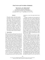
Docking of substrates and substrate analogues
into the active site
Docking of pNP-GlcNAc and pNP-GalNAc substrates
into the active site of the refined model of the PoHex,
followed by MD simulations, revealed the atomic
details of the substrate–enzyme interactions
(Fig. 7A,B). The protein showed stable behaviour after
only 1.5 ns of simulation (Fig. S3), so we used a 4-ns
simulation for substrate–enzyme complex analysis to
have at least 2 ns of equilibrated data for analysis.
Whereas pNP-GlcNAc was bound with a total of eight
hydrogen bonds, only five bonds could be identified
for pNP-GalNAc binding. In particular, the C4 posi-
tion (Fig. 7A,B, in yellow and magenta, respectively)
seems to play a key role in the specificity of these
interactions. For the pNP-GlcNAc substrate, the C4
hydroxyl hydrogen bonds to both Arg193 and Glu520,
whereas for pNP-GalNAc only a single, non-persistent,
hydrogen bond to Arg193 could be observed. This dif-
ference in binding is also reflected in the monitored
interaction energies during the MD simulations. The
standard MD simulations at pH 7 in water show an
average value of the interaction energy for the equili-
brated production phase of the simulation of 345 kJÆ-
mol
)1
for pNP-GlcNAc and 334 kJÆmol
)1
for pNP-
GalNAc.
Inhibition by excess substrate that was observed
experimentally with pNP-GlcNAc (although not with
pNP-GalNAc) (Fig. 2C,D) may be caused by the exis-
tence of additional binding sites for this compound.
To test this hypothesis, a blind docking experiment
was designed to screen the protein surface for addi-
tional potential binding sites. The docking experiment
did indeed reveal the existence of one ‘secondary’ bind-
ing site (Fig. 7C) in close proximity to the active site
of the enzyme. The interaction score of )21.5 kJÆmol
)1
given by the scoring function of autodock [31,32] is
comparable to the value measured for the substrate
docked into the active site ()21.1 kJÆmol
)1
). Hydrogen
bonds were observed between the oxygen at the C4
position of pNP-GlcNAc and residues Arg491 and
Asp443. The Asp425 residue was found to be within
0.3 nm of the docked inhibitor, a distance favourable
for electrostatic interaction; in A. oryzae Hex, this resi-
due is substituted by Glu424 and thus has a longer
side chain (see Fig. 7). The PoHex residue Asp443
belongs to the same turn as the active site residue
Tyr446, participating in the formation of a substrate–
enzyme intermediate [4]. autodock was able to
dock pNP-GalNAc into the enzyme active site ()18.0
kJÆmol
)1
) but was unable to identify an additional
binding site for it with favourable binding energy.
A similar procedure was used to investigate the
mechanism of inhibition by the reaction products Glc-
NAc and GalNAc that is observed experimentally
(Fig. 2E–H). Blind docking with GlcNAc shows a
clear preference for the ‘secondary’ binding site
(Fig. 7C), with an autodock score of )23.9 kJÆmol
)1
,
whereas the value for docking into the active site was
only )14.2 kJÆmol
)1
, which is significantly lower than
with pNP-GlcNAc or pNP-GalNAc as substrate. On
the other hand, the results for GalNAc suggest that
docking is only favourable at the active site ()22.0
kJÆmol
)1
). This active site interaction score is even
slightly higher than with pNP-GalNAc, indicating a
H. Rys
ˇ
lava
´
et al. b-N-Acetylhexosaminidase from Penicillium oxalicum
FEBS Journal 278 (2011) 2469–2484 ª 2011 The Authors Journal compilation ª 2011 FEBS 2477
clear competition between pNP-GalNAc and GalNAc
for the active site of the enzyme.
The amino acids of the loop that come into close
proximity to pNP-GlcNAc and GlcNAc at the ‘sec-
ondary’ binding site in the P. oxalicum enzyme differ
from those in the A. oryzae Hex. This difference in
models, determined by substitution of residues 503–505
and 424–428 from PoHex in the sequence of the hexos-
aminidase from A. oryzae, leads to a decrease in the
size of this region in A. oryzae compared with the
P. oxalicum enzyme (Fig. 7D). A shift in the position
of the loops to accommodate the secondary sub-
strate ⁄ product binding site may explain the differences
in kinetics observed between the two enzymes
(Table 1).
Hex substrates modified at their N-acyl residues fall
into two different categories. The trifluoroacetyl deriva-
tive of pNP-GlcNAc is not hydrolysed by the enzyme
from either tested species, whereas three other
substrates with N-acyl modifications are much better
hydrolysed by the P. oxalicum enzyme than the A. ory-
zae enzyme. Thus, we performed additional docking
experiments in which we docked all four modified sub-
strates in their standard form into the structures of Hex
from both P. oxalicum and A. oryzae. Substrates bear-
ing smaller N-acyl groups docked into the structure of
the enzymes with significantly decreased docking
energy. For example, the N-formyl substrate, in which
the methyl group has been replaced by a much smaller
hydrogen atom, docked with a docking energy of
339 kJÆmol
)1
compared with the standard N-acetyl sub-
strate, which yielded a docking energy of 345 kJÆmol
)1
(Fig. 8A). The accommodation of this substrate into
the substrate binding site is otherwise unaffected and
proceeds in the same way as the standard N-acetyl sub-
strate. However, the substrate is shifted in the active
site of the enzyme, making the hydrolysed glycosidic
bond more distant from the attacking catalytic residues
(Table S4). Moreover, we observed a change in the dis-
tance from atom O28 (at the C3 atom of the pyranose
ring) of this non-reducing sugar to the catalytic aspartic
acid (Asp) responsible for proper orientation of the
acetyl group during the formation of the oxazo-
linium ring. This distance shortened from 4.4 A
˚
in the
Fig. 7. Docking of N-acetylhexosamine substrates into the active site of the PoHex. (A) Active site with docked pNP-GlcNAc. The C4 atom
is shown in yellow, and hydrogen bonds are shown by yellow dotted lines. (B) Active site with docked pNP-GalNAc. Hydrogen bonds are
again yellow and the C4 atom is magenta. (C) Molecular surface representation of the protein with ‘secondary’ binding site (yellow) and
active site of the enzyme (magenta) with bound pNP-GlcNAc. The position of amino acids responsible for the creation of hydrogen bonds
(magenta lines) with the substrate at the secondary site are schematically depicted by blue sticks. (D) Secondary binding pocket of the Po-
Hex with docked pNP-GlcNAc overlaid with the Hex from Aspergillus oryzae (yellow surface). In the upper right corner is a list of the por-
tions of the sequence alignment that differ between the two enzymes in the vicinity of the secondary binding site.
b-N-Acetylhexosaminidase from Penicillium oxalicum H. Rys
ˇ
lava
´
et al.
2478 FEBS Journal 278 (2011) 2469–2484 ª 2011 The Authors Journal compilation ª 2011 FEBS
standard substrate (3.23 A
˚
in A. oryzae) to 2.77 A
˚
in
the N-formyl derivative (2.56 A
˚
in A. oryzae), enabling
the formation of a hydrogen bond between the O28
and the O-Asp, which competes with the hydrogen
bond formed by the N18(from the acetyl group)-H and
the O-Asp (Fig. 8B).
A completely different mechanism applies for sub-
strate analogues bearing longer acyls. These modified
substrates generally displayed docking energies compa-
rable with the acetyl-bearing substrate (e.g. the dock-
ing energy for N-propionyl substrate was
370.97 kJÆmol
)1
). Such a high binding energy is the
result of a hydrophobic effect that makes burying a
long propionyl inside the protein energetically favour-
able. The accommodation of the larger substrates into
the active site of the enzyme leads to a shift in their
position, resulting in decreased access of the hydroly-
sing groups of the active-site amino acids to the glyco-
sidic bond (Fig. 8C). The substrate in the active site is
thus more exposed to water (Fig. 8D). This effect was
also observed with the N-trifluoroacetyl substrate
(Fig. 8D). Despite the fact that the binding energy of
the N-trifluoroacetyl substrate was comparable with
that observed in the N-glycoloyl substrate, the cleavage
of the former substrate is complicated by electrostatic
repulsion between the catalytic aspartic acid and the
fluorines, which, taking into account the role of the
Asp residue in the mechanism [33], can prevent the for-
mation of the oxazolinium ring necessary for creating
the intermediate structure.
Discussion
Fungal hexosaminidases have proved useful in biotech-
nology and chemoenzymatic syntheses of novel oligo-
saccharide sequences. Unique features of the PoHex
among the secreted fungal hexosaminidases (although
matched in mammalian HexD and hexosaminidases
from C. elegans belonging to clade B [28,29]) such as a
high ratio of GalNAc-ase ⁄ GlcNAc-ase activity and its
Fig. 8. Docking of modified N-acylhexos-
amine substrates into the active site of
PoHex. (A) Binding energies of N-acyl
modified substrates with PoHex during 4 ns
of MD simulation. (B) Overlay of positions
of N-formyl modified (light blue) and stan-
dard (magenta) substrates after 4 ns of MD.
(C) Overlay of positions of N-propionyl
modified (light blue) and standard (magenta)
substrates after 4 ns of MD. (D) Molecular
surface of the binding pocket of PoHex with
bound N-trifluoroacetyl substrate after 4 ns
of MD. (E) Molecular surface of the binding
site of PoHex with bound pNP-GlcNAc after
4 ns of MD.
H. Rys
ˇ
lava
´
et al. b-N-Acetylhexosaminidase from Penicillium oxalicum
FEBS Journal 278 (2011) 2469–2484 ª 2011 The Authors Journal compilation ª 2011 FEBS 2479
ability to cleave substrate analogues with various
functional groups found for crude preparations
appeared interesting, and deserved detailed molecular
investigations using purified enzymes. To obtain stan-
dardized preparations, we used two publicly available
collection strains, tested several media and cultivation
conditions, and optimized a complete purification
protocol for PoHex. The highest specific activity
(35.6 UÆmg
)1
protein) and total yield (48 UÆL
)1
med-
ium) was obtained using CCF 3438 strain cultivated
for 7 days using medium M5. The PoHex isolated
from the CCF 1959 strain had a significantly lower
specific activity (10.8 UÆmg
)1
protein) despite its
identical primary structure. These results may be
explained by the low content of the propeptide in the
latter preparation. We have previously shown for the
Hex from A. oryzae that the propeptide represents an
important enzyme regulator that associates with the
catalytic subunit during its intracellular processing.
The amount of propeptide present is related to indi-
vidual stages of the life-cycle of the producing fungus
[10]. Only catalytic subunits that are supplemented
with the propeptide possessing proper post-transla-
tional modification (proteolysis at the dibasic site, spe-
cific glycosylation) can achieve full enzymatic activity
and be secreted into the extracellular environment
[9,10].
Previously published data describing the results of
enzyme kinetics measurements using PoHex [18,19]
are mostly in agreement with our results. However,
working with the highly purified enzyme under strictly
defined chemical conditions, we were able to find a
number of hitherto undescribed characteristics that
have escaped the attention of previous investigations.
First, the inhibition of the enzyme by an excess of
pNP-GlcNAc (but not pNP-GalNAc) is a completely
novel finding, as is the fact that the ratio of GalNA-
case to GlcNAcase activities is highly dependent on
the concentration of the particular substrates. Second,
the inhibition of the enzyme by its hydrolytic prod-
ucts (GlcNAc, GalNAc) has been studied in greater
detail. Moreover, we now propose a mechanism for
both of these phenomena based on the identification
of a secondary binding site for pNP-GlcNAc (see
below). Finally, the ratio of the two enzyme activities
has been found to depend on the concentration of
certain salts in the reaction buffer, especially ammo-
nium sulfate, a common salt used for enzyme storage
and stabilization. Based on these results, 0.4 mm
pNP-GlcNAc substrate in buffers lacking ammonium
sulfate or saturating concentrations of pNP-GalNAc
in buffers with high ammonium sulfate concentrations
appear optimal for practical use.
The purified PoHex displays a number of unique
properties in terms of stability and substrate specificity.
We have found that, unlike some Hex that are stable
under mildly acidic conditions [1,10], PoHex has an
optimum stability at pH 7–8. Additional stability max-
ima were detected for the native enzyme at pH 3 previ-
ously [19] and at pH 3 and pH 5 (this work). We have
found that the pH stability profile is significantly influ-
enced by enzyme glycosylation: Endo H-treated
enzyme has a single stability maximum between pH 5
and 8 and remains completely inactive outside this
range. The temperature optimum of the enzyme is
somewhat lower than the optimum for the A. oryzae
Hex (50 and 60 °C, respectively). The P. oxalicum
enzyme cleaves substrates with N-acyl modifications
much better (15% hydrolysis of N-formyl, N-glycolyl
and N-propionyl derivatives of the standard N-acetyl
substrate) than A. oryzae (2–5% hydrolysis).
The cloning and sequencing of PoHex and analysis
of the corresponding gene confirmed a significant pri-
mary structure similarity to the Hex from A. oryzae
published previously [10]. However, due to much
higher quality of the sequence in the promoter
upstream region of the hex gene from P. oxalicum,we
have now been able to identify several regulatory
sequences that might be responsible for the induction
of enzyme synthesis by GlcNAc and related inducers
[23]. Detailed comparisons of the primary structure of
the Hex from P. oxalicum to A. oryzae revealed very
small differences between the two enzymes, although
the evolutionary pressure, and thus the rate of diver-
gence, differed significantly within the individual seg-
ments of the sequence. Signal peptides are known to
diverge very rapidly and, indeed, we could not find
any similarity between the sequence of the signal pep-
tide of the PoHex and other hexosaminidases. Large
propeptides appear to be unique for the subfamily of
fungal Hex and also appear to diverge relatively rap-
idly with 59% identity (70% similarity) to the evolu-
tionarily closest propeptide from A. terreus NIH2624.
The sequence similarity was particularly high for the
catalytic subunit, with 76% identity (88% similarity)
to the most closely related Hex from P. chrysogenum
and 74% identity (87% similarity) to the Hex from
A. oryzae CCF 1066 studied previously [10,25]. More-
over, important post-translational modifications such
as disulfide bond formation [24] and N-glycosylation
were also rather similar. The positions of all the cyste-
ine residues, as well as the critical catalytic residues,
were identical, and there were only two regions with
significant differences in their primary structures.
Molecular modelling techniques that we employed
contributed significantly to a better understanding of
b-N-Acetylhexosaminidase from Penicillium oxalicum H. Rys
ˇ
lava
´
et al.
2480 FEBS Journal 278 (2011) 2469–2484 ª 2011 The Authors Journal compilation ª 2011 FEBS
the unique catalytic properties of the P. oxalicum
enzyme that would not have been obvious otherwise
considering the minimal differences in primary struc-
ture between PoHex and the Hex from A. oryzae.
Using advanced techniques of contemporary molecular
modelling, we were able to construct a three-dimen-
sional model of the complete native Hex in the form
of a fully glycosylated dimeric structure. The most dra-
matic differences between this three-dimensional model
and the model of the Hex from A. oryzae published
previously [25] are in the structure of the lids covering
the active sites of the enzymes and the structurally
adjacent protein loops. In the P. oxalicum enzyme, the
corresponding amino acid residues are smaller and
more flexible, and the lid loop tends to be in a more
open conformation due, in part, to a direct binding to
the same monomeric subunit (Fig. 6).
Moreover, techniques of MD simulations and ligand
docking allowed us to provide a mechanistic under-
standing of the details of the kinetics of these compli-
cated enzymes. In particular, the docking of both pNP-
GlcNAc and pNP-GalNAc substrates allowed us to
understand important features of Hex found experimen-
tally, such as the values of K
m
. In addition, the inhibi-
tion by an excess of pNP-GlcNAc could be understood
by the identification of the ‘secondary’ binding site close
to the active site of the enzyme. The fact that GlcNAc is
bound preferentially to this secondary binding site while
GalNAc binds only to the active site of the enzyme
allows us to understand the molecular nature of the type
of inhibition by the products (GlcNAc and GalNAc as
non-competitive and competitive inhibitors, respec-
tively). At the same time, we have become aware of cer-
tain limits of these techniques that could not fully
explain the experimental results obtained with N-acyl
modified substrates. In order to understand these experi-
mental data, the catalytic process will have to be consid-
ered in its entirety, including not only the binding and
hydrolysis of the substrate in the enzyme active site, but
also the diffusion and access of the substrate to the bind-
ing site of the enzyme, as well as the release of the prod-
ucts. In conclusion, in this paper we have clarified the
chemical nature of the unique catalytic properties of the
PoHex, which will prove useful for the future use of
this enzyme in biotechnology and chemoenzymatic
synthesis.
Materials and methods
Microbial strains and growth conditions
The strains of P. oxalicum CCF 1959 and CCF 3438 were
obtained from the publicly available Culture Collection of
Fungi at the Department of Botany, Faculty of Science,
Charles University Prague, Czech Republic (-
tur.cuni.cz/fccm/collecze.htm#ccf). The following culture
media were used: M1 was made up of (per L) 0.3 g
KH
2
PO
4
, 0.5 g NH
4
H
2
PO
4
, 0.2 g (NH
4
)
2
SO
4
, 0.1 g yeast
extract, 0.5 g GlcNAc, 5 g NaCl, 0.05 g MgSO
4
,pH6
(adjusted with KOH); M2 was identical to M1 except that
the yeast extract was replaced by 0.5 gÆL
)1
peptone; M3
was identical to M1 except that the yeast extract was
replaced by 0.1 gÆL
)1
peptone; M4 was made up of (per L)
0.2 g NaNO
3
, 0.05 g KCl, 0.001 g FeSO
4
, 0.1 g KH
2
PO
4
,
1.0 g GlcNAc, 0.5 g MgSO
4
, pH 5.5 (adjusted with HCl);
M5 was identical to M4 except that the pH was adjusted to
4.5; M6 was identical to M5 except that the pH was set to
6.5. The fungi were cultivated at 28 °C with constant reci-
procal shaking (200 r.p.m.) for the times indicated in indi-
vidual experiments. After the end of the cultivation period,
mycelia were removed by filtration, and the remaining med-
ium was used for protein and enzyme activity determina-
tions. The medium used for enzyme purification was
collected after 12 and 7 days of cultivation for the CCF
1959 and CCF 3438 strains, respectively.
Purification of PoHex
The crude enzyme obtained by precipitation of the culture
medium with ammonium sulfate (80% saturation) was dial-
ysed against 0.6 m ammonium sulfate in 20 mm sodium
phosphate buffer (pH 6.8). The enzyme solution was
applied to a Phenyl-Sepharose HP column (2.0 · 20 cm;
GE Healthcare, Fairfield, CT, USA), equilibrated using
the same buffer. The enzyme was eluted with a linear salt
gradient decreasing the content of ammonium sulfate
(10 mmÆmin
)1
). The enzyme was further purified on a
Mono Q column (1.5 · 15 cm; GE Healthcare) to near
homogeneity using a linear gradient from 0 to 0.5 m NaCl
in 20 mm Bistris buffer (pH 7.0). Final purification was
achieved on a Superdex 200 HR column (1 · 30 cm; GE
Healthcare), equilibrated and eluted in 10 mm Bistris buffer
(pH 7.0) with 0.5 m (NH
4
)
2
SO
4
. The enzyme was con-
centrated to 6 mgÆmL
)1
using a Centricon 30 (Millipore,
Billerica, MA, USA) and stored at 4 °C.
Enzyme assay
The enzyme activity was monitored using pNP-GlcNAc or
pNP-GalNAc substrates continuously at 348 nm [34] or via
the end-point method at 405 nm. The enzyme activity was
expressed in units (U, lmol of product formed per minute).
The reaction mixture contained 50 mm citrate buffer (pH
3.0) and the corresponding substrate at the concentration
corresponding to the saturated reaction rate (0.4 mm pNP-
GlcNAc, 2 m m pNP-GalNAc). After incubation for an
appropriate time, 0.2 m sodium carbonate (pH 11.0) was
added, and the concentration of liberated p-nitrophenol
H. Rys
ˇ
lava
´
et al. b-N-Acetylhexosaminidase from Penicillium oxalicum
FEBS Journal 278 (2011) 2469–2484 ª 2011 The Authors Journal compilation ª 2011 FEBS 2481
was determined. Steady-state initial velocity studies were
performed at 20 ° C in a final volume of 0.2 mL in 50 mm
McIlvaine buffer (pH 3.0) containing 0.02–2 mm substrate.
These data were fitted to the Michaelis–Menten equation or
an equation characterizing substrate inhibition; Michaelis
constant, maximal reaction rate, catalytic constant, cata-
lytic efficiency and substrate inhibition constants were
calculated. With 4-MU-GlcNAc substrate, the fluorescence
of the product (4-MU) was measured (k
ex
380 nm, k
em
520 nm).
pH optimum, pH stability, temperature optimum
pH optimum of the b-hexosaminidase was determined using
different buffers for various pH ranges: 0.1 m HCl ⁄ KCl
buffer (pH 1–2), 0.1 m citrate ⁄ phosphate buffer (pH 3–7),
0.1 m Tris ⁄ HCl buffer (pH 7–9) and 0.1 m glycine ⁄ NaOH
buffer (pH 9–11). pH stability of the enzyme was moni-
tored in long-term assays upon incubation in the same ser-
ies of buffers used for determining the pH optimum. The
hexosaminidase was kept in these buffers at 4 °C, and
aliquots were screened for enzyme activity at regular inter-
vals. The activity was measured at the pH optimum (3.0).
The temperature optimum was measured over the range
25–80 °Cin10°C increments.
Molecular modelling
To identify homologues for the protein, a blast search
() in the databases of non-
redundant protein sequences and some protein data banks
were used. High-scoring templates were extracted from the
Protein Data Bank results (): 1NOW,
1C7S and 1JAK. The identified templates were used for
making a structure-based multiple sequence alignment with
the tcoffee server ( />tcoffee_cgi ⁄ index.cgi) [35] using Expresso mode. This
method incorporates a consistency score to evaluate align-
ment. A structural alignment of the three templates was
additionally made with the sheba plug-in and visualized
with the program yasara [36]. Structure alignment was fur-
ther used for checking and correcting the tcoffee output
according to secondary structure elements and conserved
residues in known structures. To gain more information
from the alignments, we also used a secondary structure
prediction method ( />automat.pl?page=/NPSA/npsa_seccons.html) and two HMM
models to gain reliable secondary structure prediction.
Additionally, the older alignment used for modelling of the
A. oryzae enzyme [25] that was based on a careful analysis
of the whole protein family was used for comparison.
Three-dimensional models were built with the package
modeller 9.1 [37]. The stereochemical parameters of the
models were assessed with the program procheck [38], and
energetic parameters were analysed by prosa [39]. After
several iterations of a (re)alignment model building valida-
tion process, the best model was selected. Loops were mod-
elled using modloop [27]. The improvement of the quality
of models after loop modelling was analysed by procheck
and visual control.
The dimeric structure of the P. oxalicum enzyme was
built by overlying the monomers with the dimeric struc-
ture of human b-hexosaminidase 1O7A. Glycosylation was
performed online at with
sweet [40]. Selected glycan antennae were cut down to
leave two b-N-acetylglucosamine residues. The monomer
was further refined with yasara. The three-dimensional
structure was placed in a box with periodic boundary con-
ditions; the cell was filled with TIP3P water and neutral-
ized by placing counter-ions (sodium ions). To remove
steric overlaps and correct the covalent geometry, the
energy of the complex was minimized with the yamber 2
force field and default parameters, followed by a short
simulated annealing protocol (atom velocities scaled down
by 0.9 every tenth step) until convergence was reached
[25,34]. Docking (done by yasara [36] and autodock
[31,32]) and MD simulations by yasara are described in
supplementary material.
Acknowledgements
The authors acknowledge helpful comments and sug-
gestions by the referees, and technical help by Daniel
Kavan, Anna Hodkova
´
and Jir
ˇ
ı
´
Janovsky´ . This work
was supported in part by the Institutional Research
Concepts AVOZ50200510 for the Institute of Microbi-
ology and AVOZ60870520 for GCRC, by the Ministry
of Education of the Czech Republic (LC06010, MSM
21620808, MSM6007665808 and 1M0505) and by the
Grant Agency of the Czech Republic (303 ⁄ 09 ⁄ 0477,
203 ⁄ 09 ⁄ P024 and 305 ⁄ 09 ⁄ H008). Additionally, N.K.
was supported by the University of South Bohemia,
grant GAJU 170 ⁄ 2010 ⁄ P.
References
1 Sla
´
mova
´
K, Bojarova
´
P, Petra
´
skova
´
L&Kr
ˇ
en V (2010)
b-N-Acetylhexosaminidase: what’s in a name? Biotech-
nol Adv 28, 682–693.
2 Proia RL (1988) Gene encoding the human b-hexosa-
minidase b-chain – extensive homology of intron place-
ment in the a-chain and b-chain genes. Proc Natl Acad
Sci USA 85, 1883–1887.
3 Knapp S, Vocadlo D, Gao ZN, Kirk B, Lou JP &
Withers SG (1996) NAG-thiazoline, an N-acetyl-b-hex-
osaminidase inhibitor that implicates acetamido partici-
pation. J Am Chem Soc 118, 6804–6805.
4 Tews I, Perrakis A, Oppenheim A, Dauter Z, Wilson
KS & Vorgias CE (1996) Bacterial chitobiase structure
b-N-Acetylhexosaminidase from Penicillium oxalicum H. Rys
ˇ
lava
´
et al.
2482 FEBS Journal 278 (2011) 2469–2484 ª 2011 The Authors Journal compilation ª 2011 FEBS
provides insight into catalytic mechanism and the basis
of Tay–Sachs disease. Nat Struct Biol 3, 638–648.
5 Gooday GW, Zhu WY & Odonnell RW (1992) What
are the roles of chitinases in the growing fungus? FEMS
Microbiol Lett 100, 387–391.
6 Reyes F, Calatayud J, Vazquez C & Martinez MJ
(1989) b-N-Acetylglucosaminidase from Aspergil-
lus nidulans which degrades chitin oligomers during
autolysis. FEMS Microbiol Lett 65, 83–87.
7Kr
ˇ
en V & Martı
´
nkova
´
L (2001) Glycosides in medicine:
the role of glycosidic residue in biological activity. Curr
Med Chem 8, 1303–1328.
8 Bojarova
´
P&Kr
ˇ
en V (2009) Glycosidases: a key to tai-
lored carbohydrates. Trends Biotechnol 27, 199–209.
9 Plı
´
hal O, Sklena
´
r
ˇ
J, Kmonı
´
c
ˇ
kova
´
J, Man P, Pompach
P, Havlı
´
c
ˇ
ek V, Kr
ˇ
en V & Bezous
ˇ
ka K (2004) N-Gly-
cosylated catalytic unit meets O-glycosylated propep-
tide: complex protein architecture in a fungal
hexosaminidase. Biochem Soc Trans 32, 764–765.
10 Plı
´
hal O, Sklena
´
r
ˇ
J, Hofbauerova
´
K, Nova
´
k P, Man P,
Pompach P, Kavan D, Rys
ˇ
lava
´
H, Weignerova
´
L,
Charva
´
tova-Pis
ˇ
vejcova
´
A
et al. (2007) Large propep-
tides of fungal b-N-acetylhexosaminidases are novel
enzyme regulators that must be intracellularly processed
to control activity, dimerization, and secretion into the
extracellular environment. Biochemistry 46, 2719–2734.
11 Weignerova
´
L, Vavrus
ˇ
kova
´
P, Pis
ˇ
vejcova
´
A, Thiem J &
Kr
ˇ
en V (2003) Fungal b-N-acetylhexosaminidases with
high b-N-acetylgalactosaminidase activity and their use
for synthesis of b-GalNAc-containing oligosaccharides.
Carbohydr Res 338, 1003–1008.
12 Fialova
´
P, Weignerova
´
L, Rauvolfova
´
J, Pr
ˇ
ikrylova
´
V,
Pis
ˇ
vejcova
´
A, Ettrich R, Kuzma M, Sedmera P & Kr
ˇ
en
V (2004) Hydrolytic and transglycosylation reactions of
N-acyl modified substrates catalysed by b-N-acetylh-
exosaminidases. Tetrahedron 60, 693–701.
13 Fialova
´
P, Namdjou DJ, Ettrich R, Pr
ˇ
ikrylova
´
W, Rau-
volfova
´
J, Kr
ˇ
enek K, Kuzma M, Elling L, Bezous
ˇ
ka K
&Kr
ˇ
en V (2005) Combined application of galactose
oxidase and b-N-acetylhexosaminidase in the synthesis
of complex immunoactive N-acetyl-D-galactos-aminides.
Adv Synth Catal 347, 997–1006.
14 Hus
ˇ
a
´
kova
´
L, Riva S, Casali M, Nicotra S, Kuzma M,
Hun
ˇ
kova
´
Z&Kr
ˇ
en V (2001) Enzymatic glycosylation
using 6-O-acylated sugar donors and acceptors:
b-N-acetylhexosaminidase-catalysed synthesis of
6-O,N,N’-triacetylchitobiose and 6¢-O,N,N’-triacetylchi-
tobiose. Carbohydr Res 331, 143–148.
15 Sla
´
mova
´
K, Gazˇ a
´
k R, Bojarova
´
P, Kulik N, Ettrich R,
Pelantova
´
H, Sedmera P & Kr
ˇ
en V (2010) 4-Deoxy-
substrates for b-N-acetylhexosaminidases: how to make
use of their loose specificity. Glycobiology 20, 1002–
1009.
16 Di Giambattista R, Federici F, Petruccioli M & Fenice
M (2001) The chitinolytic activity of Penicillium janthin-
ellum P9: purification, partial characterization and
potential applications. J Appl Microbiol 91, 498–505.
17 Diez B, Rodriguez-Saiz M, de la Fuente JL, Moreno
MA & Barredo JL (2005) The nagA gene of Penicil-
lium chrysogenum encoding b-N-acetylglucosaminidase.
FEMS Microbiol Lett 242, 257–264.
18 Rodriguez J, Copapatino JL, Reyes F & Perezleblic MI
(1994) A b-N -acetylhexosaminidase from Penicillium
oxalicum implicated in its cell-wall degradation. Lett
Appl Microbiol 19, 217–220.
19 Yamamoto K, Lee KM, Kumagai H & Tochikura T
(1985) Purification and characterization of b-N-acetyl-
hexosaminidase from Penicillium oxalicum. Agric Biol
Chem 49, 611–619.
20 Hun
ˇ
kova
´
Z, Kr
ˇ
en V, S
ˇ
c
ˇ
igelova
´
M, Weignerova
´
L,
Scheel O & Thiem J (1996) Induction of b-N-acetylhex-
osaminidase in Aspergillus oryzae. Biotechnol Lett 18,
725–730.
21 Laemmli UK (1970) Cleavage of structural proteins
during assembly of head of bacteriophage-T4. Nature
227, 680–685.
22 Lee DH & Lee CB (2000) Chilling stress-induced
changes of antioxidant enzymes in the leaves of cucum-
ber: in gel enzyme activity assays. Plant Sci 159, 75–85.
23 Peterbauer CK, Brunner K, Mach RL & Kubicek CP
(2002) Identification of the N -acetyl-D-glucosamine-
inducible element in the promoter of the
Trichoderma atroviride nag1 gene encoding N-acetyl-glu-
cosaminidase. Mol Gen Genomics 267, 162–170.
24 Pompach P, Man P, Kavan D, Hofbauerova
´
K, Kumar
V, Bezous
ˇ
ka K, Havlı
´
c
ˇ
ek V & Novak P (2009) Modified
electrophoretic and digestion conditions allow a simpli-
fied mass spectrometric evaluation of disulfide bonds.
J Mass Spectrom 44, 1571–1578.
25 Ettrich R, Kopecky´ V, Hofbauerova
´
K, Baumruk V,
Nova
´
k P, Pompach P, Man P, Plı
´
hal O, Kuty´ M, Kulik
N et al. (2007) Structure of the dimeric N-glycosylated
form of fungal b-N-acetylhexosaminidase revealed by
computer modeling, vibrational spectroscopy, and
biochemical studies. BMC Struct Biol 7, 32.
26 Schuck P (2003) On the analysis of protein self-associa-
tion by sedimentation velocity analytical ultracentrifu-
gation. Anal Biochem 320, 104–124.
27 Fisher A, Do RK & Sali A (2000) Modeling of loops in
protein structures. Protein Sci 9, 1753–1773.
28 Intra J, Pavesi G & Horner DS (2008) Phylogenetic
analyses suggest multiple changes of substrate specificity
within the glycosyl hydrolase 20 family. BMC Evol Biol
8, 214.
29 Gutternigg M, Rendic
ˇ
D, Voglauer R, Iskratsch TI &
Wilson IBN (2009) Mammalian cells contain a second
nucleocytoplasmic hexosaminidase. Biochem J 419,
83–90.
30 Prag G, Papanikolau Y, Tavlas G, Vorgias CE, Petra-
tos K & Oppenheim AB (2000) Structures of chitobiase
H. Rys
ˇ
lava
´
et al. b-N-Acetylhexosaminidase from Penicillium oxalicum
FEBS Journal 278 (2011) 2469–2484 ª 2011 The Authors Journal compilation ª 2011 FEBS 2483
mutants complexed with the substrate di-N-acetyl-D-
glucosamine: the catalytic role of the conserved acidic
pair, aspartate 539 and glutamate 540. J Mol Biol 300,
611–617.
31 Goodsell DS & Olson AJ (1990) Automated docking of
substrates to proteins by simulated annealing. Proteins
8, 195–202.
32 Morris GM, Goodsell DS, Halliday RS, Huey R, Hart
WE, Belew RK & Olson AJ (1998) Automated docking
using a Lamarckian genetic algorithm and empirical
binding free energy function. J Comput Chem 19, 1639–
1662.
33 Mark BL, Mahuran DJ, Cherney MM, Zhao DL,
Knapp S & James MNG (2003) Crystal structure of
human b-hexosaminidase B: understanding the molecu-
lar basis of Sandhoff and Tay–Sachs disease. J Mol Biol
327, 1093–1109.
34 Brumer H, Sims PFG & Sinnott ML (1999) Lignocellu-
lose degradation by Phanerochaete chrysosporium:
purification and characterization of the main a-galacto-
sidase. Biochem J 339, 43–53.
35 Notredame C, Higgins DG & Heringa J (2000)
T-Coffee: a novel method for fast and accurate multiple
sequence alignment. J Mol Biol 302, 205–217.
36 Krieger E, Darden T, Nabuurs SB, Finkelstein A &
Vriend G (2004) Making optimal use of empirical
energy functions: force-field parameterization in crystal
space. Proteins 57, 678–683.
37 Sali A & Blundell TL (1993) Comparative protein mod-
eling by satisfaction of spatial restraints. J Mol Biol
234, 779–815.
38 Laskowski RA, Macarthur MW, Moss DS & Thornton
JM (1993) Procheck – a program to check the stereo-
chemical quality of protein structures. J Appl Crystal-
logr 26, 283–291.
39 Sippl MJ (1993) Recognition of errors in three-dimen-
sional structures of proteins. Proteins 17, 355–362.
40 Bohne-Lang A & von der Lieth CW (2005) GlyProt:
in silico glycosylation of proteins. Nucleic Acids Res 33,
W214–W219.
Supporting information
The following supplementary material is available:
Doc. S1. Supporting methods.
Fig. S1. Results of the validation of the refined model
by procheck.
Fig. S2. Relative mobility of surface residues shown as
a function of pH.
Fig. S3. RMSD of C-alpha atoms of the model of
PoHex during MD with different substrates.
Fig. S4. Secondary structure comparison with HMM
predictions.
Table S1. Purification of the hexosaminidase from
Penicillium oxalicum.
Table S2. Occurrence of contaminating glycosidases in
the course of purification.
Table S3. Occupancy of individual sites of N-glycosyl-
ation in PoHex by high mannose glycans identified by
mass spectrometry.
Table S4. Trp and position of the catalytic residues for
docking of the modified substrates.
This supplementary material can be found in the
online version of this article.
Please note: As a service to our authors and readers,
this journal provides supporting information supplied
by the authors. Such materials are peer-reviewed and
may be reorganized for online delivery, but are not
copy-edited or typeset. Technical support issues arising
from supporting information (other than missing files)
should be addressed to the authors.
b-N-Acetylhexosaminidase from Penicillium oxalicum H. Rys
ˇ
lava
´
et al.
2484 FEBS Journal 278 (2011) 2469–2484 ª 2011 The Authors Journal compilation ª 2011 FEBS