Tài liệu Báo cáo khoa học: Receptor association and tyrosine phosphorylation of S6 kinases pdf
Bạn đang xem bản rút gọn của tài liệu. Xem và tải ngay bản đầy đủ của tài liệu tại đây (1.07 MB, 14 trang )
Receptor association and tyrosine phosphorylation
of S6 kinases
Heike Rebholz
1,2
, Ganna Panasyuk
3
, Timothy Fenton
1,2
, Ivan Nemazanyy
3
, Taras Valovka
4
,
Marc Flajolet
5
, Lars Ronnstrand
6
, Len Stephens
7
, Andrew West
7
and Ivan T. Gout
2,3
1 Ludwig Institute for Cancer Research, London, UK
2 Department of Biochemistry and Molecular Biology, University College London, UK
3 The Institute of Molecular Biology and Genetics, Kyiv, Ukraine
4 Institute of Veterinary Biochemistry and Molecular Biology, University Zurich, Switzerland
5 Rockefeller University, New York, NY, USA
6 Lund University, Department of Experimental Clinical Chemistry, Malmo, Sweden
7 Babraham Institute, Cambridge, UK
8 GlaxoSmithKline, Harlow, UK
Keywords
AGC kinases; platelet-derived growth factor
receptor; receptor tyrosine kinases;
ribosomal protein S6 kinase; src
Correspondence
H. Rebholz, Box 296, Rockefeller University,
1230 York Ave, New York, NY 10021, USA
Fax: +1 212 327 7888
Tel: +1 212 327 8486
E-mail:
(Received 17 August 2005, revised 6
February 2006, accepted 8 March 2006)
doi:10.1111/j.1742-4658.2006.05219.x
Ribosomal protein S6 kinase (S6K) is activated by an array of mitogenic
stimuli and is a key player in the regulation of cell growth. The activation
process of S6 kinase involves a complex and sequential series of multiple
Ser ⁄ Thr phosphorylations and is mainly mediated via phosphatidylinositol
3-kinase (PI3K)-3-phosphoinositide-dependent protein kinase-1 (PDK1)
and mTor-dependent pathways. Upstream regulators of S6K, such as
PDK1 and protein kinase B (PKB ⁄ Akt), are recruited to the membrane via
their pleckstrin homology (PH) or protein–protein interaction domains.
However, the mechanism of integration of S6K into a multi-enzyme com-
plex around activated receptor tyrosine kinases is not clear. In the present
study, we describe a specific interaction between S6K with receptor tyrosine
kinases, such as platelet-derived growth factor receptor (PDGFR). The
interaction with PDGFR is mediated via the kinase or the kinase extension
domain of S6K. Complex formation is inducible by growth factors and
leads to S6K tyrosine phosphorylation. Using PDGFR mutants, we have
shown that the phosphorylation is exerted via a PDGFR-src pathway. Fur-
thermore, src kinase phosphorylates and coimmunoprecipitates with S6K
in vivo. Inhibitors towards tyrosine kinases, such as genistein and PP1, or
src-specific SU6656, but not PI3K and mTor inhibitors, lead to a reduction
in tyrosine phosphorylation of S6K. In addition, we mapped the sites of
tyrosine phosphorylation in S6K1 and S6K2 to Y39 and Y45, respectively.
Mutational and immunofluorescent analysis indicated that phosphorylation
of S6Ks at these sites does not affect their activity or subcellular localiza-
tion. Our data indicate that S6 kinase is recruited into a complex with
RTKs and src and becomes phosphorylated on tyrosine ⁄ s in response to
PDGF or serum.
Abbreviations
btk, Bruton’s tyrosine kinase; CSFR, colony stimulating factor receptor; DBS, donor bovine serum; DMEM, Dulbecco’s modified Eagle’s
medium; FBS, fetal bovine serum; FITC, fluoroscein isothiocyanate; HGFR, hepatocyte-growth factor receptor; PDGF, platelet-derived
growth factor; PDGFR, platelet-derived growth factor receptor; PDK1, 3-phosphoinositide-dependent protein kinase-1; PH, pleckstrin
homology; PI3K, phosphatidylinositol 3-kinase; PIP3, phosphatidylinositol-3,4,5-trisphosphate; PKB ⁄ Akt, protein kinase B; PKC, protein kinase
C; PTB, phosphotyrosine binding domain; RTK, receptor tyrosine kinase; S6K, ribosomal protein S6 kinase; SH2, Src homology 2.
FEBS Journal 273 (2006) 2023–2036 ª 2006 The Authors Journal compilation ª 2006 FEBS 2023
Ribosomal protein S6 kinase (S6K) is a serine ⁄ threon-
ine kinase belonging to the family of AGC kinases,
which includes protein kinase A (PKA), protein kinase
B (PKB ⁄ Akt), protein kinase C (PKCs), p90 ribosomal
S6 kinase and 3-phosphoinositide-dependent protein
kinase-1 (PDK1). AGC kinases share a high homology
in their kinase domains and have a similar mode of
activation [1].
There are two isoforms of S6 kinase, S6K1 and 2.
Both have highly homologous kinase and kinase
extension domains flanked by the less conserved
N- and C-terminal regulatory regions which are
responsible for their differential regulation [2,3]. S6K1
and S6K2 have cytoplasmic and nuclear isoforms,
which originate from different translational start sites.
Nucleocytoplasmic shuttling has been shown for both
cytoplasmic forms of S6Ks. All four isoforms lack
canonical protein–protein interaction domains, such as
Src homology 2 (SH2), phosphotyrosine binding
domain (PTB), Src homology 3 and WW, and have no
pleckstrin homology (PH) domain, which would enable
membrane association via lipid-binding. Instead, in
their C-terminal regions, S6K1 and S6K2 possess
either a PDZ domain-binding motif or a proline-rich
region, respectively, through which S6Ks could bind
other signaling molecules.
S6 kinases are activated through mitogen- and nutri-
ent-mediated pathways. Growth factor-activated recep-
tor tyrosine kinases (RTKs) recruit PI3K which, via its
effectors PKB ⁄ Akt and PDK1, mediates S6K activa-
tion [4]. Another major player in the activation of S6K
is the mammalian target of rapamycin, mTor (FRAP)
which senses the level of amino acids and possibly
other nutrients within a cell [5]. The activation of S6K
is a multistep phosphorylation event, involving several
ser ⁄ thr kinases. Initially, a series of serines and threo-
nines in the C-terminal autoinhibitory domain become
phosphorylated, followed by two sites within the
hydrophobic linker domain (S371 and T389) [6,7].
Phosphorylation at T389 by mTor or an mTor-
dependent kinase enables PDK1 to bind to S6K via its
PIF binding pocket [8]. Finally, PDK1 phosphorylates
T229 in the activation loop and hereby fully activates
S6K [8]. Protein phosphatases PP2A and PP1 have
been found in a complex with S6Ks [9,10]. PP2A has
further been shown to be the major phosphatase
responsible for the dephosphorylation and inactivation
of S6K [11] and its activity is stimulated upon inhibi-
tion of mTor [12].
The main known physiological substrate of S6 kin-
ases is the 40S ribosomal protein S6. Several other
in vitro and in vivo substrates have been recently identi-
fied, including pro-apoptotic protein Bad1 [13], cyto-
skeletal protein neurabin [14] and transcriptional
activator CREM [15].
Knockout studies in mice and Drosophila provided
evidence that S6K is an important regulator of cell size
and growth [16,17]. In S6K2(– ⁄ –) cells S6 phosphoryla-
tion is strongly reduced whereas in S6K1(– ⁄ –) almost
no reduction can be observed. This finding indicates
that S6 protein is not the major substrate for S6K1
in vivo as it cannot compensate for the lack of S6K2.
Hence, it is possible to imagine that S6K1 exerts some
effects via other substrates. It is also plausible that
changes in subcellular localization bring S6K in
contact with different substrates. Indeed, we have
shown that PKC-mediated phosphorylation of S6K2
at S486 leads to a retention of the kinase in the cyto-
plasm [2].
Here we report, for the first time, that both isoforms
of S6 kinase, S6K1 and S6K2, are associated with
RTKs and recruited to membrane ruffles upon growth
factor stimulation. Furthermore, we have shown that
S6Ks become phosphorylated on tyrosine in response
to mitogenic stimuli and that this phosphorylation
coincides with receptor recruitment. The use of platelet
derived growth factor (PDGF) receptor mutants defici-
ent in signaling via specific pathways and SU6656, a
src-specific inhibitor, indicated that both, RTK and src
activities are needed for tyrosine phosphorylation of
S6Ks. We have mapped the major src-dependent tyro-
sine phosphorylation site to a tyrosine in the N-termi-
nus of S6K1 and 2. Tyrosine phosphorylation does not
affect the activity or subcellular localization of S6Ks.
Results
S6 kinases are tyrosine phosphorylated by various
receptor and nonreceptor tyrosine kinases
In the present study, we addressed whether S6K acti-
vation involves tyrosine phosphorylation and trans-
location to the plasma membrane. In recent years, a
number of AGC kinases such as PKB ⁄ Akt, PDK1,
various PKCs and PKD but not S6Ks have been
shown to be tyrosine phosphorylated [18–23]. Initially,
we used a baculoviral expression system in Sf9 insect
cells. We infected Sf9 cells with viruses expressing
either cytoplasmic EE[Glu-Glu]-tagged S6K1 or S6K2
together with a panel of RTKs or the cytosolic tyro-
sine kinase fyn. When we immunoprecipitated S6Ks
with an anti-EE-tag IgG and probed the membrane
with phosphotyrosine antibody (4G10), tyrosine phos-
phorylation of S6K1 and 2 was reproducibly observed
when HGFR (hepatocyte-growth factor receptor),
PDGFR (platelet-derived growth factor receptor) and
S6K tyrosine phosphorylation H. Rebholz et al.
2024 FEBS Journal 273 (2006) 2023–2036 ª 2006 The Authors Journal compilation ª 2006 FEBS
CSFR (colony stimulating factor receptor) were coex-
pressed. The cytoplasmic tyrosine kinase fyn induced
tyrosine phosphorylation of S6K2 but not S6K1
(Fig. 1A).
Next, we investigated tyrosine phosphorylation of
S6Ks in an in vitro kinase assay with a panel of recom-
binant tyrosine kinases. As PDGFRb induced a strong
phosphotyrosine signal for S6K in insect cells, we tes-
ted this receptor for the ability of its kinase domain to
phosphorylate S6Ks in vitro. As shown in Fig. 1B,
recombinant PDGFRb kinase domain phosphorylated
both S6Ks. We further tested a panel of nonreceptor
tyrosine kinases, including src and fyn, Bruton’s tyro-
sine kinase (btk) and syk in an in vitro kinase assay
using S6K1 and 2 as substrates. As shown in Fig. 1C,
all tested tyrosine kinases, in particular src, phosphor-
ylated both isoforms of S6K in vitro. When tyrosine
kinases were not present in the assay, autophosphory-
lation of S6Ks was hardly detectable. Src kinase and
S6K2 both migrate at 60 kDa in a SDS ⁄ PAGE gel.
Therefore, both autoradiography signals are merged in
the S6K2 sample treated with src. However, when
S6K1 is treated with src, the src autophosphorylation
signal is low in our experiment. For this reason, the
autoradiography signal from the S6K2 plus src sample
should stem mainly from S6K2 phosphorylation.
S6Ks are tyrosine phosphorylated and associated
with receptor tyrosine kinases upon growth
factor stimulation
To test whether tyrosine phosphorylation would also
occur in mammalian cells, we transiently transfected
Cos7 cells with S6Ks and PDGFR. Cells were starved
for 24 h and stimulated for 30, 60 or 180 min with
PDGF-BB. When S6Ks were immunoprecipitated via
their EE-tag, we found PDGF-dependent tyrosine
phosphorylation of S6Ks. Tyrosine phosphorylation
reached its maximum at 30 min of stimulation and
decreased after 1 h (Fig. 2A). Time course experiments
using 5 and 10 min of stimulation were also performed
and indicated that S6Ks are already tyrosine phos-
phorylated within 5 min (data not shown). Further-
more, PDGFR was found to coimmunoprecipitate
with S6Ks. In this system, the association appears to
be constitutive. However, one has to take into account
that the receptor is strongly overexpressed and there-
fore may be partially active even in starved cells. The
fact that the top band of the coimmunoprecipitated
PDGFR (representing the mature receptor) exhibits a
slightly weaker pY signal in the starved sample than in
the PDGF-treated sample further indicates that the
receptor is partially but not fully active when cells are
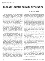
A
C
B
Fig. 1. Tyrosine phosphorylation of S6K1 and S6K2 in Sf9 cells and in vitro. (A) Sf9 cells were infected with baculoviruses encoding either
EE-tagged S6K1 or S6K2 and a receptor tyrosine kinase (EGFR, HGFR, PDGFR) or the cytosolic tyrosine kinase fyn. Cells were lyzed 2 days
postinfection and S6Ks were immunoprecipitated with anti-EE IgG. Samples were resolved by SDS ⁄ PAGE, transferred onto nitrocellulose
membrane and analyzed by immunoblotting with monoclonal antibodies against phosphotyrosine (4G10). (B) In vitro tyrosine phosphorylation
of S6K by PDGFR. S6Ks were immunoprecipitated from Sf9 cells (using anti-EE IgG) and then subjected to an in vitro tyrosine kinase assay
with PDGFR as kinase for 30 min at 30 °C. 100 ng of PDGFR were used per sample. An autoradiograph and the Coomassie-stained gel are
shown. (C) In vitro tyrosine phosphorylation of S6K by cytosolic tyrosine kinases. P70S6Ks were immunoprecipitated from Sf9 cells (using
anti-EE IgG), then subjected to an in vitro tyrosine kinase assay for 30min at 30 °C. Per sample, 7 pmol of the different tyrosine kinases src,
lyn, syk and btk were used. Autoradiograph and the Coomassie-stained gel are shown.
H. Rebholz et al. S6K tyrosine phosphorylation
FEBS Journal 273 (2006) 2023–2036 ª 2006 The Authors Journal compilation ª 2006 FEBS 2025
starved. This activation may be sufficient for S6K
recruitment but not for maximal S6K activation and
tyrosine phosphorylation. The anti-S6K western blot
confirms this hypothesis as in this system S6K is parti-
ally active in starved cells as indicated by the partial
bandshift, with most S6K being the bottom inactive
S6K. With PDGF there is a stronger band shift which
is decreased again after 180 min of stimulation. In a
control experiment it was established that PDGFR,
when expressed alone, does not precipitate with Pro-
tein A Sepharose beads coupled with anti-EE IgG
(data not shown, or also see Fig. S1).
To strengthen our observation, we tested whether
endogenous S6K would also become tyrosine phos-
phorylated. Since NIH3T3 cells express high levels of
endogenous S6K we used them in this study. To
achieve maximal stimulation of multiple RTKs, we
stimulated the cells with serum rather than PDGF.
Endogenous S6K1 was immunoprecipitated from cells
after 30, 60 and 180 min of serum-stimulation. As
shown in Fig. 2B, both variants of S6K1 (p70 and
p85) are phosphorylated on tyrosine in an inducible
manner. Interestingly, the phosphorylation of the nuc-
lear isoform, p85 S6K1 appears delayed compared to
p70 S6K1. Activated S6K usually migrates as four dis-
tinct bands on a SDS ⁄ PAGE gel due to multiple phos-
phorylation. The tyrosine phosphorylated bands of
S6K overlap with two of the activated and slower
migrating bands.
We have shown that S6Ks can be detected in a
complex with PDGFR when they are transiently
expressed. We further tested the interaction between
endogenous S6Ks and PDGFR (Fig. 2C). We found
that PDGFR is specifically associated with S6K1 in a
serum-inducible manner indicating that, under physio-
logical circumstances, S6K is only recruited to acti-
vated RTKs.
S6K translocates to PDGF-induced membrane
ruffles
Immunofluorescence studies in fibroblasts showed that
PKB ⁄ Akt is recruited to membrane ruffles upon mito-
gen treatment [24]. We therefore decided to investigate
if S6K is also recruited to the plasma membrane upon
mitogenic stimulation. We used PDGF as a stimulus
as it is well known to generate ruffling in NIH3T3
cells. Serum-starved NIH3T3 cells were stimulated for
various times, fixed and stained with an antibody
against the C-terminus of S6K1. Using an fluoroscein
isothiocyanate (FITC)-labeled secondary anti-rabbit
IgG and phalloidin to stain actin, S6K was shown to
be evenly distributed in the cytoplasm of starved
cells. We could also detect colocalization with stress
fibers which has been described previously [25] (data
not shown). Platelet-derived growth factor (PDGF)
Fig. 2. S6Ks are tyrosine phosphorylated and associated with RTKs.
(A) PDGFR and S6K1 or 2 were expressed in Cos7 cells. Twenty-
four hour post-transfection cells were starved for 20 h and stimul-
ated with 40 ngÆmL
)1
PDGF as indicated. Immunoprecipitated
EE-S6Ks and complexed were separated by SDS ⁄ PAGE, trans-
ferred onto nitrocellulose and blotted with phosphotyrosine (4G10)
antibodies. The upper half of the membrane was reprobed with
anti-PDGFR IgG and the lower part with anti-EE IgG. (B) Tyrosine
phosphorylation of endogenous S6K1. NIH 3T3 cells were starved
in 0.3% DBS for 24 h and stimulated with 10% DBS as indicated.
Endogenous S6K1 was immunoprecipitated using an antibody
against its C-terminus. The immunoprecipitates were treated as in
(A) and the membrane reprobed with the C-terminal antibody. In
this experiment we focused on S6K1 as NIH3T3 cells do not
express S6K2. The results of three individual experiments for
p70 S6K1 were quantified and are shown as histogram. (C) Endo-
genous PDGF receptor coimmunoprecipitates with S6K1 in a stim-
ulation-dependent manner. NIH3T3 cells were starved and
stimulated with 10% DBS for the indicated times. Endogenous
S6K1 was immunoprecipitated with antibody against the C-termin-
us of S6K and immunocomplexes were analyzed by immunoblot-
ting using anti-S6K or anti-PDGFR IgG.
S6K tyrosine phosphorylation H. Rebholz et al.
2026 FEBS Journal 273 (2006) 2023–2036 ª 2006 The Authors Journal compilation ª 2006 FEBS
treatment leads to a redistribution of the bulk of
S6K towards the nucleus or the perinuclear region.
In addition, we reproducibly observed a small frac-
tion of S6K1 in membrane ruffles for various time
points tested. Fig. 3A shows a 5-min treatment with
PDGF.
In addition, we used v-src transformed Swiss 3T3
cells in this study as they show very strong PDGF-
inducible ruffling. Serum-starved Swiss 3T3 cells were
stimulated with PDGF for 5 min, fixed and stained
with antibody against the C-terminus of S6K1. Simi-
larly to NIH3T3 cells, PDGF treatment lead to a
redistribution of the bulk of S6K towards the nucleus
or the perinuclear region and of a small fraction of
S6K1 to membrane ruffles (Fig. 3B). In a western blot
on total cell lysate from NIH3T3, this S6K-antibody is
very specific and solely recognizes S6K1 (p70 and p85).
We used the same antibody for immunofluorescence
studies. These data suggest that S6K may translocate
towards the membrane where it could participate in
multienzyme complexes consisting of RTKs and other
signaling molecules.
Tyrosine phosphorylation is dependent on
PDGFR-src signaling
Upon stimulation, the PDGF receptor dimerizes and
autophosphorylates. The generated phosphotyrosine
sites constitute binding sites for a variety of down-
stream proteins with SH2 domains. In order to deter-
mine the signaling pathways resulting in S6K tyrosine
phosphorylation, we utilized a panel of PDGFR
mutants where specific tyrosine sites were mutated to
phenylalanines. The PDGFRb Y763 ⁄ 1009F mutant is
deficient in signaling via Shp2 phosphatase, while
PDGFRb Y579 ⁄ 581F is unable to bind and activate
src [26,27]. PDGFRb K634A is kinase dead. We
transfected Cos7 cells with S6K1 ⁄ 2 and various
PDGFRb mutants. After serum starvation, cells were
stimulated with PDGF and both S6Ks were immuno-
precipitated and analyzed by western blotting. In the
control experiment with kinase dead receptor
(PDGFRb K634A), there was no detectable S6K tyro-
sine phosphorylation (Fig. 4A). Notably, S6K expres-
sion was always reduced when expressed together with
Fig. 3. S6K1 is localized in membrane ruf-
fles upon PDGF stimulation in NIH3T3 cells.
NIH3T3 cells were starved for 24 h,
followed by stimulation with PDGF
(10 ngÆmL
)1
) for 5 min. Cells were fixed,
permeabilized, blocked and probed with
anti-C-terminal S6K1 IgG and secondary
FITC-anti-rabbit IgG. Actin was visualized by
phalloidin staining which was added during
the last 10 min of incubation with FITC-anti-
rabbit IgG. Arrows indicate membrane ruf-
fles in which S6K is present. We also used
v-src transformed Swiss3T3 cells as they
generate very strong PDGF-induced ruffles.
Cells were grown at 35 °C and treated simi-
larly to NIH3T3 cells.
H. Rebholz et al. S6K tyrosine phosphorylation
FEBS Journal 273 (2006) 2023–2036 ª 2006 The Authors Journal compilation ª 2006 FEBS 2027
KD PDGFR. However, in the S6K2 ⁄ KD PDFGR
sample the expression level is comparable to
S6K ⁄ wtPDGFR of the starved sample. Expression of
the Y763 ⁄ 1009F mutant when compared to
wtPDGFR did not alter tyrosine phosphorylation of
S6K. Interestingly, the phosphotyrosine signal of
S6Ks from cells expressing the Y579 ⁄ 581F receptor is
strongly reduced. This result suggests that src kinase
may be involved in tyrosine phosphorylation of
S6 kinase. When the membrane was re-probed with
anti-S6K1 IgG, the migration of multiple bands
representing S6K1 was similar in wtPDGFR and the
Y579 ⁄ 581F mutant hinting that the activation process
was probably not altered by the lack of tyrosine phos-
phorylation.
To investigate the involvement of src and PDGFR
in tyrosine phosphorylation of S6K further, we studied
the effect of inhibitors on tyrosine phosphorylation of
S6Ks. As expected, genistein, a broad-range tyrosine
kinase inhibitor, reduced the PDGF-induced phospho-
tyrosine signal of both S6Ks. Similarly, PP1, an inhib-
itor acting on src, but also PDGFR, c-kit and abl [28]
reduced tyrosine phosphorylation of S6K very strongly.
Finally, the src-specific SU6656 also showed an inhibi-
tory effect on the phosphotyrosine signal in S6K
(Fig. 4B). Interestingly, LY294002 and rapamycin,
inhibitors of PI3Kand mTor, respectively, while being
effective in inhibiting S6K activity (as shown by phos-
pho-S6 blot), did not reduce but rather slightly
enhanced tyrosine phosphorylation of S6K (supple-
mentary Fig. S2).
To further investigate if tyrosine phosphorylation
was mediated by the action of src in vivo, we transi-
ently expressed various mutants of src together with
S6K. Expression of wild-type src leads to weak basal
tyrosine phosphorylation which could be enhanced by
serum ⁄ vanadate stimulation. A constitutively active src
(Y527F) induced a much stronger tyrosine phosphory-
lation of S6K1 (Fig. 5A). Dominant-negative src lead
to a complete loss of the phosphotyrosine signal in
immunoprecipitated S6Ks. Interestingly, in starved
cells we could observe that overexpression of a consti-
tutively active version of src (527F) led to a band shift
of S6K1 that was similar to the shift in stimulated
cells. Furthermore the pT389 signal, a marker of S6K
activity, in these starved cells was equal to the signal
from the stimulated cells. This activation was not
reflected by the state of tyrosine phosphorylation,
which was significantly lower in starved than in stimu-
lated cells. In serum-stimulated cells we did not see a
significant effect of src 527F on the activity of S6K
even though src 527F led to its strong tyrosine phos-
phorylation. Phospho-T389 levels and the band shift
of S6K were similar and independent of the src variant
(DN, WT, 527F) in stimulated cells. The most highly
tyrosine phosphorylated S6K from src 527F expres-
sing, stimulated cells was no more active than the
non-tyrosine phosphorylated S6K derived from cells
overexpressing DN src. Interestingly, this constitutively
active src variant still needed stimulation in order to
generate a maximal phosphotyrosine signal on S6K1.
The reason therefore may be that stimulation leads to
S6K translocation towards the plasma membrane
where it may interact with src. Furthermore, we could
B
A
Fig. 4. Tyrosine phosphorylation of S6K is mediated via a PDGFR-
src pathway. (A) Cos7 cells transfected with wt or mutant
forms of PDGFRb (KD PDGFRb K634A, PDGFRb579 ⁄ 581F,
PDGFRbY763 ⁄ 1009F) and EE-tagged S6K1 or 2, starved and stimul-
ated with PDGF (40 ngÆmL
)1
) for 15 min. Lysates were incubated
with anti-EE IgG bound to protein A-sepharose followed by western
blot analysis using anti-pY IgG. The membrane was stripped and
reprobed with anti-S6K IgG. The total lysate (30 lg) was also tested
for PDGFR expression. (B) Effect of inhibitors on tyrosine phos-
phorylation of S6Ks. Hek293 cells transiently expressing PDGFR
and either S6K1 or S6K2 were starved for 24 h. Sixty minutes
before stimulation, cells were incubated with a panel of inhibitors
(genistein 100 l
M, PP1 50 lM and SU6656 4 lM), then stimulated
with PDGF (40 ngÆmL
)1
). EE-S6Ks were immunoprecipitated with
anti–EE IgG, transferred to nitrocellullose membrane and probed
with antiphosphotyrosine (4G10) followed by anti–S6K IgG. Total
lysate (30 lg) was tested for PDGFR expression.
S6K tyrosine phosphorylation H. Rebholz et al.
2028 FEBS Journal 273 (2006) 2023–2036 ª 2006 The Authors Journal compilation ª 2006 FEBS
detect endogenous S6K1 and src in a complex in expo-
nentially growing Hek293 cells (Fig. 5B), strengthening
the hypothesis that src kinase, which localizes to an
activated receptor tyrosine kinase, is a major kinase
responsible for tyrosine phosphorylation of S6K
in vivo.
We also found endogenous S6K1 to be tyrosine
phosphorylated in v-src transformed Swiss3T3 cells
but not in the parental cell line. The src-specific inhib-
itor SU6656 could inhibit this phosphorylation
(Fig. 5C). This is another indication that the phos-
phorylation of native S6K occurs in cells in a src-
dependent manner. It is possible to imagine that S6K
tyrosine phosphorylation occurs during the process of
oncogenic transformation. In these Swiss3T3 cells we
could also observe higher levels of phospho-S6 than in
parental cells confirming earlier reports of elevated
S6K activity [29] (data not shown).
Src kinase phosphorylates S6K in the N-terminus
In order to determine the sites of tyrosine phosphory-
lation, we used N- and C-terminally truncated S6K1.
When these mutants were immunoprecipitated from
Hek293 cells that also transiently expressed activated
src (Y527F), S6K1DC was tyrosine phosphorylated but
not the S6K1DN mutant (Fig. 6A). This indicated that
the major tyrosine phosphorylation site ⁄ s may be
located at the N-terminus of S6K1. To verify our
hypothesis and to exclude that the lack of tyrosine
phosphorylation in the S6KDN mutant might be due
to a conformational change that hinders the access of
tyrosine kinases to their substrate residues, we gener-
ated and purified recombinant S6K1 N-terminal
domain and subjected it to an in vitro kinase assay
with several cytoplasmic tyrosine kinases such as src,
lyn, syk and btk. As a result, all kinases were able to
phosphorylate the S6K1 N-terminal domain (Fig. 6B).
An almost complete mobility shift of the domain could
be seen in the presence of src. Even though N-terminal
sequences of S6K1 and S6K2 are only conserved to
38%, both contain a tyrosine residue, S6K1Y39 and
S6K2Y45, equally followed by a glutamate at +1
indicative for a src phosphorylation site. Using mass
spectrometry, we could confirm the S6K1Y39 site as
being tyrosine phosphorylated in vitro (supplementary
Fig. S3). In order to determine if these residues consti-
tute major phosphorylation sites in full length S6K we
generated EE-tagged phenylalanine mutants. When
these mutants were subjected to an in vitro tyrosine
kinase assay, they were much less tyrosine phosphoryl-
ated by src than wt S6K (Fig. 6C). The level of S6K
autophosphorylation was also assessed and was hardly
detectable under the experimental conditions. More
importantly, overexpression of the mutants together
with src (527F) in Hek293 cells led to a strongly
Fig. 5. Tyrosine phosphorylation of S6K is dependent on Src activity. (A) Hek293 cells were transfected with S6K1 and either pcDNA3.1 or
wild-type src, dominant negative src (DN) or constitutively active src (Y527F). Starved cells were stimulated with 10% FBS (15 min) followed
by a brief treatment with pervanadate (2 min). Phosphotyrosine levels of S6K were assessed by western blot using 4G10 antibody. The
membrane was stripped and reprobed twice with antibodies against pT389 and S6K1. Total lysates (30 lg) were probed with anti-src IgG.
(B) Exponentially growing Hek293 cells were lyzed. Endogenous S6K1 was immunoprecipitated using an anti-S6K1 IgG, immunocomplexes
were separated by SDS ⁄ PAGE and membrane was probed with anti-src IgG. As a control, we used ProteinA-sepharose beads to test for
the specificity of the coimmunoprecipitation. (C) S6K is tyrosine phosphorylated in v-src transformed cells. Exponentially growing v-src trans-
formed Swiss 3T3 and parental cells were treated with 4 l
M SU6656 for 16 h before lysis. S6K1 was immunoprecipitated and blotted with
4G10 antibody. The membrane was reprobed with anti-S6K1 IgG.
H. Rebholz et al. S6K tyrosine phosphorylation
FEBS Journal 273 (2006) 2023–2036 ª 2006 The Authors Journal compilation ª 2006 FEBS 2029
reduced phosphotyrosine signal (by 88 and 95% for
S6K1 and S6K2, respectively) (Fig. 6D) indicating that
the N-terminal site is the major phosphorylation site
in vivo. However, the possibility that another minor
site exists cannot be excluded.
As tyrosine phosphorylation was detectable upon
growth factor stimulation and therefore paralleled the
activation by S ⁄ T phosphorylation, it was logical to
hypothesize that tyrosine phosphorylation may be
involved in the regulation of S6K activity. As previ-
ously shown, tyrosine phosphorylation is strongly
reduced when the src signaling-deficient PDGFRY579 ⁄
581F mutant is expressed (Fig. 4A). We assayed the
in vitro activity of S6K coexpressed with wild-type
PDGFR or Y579 ⁄ 581F in starved or PDGF-stimula-
ted cells. S6K activity was not altered in the presence
of the src signaling deficient mutant when compared
with wild type (supplementary Fig. S4). Next, we tes-
ted if mutation of Y39 ⁄ Y45 to phenylalanine would
affect the activity of S6Ks. No difference between
wild-type and mutant activities could be observed in
stimulated or starved cells in an in vitro kinase assay,
indicating that tyrosine phosphorylation of this site
does not modulate kinase activity (Fig. 6E). The
S6K1Y39D mutant was also tested and had similar
activity to the wild type (data not shown).
Src-induced tyrosine phosphorylation of atypical PKC
has been shown to alter its subcellular localization.
Therefore, we tested the subcellular localization of
wild-type S6K and mutants (S6K1Y39F, S6K2Y45F)
by confocal microscopy in NIH3T3 cells but did not
observe significant differences. In addition, the subcellu-
lar localization of S6K1 was similar in src-deficient (syf)
or syf + src fibroblasts (data not shown). This data
A
D
B
C
E
Fig. 6. Determination of a N-terminal tyrosine as src-dependent phosphorylation site. (A) Deletion of the N-terminus leads to a loss of phos-
photyrosine in S6K. Hek293 cells were transiently transfected with WT and truncated mutants of S6K (S6K1, S6K1 DN and S6K1DC) and src
527F. Cells were starved for 24 h and stimulated with FBS (15 min) followed by a 2-min treatment with Na
3
VO
4
. S6Ks were precipitated,
immunocomplexes separated via SDS ⁄ PAGE and blotted with pY antibody. Membrane was stripped and reprobed with anti-EE IgG. Total
lysate (30 lg) was also analyzed for src expression. Arrows indicate the truncated S6Ks. (B) The N-terminal domain of S6K1 is a substrate
for tyrosine kinases. One microgram of the purified recombinant N-terminal fragment was used for an in vitro kinase assay using 7 pmol of
cytosolic tyrosine kinases src, btk, lyn and syk. (C) Tyrosine Y39 ⁄ 45 in S6K1 ⁄ 2 is a substrate for src kinase in vitro. S6K1 ⁄ S6K2 and
S6K1Y39F ⁄ S6K2Y45F mutants were immunopurified from Hek293 cells and subjected to an in vitro kinase assay using recombinant src
kinase. Reaction products were analyzed by autoradiography and Coomassie staining as indicated. (D) Tyrosine Y39 ⁄ 45 in S6K1 ⁄ 2 is a sub-
strate for src kinase in vivo. S6K WT and mutants and src kinase were overexpressed in Hek293 cells, which were starved and stimula-
ted with 10% FBS for 15 min and for 2 min with Na
3
VO
4
. Immunoprecipitated S6Ks were tested with anti-pY IgG and membrane was
reprobed with S6K antibody. Total lysate was also analyzed for src expression. (E) The activity of S6K1 ⁄ 2 Y39F ⁄ Y45F mutants is not altered.
Hek293 cells were transfected with S6K1 ⁄ 2 or Y39F ⁄ 45F. Cells were starved and stimulated as indicated (15 min FBS). S6K was immuno-
precipitated from these cells, subjected to an in vitro kinase assay using S6 as a substrate. The expression of S6Ks was assessed by
western blotting.
S6K tyrosine phosphorylation H. Rebholz et al.
2030 FEBS Journal 273 (2006) 2023–2036 ª 2006 The Authors Journal compilation ª 2006 FEBS
indicate that src-mediated tyrosine phosphorylation of
S6K does not affect its subcellular localization.
Taken together, we found that S6K becomes tyro-
sine phosphorylated in a PDGFR-src mediated path-
way which involves membrane recruitment of S6K. We
have shown that a subpopulation of S6K is present at
the membrane upon PDGF stimulation and thus in
the vicinity of PKB and PDK1 which are the major
activators of S6K.
Discussion
In this study, we have shown for the first time that
S6Ks become tyrosine phosphorylated and associated
with PDGFR in a ligand-induced manner. In mamma-
lian cells, both events, receptor association and tyro-
sine phosphorylation occur simultaneously and peak
within the first 30 min after stimulation.
Membrane translocation in response to mitogenic
stimuli has been shown for a variety of AGC kinases,
including PKB ⁄ Akt, PDK1, PKD and various iso-
zymes of the PKC family. This is mainly thought to
occur via binding to second messengers such as phos-
pholipids or via binding to phosphotyrosine residues
on activated RTKs. Translocation of PKB ⁄ Akt or
PDK1 is mediated through PH domains which specif-
ically recognize the second messenger PIP3 [30,31]. A
variety of signaling molecules such as PI3K, IRS1, Src
or GRB2 translocate to the membrane and associate
with activated receptors via their SH2 or PTB
domains. PKC translocation is mediated by a variety
of isoform-specific RACKs (receptors for activated
C-kinase) [32]. In addition, many AGC kinases have
been shown to be substrates for src kinase which itself
associates with activated RTKs. Even though the
phosphorylation events leading to full activation of
S6K have been thoroughly studied, it is not clear if
they involve translocation of S6K to the membrane.
However, S6K, in order to be phosphorylated by
PDK1, may be in the vicinity of the membrane. Fur-
thermore, Rho family G proteins Rac and Cdc42,
which control cytoskeletal organization, were shown to
associate with and activate S6K [33]. As these small
GTPases are most active when they are membrane-
bound, it would be logical for S6K to be colocalized
with its upstream effectors. Finally, it was reported
that S6K is complexed with the receptor-associated
p85 subunit of PI3K and that this complex formation
is needed for mTor and PI3K-mediated activation of
S6K [34]. We showed that PDGFR can specifically im-
munoprecipitate with S6K which, to our knowledge, is
the first report of coimmunoprecipitation of an RTK
with an AGC kinase. We further used immunofluores-
cence microscopy to show that S6K1 can be localized
at the plasma membrane. In starved cells, S6K1 is
evenly distributed within the cytoplasm and can also
be detected along stress fibers. Upon stimulation, the
majority of S6K1 molecules translocate to the nucleus,
whereas a subpopulation is reproducibly found in
membrane ruffles.
The association between receptor and nonreceptor
tyrosine kinases and S6K leads to its tyrosine phos-
phorylation in vitro and in vivo. The recombinant
kinase domain of PDGFR, as well as cytoplasmic
tyrosine kinases such as src, is able to phosphorylate
S6Ks on tyrosine. In vivo, using PDGFR mutants that
are deficient in signaling via src kinase, we found that
both PDGFR and src kinase activities are needed for
maximal tyrosine phosphorylation of S6Ks. Studies
employing tyrosine kinase inhibitors such as PP1 and
SU6656 validated this finding. PI3K and mTor do not
influence tyrosine phosphorylation of S6K as demon-
strated by the use of the inhibitors LY294002 or rapa-
mycin. This finding is in congruence with the finding
that PDK1 tyrosine phosphorylation is independent of
PI3K activity [20]. The major src-dependent phos-
phorylation sites, S6K1 Y39 and S6K2 Y45 are located
at the N-terminus of S6K.
We observed a difference in phosphorylation kinetics
of the p70 and p85 isoforms of endogenous S6K1
in NIH3T3 cells: Whereas P70 was already phosphoryl-
ated after 30 min, we could only detect p85 phosphory-
lation after 60 min of stimulation. In contrast to
p70 S6K, the p85 isoform is thought to be exclusively
localized in the nucleus, and thus, the delayed tyrosine
phosphorylation may result from activation and ⁄ or
translocation of the respective kinase. For example, as
c-src was shown to be in part localized in the nucleus
[35], one possibility could be that src translocates to the
nucleus where it can phosphorylate p85 S6K. Very poss-
ibly both isoforms are part of distinct feedback mecha-
nisms via tyrosine phosphatases.
For several AGC kinases such as PKB ⁄ Akt, PDK1,
PKCs and PKD it was shown that tyrosine phosphory-
lation results in increased kinase activity [19]
[20,23,36]. It is known that PI3K activity is involved in
v-src transformation and the level of PIP3 is elevated
in v-src transformed cells [37]. In v-src transformed
cells PKB ⁄ Akt activity is enhanced, due to elevated
PIP3 levels [38,39]. In the case of S6K, there is also
evidence pointing towards src-induced S6K activation:
Src inhibitor PP1 interferes with S6K activation after
insulin, IGF1 and pervanadate stimulation [40]. Fur-
thermore, S6K activity in v-src transformed cells is
higher than in nontransformed cells [29]. We could
confirm that the level of phospho-S6 is higher in v-src
H. Rebholz et al. S6K tyrosine phosphorylation
FEBS Journal 273 (2006) 2023–2036 ª 2006 The Authors Journal compilation ª 2006 FEBS 2031
transformed cells. We also found that S6K in v-src
transformed cells but not the parental cells is tyrosine
phosphorylated. However, our experimental data indi-
cate that S6K tyrosine phosphorylation does not corre-
late with its activity. We propose that in v-src
transformed cells S6K could be activated indirectly via
the enhanced action of upstream kinases PKB ⁄ Akt,
PDK1 or PI3K or via the inhibition of ser ⁄ thr phos-
phatases [41,42].
It was shown that some PKCs act in a negative feed-
back loop which controls kit tyrosine kinase activity by
directly phosphorylating two serine residues in the kin-
ase insert of the receptor in a stem cell factor-dependent
manner [43]. Similarly, it was recently published that
S6K activity is required in a negative feedback loop
which down-regulates insulin receptor signaling via
phosphorylation of IRS1 [44,45]. In order to achieve
this, S6K must be recruited to IRS1 and therefore be in
membrane vicinity. It is plausible to speculate that S6K
might not only receive signaling information from acti-
vated PDGF receptors or associated second messengers,
but could regulate their function by phosphorylation.
Bioinformatic analysis of PDGFR kinase domain does
not show the presence of S6K phosphorylation motifs.
An in vitro kinase assay indicated no obvious phos-
phorylation of recombinant PDGFR kinase domain by
S6K. One could speculate that tyrosine phosphorylation
may create an SH2 recognition site and thus may alter
the binding affinities of S6K.
In this study, and for the first time, we demonstrate
receptor association and tyrosine phosphorylation of
S6Ks. Both events occur simultaneously and can be
induced by growth factor stimulation.
Experimental procedures
Materials
Monoclonal antibody to the EE-tag was a gift from J.
Downward, Cancer Research UK. The antiphosphotyrosine
4G10 antibody, polyclonal phosphospecific S6 protein
(S235 ⁄ 236) and anti-src IgG were from Upstate (Lake
Placid, NY, USA). Phosphospecific antibody against
p70S6Kinase (pT389) was purchased from Cell Signaling
(Danvers, MA, USA). Anti-flag (M2) IgG and anti- b -actin
were from Sigma (St. Louis, MO, USA). Polyclonal anti-
bodies against the C-terminus of S6K1 and 2 were des-
cribed previously [2]. Recombinant human PDGF-BB was
purchased from AutogenBioclear (Calne, UK). LY294002
and rapamycin were from Calbiochem (Nottingham, UK),
genistein from Oxford Biomedical Research (Oxford, MI,
USA), PP1 from Biomol (Exeter, UK), SU6656 and phal-
loidin from Sigma.
Construction of expression vectors
Baculoviruses containing S6K1 and S6K2, fyn and RTKs
have been made as described elsewhere [46]. The con-
struction of mammalian expression vectors encoding wt
S6Ks1 ⁄ 2, activated and kinase-dead forms of S6K
(p70S6K1T389D, p70S6K2T388D and p70S6K1K100R)
was previously reported [2]. The flag–tagged truncated
S6Ks (S6K1DNDC and S6K2DNDC) were from K. Yone-
zawa (Kobe University, Japan). The mammalian expres-
sion constructs for wild-type PDGFRb and kinase dead
PDGFR, PDGFR Y579 ⁄ 581F, PDGFR Y763 ⁄ 1009F
were made as reported [26] [27]. Mouse ⁄ chicken activated
Src (Y527F) and DN src (mouse K296R, Y528F) mam-
malian expression constructs were purchased from
Upstate.
Expression of recombinant proteins in bacteria
and Sf9 cells
EE-tagged S6Ks were expressed in Sf9 cells, affinity purified
using monoclonal EE-antibody and eluted with EE-peptide.
PDGFRb cytoplasmic domain recombinant protein was
purchased from Upstate. Tyrosine kinases src, fyn, btk and
syk were purified as described [47]. The N-terminal domain
of S6K1 was subcloned into pET42a (Novagen, Notting-
ham, UK) in frame with a C-terminal His-tag, expressed
in BLR21 DE3 cells, induced and purified with NiNTA
agarose and eluted with 400 mm imidazole.
Cell culture and transfection
Sf9 cells were maintained at 27 °C in IPL41 insect medium
(Invitrogen, Paisley, UK) with yeastolate ultrafiltrate (Gib-
co ⁄ Invitrogen), lipid concentrate and gentamycin (Invitro-
gen). NIH3T3 cells were grown in Dulbecco’s modified
Eagle’s medium (DMEM) supplemented with 10% donor
bovine serum (DBS, Invitrogen), 50 lgÆmL
)1
streptomycin,
50 UÆmL
)1
penicillin and 2 mml-glutamine. Cos7 and
Hek293 cells were cultured in the same conditions than
NIH3T3, but 10% fetal bovine serum (FBS, Invitrogen)
was added instead of DBS. Swiss 3T3 parental and tem-
perature-sensitive v-src transformed cells (F29) were a gift
from M. Frame (Beatson Institute, Glasgow, UK) and were
grown at 35 °C. Cos7 cells were electroporated as described
previously [31]. Hek293 cells were transiently transfected
with LipofectAMINE (Qiagen, Crawley, UK).
Immunoprecipitation
Two days postinfection, Sf9 cells were lyzed in 50 mm Tris-
HCl (pH 7.6), 150 mm NaCl, 5 mm EDTA, 1 mm EGTA,
1% Triton X-100, 20 mm NaF, 50 lgÆmL
)1
leupeptin,
0.5% aprotinin, 1 mm PMSF, 3 mm benzamidine and
S6K tyrosine phosphorylation H. Rebholz et al.
2032 FEBS Journal 273 (2006) 2023–2036 ª 2006 The Authors Journal compilation ª 2006 FEBS
1mm Na
3
VO
4.
Whole cell extracts were cleared by centrifu-
gation at 9000 g for 15 min at 4 °C and recombinant EE-
S6Ks were immunoprecipitated for four hours with anti-EE
IgG immobilized on Protein A-sepharose beads (Amersham
Pharmacia Biotech, Little Chalfont, UK). Immune com-
plexes were washed and subjected to either S6 kinase assay
or separation on a 7.5% SDS ⁄ PAGE for western blot ana-
lysis. Cos7 or Hek293 cells were lyzed in 20 mm Tris
pH 7.5, 150 mm NaCl, 1% NP40, 5 mm EDTA, 20 mm
NaF, 50 lgÆmL
)1
leupeptin, 0.5% aprotinin, 1 mm phenyl-
methylsulfonyl fluoride, 3 mm benzamidine and 1 mm
Na
3
VO
4
. Immunoprecipitation was performed as described
for Sf9 cells, using either anti-EE IgG or polyclonal anti-
body raised against the C-terminus of S6K1 ⁄ 2. NIH3T3
cells were lyzed and subjected to immunoprecipitation in
low salt association buffer (100 mm NaCl, 100 mm
Tris ⁄ HCl pH 8.0, 1% NP40 plus above mentioned inhibi-
tors).
Immunoblot analysis
Proteins were subjected to SDS ⁄ PAGE gel electrophoresis
and transferred onto nitrocellulose membrane. For phos-
photyrosine immunoblots membranes were blocked in 2%
bovine serum albumine (Fraction 5, Sigma) in TBS contain-
ing 0.05% Tween-20, then probed with anti-phosphotyro-
sine IgG (4G10), washed extensively and incubated with
peroxidase-conjugated secondary antibodies (Promega,
Southampton, UK). The antigen–antibody complexes were
detected using enhanced chemiluminescence (ECL, Amer-
sham Pharmacia Biotech).
In vitro S6 kinase assay and tyrosine kinase
assay
The in vitro kinase assay was performed with immunopuri-
fied S6Ks and 40S ribosomes as substrate, which we des-
cribed previously [2]. To test for tyrosine kinase activity
towards S6 kinase, EE-tagged S6Ks were immunoprecipitat-
ed from Sf9 cells with anti-EE IgG immobilized on protein
A-Sepharose. Immunocomplexes bound to beads were
washed twice in lysis buffer and onc0.5 mm EGTA, 10 mm
MnCl
2
, 120 mm KCl, 0.05% TritonX100, e in tyrosine kinase
buffer (25 mm Tris HCl pH 7.5, 30 mm MgCl
2
, 0.5 mm
dithiothreitol). For PDGFR tyrosine kinase assay the follow-
ing buffer was used: 20 mm Mops pH 7.5, 0.5 mm EDTA,
0.5 mm EGTA, 0.5% glycerol, 0.01% TritonX100, 30 mm
MnCl
2.
100 ng PDGFR cytoplasmic domain or 7 pmol of
purified tyrosine kinases per sample were added to the kinase
buffer including 0.5 mm Na
3
VO
4
, 100 lm ATP plus 5 lCi of
[c-
32
P] ATP to give a final volume of 40 lL which was added
to immune complexes. After 30 min at 30 °C, reactions were
stopped by one wash with cold 20 mm Tris HCl
pH 7.5 ⁄ 150 mm NaCl and the addition of SDS ⁄ PAGE sam-
ple buffer. Samples were subjected to 10% SDS ⁄ PAGE, and
the amount of
32
P incorporated into S6 was assessed by auto-
radiography.
Immunofluorescent staining and microscopy
NIH3T3 or Swiss3T3 cells were plated on coverslips in
24-well dishes at a density of 1.2 · 10
4
cells per well and
cultured overnight. The cells were starved in 0.3%
DBS ⁄ DMEM for 24 h and then stimulated with 10 ngÆmL
)1
PDGF for the indicated times. Cells were fixed with 4% for-
maldehyde and permeabilized with 0.2% TritonX-100 in
NaCl ⁄ P
i
. The coverslips were blocked by incubation with
0.5% bovine serum albumin in NaCl ⁄ P
i
, incubated with
rabbit polyclonal antibody against the S6K1, washed and
incubated with goat FITC anti-rabbit IgG. Rhodamin-
phalloidin (Sigma) was added for 10 min. After washing,
the slips were mounted onto microscope slides using moviol
(Sigma). Immunofluorescent staining was analyzed with a
Laser Scanning Microscope LSM510 (Zeiss, Oberkochen,
Germany), using 40 ·⁄1.30 oil Plan-Neofluar immersion
objective (Zeiss, Germany).
Acknowledgements
The authors would like to thank Mike Waterfield for
his critical comments and suggestions, Richard Foxon
for excellent technical assistance, Margaret Frame for
the gift of the v-src transformed cell line and Claus
Spitzfaden for mass spectrometry work. H.R. was sup-
ported by GlaxoSmithKline, G.P. by FEBS Collabor-
ative Experimental Scholarships for Central & Eastern
Europe and I.N. by EMBO Short-term fellowship.
References
1 Hanks SK & Hunter T (1995) Protein kinases 6. The
eukaryotic protein kinase superfamily: kinase (catalytic)
domain structure and classification. FASEB J 9, 576–596.
2 Valovka T, Verdier F, Cramer R, Zhyvoloup A, Fen-
ton T, Rebholz H, Wang ML, Gzhegotsky M, Lutsyk
A, Matsuka G et al. (2003) Protein kinase C phos-
phorylates ribosomal protein S6 kinase betaII and
regulates its subcellular localization. Mol Cell Biol 23,
852–863.
3 Martin KA, Schalm SS, Romanelli A, Keon KL & Ble-
nis J (2001) Ribosomal S6 kinase 2 inhibition by a
potent C-terminal repressor domain is relieved by mito-
gen-activated protein-extracellular signal-regulated
kinase kinase-regulated phosphorylation. J Biol Chem
276, 7892–7898. Epub 2000 December 6.
4 Chung J, Grammer TC, Lemon KP, Kazlauskas A &
Blenis J (1994) PDGF- and insulin-dependent pp70S6k
activation mediated by phosphatidylinositol-3-OH
kinase. Nature 370, 71–75.
H. Rebholz et al. S6K tyrosine phosphorylation
FEBS Journal 273 (2006) 2023–2036 ª 2006 The Authors Journal compilation ª 2006 FEBS 2033
5 Hara K, Yonezawa K, Weng QP, Kozlowski MT, Bel-
ham C & Avruch J (1998) Amino acid sufficiency and
mTOR regulate p70, S6 kinase and eIF-4E BP1 through
a common effector mechanism. J Biol Chem 273,
14484–14494.
6 Dennis PB, Pullen N, Pearson RB, Kozma SC & Tho-
mas G (1998) Phosphorylation sites in the autoinhibi-
tory domain participate in p70 (s6k) activation loop
phosphorylation. J Biol Chem 273, 14845–14852.
7 Isotani S, Hara K, Tokunaga C, Inoue H, Avruch J &
Yonezawa K (1999) Immunopurified mammalian target
of rapamycin phosphorylates and activates p70, S6
kinase alpha in vitro. J Biol Chem 274, 34493–34498.
8 Biondi RM, Kieloch A, Currie RA, Deak M & Alessi
DR (2001) The PIF-binding pocket in PDK1 is essential
for activation of S6K and SGK, but not PKB. EMBO J
20, 4380–4390.
9 Bettoun DJ, Buck DW, 2nd Lu J, Khalifa B, Chin WW
& Nagpal S (2002) A vitamin d receptor-Ser ⁄ Thr phos-
phatase-p70, S6 kinase complex and modulation of its
enzymatic activities by the ligand. J Biol Chem 277,
24847–24850. Epub 2002 May 29.
10 Peterson RT, Desai BN, Hardwick JS & Schreiber SL
(1999) Protein phosphatase 2A interacts with the
70-kDa S6 kinase and is activated by inhibition of
FKBP12-rapamycinassociated protein. Proc Natl Acad
Sci USA 96, 4438–4442.
11 Petritsch C, Beug H, Balmain A & Oft M (2000) TGF-
beta inhibits p70, S6 kinase via protein phosphatase 2A
to induce G(1) arrest. Genes Dev 14 , 3093–3101.
12 Hartley D & Cooper GM (2002) Role of mTOR in the
degradation of IRS-1: regulation of PP2A activity.
J Cell Biochem 85, 304–314.
13 Harada H, Andersen JS, Mann M, Terada N & Kors-
meyer SJ (2001) p70S6 kinase signals cell survival as
well as growth, inactivating the pro-apoptotic molecule
BAD. Proc Natl Acad Sci USA 98, 9666–9670. Epub
2001 August 7.
14 Burnett PE, Blackshaw S, Lai MM, Qureshi IA, Burnett
AF, Sabatini DM & Snyder SH (1998) Neurabin is a
synaptic protein linking p70, S6 kinase and the neuronal
cytoskeleton. Proc Natl Acad Sci USA 95, 8351–8356.
15 de Groot RP, Ballou LM & Sassone-Corsi P (1994)
Positive regulation of the cAMP-responsive activator
CREM by the p70, S6 kinase: an alternative route to
mitogen-induced gene expression. Cell 79, 81–91.
16 Pende M, Um SH, Mieulet V, Sticker M, Goss VL,
Mestan J, Mueller M, Fumagalli S, Kozma SC &
Thomas G (2004) S6K1 (– ⁄ –) ⁄ S6K2 (– ⁄ –) mice exhibit
perinatal lethality and rapamycin-sensitive 5¢-terminal
oligopyrimidine mRNA translation and reveal a mito-
gen-activated protein kinase-dependent S6 kinase path-
way. Mol Cell Biol 24, 3112–3124.
17 Shima H, Pende M, Chen Y, Fumagalli S, Thomas G &
Kozma SC (1998) Disruption of the p70(s6k) ⁄ p85(s6k):
gene reveals a small mouse phenotype and a new func-
tional S6 kinase. EMBO J 17, 6649–6659.
18 Grillo S, Gremeaux T, Casamayor A, Alessi DR, Le
Marchand-Brustel Y & Tanti JF (2000) Peroxovanadate
induces tyrosine phosphorylation of phosphoinositide-
dependent protein kinase-1 potential involvement of src
kinase. Eur J Biochem 267, 6642–6649.
19 Konishi H, Yamauchi E, Taniguchi H, Yamamoto T,
Matsuzaki H, Takemura Y, Ohmae K, Kikkawa U &
Nishizuka Y (2001) Phosphorylation sites of protein
kinase C delta in H
2
O
2
-treated cells and its activation
by tyrosine kinase in vitro. Proc Natl Acad Sci USA 98,
6587–6592. Epub 2001 May 29.
20 Park J, Hill MM, Hess D, Brazil DP, Hofsteenge J &
Hemmings BA (2001) Identification of tyrosine phos-
phorylation sites on 3-phosphoinositide-dependent pro-
tein kinase-1 and their role in regulating kinase activity.
J Biol Chem 276, 37459–37471. Epub 2001 July 31.
21 Chen R, Kim O, Yang J, Sato K, Eisenmann KM,
McCarthy J, Chen H & Qiu Y (2001) Regulation of
Akt ⁄ PKB activation by tyrosine phosphorylation. J Biol
Chem 276, 31858–31862. Epub 2001 July 9.
22 Conus NM, Hannan KM, Cristiano BE, Hemmings BA
& Pearson RB (2002) Direct identification of tyrosine
474 as a regulatory phosphorylation site for the Akt
protein kinase. J Biol Chem 277, 38021–38028. Epub
2002 July 30.
23 Wooten MW, Vandenplas ML, Seibenhener ML, Gee-
tha T & Diaz-Meco MT (2001) Nerve growth factor
stimulates multisite tyrosine phosphorylation and activa-
tion of the atypical protein kinase C’s via a src kinase
pathway. Mol Cell Biol 21, 8414–8427.
24 Watton SJ & Downward J (1999) Akt ⁄ PKB localisation
and 3¢ phosphoinositide generation at sites of epithelial
cell-matrix and cell–cell interaction. Curr Biol 9, 433–
436.
25 Crouch MF (1997) Regulation of thrombin-induced
stress fibre formation in Swiss 3T3 cells by the 70-
kDa S6 kinase. Biochem Biophys Res Commun 233,
193–199.
26 Ronnstrand L, Arvidsson AK, Kallin A, Rorsman C,
Hellman U, Engstrom U, Wernstedt C & Heldin CH
(1999) SHP-2 binds to Tyr763 and Tyr1009 in the
PDGF beta-receptor and mediates PDGF-induced acti-
vation of the Ras ⁄ MAP kinase pathway and chemo-
taxis. Oncogene 18, 3696–3702.
27 Mori S, Ronnstrand L, Yokote K, Engstrom A, Court-
neidge SA, Claesson-Welsh L & Heldin CH (1993) Iden-
tification of two juxtamembrane autophosphorylation
sites in the PDGF beta-receptor; involvement in the
interaction with Src family tyrosine kinases. EMBO J
12, 2257–2264.
28 Waltenberger J, Uecker A, Kroll J, Frank H, Mayr U,
Bjorge JD, Fujita D, Gazit A, Hombach V, Levitzki A
et al. (1999) A dual inhibitor of platelet-derived growth
S6K tyrosine phosphorylation H. Rebholz et al.
2034 FEBS Journal 273 (2006) 2023–2036 ª 2006 The Authors Journal compilation ª 2006 FEBS
factor beta-receptor and Src kinase activity potently
interferes with motogenic and mitogenic responses to
PDGF in vascular smooth muscle cells. A novel candi-
date for prevention of vascular remodeling. Circ Res 85,
12–22.
29 Blenis J & Erikson RL (1985) Regulation of a riboso-
mal protein S6 kinase activity by the Rous sarcoma
virus transforming protein, serum, or phorbol ester.
Proc Natl Acad Sci USA 82, 7621–7625.
30 Alessi DR, James SR, Downes CP, Holmes AB, Gaff-
ney PR, Reese CB & Cohen P (1997) Characterization
of a 3-phosphoinositide-dependent protein kinase which
phosphorylates and activates protein kinase Balpha.
Curr Biol 7, 261–269.
31 Anderson KE, Coadwell J, Stephens LR & Hawkins PT
(1998) Translocation of PDK-1 to the plasma mem-
brane is important in allowing PDK-1 to activate pro-
tein kinase B. Curr Biol 8, 684–691.
32 Schechtman D & Mochly-Rosen D (2001) Adaptor pro-
teins in protein kinase C-mediated signal transduction.
Oncogene 20, 6339–6347.
33 Chou MM & Blenis J (1996) The 70 kDa S6 kinase
complexes with and is activated by the Rho family G
proteins Cdc42 and Rac1. Cell 85, 573–583.
34 Gonzalez-Garcia A, Garrido E, Hernandez C, Alvarez
B, Jimenez C, Cantrell DA, Pullen N & Carrera AC
(2002) A new role for the p85-phosphatidylinositol
3-kinase regulatory subunit linking FRAP to p70, S6
kinase activation. J Biol Chem 277, 1500–1508. Epub
2001 October 29.
35 David-Pfeuty T & Nouvian-Dooghe Y (1995) Highly
specific antibody to Rous sarcoma virus src gene pro-
duct recognizes nuclear and nucleolar antigens in
human cells. J Virol 69, 1699–1713.
36 Storz P, Doppler H, Johannes FJ & Toker A (2003)
Tyrosine phosphorylation of protein kinase D in the
pleckstrin homology domain leads to activation. J Biol
Chem 278, 17969–17976. Epub 2003 March 11.
37 Whitman M, Kaplan D, Cantley L, Roberts TM &
Schaffhausen B (1986) Phosphoinositide kinase activity
and transformation. Fed Proc 45 , 2647–2652.
38 Datta K, Bellacosa A, Chan TO & Tsichlis PN (1996)
Akt is a direct target of the phosphatidylinositol
3-kinase. Activation by growth factors, v-src and v-Ha-
ras, in Sf9 and mammalian cells. J Biol Chem 271,
30835–30839.
39 Liu AX, Testa JR, Hamilton TC, Jove R, Nicosia SV &
Cheng JQ (1998) AKT2, a member of the protein kinase
B family, is activated by growth factors, v-Ha-ras, and
v-src through phosphatidylinositol 3-kinase in human
ovarian epithelial cancer cells. Cancer Res 58, 2973–2977.
40 Shah OJ, Kimball SR & Jefferson LS (2002) The Src-
family tyrosine kinase inhibitor PP1 interferes with the
activation of ribosomal protein S6 kinases. Biochem J
366, 57–62.
41 Belandia B, Brautigan D & Martin-Perez J (1994)
Attenuation of ribosomal protein S6 phosphatase activ-
ity in chicken embryo fibroblasts transformed by Rous
sarcoma virus. Mol Cell Biol 14, 200–206.
42 Villa-Moruzzi E & Puntoni F (1996) Phosphorylation of
phosphatase-1alpha in cells expressing v-src. Biochem
Biophys Res Commun 219, 863–867.
43 Blume-Jensen P, Wernstedt C, Heldin CH & Ronnstrand
L (1995) Identification of the major phosphorylation sites
for protein kinase C in kit ⁄ stem cell factor receptor
in vitro and in intact cells. J Biol Chem 270, 14192–14200.
44 Harrington LS, Findlay GM, Gray A, Tolkacheva T,
Wigfield S, Rebholz H, Barnett J, Leslie NR, Cheng S,
Shepherd PR et al. (2004) The TSC1-2 tumor suppres-
sor controls insulin-PI3K signaling via regulation of
IRS proteins. J Cell Biol 166, 213–223. Epub 2004 July
12.
45 Shah OJ, Wang Z & Hunter T (2004) Inappropriate
activation of the TSC ⁄ Rheb ⁄ mTOR ⁄ S6K cassette indu-
ces IRS1 ⁄ 2 depletion, insulin resistance, and cell survi-
val deficiencies. Curr Biol 14, 1650–1656.
46 Savinska LO, Lyzogubov VV, Usenko VS, Ovcharenko
GV, Gorbenko ON, Rodnin MV, Vudmaska MI, Pog-
ribniy PV, Kyyamova RG, Panasyuk GG et al. (2004)
Immunohistochemical analysis of S6K1 and S6K2
expression in human breast tumors. Eksp Onkol 26,
24–30.
47 Stephens LR, Anderson KE & Hawkins PT (2001) Src
family kinases mediate receptor-stimulated, phosphoino-
sitide 3-kinase-dependent, tyrosine phosphorylation of
dual adaptor for phosphotyrosine and 3-phosphoinosi-
tides-1 in endothelial and B cell lines. J Biol Chem 276,
42767–42773.
Supplementary material
The following supplementary material is available
online:
Fig. S1. The kinase or kinase extension domain of S6K
mediates the interaction with PDGFR. To determine
which domain of S6K interacts with the cytoplasmic
domain of PDGFR, we transfected Hek293 cells with
PDGFR and either full-length S6K1 ⁄ 2 (EE-S6K1 ⁄ 2
WT) or deletion mutants which lack both N- and C-ter-
minal regulatory domains (flag-p70S6K1 ⁄ 2DNDC).
S6Ks were immunoprecipitated via their tags and west-
ern blotting was performed with an EE ⁄ anti-flag anti-
body and anti-PDGFR antibody. Both full-length S6Ks
and mutants associated with PDGFR. This suggests
that the interaction is exerted via the S6K kinase and ⁄ or
the kinase extension domain. The positions of the trun-
cated S6Ks are indicated with arrows. Similarly, when
mutants lacking only one regulatory domain, either N-
or C-terminus, were coexpressed with PDGF receptor,
H. Rebholz et al. S6K tyrosine phosphorylation
FEBS Journal 273 (2006) 2023–2036 ª 2006 The Authors Journal compilation ª 2006 FEBS 2035
the interaction between the receptor and S6K mutants
could be detected (data not shown).
Fig. S2. Tyrosine phosphorylation occurs independ-
ently of PI3K and mTor. LY 294002 (20 lm) and
rapamycin (30 nm) were added 60 min before starved
Hek293 cells were PDGF stimulated. Transiently
expressed EE-S6Ks were immunoprecipitated with
anti-EE IgG, transferred to nitrocellullose and probed
with pY antibody followed by anti-S6K IgG. Total
lysate (30 lg) was tested for equal expression of
PDGFR. The total lysate was analyzed for S6 protein
phosphorylation (pS235, 236), S6K and PDGFR
expression.
Fig. S3. Identification of pY site by mass spectrometry.
The recombinant S6K1 N-terminal domain was phos-
phorylated by src as described in Experimental proced-
ures. Dithiothreitol (2 mm) and EDTA (5 mm) were
added to the in vitro kinase assay mix which was puri-
fied with C3 beads. The sample was analyzed by a
Bruker Tof instrument in reflectron mode. The ISD
spectrum of the control sample that had been mock-
treated without src kinase is shown in the upper panel,
the lower panel stems from the tyrosine phosphorylat-
ed S6K fragment. Phosphotyrosine remained stable
and could be detected within the series as a 243 m ⁄ z)1
fragment (163 + 80). The black arrow indicates the
+16 shift caused by oxidized methionine.
Fig. S4. Tyrosine phosphorylation mediated by
PDGFR does not affect S6K activity. Hek293 cells
were transfected with WT or mutant PDGFR and
either isoform of S6K1 or 2, starved and stimulated
with PDGF (40 ngÆmL)1) for 15 min (and Na
3
VO
4
for 2 min). S6Ks were immunoprecipitated with the
EE-antibody, then used for an in vitro kinase assay
with ribosomal protein S6 as a substrate. The total ly-
sate (30 lg) was tested for S6K and PDGFR expres-
sion and b -actin levels.
This material is available as part of the online article
from
S6K tyrosine phosphorylation H. Rebholz et al.
2036 FEBS Journal 273 (2006) 2023–2036 ª 2006 The Authors Journal compilation ª 2006 FEBS