Báo cáo khoa học: Catalytic mechanism of the primary human prostaglandin F2asynthase, aldo-keto reductase 1B1 – prostaglandin D2 synthase activity in the absence of NADP(H) pptx
Bạn đang xem bản rút gọn của tài liệu. Xem và tải ngay bản đầy đủ của tài liệu tại đây (657.09 KB, 11 trang )
Catalytic mechanism of the primary human prostaglandin
F
2a
synthase, aldo-keto reductase 1B1 – prostaglandin D
2
synthase activity in the absence of NADP(H)
Nanae Nagata
1
, Yukiko Kusakari
2
, Yoshifumi Fukunishi
3
, Tsuyoshi Inoue
2
and Yoshihiro Urade
1
1 Department of Molecular Behavioral Biology, Osaka Bioscience Institute, Japan
2 Department of Materials Chemistry, Osaka University, Japan
3 Biomedicinal Information Research Center, National Institute of Advanced Industrial Science and Technology, Tokyo, Japan
Introduction
Aldo-keto reductases (AKRs) are soluble monomeric
proteins with molecular masses of 37 kDa with
NADPH-dependent oxidoreductase activity. AKR
proteins are widely distributed in prokaryotes and
eukaryotes, fall into 15 families [1] and metabolize a
number of substrates, including aldehydes, monosaccha-
rides, steroids, polycyclic hydrocarbons, isoflavonoids
and prostaglandins (PGs) in the presence of NADPH
[2]. Aldose reductase (EC 1.1.1.21), named AKR1B1 in
human and AKR1B3 in mouse, is considered to be the
prototypical enzyme of the AKR superfamily. In addi-
tion to these conical aldose reductases, a second group,
named aldose reductase-like proteins, has been charac-
terized on the basis of sequence homology (at least
Keywords
aldo-keto reductase; His; prostaglandin D
2
synthase; prostaglandin F
2a
synthase;
prostaglandin H
2
Correspondence
Y. Urade, Department of Molecular
Behavioral Biology, Osaka Bioscience
Institute, 6-2-4 Furuedai, Suita,
Osaka 565-0874, Japan
Fax: +81 6 6872 2841
Tel: +81 6 6872 4851
E-mail:
(Received 21 October 2010, revised 1
February 2011, accepted 7 February 2011)
doi:10.1111/j.1742-4658.2011.08049.x
Aldo-keto reductase 1B1 and 1B3 (AKR1B1 and AKR1B3) are the pri-
mary human and mouse prostaglandin F
2a
(PGF
2a
) synthases, respectively,
which catalyze the NADPH-dependent reduction of PGH
2
, a common
intermediate of various prostanoids, to form PGF
2a
. In this study, we
found that AKR1B1 and AKR1B3, but not AKR1B7 and AKR1C3, also
catalyzed the isomerization of PGH
2
to PGD
2
in the absence of NADPH
or NADP
+
. Both PGD
2
and PGF
2a
synthase activities of AKR1B1 and
AKR1B3 completely disappeared in the presence of NADP
+
or after heat
treatment of these enzymes at 100 °C for 5 min. The K
m
, V
max
,pK and
optimum pH values of the PGD
2
synthase activities of AKR1B1 and
AKR1B3 were 23 and 18 l
M, 151 and 57 nmolÆmin
)1
Æ(mg protein)
)1
, 7.9
and 7.6, and pH 8.5 for both AKRs, respectively, and those of PGF
2a
syn-
thase activity were 29 and 33 l
M, 169 and 240 nmolÆmin
)1
Æ(mg protein)
)1
,
6.2 and 5.4, and pH 5.5 and pH 5.0, respectively, in the presence of 0.5 m
M
NADPH. Site-directed mutagenesis of the catalytic tetrad of AKR1B1,
composed of Tyr, Lys, His and Asp, revealed that the triad of Asp43,
Lys77 and His110, but not Tyr48, acts as a proton donor in most AKR
activities, and is crucial for PGD
2
and PGF
2a
synthase activities. These
results, together with molecular docking simulation of PGH
2
to the crystal-
lographic structure of AKR1B1, indicate that His110 acts as a base in con-
cert with Asp43 and Lys77 and as an acid to generate PGD
2
and PGF
2a
in
the absence of NADPH or NADP
+
and in the presence of NADPH,
respectively.
Abbreviations
AKR, aldo-keto reductase; PG, prostaglandin; PGDS, prostaglandin D synthase; PGFS, prostaglandin F
2a
synthase; TLC, thin-layer
chromatography.
1288 FEBS Journal 278 (2011) 1288–1298 ª 2011 The Authors Journal compilation ª 2011 FEBS
60–70% identity with aldose reductase). AKR1B7,
initially characterized as a mouse vas deferens andro-
gen-dependent protein, belongs to the aldose reductase-
like proteins. X-Ray crystallographic structures of
members of the AKR superfamily have shown these
enzymes to have a common three-dimensional fold,
known as the (a ⁄ b)
8
-barrel fold [3–6]. The nucleotide
cofactor binds in an extended conformation at the top
of the a ⁄ b-barrel, with the nicotinamide ring projecting
down into the center of the barrel and pyrophosphate
straddling the barrel lip [7]. Kubiseski et al. [8] have
established that the enzyme follows a sequential ordered
mechanism in which NADPH binds before the aldehyde
substrate and NADP
+
is released after the alcohol
product is formed. When, in 1992, the first complete
crystal structure of human AKR1B1 was solved, the
conserved Tyr48 was shown to fulfill the role of a
catalytic acid for NADPH-dependent reduction [5].
Recently, we have reported that human AKR1B1,
mouse AKR1B3 and mouse AKR1B7 are associated
with PGF
2a
synthase (PGFS; EC 1.1.1.188) activity,
which catalyzes the reduction of PGH
2
, a common
intermediate of various prostanoids of the two series,
to PGF
2a
[9]. PGF
2a
plays a variety of physiological
roles in the body, such as the contraction of uterus,
bronchial, vascular and arterial smooth muscles [10],
regulation of pressure in the eye [11], renal filtration
[12], stimulation of hair growth [13] and regulation of
the ovarian cycle through the induction of luteolysis
[14]. More recently, human AKR1B1 and mouse
AKR1B3 were identified to be the primary PGFS
[15,16]. Three different pathways have been reported
for PGF
2a
production [10], i.e. 9,11-endoperoxide
reduction of PGH
2
, 9-ketoreduction of PGE
2
and
11-ketoreduction of PGD
2
, although the latter results
in the production of a PGF
2a
stereoisomer, 9a,11b-
PGF
2
, not PGF
2a
[17]. PGFS was first isolated from
mammals as an enzyme that catalyzes the reduction of
PGH
2
to PGF
2a
, and of PGD
2
to 9a,11b-PGF
2
[18].
The first identified mammalian PGFS belongs to the
AKR1C family [19,20], and protozoan PGFS to the
AKR5A subfamily [21,22]. PGF ethanolamide syn-
thase, which belongs to the thioredoxin-like superfam-
ily, has also recently been found to convert PGH
2
to
PGF
2a
[23].
In this study, we introduced site-directed mutagene-
sis into the catalytic tetrad of AKR1B1, and found
that His110, not Tyr48, was crucial for PGFS activity
in the presence of NADPH. Furthermore, we found
that AKR1B1 and AKR1B3, but not AKR1B7 and
AKR1C3, also catalyzed the isomerization of PGH
2
to PGD
2
in the absence of NADPH or NADP
+
.In
combination with the mutagenesis analyses and pH
titration studies, we found that His110 acted as a base
to generate PGD
2
in the absence of NADPH or
NADP
+
and as an acid to generate PGF
2a
in the pres-
ence of NADPH. Thus, this is the first report demon-
strating the proton donor ⁄ acceptor function of His110
during the conversion of PGH
2
catalyzed by AKR1B1.
Results
Formation of PGF
2a
and PGD
2
from PGH
2
by
AKR1B1
Recombinant human AKR1B1, mouse AKR1B3,
mouse AKR1B7 and human AKR1C3 were expressed
in Escherichia coli and purified to be a single band as
judged by SDS ⁄ PAGE. We incubated these purified
AKR proteins with 5 lm [1-
14
C]PGH
2
in the presence
or absence of 0.5 mm NADPH or NADP
+
and ana-
lyzed the reaction products by thin-layer chromatogra-
phy (TLC). AKR1B1 catalyzed the reduction of the
9,11-endoperoxide group of PGH
2
to produce PGF
2a
in the presence of NADPH, which was defined as the
PGFS activity, and the isomerization of PGH
2
to
PGD
2
in the absence of NADPH or NADP
+
, which
was defined as the PGD
2
synthase (PGDS) activity
(Fig. 1A). Both PGDS and PGFS activities were not
found in the presence of NADP
+
at all and were com-
pletely inactivated by heat treatment of AKR1B1 at
100 °C for 5 min. The PGFS and PGDS activities
catalyzed by AKR1B1 were calculated to be 2.4 and
3.7 nmolÆmin
)1
Æ(mg protein)
)1
, respectively (Fig. 1B).
AKR1B3 with 85.8% identity of the amino acid
sequence of AKR1B1 also catalyzed both PGFS activ-
ity [3.6 nmolÆmin
)1
Æ(mg protein)
)1
] in the presence of
NADPH and PGDS activity [3.3 nmolÆmin
)1
Æ(mg pro-
tein)
)1
] in the absence of NADPH or NADP
+
. How-
ever, AKR1B7 (71.2% and 69.6% identity with
AKR1B1 and AKR1B3, respectively) and AKR1C3
(47.4% and 47.1% identity with AKR1B1 and
AKR1B3, respectively) did not catalyze PGDS activity,
although these AKRs showed PGFS activity [3.9 and
0.9 nmolÆmin
)1
Æ(mg protein)
)1
, respectively] in the
presence of NADPH. These results suggest that PGDS
activity is selective to AKR1B1 and AKR1B3 among
these mammalian AKR proteins.
Kinetic analysis of the PGFS and PGDS activities
of AKR1B1
Figure 2A shows the pH–rate profiles of AKR1B1 for
PGFS and PGDS activities. The PGFS activity
decreased with increasing pH, with an optimum of
pH 5.5. The pK
b
value of PGFS activity was calculated
N. Nagata et al. Prostaglandin D
2
synthase activity of AKR1B1
FEBS Journal 278 (2011) 1288–1298 ª 2011 The Authors Journal compilation ª 2011 FEBS 1289
to be 6.19 ± 0.05 by nonlinear fitting of Eqn (1) (see
Materials and methods section) to the pH–rate profile
data. However, the PGDS activity of AKR1B1
increased with increasing pH, with an optimum of
pH 8.5. The pK
a
value of PGDS activity was calculated
by Eqn (1) to be 7.94 ± 0.07. Nonenzymatic autode-
gradation of PGH
2
was almost constant in a range
from pH 4 to pH 9 and increased at alkaline pH val-
ues, especially at pH > 11 [24], suggesting that the pK
a
value of C11 may be higher than pH 9. As PGH
2
does
not ionize in the pH range examined, the pH profiles of
the reaction velocity reflect the pH-dependent ioniza-
tion of the catalytic residue for the PGFS and PGDS
activities of AKR1B1. AKR1B3 also showed similar
pH–rate profiles to AKR1B1 for PGFS and PGDS
activities, although the PGDS activity of AKR1B3 was
24% of the PGFS activity (Fig. S1A). The optimum
pH values were found to be pH 5.0 for PGFS activity
and pH 8.5 for PGDS activity of AKR1B3. The pK
b
value of PGFS activity and the pK
a
value of PGDS
activity were calculated by Eqn (1) to be 5.39 ± 0.09
and 7.57 ± 0.04, respectively, by nonlinear fitting of
Eqn (1) to the pH–rate profile data.
PGDS and PGFS activities of AKR1B1 were char-
acterized at their optimum pH values (pH 5.5 for
PGFS and pH 8.5 for PGDS) by Michaelis–Menten
kinetics (Fig. 2B). AKR1B1 exhibited a K
m
value for
PGH
2
of 29 lm and a V
max
value of 169 nmolÆ
min
)1
Æ(mg protein)
)1
for PGFS activity in the presence
of NADPH at pH 5.5, and values of 23 lm and
Fig. 2. Kinetic analysis of PGFS and PGDS activities of wild-type AKR1B1. (A) V value versus pH for PGH
2
conversion catalyzed by wild-type
AKR1B1. Wild-type AKR1B1 was incubated with 5 l
M 1-[
14
C]PGH
2
in the presence of 0.5 mM NADPH for PGFS activity (s) or in the
absence of NADPH or NADP
+
for PGDS activity (d)at37°C for 1 min. PGH
2
conversion to PGD
2
or PGF
2a
was analyzed by TLC and autora-
diography. The corresponding values of the spot densities were plotted. (B) Michaelis–Menten plots of PGFS (s) and PGDS (d) activities for
AKR1B1 on PGH
2
at their optimum pH. Wild-type AKR1B1 protein was incubated with various concentrations of PGH
2
in the presence of
0.5 m
M NADPH for PGFS activity or in the absence of NADPH or NADP
+
for PGDS activity. For PGDS activity, the enzymes were incubated
at 37 °C for 1 min with 5 l
M 1-[
14
C]PGH
2
in the absence of NADPH or NADP
+
at pH 8.5, and, for PGFS activity, in the presence of 0.5 mM
NADPH at pH 5.5.
Fig. 1. PGDS and PGFS activities of recombinant AKR1B1 protein. (A) Autoradiogram of TLC after incubation of AKR1B1 (each 10 lg pro-
tein) with 5 l
M 1-[
14
C]PGH
2
in the presence or absence of cofactor at 37 °C for 2 min at pH 7.0 with or without heat treatment at 100 °C
for 5 min. (B) Enzyme activities of wild-type AKR1B1, AKR1B3, AKR1B7 and AKR1C3 obtained from the respective PGDS and PGFS assays
at pH 7.0. Data are presented as the mean ± SD from four to six independent experiments.
Prostaglandin D
2
synthase activity of AKR1B1 N. Nagata et al.
1290 FEBS Journal 278 (2011) 1288–1298 ª 2011 The Authors Journal compilation ª 2011 FEBS
151 nmolÆmin
)1
Æ(mg protein)
)1
, respectively, for PGDS
activity in the absence of NADPH or NADP
+
at
pH 8.5. However, AKR1B3 showed a K
m
value of
33 lm and V
max
value of 240 nmolÆmin
)1
Æ(mg pro-
tein)
)1
for PGFS activity at pH 5.0, and 18 lm and
57 nmolÆmin
)1
Æ(mg protein)
)1
, respectively, for PGDS
activity at pH 8.5 (Fig. S1B). Similar affinities for the
substrate PGH
2
and V
max
values of PGFS and PGDS
activities of AKR1B1 and AKR1B3 suggest that the
substrate is bound in a similar fashion.
Mutagenesis analyses of the effect of the AKR
tetrad in AKR1B1 on PGDS and PGFS activities
X-Ray crystallographic and biochemical analyses of
AKR1B1 revealed that this protein contains a catalytic
tetrad composed of Asp43, Tyr48, Lys77 and His110,
which is highly conserved among members of the
AKR family and constructs the common active site
with a hydrophobic core in this family [3–6]. To iden-
tify the catalytic residues involved in the PGDS and
PGFS activity catalyzed by AKR1B1, we introduced
site-directed mutagenesis into the tetrad, generating the
D43N, D43E, Y48F, K77L, K77R, H110F and
H110A mutants, and assessed their PGDS and PGFS
activities with 5 lm [1-
14
C]PGH
2
at the optimum
pH 8.5 for PGDS activity and pH 5.5 for PGFS activ-
ity in the absence and presence of 0.5 mm NADPH,
respectively. The typical autoradiograms of TLC used
for PGDS and PGFS assays are shown in Fig. 3A,B,
respectively. Under these conditions, the Y48F mutant
changed slightly both PGDS and PGFS activities from
wild-type AKR1B1 (138% and 69% of wild-type
AKR1B1, respectively), although this mutant decreased
the p-nitrobenzaldehyde reductase activity to 0.2%
(Fig. 3C), indicating that the catalytic Y48 residue is
essential for AKR activity but not necessary for either
PGDS or PGFS activity. However, all other mutants
of the tetrad, including H110, showed some trace
activity on both PGDS and PGFS activities (Fig. 3C),
suggesting that the triad of Asp43, Lys77 and His110
residues is essential for these activities.
When site-specific mutagenesis was introduced to
Y48 and H110 of AKR1B3 (Fig. S2C), the Y48F
mutant changed slightly both PGDS and PGFS activi-
ties (133% and 286% of wild-type AKR1B3, respec-
tively), and the H110F mutant decreased the PGDS
and PGFS activities to 37% and 1%, respectively. The
double mutant Y48F ⁄ H110F completely lost PGDS
activity and showed a weak PGFS activity (2.9%).
These results suggest that both Tyr48 and His110 resi-
dues are essential for PGDS activity in the case of
AKR1B3 (Fig. S2A–C).
The p-nitrobenzaldehyde reductase activity of
AKR1B1 [291 nmolÆmin
)1
Æ(mg protein)
)1
at pH 7.0,
Fig. 3C] was decreased remarkably to less than 1% in
the Y48F, K77L and H110F mutants, to 4% in the
D43N mutant and to 12% in the H110A mutant. The
AKR activity of the D43N and K77L mutants was
partly restored in the charge-recovered (D43E and
K77R) mutants to 41% and 20%, respectively
(Fig. 3C). However, the p-nitrobenzaldehyde reductase
activity of AKR1B3 [542 nmolÆmin
)1
Æ(mg protein)
)1
at
pH 7.0] was also decreased to less than 1% in the
Y48F and H110F mutants (Fig. S2C). These results
are consistent with previous reports that the p-nitro-
benzaldehyde reductase activity of AKR1B1 is
Fig. 3. Mutagenesis analyses of the catalytic tetrad of AKR1B1.
Typical autoradiogram of TLC used for the PGDS assay at optimum
pH 8.5 (A) and the PGFS assay at the optimum pH 5.5 (B) for
AKR1B1. (C) Summarized enzyme activities obtained from the
respective PGDS and PGFS assays at the optimum pH and the
NADPH-dependent p-nitrobenzaldehyde reductase (PNBR) activity
at pH 7.0 by the wild-type and mutants. (D) Typical fluorescence
quenching curves of wild-type AKR1B1 and its mutants. The bind-
ing of NADP
+
to these proteins was determined by measuring the
decrease in fluorescence emission at 338 nm (excitation at
282 nm). Data are presented as the mean ± SD from three to six
independent experiments.
N. Nagata et al. Prostaglandin D
2
synthase activity of AKR1B1
FEBS Journal 278 (2011) 1288–1298 ª 2011 The Authors Journal compilation ª 2011 FEBS 1291
catalyzed by the triad composed of Tyr48, Lys77 and
His110 and assisted by ionic interaction with Asp43
[25,26], and suggest that Tyr48 is also crucial for the
p-nitrobenzaldehyde reductase activity of AKR1B3
(Fig. S2C).
Furthermore, all these mutants of AKR1B1 and
AKR1B3 showed fluorescence quenching of intrinsic
Trp residues after incubation with NADP
+
in a con-
centration-dependent manner. The K
d
values of
NADP
+
for AKR1B1 and AKR1B3 are summarized
in Tables 1 and S1, respectively, and the typical fluo-
rescence quenching curves of AKR1B1 and its mutants
are shown in Fig. 3D. The K
d
value of the K77L
mutant of AKR1B1 was similar to that of the wild-
type enzyme (0.3 lm) and the values of the D43N,
D43E, Y48F, K77R, H110F and H110A mutants were
12–360 times higher than that of the wild-type
AKR1B1. However, the K
d
value of the Y48F ⁄ H110F
double mutant of AKR1B3 was similar to that of the
wild-type enzyme (5.7 lm) and the values of the Y48F
and H110F mutants were 12 times higher than that of
the wild-type AKR1B3. These results confirm that
these mutations do not affect significantly the overall
three-dimensional structure of the cofactor-binding site
within the catalytic pocket.
Discussion
Catalytic mechanism of PGDS and PGFS
activities of AKR1B1
Mutational analysis of the catalytic tetrad of AKR1B1
and pH titration analysis revealed that the His110 resi-
due functioned as a proton acceptor and donor during
the conversion of PGH
2
to PGD
2
and PGF
2a
, respec-
tively. pH titration analysis of PGDS and PGFS activ-
ities demonstrated that PGD
2
formation required a
deprotonated group with a pK
a
value of 7.9 for
AKR1B1, and PGF
2a
formation required a protonated
group with a pK
b
value of 6.2 (Fig. 2A). In the light
of the expected acidity, His110 was deduced to act as
the proton acceptor and donor for PGH
2
to produce
PGD
2
and PGF
2a
, respectively, at physiologic pH,
because the imidazolium side chain of His has a pK
a
value in the range 6–7, whereas the value for the
hydroxyl group of Tyr is about 10, and those of Asp
and Lys are about 3.6 and 10.5, respectively.
Molecular docking simulation of PGH
2
to the cry-
stallographic structure of AKR1B1 (PDB code, 2qxw;
resolution, 0.8 A
˚
) demonstrated that the substrate
PGH
2
was bound to the substrate-binding cavity in an
extended conformation at the top of the (a ⁄ b)
8
-barrel
(Fig. 4A,B). The docking calculation, including molec-
ular dynamics, revealed that the 11-endoperoxide oxy-
gen atom of PGH
2
was accessible to His110 within the
AKR tetrad at a distance of 2.9 A
˚
, and the substrate
PGH
2
was stabilized by both hydrophobic and hydro-
philic interactions with Trp20, Val47, Trp79, Trp111,
Phe122, Pro218, Trp219 and Leu300 (Fig. 4C). In the
presence of NADP
+
, when H atoms were added to
the protein crystal structure of AKR1B1 by the myP-
resto ⁄ tplgene program [27] and used for the construc-
tion of the energy minimization model by the cosgene
molecular dynamics simulation program, the distance
between the H atom of the OH group (O34) of
NADP
+
and the carboxyl O atom of Asp43 was
2.61 A
˚
, within a hydrogen-bonding distance.
These results suggest that the His110 residue is the
catalytic residue of PGDS and PGFS activity. The role
of Lys77 could be to deprotonate the protonated
His110, but it might just form a stable hydrogen bond,
or a hydrogen bond network around the active site, to
assist acid–base catalysis. The Asp43 residue is also
important for the hydrogen bond network. Further-
more, the observation that K
m
for NADP
+
is signifi-
cantly altered in these mutants also suggests that
Lys77 and Asp43 may have roles in NADPH binding
as well as catalysis. The hypothetical catalytic mecha-
nisms of PGDS and PGFS activities of AKR1B1 are
shown schematically in Fig. 5. In the absence of
NADPH, the concerted reaction of Asp43, Lys77 and
His110 increases the basicity of His110 and extracts
the proton C11 of PGH
2
. Another proton is provided
to the O9 atom of PGH
2
from an unidentified proton
donor (EnzA-H) to produce PGD
2
. However, in the
presence of NADPH, the hydride ion is transferred
from NADPH to the O9 atom of the peroxide oxygen
of PGH
2
, and a proton is provided from His110 to
O11 to produce PGF
2a
. In the presence of NADP
+
,
Asp43 forms a hydrogen bond with NADP
+
and dis-
rupts the catalytic triad, which is essential for the pro-
duction of PGD
2
. However, the function of Tyr48 is
not clear at present.
Table 1. NADP
+
-binding affinities of wild-type (WT) and mutants of
AKR1B1.
K
d
(lM)
AKR1B1 WT 0.31 ± 0.06
D43N 88 ± 39
D43E 110 ± 49
Y48F 22 ± 4
K77L 0.69 ± 0.16
K77R 45 ± 9
H110F 27 ± 9
H110A 5.2 ± 1.0
Prostaglandin D
2
synthase activity of AKR1B1 N. Nagata et al.
1292 FEBS Journal 278 (2011) 1288–1298 ª 2011 The Authors Journal compilation ª 2011 FEBS
Comparison of AKR1B1- and AKR1B3-catalyzed
PGDS and PGFS activities with other
AKR-mediated reactions
In the p-nitrobenzaldehyde reductase activity of
AKR1B1 and AKR1B3 in the presence of NADPH,
Tyr48 acts as the proton donor, consistent with previ-
ous reports from the mutational analysis of AKR1B1
[25,26] and various other members of the AKR super-
family [28,29], in which all AKRs have been shown to
retain the same active site, and the conserved Tyr resi-
due in the catalytic tetrad has been identified to play a
crucial role in the catalysis of NADPH-dependent
reduction. Alternatively, we propose a mechanism in
the PGDS and PGFS reactions catalyzed by AKR1B1
in which His110 acts as a base in concert with Asp43
and Lys77 to generate PGD
2
in the absence of
NADPH or NADP
+
, and as an acid to generate
PGF
2a
in the presence of NADPH.
However, the H110F mutant of AKR1B3 retained
more than 25% of the wild-type PGDS activity, so
that we generated a Y48F ⁄ H110F double mutant of
AKR1B3. This double mutant completely lost PGDS
activity and showed only 2.9% of PGFS activity
(Fig. S2C). These results suggest that both Tyr48 and
His110 residues are essential for PGDS activity in the
case of AKR1B3, different from AKR1B1. The deter-
mination of the X-ray crystallographic structure of
AKR1B3 is needed to elucidate the catalytic mecha-
nism of PGDS and PGFS activities of AKR1B3.
We have reported previously that Trypanosoma bru-
cei PGFS (AKR5A2 with 40.1% amino acid sequence
identity with human AKR1B1) utilizes His110, but not
Tyr48, as the catalytic residue for the reduction of
PGH
2
to PGF
2a
in the presence of NADPH. There-
fore, the catalytic mechanism of PGFS activity of
AKR1B1, AKR1B3 and T. brucei PGFS may be con-
sidered to be identical. However, T. brucei PGFS did
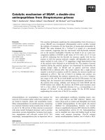
Fig. 4. Overall views of the ternary complex
of human AKR1B1–NADPH–PGH
2
. The
three-dimensional structure was calculated
by the program sievgene ⁄ myPresto [http://
medals.jp/myPresto/index.html; http://
presto.protein.osaka-u.ac.jp/myPresto4/].
(A) Schematic drawing of the ternary
structure using the program PyMOL version
1.0 [39] showing the TIM barrel structure of
AKR1B1 (PDB code, 2QXW; resolution,
0.8 A
˚
; green), NADP
+
(yellow), the sub-
strate, PGH
2
(orange), Tyr48 (purple), Asp43
(green), Lys77 (cyan) and His110 (magenta).
(B) Stereoview of the active site model with
the NADPH (yellow) and PGH
2
(orange) mol-
ecules (stick models) bound in the active-
site cleft consisting of Asp43 (green), Tyr48
(purple), Lys77 (cyan) and His110 (magenta).
(C) Schematic representation of the interac-
tions between PGH
2
(orange) and human
AKR1B1. PGH
2
is predicted to be stabilized
by both hydrophobic (blue) and hydrophilic
(red) interactions with Trp20, Val47, Trp79,
Trp111, Phe122, Pro218, Trp219 and
Leu300.
N. Nagata et al. Prostaglandin D
2
synthase activity of AKR1B1
FEBS Journal 278 (2011) 1288–1298 ª 2011 The Authors Journal compilation ª 2011 FEBS 1293
not catalyze PGDS activity in the absence of NADPH
or NADP
+
(N. Nagata & Y. Urade, unpublished
results). These results indicate that PGDS activity is
selective to AKR1B1 and AKR1B3, but not AKR1B7,
AKR1C3 and AKR5A2, and suggest that the tertiary
structure of the catalytic pocket, especially the PGH
2
-
binding site, of AKR1B1 and AKR1B3 is very similar
and different from that of other members of the AKR
family.
Human AKR1B1 has been reported recently to func-
tion as PGFS in the endometrium and is a potential
target for the treatment of menstrual disorders [15],
and mouse AKR1B3 has been reported to be involved
in the suppression of adipogenesis through FP recep-
tors [16]. Further characterization of the in vivo func-
tion of AKR1B1 in the endometrium and AKR1B3 in
adipocytes as PGFS is essential to understand the
development of menstrual disorders and metabolic dis-
orders, such as diabetes and obesity, respectively. How-
ever, the catalytic mechanisms of PGFS catalyzed by
several isozymes of mammalian AKRs are not clearly
understood, because the X-ray crystallographic struc-
tures of AKR1B3 and AKR1B7 have not yet been
determined. Our findings are useful for the design of
inhibitors selective to AKR1B1, which can be employed
for the evaluation of its contribution to the biosynthesis
of PGF
2a
in various systems.
Materials and methods
Expression and purification of recombinant AKR
enzymes
Open reading frames of the wild-type enzymes of AKR1B1,
AKR1B3, AKR1B7 and AKR1C3, and their mutants, were
inserted between Nde I and BamH1 ⁄ EcoRI sites of the
expression vector pET-28a, as described previously [30,31],
and used for the transformation of E. coli BL21DE3 (Invi-
trogen, Carlsbad, CA, USA). The outside primers used for
PCR amplifications of the inserts were as follows: 5¢-1B1
NdeI primer (5¢-CGGCAGCCATATGGCAAGCCGTC-3¢)
and 3¢-1B1 EcoRI primer (5¢-CGGAATTCGGGCTTCAA
AACTCTTCATGG-3¢); 5¢-1B3 NdeI primer (5¢-CGGCA
GCCATATGGCCAGCCATC-3¢) and 3¢-1B3 EcoRI
primer (5¢-CACGAATTCCAGAGAGACACAGGACACT
TGC-3¢); 5¢-1B7 NdeI primer (5¢-CGGCAGCCATATGGC
CACCTTCGT-3¢) and 3¢-1B7 BamHI primer (5¢-CGGG
ATCCCGTCAGTATTCCTCGTGG-3¢); and 5¢-1C3 NdeI
primer (5¢-GGAATTCCATATGGATTCCAAACACCAG
TG-3¢) and 3¢-1C3 EcoRI primer (5¢-CGGAATTCTTAA
Fig. 5. Schematic drawings of the PGDS
(top) and PGFS (bottom) activities in the
absence and presence of NADPH,
respectively (see Discussion for detailed
description).
Prostaglandin D
2
synthase activity of AKR1B1 N. Nagata et al.
1294 FEBS Journal 278 (2011) 1288–1298 ª 2011 The Authors Journal compilation ª 2011 FEBS
TATTCATCTGAATATG-3¢). Site-directed mutagenesis
was performed using a QuikChange
Ò
site-directed muta-
genesis kit (Agilent Technologies, Santa Clara, CA, USA).
The D43N-, D43E-, Y48F-, K77L-, K77R-, H110A- and
H110F-substituted recombinant enzymes for AKR1B1 and
the Y48F- and H110F-substituted recombinant enzymes for
AKR1B3 were obtained using the following respective
oligonucleotide primer pairs: AKR1B1 D43N forward (F)
(5¢-GTACCGCCACATCAACTGTGCCCATGTG-3¢) and
reverse (R) (5¢-CACATGGGCACAGTTGATGTGGCGG
TACC-3¢); AKR1B1 D43E (F) (5¢-GGGTACCGCCACA
TCGAATGTGCCCATGTG-3¢) and (R) (5¢-CACATGGG
CACATTCGATGTGGCGGTACCC-3¢); AKR1B1 Y48F (F)
(5¢-CTGTGCCCATGTGTTCCAGAATGAGAATG-3¢)and
(R) (5¢-CATTCTCATTCTGGAACACATGGGCACAG-3¢);
AKR1B1 K77L (F) (5¢-CTTCATCGTCAGCCTGCTGTG
GTGCACG-3¢) and (R) (5¢-CGTGCACCACAGCAGGCT
GACGATGAAG-3¢); AKR1B1 K77R (F) (5¢-CTCTTCA
TCGTCAGCAGGCTGTGGTGCACG-3¢) and (R) (5¢-CG
TGCACCACAGCCTGCTGACGATGAAGAG-3¢); AKR1
B1 H110F (F) (5¢-CCTCTACCTTATTTTCTGGCCGACT
GGC-3¢) and (R) (5¢-GCCAGTCGGCCAGAAAATAAG
GTAGAGG-3¢); AKR1B1 H110A (F) (5¢-CCTCTACCTT
ATTGCCTGGCCGACTGGC-3¢) and (R) (5¢-GCCAGTC-
GGCCAGGC
AATAAGGTAGAGG-3¢); AKR1B3 Y48 F (F)
(5¢-GACTGCGCCCAGGTGTTCCAGAATGAGAAG-3¢)
and (R) (5¢-CTTCTCATTCTGGAACACCTGGGCGCAG
TC-3¢); AKR1B3 H110F (F) (5¢-GATCTCTACCTTATT
TTCTGGCCAACGGGG-3¢) and (R) (5¢-CCCCGTTGGC
CAGAAAATAAGGTAGAGATC-3¢) (the italic codons
indicate the sites of mutations). Transformed cells were pre-
cultured overnight at 30 °C. Induction was started by the
addition of 1 mm isopropyl thio-b-d-galactoside (final con-
centration, 1 mm) when the absorbance (A) at 600 nm of
the culture had reached 0.5–0.6, and further cultivation was
carried out for 6 h at 30 °C. The recombinant protein was
purified by chromatography with nickel nitrilotriacetate
His-Bind resin (Merck, Darmstadt, Germany) according to
the manufacturer’s protocol, followed by digestion with
thrombin to remove the 6· His tag. The recombinant
protein was further purified by gel filtration chromatogra-
phy with Hiload 16 ⁄ 60 Superdex 75 pg (GE Healthcare,
Amersham, Buckinghamshire, UK) in Dulbecco’s phos-
phate-buffered saline. Protein purity was assessed by
SDS ⁄ PAGE on 10–20% gradient gels after staining with
Coomassie Brilliant Blue. Protein concentrations were mea-
sured using a BCA Protein Assay Kit (Pierce Biotechnol-
ogy, Rockford, IL, USA).
Enzyme activity assays
The PGFS and PGDS activities of AKR proteins were
determined as described previously [32]. In brief, the purified
recombinant enzymes were incubated at 37 °C for 2 min
with 5 lm 1-[
14
C]PGH
2
as a substrate in the presence or
absence of 0.5 mm NADPH in 50 mm sodium phosphate,
pH 7.0. The reaction was terminated by the addition of
300 lL of diethyl ether–methanol–2 m citric acid (30 : 4 : 1,
v ⁄ v ⁄ v). The reaction products recovered into the organic
phase were separated by TLC. The conversion rate from
14
C-labeled substrate to
14
C-labeled product was calculated
using an imaging plate system (Fuji Film, Tokyo, Japan).
The kinetic constants were determined from Lineweaver–
Burk plots prepared with sigmaplot software (version 10.0
for Windows; Systat Software, Inc., San Jose, CA, USA).
For pH–rate profiles, K
m
values were calculated from ini-
tial velocity studies over a wide range of pH values using a
triple buffer system containing 50 mm sodium phosphate,
50 mm sodium pyrophosphate and 50 mm 3-[(1,1-dimethyl-
2-hydroxyethl)amino-2-hydroxypropanesufonic acid. In
analyzing these data, the pK
a
and pK
b
values were esti-
mated using the fitting equation
y ¼½C=ð1 þ 10
ðpKaÀpHÞ
þ10
ðpHÀpKbÞ
ð1Þ
prepared with sigmaplot software. C is the pH-indepen-
dent value of V. The p-nitrobenzaldehyde reductase activity
of AKR1B1 was measured with 0.2 mm NADPH and
1mm p-nitrobenzaldehyde in 100 mm sodium phosphate
(pH 7.0). The reaction was initiated by the addition of the
substrate, and the decrease in the absorbance at 340 nm
was monitored at 25 °C [9].
Fluorescence quenching assay
The binding of NADP
+
to wild-type and mutant proteins
of AKR1B1 was determined by performing a fluorescence
quenching assay, in which various concentrations of coen-
zyme were incubated with AKR1B1 proteins in 300 lLof
50 mm sodium phosphate (pH 7.0) at 25 °C for 2 min. The
intrinsic Trp fluorescence was measured using an FP-6200
spectrofluorometer (JASCO, Tokyo, Japan) operated at an
excitation wavelength of 282 nm and an emission wave-
length of 338 nm [33]. The K
d
values for coenzyme binding
to AKR1B1 proteins were calculated from the difference in
fluorescence signal observed in the presence and absence
of coenzyme, as reported previously [26], with sigmaplot
software.
Molecular docking simulation
The docking study was performed by sievgene ⁄ myPresto
( -
tein.osaka-u.ac.jp/myPresto4/) [27]. The prediction accura-
cies of the sievgene program have already been reported to
be 19.2%, 50.78% and 60.0% with rmsd values of less than
1A
˚
,2A
˚
and 3 A
˚
, respectively, in a total of 130 complexes.
Among the top 10 docking models, the probabilities
increase to 28.5%, 63.1% and 76.9% with rmsd values of
less than 1 A
˚
,2A
˚
and 3 A
˚
, respectively [27]. The initial
N. Nagata et al. Prostaglandin D
2
synthase activity of AKR1B1
FEBS Journal 278 (2011) 1288–1298 ª 2011 The Authors Journal compilation ª 2011 FEBS 1295
three-dimensional coordinates of the small compounds were
generated by the Chem3D program (cambridge Software,
Cambridge, MA, USA) manually. We used the general
AMBER force field [34], and the molecular topology files
were generated by tplgeneL ⁄ myPresto. The energy optimi-
zation of the coordinates of small compounds was per-
formed using cosgene ⁄ myPresto [35]. The atomic charges
were calculated by the Gasteiger method of Hgene ⁄ myPres-
to [36,37]. The atomic charges of the proteins were the
same as the atomic charges of AMBER parm99 [38]. For
flexible docking, the sievgene program generated up to 1000
conformers for each compound. We predicted that the C4
atom of nicotinamide reacts with the O atom of PGH
2
and
that these two atoms should be close to each other. Among
the top 10 docking models, two models similar to each
other showed C4–O distances of 2.0 and 2.1 A
˚
, consistent
with the experimental results, whereas the other eight mod-
els showed C4–O distances of more than 6.5 A
˚
. The com-
plex structure depicted in Fig. 4 was given by the energy
minimization calculation based on the model with a C4–O
distance of 2.0 A
˚
. The position of the ring structure of PG
should be 70–80% accurate based on the prediction accu-
racy of ‘sievgene’. Although it is difficult to predict the
position of side chains of PG, the side chains are not
important for the discussion of the reaction mechanism.
Acknowledgements
This work was supported in part by the Japan
Aero-space Exploration Agency (JAXA), the Program
of Basic and Applied Researches for Innovations in
Bio-oriented Industry of Japan, Takeda Science Foun-
dation, and Osaka City (to Y.U.) and Grant-in-Aid for
Scientific Research (No. 22550152) from the Ministry
of Education, Culture, Sports, Science and Technology
of Japan (to T.I.). We thank Dr Michele Manin (CNRS
UMR6247-GReD, France) for kindly providing the
AKR1B1 expression vector; Drs Kenji Mizuguchi and
Sukanta Mondal (National Institute of Biomedical
Innovation, Ibaraki, Japan) for homology modeling;
Dr Toshiyoshi Yamamoto (Department of Molecular
Biology and Medicine, Research Center for Advanced
Science and Technology, University of Tokyo, Japan)
for kinetic analysis; Dr Zakayi Kabututu, Nobuko
Uodome and Toshiharu Tsurumura (Osaka Bioscience
Institute, Japan) for assistance during the early stage of
this research; and Megumi Yamaguchi, Naoko Takah-
ashi and Taeko Nishimoto (Osaka Bioscience Institute,
Japan) for secretarial assistance.
References
1 Penning TM & Drury JE (2007) Human aldo-keto
reductases: function, gene regulation, and single
nucleotide polymorphisms. Arch Biochem Biophys 464,
241–250.
2 Jez JM, Flynn TG & Penning TM (1997) A new
nomenclature for the aldo-keto reductase superfamily.
Biochem Pharmacol 54, 639–647.
3 Hoog SS, Pawlowski JE, Alzari PM, Penning TM &
Lewis M (1994) Three-dimensional structure of rat liver
3 alpha-hydroxysteroid ⁄ dihydrodiol dehydrogenase: a
member of the aldo-keto reductase superfamily. Proc
Natl Acad Sci USA 91, 2517–2521.
4 Kilunga KB, Inoue T, Okano Y, Kabututu Z, Martin
SK, Lazarus M, Duszenko M, Sumii Y, Kusakari Y,
Matsumura H et al. (2005) Structural and mutational
analysis of Trypanosoma brucei prostaglandin H
2
reduc-
tase provides insight into the catalytic mechanism of
aldo-ketoreductases. J Biol Chem 280, 26371–26382.
5 Wilson DK, Bohren KM, Gabbay KH & Quiocho FA
(1992) An unlikely sugar substrate site in the 1.65 A
˚
structure of the human aldose reductase holoenzyme
implicated in diabetic complications. Science 257,
81–84.
6 Wilson DK, Nakano T, Petrash JM & Quiocho FA
(1995) 1.7 A
˚
structure of FR-1, a fibroblast growth
factor-induced member of the aldo-keto reductase
family, complexed with coenzyme and inhibitor.
Biochemistry 34, 14323–14330.
7 Petrash JM (2004) All in the family: aldose reductase
and closely related aldo-keto reductases. Cell Mol Life
Sci 61, 737–749.
8 Kubiseski TJ, Hyndman DJ, Morjana NA & Flynn TG
(1992) Studies on pig muscle aldose reductase. Kinetic
mechanism and evidence for a slow conformational
change upon coenzyme binding. J Biol Chem 267,
6510–6517.
9 Kabututu Z, Manin M, Pointud JC, Maruyama T,
Nagata N, Lambert S, Lefrancois-Martinez AM,
Martinez A & Urade Y (2009) Prostaglandin F
2a
synthase activities of aldo-keto reductase 1B1, 1B3 and
1B7. J Biochem 145, 161–168.
10 Watanabe K (2002) Prostaglandin F synthase. Prosta-
glandins Other Lipid Mediat 68–69, 401–407.
11 Crawford K, Kaufman PL & Gabelt BT (1987) Effects
of topical PGF
2a
on aqueous humor dynamics in cyno-
molgus monkeys. Curr Eye Res 6, 1035–1044.
12 Weber PC (1980) Renal prostaglandins, kidney function
and essential hypertension. Contrib Nephrol 23, 83–92.
13 Sasaki S, Hozumi Y & Kondo S (2005) Influence of
prostaglandin F
2a
and its analogues on hair regrowth
and follicular melanogenesis in a murine model. Exp
Dermatol 14, 323–328.
14 McCracken JA, Custer EE & Lamsa JC (1999) Luteoly-
sis: a neuroendocrine-mediated event. Physiol Rev 79,
263–323.
15 Bresson E, Boucher-Kovalik S, Chapdelaine P, Madore
E, Harvey N, Laberge PY, Leboeuf M & Fortier MA
Prostaglandin D
2
synthase activity of AKR1B1 N. Nagata et al.
1296 FEBS Journal 278 (2011) 1288–1298 ª 2011 The Authors Journal compilation ª 2011 FEBS
(2011) The human aldose reductase AKR1B1 qualifies
as the primary prostaglandin F synthase in the endome-
trium. J Clin Endocrinol Metab 96, 210–219.
16 Fujimori K, Ueno T, Nagata N, Kashiwagi K, Aritake
K, Amano F & Urade Y (2010) Suppression of
adipocyte differentiation by aldo-keto reductase 1B3
acting as prostaglandin F
2a
synthase. J Biol Chem 285,
8880–8886.
17 Liston TE, Oates JA & Roberts LJ II (1985) Prosta-
glandin D
2
is metabolized in humans to 9 alpha,11
beta-prostaglandin F
2
, a novel biologically active pros-
taglandin. Adv Prostaglandin Thromboxane Leukot Res
15, 365–367.
18 Watanabe K, Yoshida R, Shimizu T & Hayaishi O
(1985) Enzymatic formation of prostaglandin F
2a
from
prostaglandin H
2
and D
2
. Purification and properties of
prostaglandin F synthetase from bovine lung. J Biol
Chem 260, 7035–7041.
19 Hayaishi O, Watanabe K, Fujii Y, Nakayama K,
Ohkubo H, Kuramitsu S, Kagamiyama H & Nakanishi
S (1988) Prostaglandin F synthetase, a dual function
enzyme. Prog Clin Biol Res 274, 577–587.
20 Watanabe K, Fujii Y, Nakayama K, Ohkubo H, Ku-
ramitsu S, Kagamiyama H, Nakanishi S & Hayaishi O
(1988) Structural similarity of bovine lung prostaglan-
din F synthase to lens epsilon-crystallin of the Euro-
pean common frog. Proc Natl Acad Sci USA 85, 11–15.
21 Kubata BK, Duszenko M, Kabututu Z, Rawer M,
Szallies A, Fujimori K, Inui T, Nozaki T, Yamashita
K, Horii T et al. (2000) Identification of a novel prosta-
glandin F
2a
synthase in Trypanosoma brucei. J Exp Med
192, 1327–1338.
22 Kubata BK, Kabututu Z, Nozaki T, Munday CJ,
Fukuzumi S, Ohkubo K, Lazarus M, Maruyama T,
Martin SK, Duszenko M et al. (2002) A key role for
old yellow enzyme in the metabolism of drugs by
Trypanosoma cruzi. J Exp Med 196, 1241–1251.
23 Moriuchi H, Koda N, Okuda-Ashitaka E, Daiyasu H,
Ogasawara K, Toh H, Ito S, Woodward DF & Watan-
abe K (2008) Molecular characterization of a novel type
of prostamide ⁄ prostaglandin F synthase, belonging to
the thioredoxin-like superfamily. J Biol Chem 283,
792–801.
24 Urade Y, Fujimoto N & Hayaishi O (1985) Purification
and characterization of rat brain prostaglandin D syn-
thetase. J Biol Chem 260 , 12410–12415.
25 Bohren KM, Grimshaw CE, Lai CJ, Harrison DH,
Ringe D, Petsko GA & Gabbay KH (1994) Tyrosine-48
is the proton donor and histidine-110 directs substrate
stereochemical selectivity in the reduction reaction of
human aldose reductase: enzyme kinetics and crystal
structure of the Y48H mutant enzyme. Biochemistry 33,
2021–2032.
26 Tarle I, Borhani DW, Wilson DK, Quiocho FA &
Petrash JM (1993) Probing the active site of human
aldose reductase. Site-directed mutagenesis of Asp-43,
Tyr-48, Lys-77, and His-110. J Biol Chem 268, 25687–
25693.
27 Fukunishi Y, Mikami Y & Nakamura H (2005) Similar-
ities among receptor pockets and among compounds:
analysis and application to in silico ligand screening.
J Mol Graph Model 24, 34–45.
28 Klimacek M, Szekely M, Griessler R & Nidetzky B
(2001) Exploring the active site of yeast xylose reductase
by site-directed mutagenesis of sequence motifs charac-
teristic of two dehydrogenase ⁄ reductase family types.
FEBS Lett 500, 149–152.
29 Schlegel BP, Jez JM & Penning TM (1998) Mutagenesis
of 3 alpha-hydroxysteroid dehydrogenase reveals a
‘push-pull’ mechanism for proton transfer in aldo-keto
reductases. Biochemistry 37, 3538–3548.
30 Lefrancois-Martinez AM, Bertherat J, Val P, Tournaire
C, Gallo-Payet N, Hyndman D, Veyssiere G, Bertagna
X, Jean C & Martinez A (2004) Decreased expression
of cyclic adenosine monophosphate-regulated aldose
reductase (AKR1B1) is associated with malignancy in
human sporadic adrenocortical tumors. J Clin Endocri-
nol Metab 89, 3010–3019.
31 Lefrancois-Martinez AM, Tournaire C, Martinez A,
Berger M, Daoudal S, Tritsch D, Veyssiere G & Jean C
(1999) Product of side-chain cleavage of cholesterol,
isocaproaldehyde, is an endogenous specific substrate of
mouse vas deferens protein, an aldose reductase-like
protein in adrenocortical cells. J Biol Chem 274,
32875–32880.
32 Irikura D, Aritake K, Nagata N, Maruyama T,
Shimamoto S & Urade Y (2009) Biochemical,
functional, and pharmacological characterization of
AT-56, an orally active and selective inhibitor of
lipocalin-type prostaglandin D synthase. J Biol Chem
284, 7623–7630.
33 Jez JM, Schlegel BP & Penning TM (1996) Character-
ization of the substrate binding site in rat liver 3alpha-
hydroxysteroid ⁄ dihydrodiol dehydrogenase. The roles
of tryptophans in ligand binding and protein fluores-
cence. J Biol Chem 271, 30190–30198.
34 Wang J, Wolf RM, Caldwell JW, Kollman PA &
Case DA (2004) Development and testing of a gen-
eral amber force field. J Comput Chem 25, 1157–
1174.
35 Fukunishi Y, Mikami Y & Nakamura H (2003) The
filling potential method: a method for estimating the
free energy surface for protein–ligand docking. J Phys
Chem B 107, 13201–13210.
36 Gasteiger J & Marsili M (1980) Iterative partial equal-
ization of orbital electronegativity – a rapid access to
atomic charges. Tetrahedron 36, 3219–3228.
37 Gasteiger J & Marsili M (1978) A new model for
calculating atomic charges in molecules. Tetrahedron
Lett 34, 3181–3184.
N. Nagata et al. Prostaglandin D
2
synthase activity of AKR1B1
FEBS Journal 278 (2011) 1288–1298 ª 2011 The Authors Journal compilation ª 2011 FEBS 1297
38 Case DA, Darden TA, Cheatham TEI, Simmerling CL,
Wang J, Duke RE, Luo R, Merz KM, Wang B,
Pearlman DA et al. (2004) AMBER 8. University of
California, San Francisco, CA.
39 DeLano WL (2005) The case for open-source software
in drug discovery. Drug Discov Today 10, 213–217.
Supporting information
The following supplementary material is available:
Fig. S1. Kinetic analysis of the PGFS and PGDS
activities of wild-type AKR1B3.
Fig. S2. Mutagenesis analyses of the catalytic tetrad of
AKR1B3.
Table S1. NADP
+
-binding affinities of wild-type and
mutants of AKR1B3.
This supplementary material can be found in the
online version of this article.
Please note: As a service to our authors and readers,
this journal provides supporting information supplied
by the authors. Such materials are peer-reviewed and
may be re-organized for online delivery, but are not
copy-edited or typeset. Technical support issues arising
from supporting information (other than missing files)
should be addressed to the authors.
Prostaglandin D
2
synthase activity of AKR1B1 N. Nagata et al.
1298 FEBS Journal 278 (2011) 1288–1298 ª 2011 The Authors Journal compilation ª 2011 FEBS