Báo cáo khoa học: Lipins from plants are phosphatidate phosphatases that restore lipid synthesis in apah1Dmutant strain of Saccharomyces cerevisiae ppt
Bạn đang xem bản rút gọn của tài liệu. Xem và tải ngay bản đầy đủ của tài liệu tại đây (542.62 KB, 12 trang )
Lipins from plants are phosphatidate phosphatases that
restore lipid synthesis in a pah1
D
mutant strain of
Saccharomyces cerevisiae
Elzbieta Mietkiewska
1
, Rodrigo M. P. Siloto
1
, Jay Dewald
2
, Saleh Shah
3
, David N. Brindley
2
and
Randall J. Weselake
1
1 Department of Agricultural, Food and Nutritional Science, University of Alberta, Edmonton, Canada
2 Department of Biochemistry, Signal Transduction Research Group, School of Molecular and Systems Medicine, University of Alberta,
Edmonton, Canada
3 Plant Biotechnology, Alberta Innovates-Technology Futures, Vegreville, Canada
Keywords
lipin; phosphatidate phosphatase;
Saccharomyces cerevisiae; subcellular
localization; triacylglycerol
Correspondence
R. J. Weselake, Agricultural Lipid
Biotechnology Program, Department of
Agricultural, Food and Nutritional Science,
4–10 Agriculture ⁄ Forestry Centre, University
of Alberta, Edmonton, Alberta T6G 2P5,
Canada
Fax: +1 780 492 6739
Tel: +1 780 492 4401
E-mail:
(Received 21 September 2010, revised 10
December 2010, accepted 20 December
2010)
doi:10.1111/j.1742-4658.2010.07995.x
The identification of the yeast phosphatidate phosphohydrolase (PAH1) gene
encoding an enzyme with phosphatidate phosphatase (PAP; 3-sn-phosphati-
date phosphohydrolase, EC 3.1.3.4) activity led to the discovery of mam-
malian Lipins and subsequently to homologous genes from plants. In the
present study, we describe the functional characterization of Arabidopsis
and Brassica napus homologs of PAH1. Recombinant expression studies
confirmed that homologous PAHs from plants can rescue different pheno-
types exhibited by the yeast pah1D strain, such as temperature growth sen-
sitivity and atypical neutral lipid composition. Using this expression
system, we examined the role of the putative catalytic motif DXDXT and
other conserved residues by mutational analysis. Mutants within the
carboxy-terminal lipin domain displayed significantly decreased PAP
activity, which was reflected by their limited ability to complement different
phenotypes of pah1D. Subcellular localization studies using a green fluores-
cent protein fusion protein showed that Arabidopsis PAH1 is mostly pres-
ent in the cytoplasm of yeast cells. However, upon oleic acid stimulation,
green fluorescent protein fluorescence was predominantly found in the
nucleus, suggesting that plant PAH1 might be involved in the transcrip-
tional regulation of gene expression. In addition, we demonstrate that
mutation of conserved residues that are essential for the PAP activity of
the Arabidopsis PAH1 enzyme did not impair its nuclear localization in
response to oleic acid. In conclusion, the present study provides evidence
that Arabidopsis and B. napus PAHs restore lipid synthesis in yeast and
that DXDXT is a functional enzymic motif within plant PAHs.
Database
The nucleotide sequence data have been deposited in the GenBank database under accession
numbers HQ113853 and HQ113854
Abbreviations
C-LIP, carboxy-terminal lipin domain; DAG, diacylglycerol; DAPI, 4,6-diamidino-2-phenylindole dilactate; DGAT, diacylglycerol acyltransferase;
FAME, fatty acid methyl ester; GFP, green fluorescent protein; HAD, haloacid dehalogenase; LPP, lipid phosphate phosphatase; N-LIP,
amino-terminal lipin domain; PA, phospatidate; PAH, phosphatidate phosphohydrolase; PAP, phosphatidate phosphatase; PL, phospholipid;
TAG, triacylglycerol; WT, wild-type.
764 FEBS Journal 278 (2011) 764–775 ª 2011 The Authors Journal compilation ª 2011 FEBS
Introduction
Phosphatidate phosphatase (PAP; 3-sn-phosphatidate
phosphohydrolase, EC 3.1.3.4) catalyzes the dephos-
phorylation of phospatidate (PA), yielding diacyl-
glycerol (DAG) and inorganic phosphate [1–3]. In
eukaryotic cells, PAP activity plays a central role in both
lipid metabolism and intracellular signaling mechanisms
[4,5]. Two distinct PAP enzyme activities, referred to as
PAP1 and PAP2, have been described [2,6–8]. PAP1,
now known as PAP activity is Mg
2+
-dependent, utilizes
PA as a unique substrate and localizes in the soluble
fraction, from where it translocates to internal mem-
branes [3,9,10]. By contrast, PAP2, currently known as
a family of lipid phosphate phosphatases (LPPs) utilizes
many substrates (e.g. PA, lysophosphatidate, sphingo-
sine-1-phosphate and DAG pyrophosphate, amongst
others), does not require Mg
2+
for activity and is mem-
brane-bound [11]. Genes encoding the LPPs have been
identified and extensively studied in both yeast and
plants [12–15].
Studies in Saccharomyces cerevisiae led to the identifi-
cation of the yeast phosphatidate phosphohydrolase
(PAH1) gene encoding an enzyme with PAP activity
[16]. The S. cerevisiae pah1D strain displays severe defi-
ciency in triacylglycerol (TAG), which is apparently
caused by a decreased level of DAG, reflecting the
importance of this enzyme for de novo synthesis of neu-
tral lipids. In addition, the free fatty acid content of
pah1D yeast was significantly increased, presumably as a
result of the decreased ability of cells to utilize fatty
acids for TAG synthesis [16]. The PAH1 enzyme was
found in soluble and membrane fractions of the cell and
its association with membranes was shown to be of a
peripheral nature [2]. The S. cerevisiae PAH1 shares
sequence homology with mammalian Lipin-1 in the con-
served N-terminal and C-terminal domains of the pro-
tein known as N-LIP and C-LIP, respectively [2,3,17].
The C-LIP domain contains a haloacid dehalogenase
(HAD)-like catalytic motif found in the superfamily of
Mg
2+
-dependent PAPs. This motif consists of DXDXT
in which the first aspartate residue is responsible for
binding the phosphate moiety during the catalytic reac-
tion [18,19]. In addition, a conserved glycine within
N-LIP is required for the PAP activity of yeast PAH1
[19]. PAH1 is phosphorylated on seven residues match-
ing the minimal Cdk consensus that is required for the
efficient transcriptional derepression of key enzymes
involved in phospholipid (PL) biosynthesis [20,21]. In
mammals, highly phosphorylated forms of Lipin-1 are
enriched in the cytoplasm, whereas dephosphorylated
forms are found mostly associated with membranes
[9,10,22,23]. Homologous PAH1 genes are also present
in Arabidopsis thaliana and other plants. Nakamura
et al. [24] showed that Arabidopsis PAH1and PAH2 are
involved in the eukaryotic pathway of galactolipid bio-
synthesis. Recently, it was demonstrated in Arabidopsis
that PAHs regulate PL synthesis in a similar manner to
that described for S. cerevisiae [25].
In the present study, we demonstrate that, in addition
to AtPAH1 and AtPAH2, homologous genes from Bras-
sica napus also encode functional PAP enzymes capable
of complementing different phenotypes of yeast pah1D
strain, including TAG synthesis and temperature-sensi-
tive growth. We also provide evidence that the con-
served aspartate residues in the HAD-like motif of the
plant enzymes are required for PAP function, and
demonstrate the importance of conserved glycine and
serine residues for the activity of the enzyme. Finally, we
show that, when expressed in yeast, AtPAH1 can be
detected in cytosolic and membrane fractions, although
it is recruited to the nucleus when the cells are cultivated
in the presence of oleic acid. A preliminary account of
some of the results reported in the present study has
been presented at the 19th International Symposium on
Plant Lipids [26].
Results and Discussion
Arabidopsis and B. napus PAHs are orthologs of
yeast PAH1 and mammalian lipins
Based on previous studies demonstrating the involve-
ment of yeast PAH1 and mammalian lipins in storage
lipid accumulation [27], we were initially interested in
identifying Arabidopsis genes encoding PAP enzymes
that may play a role in de novo DAG synthesis.
Two PAH1 homologs were found in Arabidopsis ,
which are designated in the present study as AtPAH1
(At3g09560) and AtPAH2 (At5g42870), and encode
proteins with predicted molecular masses of 100.9 and
101.2 kDa, respectively. In addition, we identified two
isoforms of B. napus PAH1 using the expressed
sequence tag information available on the Internet
(). They are designated in
the present study as BnPAH1A (GenBank #
HQ113853) and BnPAH1B (GenBank # HQ113854),
and encode proteins sharing 99% sequence identity
and calculated molecular masses of 90.6 kDa. Both
Arabidopsis and B. napus PAHs have the conserved
N-LIP and C-LIP domains found in yeast PAH1 and
mammalian Lipin-1. AtPAH1 and AtPAH2 share 63%
and 58% identity in the conserved N-LIP and C-LIP
domain, respectively. B. napus PAHs display higher
E. Mietkiewska et al. Arabidopsis and B. napus PAHs homologs of PAH1
FEBS Journal 278 (2011) 764–775 ª 2011 The Authors Journal compilation ª 2011 FEBS 765
sequence identity to AtPAH1 at the level of 95% for
each N-LIP and C-LIP domain. The C-LIP domain
harbors the HAD-like catalytic motif (DXDXT) found
in the superfamily of Mg
2+
-dependent phosphatases
[17,27]. The HAD-like motif DVDGT is located at
positions 707–711, 734–738 and 616–620 of AtPAH1,
AtPAH2 and BnPAH1, respectively (Fig. 1). By con-
trast to plant PAHs, members of the family of LPPs
that do not require Mg
2+
ions for activity in Arabid-
opsis and yeast contain a catalytic motif comprising
the consensus sequences KXXXXXXRP, PSGH and
SRXXXXXHXXXD [2,5,28,29]. Analyses made with
several prediction algorithms were unable to detect the
presence of potential transmembrane domains in
Arabidopsis or B. napus PAHs. This is in agreement
with earlier studies on mammalian and yeast homologs
[3,16,17]. Unlike Arabidopsis and B. napus PAHs, six
putative transmembrane domains were predicted in
Arabidopsis LPP2 and LPP3 and in yeast diacylgly-
cerol pyrophosphate phosphatase 1 and LPP1 that
belong to the family of Mg
2+
-independent PAP2
enzymes [14,16,30].
Arabidopsis and B. napus PAHs complement
different phenotypes of pah1-deficient yeast cells
To establish the functional relationship between yeast
PAH1 and the corresponding plant homologs, the
coding region of Arabidopsis and B. napus PAHs were
linked to the galactose-inducible GAL1 promoter in
the yeast expression vector pYES2 ⁄ NT, which also
provides an N-terminal His-tag. The resulting con-
structs were transformed into the S. cerevisiae pah1D
strain, which displays different phenotypes, such as a
reduced level of PAP activity, severe growth deficiency
at 37 °C, elevated levels of PA and decreased levels of
DAG and TAG [16,19,31]. Transformed yeast cells
were cultivated for 16 h after induction with galactose.
Immunoblot analysis of yeast homogenates showed
that the His-tagged Arabidopsis PAH1 and B. napus
PAHs migrated as 130 and 118 kDa proteins,
respectively, upon SDS ⁄ PAGE (Fig. 2A, B), and these
values are higher than the molecular weights of the
predicted polypeptides. A similar shift in mobility was
observed in yeast PAH1 and was attributed to post-
translational modification of the protein by phosphor-
ylation [16,20]. AtPAH2 migrated at a lower molecular
weight compared to AtPAH1 and, in this case, a sec-
ond band is clearly noticeable on SDS ⁄ PAGE
(Fig. 2A, lane 2). The same pattern has been also
observed previously for mammalian Lipin-2 [22]. Frac-
tionation of yeast homogenate indicated that AtPAH1
was detected in both membrane and soluble fractions
of the yeast cell (Fig. 2C). Therefore, to evaluate
Mg
2+
-dependent PAP activity, we used crude cell
homogenates. The pah1D strain expressing Arabidopsis
or B. napus PAHs displayed a significant increase in
ScPAH1
AtPAH1
AtPAH2
BnPAH1A
BnPAH1B
DIDGT
N-LIP HAD-like
G
DVDGT SG
DVDGTG
DVDGT
A A
G
A
DVDGT SG
AAAA
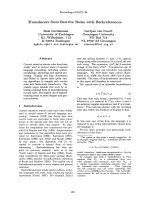
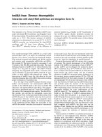
Fig. 1. PAH1 homologs from plants have similar domain organiza-
tion to yeast PAH1 (ScPAH1) polypeptide. Arabidopsis PAH1 (At-
PAH1), Arabidopsis PAH2 (AtPAH2) and B. napus PAHs (BnPAH1A
and BnPAH1B) are members of the lipin family, containing a
conserved N-terminal domain (N-LIP) and a C-terminal catalytic
domain with a HAD-like motif usually found in Mg
2+
-dependent
phosphatidate phosphatases. The conserved amino acids that were
mutated in the present study are indicated.
1212
1
ABC
2
kDa
160
kDa
120
100
kDa
120
160
120
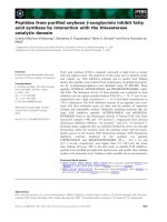
Fig. 2. Plant PAHs expressed in yeast migrate higher on SDS ⁄ PAGE than the predicted molecular masses of polypeptides and are found in
both soluble and membrane fractions. Immunoblots of Arabidopsis and B. napus PAHs were carried out using anti-HisG-HRP serum and pro-
tein extracts from yeast pah1D expressing the recombinant polypeptides after 16 h of induction. Proteins (40 lg each lane) were run on an
8% SDS ⁄ PAGE gel. Protein molecular mass was calculated using BenchMarkÔ Protein Ladder (Invitrogen). (A) Crude homogenates from
cells expressing AtPAH1 (lane 1) and AtPAH2 (lane 2). (B) Crude homogenates from cells expressing BnPAH1A (lane 1) and BnPAH1B (lane
2). (C) Subcellular fractions from cells expressing AtPAH1: microsomal fraction (lane 1) and soluble fraction (lane 2). Membrane and soluble
fractions were obtained using 100 000 g centrifugation of the 15 000 g supernatant of crude homogenate.
Arabidopsis and B. napus PAHs homologs of PAH1 E. Mietkiewska et al.
766 FEBS Journal 278 (2011) 764–775 ª 2011 The Authors Journal compilation ª 2011 FEBS
PAP activity compared to the negative control trans-
formed with LacZ (Fig. 3A). The highest difference in
PAP activity was observed for BnPAH1A, with an
increase of 21.4-fold compared to the negative control.
In the pah1D mutant, the PAP activity was reduced by
39% compared to the wild-type (WT) (Inv Sc1 strain)
control used in the present study. This was similar to
the difference between pah1D and WT parent strain
observed by Han et al. [16]. The remaining Mg
2+
-
dependent PAP activity in pah1D has been attributed
to other enzymes with unknown molecular identities
[16].
To evaluate the influence of Arabidopsis and B. na-
pus PAHs in the metabolism of neutral lipids, we ana-
lyzed the TAG and PL content of yeast cells
expressing recombinant PAHs (Fig. 3B). After 48 h of
induction, the TAG ⁄ PL ratio in pah1D cells bearing
recombinant PAHs was considerably higher compared
to the negative control. The most pronounced effect
was observed for BnPAH1A, with an increase of 40-
fold compared to the TAG ⁄ PL ratio observed in the
negative control (LacZ).
In addition to the effect on lipid composition, yeast
pah1D cells also display reduced growth when culti-
vated at 37 °C. To determine whether Arabidopsis and
B. napus PAHs could rescue this phenotype, we culti-
vated several dilutions of cells expressing plant PAHs
at 37 °C. When cells were inoculated in medium
supplemented with galactose (induced), lines expressing
PAHs from Arabidopsis and B. napus displayed growth
on dilutions as low as D
600
= 1.0 · 10
4
(Fig. 3C),
whereas cells expressing LacZ grew only at
D
600
= 1.0. In medium without galactose (not
induced), only WT cells presented appreciable growth,
indicating that complementation of temperature-sensi-
tive phenotype resulted from Arabidopsis and B. napus
PAHs expression.
Taken together, these results show that the previ-
ously characterized AtPAH1 and AtPAH2 and the two
PAH1 homologs from B. napus encode enzymes with
PAP activity. Previous work on mammalian lipins
indicated that the pah1D yeast expression system could
be used as a predictive model for confirming the func-
tions of PAH1-homolog genes [22]. Arabidopsis and
B. napus PAHs complemented the temperature sensi-
tive phenotype from the yeast pah1D strain, which was
also observed by Nakamura et al. [24] for AtPAH1
and AtPAH2. Using an Escherichia coli expression sys-
tem, Eastmond et al. [25] reported a comparatively
higher enzyme activity of AtPAH1 over AtPAH2,
which is corroborated by the results obtained in the
present study (Fig. 3A). The higher enzyme activity of
AtPAH1 compared to AtPAH2 is also evident in the
BnPAH1B
AtPAH1
AtPAH2
LacZ
LacZ
BnPAH1A
pah1Δ
Wild type
OD
1
10 10
2
10
3
10
4
Dilutions
Induced Not induced
C
A
0.0
10.0
20.0
30.0
40.0
50.0
60.0
70.0
SA (nmol·mg
–1
·min
–1
)
0.0
1.0
2.0
3.0
4.0
5.0
LacZ WT
B
0
0.1
0.2
0.3
0.4
0.5
0.6
0.7
mol TAG/mol PL
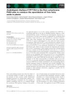
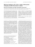
Fig. 3. Arabidopsis and B. napus PAHs complement different phe-
notypes of S. cerevisiae pah1D mutant. (A) PAP activity of crude
homogenates from yeast pah1D cells bearing AtPAH1, AtPAH2,
BnPAH1A, BnPAH1B or LacZ as a negative control. Yeast homo-
genates were prepared from cells induced for 16 h and assayed for
PAP activity in the presence of 1 m
M MgCl
2
. Total PAP activities
were calculated from measurements at three different protein con-
centrations. (B) Ratio of TAG to PL of yeast pah1D expressing
recombinant PAHs. Total lipids were extracted from yeast cells
after 48 h of induction and separated on TLC plates. Spots corre-
sponding to TAG and PL were scraped out and transmethylated.
FAMEs were analyzed by GC. Each bar represents the mean ± SD
from three determinations. (C) Complementation of temperature
sensitive phenotypes of pah1D by plant PAH homologs. Yeast
pah1D expressing AtPAH1, AtPAH2, BnPAH1A, BnPAH1B or LacZ
were cultivated in liquid medium. The density of resulting cultures
was adjusted to D
600
= 1 followed by 10-fold serial dilutions. Five
microliters of each dilution were spotted onto plates containing 2%
galactose (induced) or 2% raffinose (not induced) and incubated for
3 days at 37 °C. WT yeast and pah1D strain previously transformed
with LacZ were used as a positive and negative control, respec-
tively.
E. Mietkiewska et al. Arabidopsis and B. napus PAHs homologs of PAH1
FEBS Journal 278 (2011) 764–775 ª 2011 The Authors Journal compilation ª 2011 FEBS 767
preferential growth at 37 °C of yeast expressing
AtPAH1; this was also reported by Nakamura et al.
[24]. Taken together, these results collectively suggest
that AtPAH1 can complement phenotypes of the
pah1D strain more efficiently than AtPAH2. The
newly-characterized BnPAH1A and BnPAH1B display
relatively stronger complementation over Arabidopsis
PAHs in most aspects. Moreover, the restoration of
TAG synthesis in the yeast pah1D strain expressing
plant PAH homologs suggests an evolutionary conser-
vation of the PAP enzyme reaction between yeast and
plants.
N-LIP and C-LIP are functional domains in
Arabidopsis and B. napus PAHs
To determine the role of the conserved residues within
C-LIP and N-LIP domain of plant PAH1, we exam-
ined the mutational effect of selected residues on PAP
activity using AtPAH1 and BnPAH1A as models.
Using site-directed mutagenesis (Fig. 1), we con-
structed mutant AtPAH1 alleles (G83A, D707A,
D709A and S752A) and mutant BnPAH1A alleles
(G83A, D616A and D618A) and expressed them in the
pah1D strain. Yeast cells were harvested after 16 h of
induction in medium containing galactose. Cell homo-
genates were prepared, verified via immunoblotting
and assayed for PAP activity. Immunoblot analysis
using anti-HisG serum showed that native and mutant
AtPAH1 and BnPAH1A enzymes were expressed at
comparable levels (Fig. 4). Although the expression of
AtPAH1 and BnPAH1A resulted in PAP activity that
was significantly higher compared to LacZ control, the
corresponding mutant alleles did not restore PAP
activity to comparable levels (Fig. 4). In particular,
mutations in the predicted catalytic motif of AtPAH1
(D707A and D709A) and BnPAH1A (D616A and
D618A) abolished PAP activity, with levels compara-
ble to the negative control. These results are in agree-
ment with mutational analysis of the yeast PAH1
catalytic motif [19] and demonstrate that the conserved
aspartate residues in the plant homologs are required
for their catalytic function. In addition, substitution of
the conserved serine 752 with alanine within C-LIP
domain of AtPAH1 had a similar negative effect on
enzyme activity. The importance of the equivalent con-
served serine residue for the enzyme activity in human
Lipin-2 and mouse Lipin-1 and Lipin-2 has been
described previously [9]. For example, a rare human
mutation S734L in LIPIN-2 gene causes Majeed
syndrome, a human inflammatory disorder. Recently,
Majeed syndrome has been linked to the loss of Lipin-
2-mediated PAP activity [9].
Mutation of the conserved glycine (G83) to alanine
in the N-LIP domain of both AtPAH1 and BnPAH1A
produced less severe effects on the enzyme activity and
resulted in the loss of up to 75% and 54% of the PAP
activity of native enzyme, respectively (Fig. 4). Inter-
estingly, other mutations in the corresponding position
of PAH1 from other organisms appear to have a more
80
100
120A
B
0
20
40
60
0
AtPAH1 G83A D707A D709A S752A LacZ
130 kDa
80
100
120
20
40
60
PAP1 activity (%) PAP1 activity (%)
0
BnPAH1A G83A D616A D618A LacZ
118 kDa
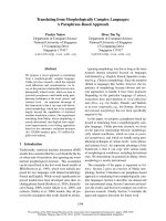
Fig. 4. Mutations within N-LIP and C-LIP domains affect PAP1
activity of plant PAH1. (A) PAP activity of yeast pah1D strain homo-
genates bearing AtPAH1 and its mutant alleles: G83A, D707A,
D709A and S752A. Lower: corresponding western blot of the site-
directed mutagenized AtPAH1. (B) PAP activity of yeast pah1D
strain homogenates bearing BnPAH1A and its mutant alleles:
G83A, D616A, D618A. Lower: corresponding western blot of the
site-directed mutagenized BnPAH1A. Yeast homogenates were
prepared from cells induced for 16 h and assayed for PAP activity
in the presence of 1 m
M MgCl
2
. The amount of PAP activity of
samples bearing AtPAH1 (34.8 nmolÆmg
)1
Æmin
)1
) and BnPAH1A
(60.0 nmolÆmg
)1
Æmin
)1
) were set at 100%. The data shown are the
mean ± SD from three determinations. Homogenates of yeast cells
expressing LacZ were used as negative controls. Western blot anal-
ysis were carried out using anti-HisG-HRP serum and protein
extracts (40 lg each lane) from yeast pah1D strain expressing the
recombinant polypeptides separated on an 8% SDS ⁄ PAGE gel.
Arabidopsis and B. napus PAHs homologs of PAH1 E. Mietkiewska et al.
768 FEBS Journal 278 (2011) 764–775 ª 2011 The Authors Journal compilation ª 2011 FEBS
pronounced effect. For example, the G80R yeast
PAH1 allele showed essentially no enzyme activity
[19]. The same substitution within N-LIP domain was
found to be crucial for the fat-regulating function of
Lipin-1 in mice [32]. Mild modifications (such as G to
A) that do not result in dramatic changes in polarity
or steric properties may not be sufficient to completely
hinder enzyme activity.
To determine whether the previous results with
respect to enzyme activity were related to other effects
on lipid metabolism, we examined the yeast lipid com-
position in stationary phase of pah1D cells expressing
mutant alleles of AtPAH1 and BnPAH1A (Fig. 5). As
demonstrated previously, the TAG ⁄ PL ratio in the
pah1D strain was increased by the expression of
AtPAH1 and BnPAH1A genes. However, the expres-
sion of their respective mutant alleles within C-LIP
domain did not affect TAG ⁄ PL ratio compared to the
negative control. Similarly, the expression of mutant
AtPAH1 and BnPAH1A C-LIP mutant alleles did not
complement the temperature sensitivity of pah1D cells
at 37 °C (Fig. 6). The effect of the G83A mutation in
BnPAH1A appears to be comparatively mild and
might be attributed to unique attributes of BnPAH1A
together with a more conservative change as outlined
above (Figs 4B, 5B and 6B). In conclusion, amino acid
substitutions in the conserved C-LIP domain, including
the HAD-like motif of AtPAH1 and BnPAH1A,
resulted in the loss of PAP activity. Mutation of G83
in the N-LIP domain also produced a significant
reduction in enzyme activity and was less severe for
BnPAH1A. The results obtained from temperature
growth sensitivity analysis correlated with changes in
lipid composition in the pah1D strain and indicate a
close relationship between enzyme activity and the
different phenotypes. The findings of the present study
are in agreement with earlier mutational analysis stud-
ies carried out with yeast PAH1 [19]. Furthermore, in
the study performed by Han et al. [19], the lack of
complementation by the D398E and D400E mutant
PAH1 alleles was linked to the specific loss of PAH1-
encoded PAP activity.
Oleic acid stimulates translocation of GFP-AtPAH1
from the cytosol to the nucleus in the yeast cell
We have previously determined that plant PAH1
homologs are present in both soluble and membrane
fractions of yeast cells (Fig. 2C). To obtain a more com-
prehensive understanding of the subcellular localization
of these enzymes, we prepared a construct encoding an
N-terminal green fluorescent protein (GFP)-fusion with
AtPAH1, and expressed this construct in pah1D under
the control of the GAL1 promoter. As shown in
Fig. 7A, the GFP-AtPAH1 fusion was present through-
out the cytoplasm as a soluble protein, in agreement
with the immunoblot on Fig. 2C, and apparently absent
in the nucleus. Previous localization studies using yeast
PAH1-GFP fusions also indicated that PAH1 was
present throughout the cytoplasm [16,33]. Confocal
microscopy of GFP-AtPAH1 fusion expressed in
Nicotiana benthamiana agrobacterium-infiltrated leaves
indicated that the fusion protein was located predomi-
nantly in the cytosol [25]. In the case of mammalian
PAH1 homologs, both mouse Lipin-1 isoforms can
localize to either the cytosol or nucleus. The majority of
Lipin-1B is present in the cytosol and the remaining
Lipin-1A is prevalent in the nucleus of mature adipo-
cytes [3]. PAP activity is primarily cytosolic but, after
fatty acid stimulation, it can be largely detected in the
0.0
0.1
0.2
0.3
0.4
0.5
mol TAG/mol PL
A
0
0.04
0.08
0.0
0.1
0.2
0.3
0.4
0.5
mol TAG/mol PL
B
0
0.04
0.08
Fig. 5. Mutations within N-LIP and C-LIP domains influence the
ability of plant PAH1 to restore TAG synthesis in yeast pah1D
strain. (A) TAG ⁄ PL ratio of the S. cerevisiae pah1D transformed
with AtPAH1 and its mutant alleles: G83A, D707A, D709A and
S752A. (B) TAG ⁄ PL ratio of S. cerevisiae pah1D transformed with
BnPAH1A and its mutant alleles: G83A, D616A, D618A. Lipids
were extracted from yeast cells after 48 h of induction and
separated on TLC plates. Spots corresponding to TAG and PL were
scraped out and transmethylated. FAMEs were analyzed by GC.
Each bar represents the mean ± SD from three determinations.
WT yeast and pah1D strain (LacZ) previously transformed with LacZ
were used as a positive and negative control, respectively.
E. Mietkiewska et al. Arabidopsis and B. napus PAHs homologs of PAH1
FEBS Journal 278 (2011) 764–775 ª 2011 The Authors Journal compilation ª 2011 FEBS 769
endoplasmic reticulum, as described previously in rat
hepatocytes [34,35], as well as in the developing seeds of
safflower, following stimulation with oleic acid [36]. The
yeast expression system used in the present study offers
the versatility of controlling environmental conditions
and stimuli. Therefore, we aimed to determine whether
oleic acid supplementation could affect the subcellular
localization of GFP-AtPAH1. When cells were
cultivated in the presence of 125 lm oleic acid, fluores-
cence was found almost exclusively in the nucleus
(Fig. 7B). This suggests that AtPAH1 might be involved
in transcriptional gene regulation, although additional
studies are required to address this hypothesis. We have
shown that conserved residues G83, D707, D709 and
S752 are essential for PAP activity of Arabidopsis
PAH1 (Fig. 4A). To determine the significance of these
conserved residues for nuclear localization, we intro-
duced point mutations into a GFP-AtPAH1 fusion at
the respective sites of AtPAH1 and investigated whether
they exhibited oleate-induced nuclear localization. As
shown in Fig. 7C–F, GFP-AtPAH1 mutant alleles
localized to the nucleus of yeast cells cultivated in the
presence of 125 lm oleic acid. These results
demonstrated that conserved amino acid residues G83,
D707, D709 and S752 are required for PAP activity of
AtPAH1, although they are not required for nuclear
localization. Previously, Santos-Rosa et al. [20]
indicated that yeast PAH1 could also play a role in
transcriptional regulation of PL synthesis. In addition,
mammalian Lipin-1 has been also suggested to act as a
transcriptional co-activator in the regulation of lipid
DICDAPI GFP Merge
A
B
C
E
D
F
Fig. 7. Localization of GFP-AtPAH1 fusion in yeast cells is influ-
enced by oleic acid supplementation. Yeast pah1D cells expressing
GFP-AtPAH1 and the ensuing mutants were cultivated for 16 h in
induction medium containing 2% galactose without or with supple-
mentation with 125 l
M oleic acid. (A) Cells expressing GFP-AtPAH1
cultivated without oleic acid. (B) Cells expressing GFP-AtPAH1 culti-
vated with oleic acid. (C–F) Cells expressing GFP-AtPAH1 G83A,
GFP-AtPAH1 D707A, GFP-AtPAH1 D709A and GFP-AtPAH1 S752A
cultivated with oleic acid, respectively. Nuclei were detected by
DNA staining with DAPI in the blue channel and recombinant
AtPAH1 was detected with GFP in the green channel. Fluorescence
signals were examined using a Leica TCS-SP5 multiphoton confo-
cal laser scanning microscope. Arrows indicate the position of a
representative nucleus in each micrograph. Scale bar = 2 lm.
DIC, Differential interference contrast.
AtPAH1
G83A
D707A
D709A
S752A
LacZ
LacZ
pah1Δ
Wild type
Induced Not induced
OD
1
10 10
2
10
3
10
4
Dilutions
BnPAH1A
G83A
D616A
D618A
LacZ
LacZ
Wild type
pah1Δ
A
B
Fig. 6. Plant PAH1 homologs containing mutations in the N-LIP and
C-LIP domains fail to rescue the temperature sensitivity of pah1D
cells. (A) Yeast pah1D expressing AtPAH1 and its mutant alleles:
G83A, D707A, D709A and S752A. (B) Yeast pah1D expressing
BnPAH1A and its mutant alleles: G83A, D616A, D618A. Yeast pah1D
expressing AtPAH1, BnPAH1A and their respective mutants were
cultivated in liquid medium. The density of resulting cultures was
adjusted to D
600
= 1 followed by 10-fold serial dilutions. Five microli-
ters of each dilution were spotted onto plates containing 2% galac-
tose (induced) or 2% raffinose (not induced) and incubated for 3 days
at 37 °C. WT yeast and pah1D strain previously transformed with
LacZ were used as a positive and negative control, respectively.
Arabidopsis and B. napus PAHs homologs of PAH1 E. Mietkiewska et al.
770 FEBS Journal 278 (2011) 764–775 ª 2011 The Authors Journal compilation ª 2011 FEBS
metabolism gene expression [27,37]. Similar to the find-
ings of the present study, Donkor et al. [9] have shown
that the equivalent serine residue in mouse Lipin-1 and
Lipin-2 was required for PAP activity, although it was
not required for transcriptional co-activator function.
The results obtained in the present study suggest that
similar mechanisms of PAH1 trafficking to the nucleus
might also be present in plants. Eastmond et al. [25]
determined that the expression of AtPAH1 and
AtPAH2 is considerably higher in developing seeds,
where a substantial flux of FAs to TAG occurs. Similar
analyses carried out in our laboratory corroborate these
findings (Fig. 8). However, analysis of lipids from seeds
of an Arabidopsis Atpah1 ⁄ Atpah2 double knockout
showed that TAG content was not substantially reduced
relative to WT [25]. Therefore, although mRNA accu-
mulation suggests that PAHs might have a key function
in seeds, their role in storage lipid metabolism remains
unclear.
Materials and methods
Plant material
A. thaliana plants (Columbia-O) were cultivated in a
growth chamber at 22 °C with an 18 h photoperiod
(120 lEÆm
)2
Æs
)1
).
Cloning and expression of Arabidopsis and
B. napus PAHs
A. thaliana PAH1 ORF was amplified from a cDNA clone
obtained from the Arabidopsis Biological Resource Center
using primers: F1: 5¢-ATA
GGTACCTATGGGGTTGGTT
GGAAGAG-3¢ (KpnI site is underlined) and R1: 5¢-CGC
GCGGCCGCTCATTCTACCTCTTCTATTGGCA-3¢ (NotI
site is underlined) and ligated into the pYES2 ⁄ NT (Invitro-
gen, Burlington, ON, Canada) yeast expression vector at
the KpnI and NotI restriction sites. A. thaliana PAH2 ORF
was amplified using a cDNA preparation from developing
seeds with primers: F2: 5¢-ATA
GGATCCAGATGAATG
CCGTCGGTAGG-3¢ (BamHI site is underlined) and R2:
5¢-CGC
GCGGCCGCTCACATAAGCGATGGAGGAG-3¢
(NotI site is underlined) and then ligated into the
pYES2 ⁄ NT vector at the BamHI and NotI sites. Under the
control of the GAL1 promoter, the PAH1 genes in the
pYES2 ⁄ NT yeast vector were expressed as an N-terminal
fusion protein to the Xpress epitope and polyhistidine
(6 · His) tag. B. napus PAHs were isolated using sequence
information identified in ESTs database (http://brassica.
bbsrc.ac.uk). These partial B. napus PAH1 homolog
sequences were used to design primers to amplify the 5¢ and
3¢ ends of the cDNA using the SMART RACE cDNA
Amplification kit (Clontech, Palo Alto, CA, USA) and a
cDNA preparation from B. napus developing seeds. After
sequence assembly to determine the full-length sequence of
the cDNA, the ORF was amplified using the primer F3:
5¢-ATA
GGTACCTATGAGTTTGGTCGGAAG-3¢ (KpnI
site is underlined) and R3: 5¢-CGC
GCGGCCGCTCAGT-
CAACCTCTTCTACCG-3¢ (NotI site is underlined), and
subsequently cloned into the KpnI and NotI sites of
pYES2.1 ⁄ NT expression vector. The Arabidopsis and
B. napus PAHs in pYES2.1 ⁄ NT were transformed into
S. cerevisiae mutant strain pah1D [19] using the S. c.
EasyComp transformation kit (Invitrogen). For yeast cells,
the pah1D mutant and WT (Inv Sc1 strain; Invitrogen)
transformed with pYES2.1 ⁄ NT ⁄ lacZ plasmid (Invitrogen),
designated as LacZ and WT in the present study, were used
as controls. The transformants were selected and grown as
described previously [38]. Briefly, yeast cultures were culti-
vated in minimal medium containing 0.67% (w ⁄ v) yeast
nitrogen base, 2% (w ⁄ v) raffinose, 20 mgÆL
)1
adenine, argi-
nine, tryptophan, methionine, histidine and tyrosine,
30 mgÆL
)1
lysine and 100 mgÆL
)1
leucine. The cultures were
grown at 30 °C in a rotary shaker at 250 r.p.m. Expression
of the recombinant genes was induced using minimal
medium containing 2% (w ⁄ v) galactose and 1% (w ⁄ v)
raffinose.
Site-directed mutagenesis studies
To introduce point mutations into the Arabidopsis PAH1
coding region, a QuikChangeÔ Site-Directed Mutagenesis
kit (Stratagene, Mississauga, ON, Canada) was used.
The primers used were: G83A (F4: 5¢-ATGTATCTTGA
TAATTCTGCTGAAGCATATTTCATCAGG-3¢ and R4:
5¢-CCTGATGAAATATGCTTCAGCAGAATTATCAAG
ATACAT-3¢); D707A (F5: 5¢- ACCAAGATAGTGATTT
0
1
2
3
4
5
6
7
8
Leaves Flowers Buds Roots Stems Siliques
Relative expression
AtPAH1 AtPAH2
Fig. 8. Expression profile of Arabidopsis PAHs. Total RNA was
obtained from tissues of mature Arabidopsis plants as well as from
developing green siliques. Equal amounts of total RNA were used
for cDNA synthesis and serial dilutions of the resulting reaction
were used for quantitative RT-PCR. Each bar represents the
mean ± SD from three determinations with individual reference
genes (At4g34270, At4g33380 and At1g58050).
E. Mietkiewska et al. Arabidopsis and B. napus PAHs homologs of PAH1
FEBS Journal 278 (2011) 764–775 ª 2011 The Authors Journal compilation ª 2011 FEBS 771
CAGCTGTTGATGGAACTATAAC-3¢, R5: 5¢-GTTATA
GTTCCATCAACAGC TGAAATCACTA TCTTGGT-3¢);
D709A (F6: 5¢-TAGTGATTTCAGATGTTGCTGGAACT
ATAACTAAATC-3¢, R6: 5¢-GATTTAGTTATAGTTCC
AGCAACATCTGAAATCACTA-3¢); and S752A (F7:
5¢-CAGTTACTGTTTTTGGCCGCTCGTGCCATCGTTC-3¢
and R7: 5¢-GAACGATGGCACGAGCGGCCAAAAA
CAGTAACTG-3¢). The primers used to introduce a point
mutation into BnPAH1A were: G83A (F8: 5¢-TATCTAGA
CAATTCCGCGGAAGCGTATTTCATC-3¢ and R8: 5¢-
GATGAAATACGCTTCCGCGGAATTGTCTAGATA-3¢);
D6161A (F9: 5¢-GATTGTAATTTCAGCTGTTGATGGA
ACTATA-3¢ and R9: 5¢-TATAGTTCCATCAACAGCTG
AAATTACAATC-3¢); and D618A (F10: 5¢-GTAATTTCA
GATGTTGCTGGAACTATAACTAAA-3¢ and R10: 5¢-TTT
AGTTATAGTTCCAGCAACATCTGAAATTAC-3¢).
Primers were complementary to opposite strands of
pYES2.1 ⁄ NT (Invitrogen) yeast expression vector contain-
ing either the Arabidopsis PAH1 or B. napus PAH1A gene.
The presence of the desired mutation was confirmed by
DNA sequencing.
Preparation of the GFP-AtPAH1 fusion construct
To prepare the GFP-AtPAH1 fusion, the coding sequences
of GFP and Arabidopsis PAH1 were PCR amplified sepa-
rately using Pfx Platinum polymerase (Invitrogen), which
was used for all PCR reactions in the present study.
The GFP fragment was generated by PCR with primers F11:
5¢-ATA
GGTACCTATGACGCACAATCCCACTATC-3¢ (Kpn I
site is underlined) and R11: 5¢-CCAACTCTTCCAACCAACCC
CATTTTGTATAGTTCATCCATGCCATG-3¢ (the sequence
found in AtPAH1 is in italics). Arabidopsis PAH1 fragment
was amplified with primers: F12: 5¢-CATGGCATGGATG
AACTATACAAAATGGGGTTGGTTGGAAGAGTTGG-
3¢ (the sequence found in GFP is in bold) and R12:
5¢-C GC
GCGGCCGCTCATT CTACCTCTTC TATTGGCA-3 ¢
(NotI site is underlined). The resulting amplicons were
combined, re-amplified with primers F11 and R12 and
then cloned into the KpnI and NotI sites of pYES2.1 ⁄ NT
(Invitrogen).
Point mutations: G83A, D707A, D709A and S752A at the
corresponding sites of Arabidopsis PAH1 coding region in
GFP-PAH1 fusion construct were introduced with primers as
described above using QuikChangeÔ Site-Directed
Mutagenesis kit (Stratagene) and their presence was con-
firmed by DNA sequencing.
Immunodetection
Total protein (40 lg) was separated onto an 8%
SDS ⁄ PAGE gel using standard protocols [39]. After elec-
trophoresis, proteins were electrotransferred (1.5 h at
180 mA and 4 °C) to poly(vinylidene difluoride) membrane
(GE Healthcare, Baie d’Urfe, Canada) using a Mini Trans-
blot (Bio-Rad, Mississauga, ON, Canada) apparatus and
transfer buffer [190 mm glycine, 25 mm Tris, 0.1% SDS,
20% (v ⁄ v) methanol]. Anti-His G-HRP serum (Invitrogen)
was used at a dilution of 1 : 10 000. The proteins were
detected using the Amersham ECL Plus Western Blotting
Detection kit (GE Healthcare). The fluorescent signal was
detected with the Tyhoon Imaging System (GE Health-
care).
Gene expression analysis
Total RNA was isolated from Arabidopsis tissues with the
RNeasy kit (Qiagen, Mississauga, ON, Canada) and used
to synthesize single-stranded cDNA with the Superscript II
reverse transcriptase followed by RNAse H treatment (both
obtained from Invitrogen). The product of these reactions
was used for quantitative RT-PCR using the Platinum
SYBR Green qPCR (Invitrogen) in accordance with the
manufacturer’s instructions. PCR was performed in a
7900HT Fast Real-Time PCR System (Applied Biosystems,
Carlsbad, CA, USA) and efficiency was calculated through
serial dilutions of the initial amount of RNA. The relative
expression level was calculated using the comparative
C
t
method after normalizing to controls using three reference
genes (At4g34270, At4g33380 and At1g58050) with stable
expression levels in Arabidopsis [40]. The pair of primers
used for each reference gene was: At4g34270 Ref1Fwd
5¢-CATACTGTGGAAGTGAAGTAGTTGAGAA-3¢ and
Ref1Rev 5¢-CTTCCCCCTT TGGATTAGC TTT-3¢; At4g33380
Ref2Fwd 5¢-TTTGAAAAG CTTTGAGGA CAAATCT- 3¢ and
Ref2Rev 5¢-TT CTCATTGC GCCACGTTT-3 ¢; At1g58050
Ref3Fwd 5¢-GAATTGCCAGTGAACTTTTCTAACG-3¢
and Ref3Rev 5¢-TCAGCAGACACATTCCAATCTTTC-3¢;
AtPAH1 AtPAH1Fwd 5¢-TCACCAGATGGCCTATTTC
CA-3¢ and AtPAH1Rev 5¢-GATCTTGAACTCATGAGG
TGCTCTT-3¢; and AtPAH2 AtPAH2Fwd 5¢-GCCTCAGT
CACAAGACAATTTCTAGT-3¢ and AtPAH2Rev 5¢-AGG
CCCATCCGGCAAT-3¢.
Lipid analysis
Total lipids were extracted from induced yeast cells by
the method of Bligh and Dyer [41]. The internal standards
of 1,2-dinonadecanoyl-sn-glycero-3-phosphocholine (19 :
0-phosphatidylcholine; 100 lg in methanol) and triheptad-
ecanoin (17 : 0-TAG; 50 lg in chloroform) were added to
each sample to permit quantitative fatty acid analysis. Lipid
extracts were separated by 1D TLC on silica gel plates (SIL
G25, 0.25 mm; Macherey-Nagel, Du
¨
ren, Germany) using
the solvent system hexane ⁄ diethyl ether ⁄ glacial acetic acid
(70 : 30 : 1 v ⁄ v). Lipid classes were visualized under UV
after spraying with 0.05% primuline solution. Spots corre-
sponding to TAG and PL were scraped out and transme-
thylated with 3 m methanolic HCl at 80 °C for 1 h.
The fatty acid methyl esters (FAMEs) were extracted with
Arabidopsis and B. napus PAHs homologs of PAH1 E. Mietkiewska et al.
772 FEBS Journal 278 (2011) 764–775 ª 2011 The Authors Journal compilation ª 2011 FEBS
hexane and dried under N
2
. Finally, FAMEs were resus-
pended in 1 mL of iso-octane with an internal standard
(21 : 0, methyl heneicosanoin, 0.1 mgÆmL
)1
). FAMEs were
analyzed on an Agilent6890N Gas Chromatograph (Agilent
Technologies, Wilmington, DE, USA) with a 5975 inert XL
Mass Selective Detector equipped with an auto sampler.
FAMEs were separated using a DB-23 capillary column
(30 m · 0.25 mm · 0.25 lm) with a constant helium flow
of 1.2 mLÆmin
)1
and the temperature program: 165 °C hold
for 4 min, 10 °CÆmin
)1
to 180 °C, hold 5 min and
10 °CÆmin
)1
to 230 °C hold 5 min. Integration events were
detected and identified between 2 and 19.5 min, and com-
pared against a Nu-Chek 463 gas-liquid chromatography
standard (Nu-Chek Prep, Inc., Elysian, MN, USA).
Preparation of yeast homogenates and PAP
enzyme assay
Yeast homogenates were prepared essentially as described
by Han et al. [16]. Briefly, cells were harvested and washed
with 5 mL of ice-cold isolation buffer (50 mm Tris ⁄ HCl,
pH 7.5, 300 mm sucrose, 2 mm dithiothreithol and 0.5 mm
phenylmethylsulfonyl fluoride), pelleted by centrifugation
and resuspended in 500 lL of isolation buffer. All buffers
were pre-treated with AG 50W-X8 (Bio-Rad) ion exchange
resin Na
+
salt form to minimize the presence of Mg
2+
.
Cells were broken using three 60-s pulses with a Mini-
BeadbeaterTM (BioSpec Products, Bartlesville, OK, USA)
using 0.5 mm glass beads. The homogenate was collected
and briefly centrifuged to remove unbroken cells. The
protein concentration of each lysate preparation was
determined using the Bio-Rad method [42].
For the PAP enzyme assay, initially, we used the procedure
described by Han et al. [16]. Essentially, this procedure was
designed to study the kinetic of purified PAP using a surface
dilution kinetic model in which PA is dispersed in micelles of
Triton X100. The Mg
2+
-dependent activity, which distin-
guishes PAP from LPP activity, was approximately half of
the total activity and therefore our differential assay was
subject to a larger error than anticipated. We then compared
this assay with one that we had designed to measure PAP in
homogenates of mammalian cells [43,44].
This latter assay maximizes the level of PAP activity and
decreases that of LPP activity. Our optimized assay system
contained in a final volume of 0.1 mL: 100 mm Tris buffer,
pH 7.5, 1 mm MgCl
2
, 200 lm tetrahydrolipstatin (to inhibit
diacylglycerol lipases), 2 mgÆmL
)1
fatty acid-poor bovine
serum albumin and 0.6 mm PA labeled with [
3
H]palmitate
( 1 · 10
5
d.p.m. ⁄ assay), which was dispersed in 0.4 mm
phosphatidylcholine, and 1 mm EDTA plus 1 mm EGTA
that was used to prepare the lipid substrate. Mg
2+
was
removed from all buffers by treating with AG 50W-X8
(Bio-Rad) ion exchange resin Na
+
salt form [44]. Reactions
were stopped after incubation at 30 °C with 2.2 mL of
chloroform containing 0.08% olive oil as a carrier for neu-
tral lipids. Next, 0.8 g of basic alumina was added to
absorb the PA and any [
3
H]palmitate formed by phospholi-
pase A type activities [44]. The tubes were centrifuged and
1 mL of the chloroform, which contained the [
3
H]DAG
product, was dried and quantified by scintillation counting.
The times of incubation (normally 30 min) were adjusted so
that < 15% of the PA was consumed during the incuba-
tion. Total PAP activities were calculated from measure-
ments at three different protein concentrations to ensure
the proportionality of the assay. Parallel incubations were
performed in the absence of Mg
2+
to block PAP activity
and to measure the LPP activity, which had to be sub-
tracted from the total to give the PAP activity. This method
gave 10-fold greater total activity than the Triton X-100
micelle assay. The Mg
2+
-independent LPP activity was
only 10% of the total activity. Therefore, this assay
provided us with a more accurate method of determining
PAP activity in homogenates where the measurement of
kinetic constants was not required.
Confocal microscopy
Yeast pah1D cells expressing the GFP-AtPAH1 fusion were
induced for 16 h using minimal medium containing 2%
(w ⁄ v) galactose, 1% (w ⁄ v) raffinose, 0.6% ethanol ⁄ tylox-
apol (5 : 1, v ⁄ v) without or with supplementation with
125 lm oleic acid. A Leica TCS-SP5 multiphoton confocal
laser scanning microscope (Leica Microsystems, Wetzlar,
Germany) was used to examine the subcellular localization
of GFP fusions in yeast cells. For the imaging of GFP, a
488 nm laser excitation was used at 30% and 520–570 nm
emission. Nuclei were identified by DNA staining with
4,6-diamidino-2-phenylindole dilactate (DAPI; Sigma-
Aldrich, Oakville, ON, Canada). Briefly, 5 lL of fresh cell
suspension were mixed with 5 lL of 80% glycerol contain-
ing 50 ngÆmL
)1
of DAPI. The mix was placed onto speci-
men slides, covered with a cover glass and visualized
immediately. Imaging of DAPI was conducted using a
405 nm laser excitation at 10% and 420–450 nm emission.
Data were acquired using a · 63 ⁄ 1.2 HCX PL APO objec-
tive.
Acknowledgements
We are grateful to Dr S. Siniossoglou for providing the
pah1D mutant yeast strains. D.N.B. is a Senior Scientist
for the Alberta Heritage Foundation for Medical
Research. This work was supported by Alberta
Innovates Bio Solutions, the Natural Sciences and Engi-
neering Research Council of Canada, the Canada
Research Chairs Program, the Canada Foundation for
Innovation and the University of Alberta. We also
thank Crystal Snyder for her critical assessment of the
manuscript.
E. Mietkiewska et al. Arabidopsis and B. napus PAHs homologs of PAH1
FEBS Journal 278 (2011) 764–775 ª 2011 The Authors Journal compilation ª 2011 FEBS 773
References
1 Kocsis MG, Weselake RJ, Eng JA, Furukawa-Stoffer
TL & Pomeroy MK (1996) Phosphatidate phosphatase
from developing seeds and microspore-derived cultures
of Brassica napus. Phytochemistry 41, 353–363.
2 Carman GM & Han G-S (2006) Roles of phosphatidate
phosphatase enzymes in lipid metabolism. Trends
Biochem Sci 31, 694–699.
3 Reue K & Brindley DN (2008) Multiple roles for
lipins ⁄ phosphatidate phosphatase enzymes in lipid
metabolism. J Lipid Res 49, 2493–2503.
4 Kocsis MG & Weselake RJ (1996) Phosphatidate
phosphatases of mammals, yeast, and higher plants.
Lipids 31, 785–802.
5 Carman GM & Henry SA (2007) Phosphatidic acid
plays a central role in the transcriptional regulation of
glycerolipid synthesis in Saccharomyces cerevisiae. J Biol
Chem 282, 37293–37297.
6 Jamal Z, Martin A, Gomez-Mun
˜
oz A, Hales P, Chang
E, Russell JC & Brindley DN (1992) Phosphatidate
phosphohydrolases in liver, heart and adipose tissue of
the JCR:LA corpulent rat and the lean genotypes:
implications for glycerolipid synthesis and signal trans-
duction. Int J Obes Relat Metab Disord 16, 789–799.
7 Athenstaedt K & Daum G (1999) Phosphatidic acid, a
key intermediate in lipid metabolism. Eur J Biochem
266, 1–16.
8 Carrasco S & Merida I (2006) Diacylglycerol, when
simplicity becomes complex. Trends Biochem Sci 32,
27–36.
9 Donkor J, Zhang P, Wong S, O’Loughlin L, Dewald J,
Kok BPC, Brindley DN & Reue K (2009) A conserved
serine residue is required for the phosphatidate phos-
phatase activity but not the transcriptional coactivator
functions of Lipin-1 and Lipin-2. J Biol Chem 284,
29968–29978.
10 Han GS & Carman GM (2010) Characterization of the
human LPIN1-encoded phosphatidate phosphatase
isoforms. J Biol Chem 285, 14628–14638.
11 Brindley DN & Pilquil C (2009) Lipid phosphate
phosphatases and signaling. J Lipid Res 50(Suppl),
S225–S230.
12 Carman GM (1997) Phosphatidate phosphatases and
diacylglycerol pyrophosphate phosphatases in
Saccharomyces cerevisiae and Echerichia coli. Biochim
Biophys Acta 1348, 45–55.
13 Pearce ML & Slabas AR (1998) Phosphatidate phos-
phatase from avocado (Persea americana) – purification,
substrate specificity and possible metabolic implications
for the Kennedy pathway and cell signalling in plants.
Plant J 14, 555–564.
14 Pierrugues O, Brutesco C, Oshiro J, Gouy M, Deveaux
Y, Carman GM, Thuriaux P & Kazmaier M (2001)
Lipid phosphate phosphatases in Arabidopsis. Regula-
tion of the AtLPP1 gene in response to stress. J Biol
Chem 276, 20300–20308.
15 Nakamura Y, Tsuchiya M & Ohta H (2007) Plastidic
phosphatidic acid phosphates identified in a distinct
subfamily of lipid phosphate phosphatases with
prokaryotic origin. J Biol Chem
282, 29013–29021.
16 Han GS, Wu WI & Carman GM (2006) The Saccharo-
myces cerevisiae lipin homolog is a Mg
2+
-dependent
phosphatidate phosphatase enzyme. J Biol Chem 281,
9210–9218.
17 Siniossoglou S (2009) Lipins, lipids and nuclear enve-
lope structure. Traffic 10, 1181–1187.
18 Collet J-F, Stroobant V, Pirard M, Delpierre G &
Van Schaftingen E (1998) A new class of phosphotrans-
ferases phosphorylated on an aspartate residue in an
amino-terminal DXDX(T ⁄ V) motif. J Biol Chem 273,
14107–14112.
19 Han GS, Siniossoglou S & Carman GM (2007) The
cellular functions of the yeast lipin homolog Pah1p are
dependent on its phosphatidate phosphatase activity.
J Biol Chem 282, 37026–37035.
20 Santos-Rosa H, Leung J, Grimsey N, Peak-Chew S &
Siniossoglou S (2005) The yeast lipin Smp2 couples
phospholipid biosynthesis to nuclear membrane growth.
EMBO J 24, 1931–1941.
21 O’Hara L, Han G-S, Peak-Chew S, Grimsey N,
Carman GM & Siniossoglou S (2006) Control of
phospholipid synthesis by phosphorylation of the
yeast lipin Pah1p ⁄ Smp2p Mg
2+
-dependent
phosphatidate phosphatase. J Biol Chem 281,
34537–34548.
22 Grimsey N, Han GS, O’Hara L, Rochford JJ, Carman
GM & Siniossoglou S (2008) Temporal and spatial reg-
ulation of the phosphatidate phosphatases lipin 1 and 2.
J Biol Chem 283, 29166–29174.
23 Harris TE, Huffman TA, Chi A, Shabanowitz J, Hunt
DF, Kumar A & Lawrence JC Jr (2007) Insulin
controls subcellular localization and multisite phosphor-
ylation of the phosphatidic acid phosphatase, lipin 1.
J Biol Chem 282, 277–286.
24 Nakamura Y, Koizumi R, Shui G, Shimojima M, Wenk
MR, Ito T & Ohta H (2009) Arabidopsis lipins mediate
eukaryotic pathway of lipid metabolism and cope criti-
cally with phosphate starvation. Proc Natl Acad Sci
USA 106, 20978–20983.
25 Eastmond PJ, Quettier A-L, Kroon JTM, Craddock C,
Adams N & Slabas AR (2010) PHOSPHATIDIC
ACID PHOSPHOHYDROLASE1 and 2 regulate
phospholipid synthesis at the endoplasmic reticulum in
Arabidopsis. Plant Cell 22, 2796–2811.
26 Mietkiewska E, Siloto RMP, Dewald J, Shah S,
Brindley DN & Weselake RJ (2010) Arabidopsis thali-
ana lipins complement pah1D mutation in Saccharomy-
ces cerevisiae.InBook of Abstracts. 19th International
Arabidopsis and B. napus PAHs homologs of PAH1 E. Mietkiewska et al.
774 FEBS Journal 278 (2011) 764–775 ª 2011 The Authors Journal compilation ª 2011 FEBS
Plant Lipid Symposium. 11–16 July 2010, Cairns,
Australia.
27 Reue K & Zhang P (2008) The lipin protein family:
dual roles in lipid biosynthesis and gene expression.
FEBS Lett 582, 90–96.
28 Stukey J & Carman GM (1997) Identification of a novel
phosphatase sequence motif. Protein Sci 6, 469–472.
29 Brindley DN & Waggoner DW (1998) Mammalian lipid
phosphate phosphohydrolases. J Biol Chem 273, 24281–
24284.
30 Brindley DN (2004) Lipid phosphate phosphatases and
related proteins: signaling functions in development, cell
division, and cancer. J Cell Biochem 92, 900–912.
31 Carman GM & Han GS (2009) Phosphatidic acid
phosphatase, a key enzyme in the regulation of lipid
synthesis. J Biol Chem 284, 2593–2597.
32 Pe
´
terfy M, Phan J, Xu P & Reue K (2001) Lipodystrophy
in the fld mouse results from mutation of a new gene
encoding a nuclear protein, lipin. Nat Genet 2, 121–124.
33 Huh WK, Falvo JV, Gerke LC, Carroll AS, Howson
RW, Weissman JS & O’Shea EK (2003) Global analysis
of protein localization in budding yeast. Nature 425,
686–691.
34 Cascales C, Mangiapane EH & Brindley DN (1984)
Oleic acid promotes the activation and translocation of
phosphatidate phosphohydrolase from the cytosol to
particulate fractions of isolated rat hepatocytes.
Biochem J 219, 911–916.
35 Gomez-Munoz A, Hatch GM, Martin A, Jamal Z,
Vance DE & Brindley DN (1992) Effects of okadaic
acid on the activities of two distinct phosphatidate
phosphohydrolases in rat hepatocytes. FEBS Lett 301 ,
103–106.
36 Ichihara K, Murota N & Fujii S (1990) Intracellular
translocation of phosphatidate phosphatase in maturing
safflower seeds: a possible mechanism of feedforward
control of triacylglycerol synthesis by fatty acids.
Biochim Biophys Acta 1043, 227–234.
37 Finck BN, Gropler MC, Chen Z, Leone TC, Croce
MA, Harris TE, Lawrence JC Jr & Kelly DP (2006)
Lipin 1 is an inducible amplifier of the hepatic
PGC-1a ⁄ PPARa regulatory pathway. Cell Metab 4,
199–210.
38 Mietkiewska E, Brost JM, Giblin EM, Barton DL &
Taylor DC (2007) Cloning and functional characteriza-
tion of the Fatty Acid Elongase 1 (FAE1) gene from
high erucic Crambe abyssinica cv. Prophet. Plant
Biotechnol J 5, 636–645.
39 Sambrook J, Fritsch EF & Maniatis T (1989) Molecu-
lar Cloning: A Laboratory Manual, 2nd edn. Cold
Spring Harbor Laboratory Press, Cold Spring Harbor,
NY.
40 Czechowski T, Stitt M, Altmann T, Udvardi MK &
Scheible W-R (2005) Genome-wide identification and
testing of superior reference genes for transcript
normalization in Arabidopsis. Plant Physiol 139,
5–17.
41 Bligh EG & Dyer WJ (1959) A rapid method for total
lipid extraction and purification. Can J Biochem Physiol
37, 911–917.
42 Bradford MM (1976) A rapid and sensitive method for
the quantification of microgram quantities of protein
utilizing the principle of protein-dye binding. Anal
Biochem 72, 248–254.
43 Jamal Z, Martin A, Gomez-Mun
˜
oz A & Brindley DN
(1991) Plasma membrane fractions from rat liver con-
tain a phosphatidate phospohydrolase distinct from that
in the endoplasmic reticulum and cytosol. J Biol Chem
266, 2988–2996.
44 Martin A, Gomez-Mun
˜
oz A, Jamal Z & Brindley DN
(1991) Characterization and assay of phosphatidate
phosphatase. Methods Enzymol 197, 553–563.
E. Mietkiewska et al. Arabidopsis and B. napus PAHs homologs of PAH1
FEBS Journal 278 (2011) 764–775 ª 2011 The Authors Journal compilation ª 2011 FEBS 775