Báo cáo khoa học: Cadmium – glutathione solution structures provide new insights into heavy metal detoxification potx
Bạn đang xem bản rút gọn của tài liệu. Xem và tải ngay bản đầy đủ của tài liệu tại đây (329.84 KB, 11 trang )
Cadmium – glutathione solution structures provide new
insights into heavy metal detoxification
Olivier Delalande
1,
*, Herve
´
Desvaux
2
, Emmanuel Godat
1,3
, Alain Valleix
4
, Christophe Junot
3
, Jean
Labarre
1
and Yves Boulard
1
1 Laboratoire de Biologie Inte
´
grative ⁄ Service de Biologie Inte
´
grative et Ge
´
ne
´
tique Mole
´
culaire ⁄ Institut de Biologie et de Technologies de
Saclay, CEA-Saclay, Gif-sur-Yvette Cedex, France
2 Laboratoire Structure et Dynamique par Re
´
sonance Magne
´
tique ⁄ Service de Chimie Mole
´
culaire, URA CEA-CNRS 331 ⁄ IRAMIS,
CEA-Saclay, Gif-sur-Yvette Cedex, France
3 Laboratoire d’Etude du Me
´
tabolisme des Me
´
dicaments ⁄ Service de Pharmacologie et d’Immuno Analyse Mole
´
culaire ⁄ Institut de Biologie
et de Technologies de Saclay, CEA-Saclay, Gif-sur-Yvette Cedex, France
4 Service de Chimie Bioorganique et de Marquage ⁄ Institut de Biologie et de Technologies de Saclay, CEA-Saclay, Gif-sur-Yvette Cedex,
France
Introduction
Cadmium is a very toxic metal with mutagenic proper-
ties. It also causes oxidative stress, but the mechanisms
involved remain unclear [1]. In most eukaryotic cells, the
first line of d efence against cadmium is thiol-containing
molecules (glutathione, phytochelatin or metallothionein
depending on the cell type) that have the property to
chelate and sequester the toxic metal. Glutathione is
a thiol-containing tripeptide, c-Glu-Cys-Gly, which
is ubiquitous and one of the most abundant cellular
metabolites in many cell types, such as yeast or
Keywords
cadmium chelation; glutathione; heavy metal
toxicity; NMR; yeast
Correspondence
Y. Boulard, CEA – Direction des Sciences
du Vivant, Institut de Biologie et de
Technologies de Saclay, Service de Biologie
Inte
´
grative et Ge
´
ne
´
tique Mole
´
culaire,
Ba
ˆ
t.144, 91101 Gif-sur-Yvette Cedex,
France
Fax: +33 1 69084712
Tel: +33 1 69083584
E-mail:
*Present address
Centre de Biophysique Mole
´
culaire, CNRS
UPR 4301, Rue Charles Sadron, 45071
Orle
´
ans Cedex 2, France
(Received 12 July 2010, revised 6 October
2010, accepted 12 October 2010)
doi:10.1111/j.1742-4658.2010.07913.x
Cadmium is a heavy metal and a pollutant that can be found in large
quantities in the environment from industrial waste. Its toxicity for living
organisms could arise from its ability to alter thiol-containing cellular com-
ponents. Glutathione is an abundant tripeptide (c-Glu-Cys-Gly) that is
described as the first line of defence against cadmium in many cell types.
NMR experiments for structure and dynamics determination, molecular
simulations, competition reactions for metal chelation by different metabo-
lites (c-Glu-Cys-Gly, a-Glu-Cys-Gly and c-Glu-Cys) combined with bio-
chemical and genetics experiments have been performed to propose a full
description of bio-inorganic reactions occurring in the early steps of cad-
mium detoxification processes. Our results give unambiguous information
about the spontaneous formation, under physiological conditions, of the
Cd(GS)
2
complex, about the nature of ligands involved in cadmium chela-
tion by glutathione, and provide insights on the structures of Cd(GS)
2
complexes in solution at different pH. We also show that c-Glu-Cys, the
precursor of glutathione, forms a stable complex with cadmium, but
biological studies of the first steps of cadmium detoxification reveal that
this complex does not seem to be relevant for this purpose.
Abbreviations
GSH, glutathione reduced form; GSSG, glutathione oxidized form.
5086 FEBS Journal 277 (2010) 5086–5096 ª 2010 The Authors Journal compilation ª 2010 FEBS
mammalian liver cells, where it is present at millimolar
range concentrations [2,3]. In vivo, it has a key role
in protecting cells against reactive oxygen species,
xenobiotics and heavy metals such as cadmium [4].
Glutathione exists in two forms: the antioxidant
reduced form conventionally called glutathione (GSH)
and its oxidized form known as glutathione disulfide
(GSSG). In vivo, the GSH ⁄ GSSG ratio is in the range
of 20–100 depending on the cell type and growth
conditions [5,6].
Driven by this biological relevance, numerous spec-
troscopic studies of cadmium(II) complexes, in particu-
lar of simple thiol-containing ligands [7–11], have been
performed, revealing a large diversity of cadmium–pep-
tide interactions varying according to pH and metal
concentration [7,12–14]. Nevertheless, the nature of the
metal binding in the case of GSH remains subject to
debate, as cadmium has been proposed to link to
amide [15], carboxylates of both glycine [13,14] and
glutamate residues [13,14,16] or the amine NH
2
lone
pair [11,13,16].
113
Cd NMR [12] was used to character-
ize this interaction, but without success, in contrast to
many
113
Cd–protein experiments [17–19]. Simulation
of theoretical chemical shifts [20,21] or EXAFS experi-
ments [22] were also performed to analyse the cad-
mium(II) sphere of co-ordination. From these studies,
it appears that the Cd(GS)
2
dimer is the major biologi-
cally active form of the complex [15,23], but it is not
necessarily the main stable form of the complex in
solution at neutral pH [7]. Also, despite the high levels
of GSH in cells, the kinetics of the formation of
Cd(GS)
2
complexes at physiological pH (6.5–7.0 in the
cytosol and 6.0–6.5 in the vacuole) have not been stud-
ied. Furthermore, it is not known whether glutathione
S-transferase activities are important for the formation
of the complex in vivo, as previously suggested [24–26].
Finally, cadmium detoxification in yeast cells is based
on export of Cd(GS)
2
complexes outside the cell or
into the vacuole compartment. These movements are
performed by ABC transporters, respectively Yor1p
[27] and Ycf1p (similar to human MRP1) [15,28].
Genetic data unambiguously indicate that the Ycf1p
vacuolar transporter has a more important role in vac-
uolar sequestration of cadmium compared with the
Yor1p transporter [27]. A third efflux recently
described is also present in some yeast strains. It con-
sists of a P
1B
-type ATPase able to directly expulse
Cd
2+
ions outside the cells [29].
Here we provide further insights into cadmium com-
plexation by metabolites. We considered four glutathi-
one-related peptides, GSH, c-Glu-Cys, a-Glu-Cys-Gly
(a-GSH) and the free GSSG oxidized form. Despite
the wide range of peptides considered for cadmium
chelation studies, c-Glu-Cys has never been studied,
even though it is a precursor used by glutathione syn-
thetase for c-GSH production. Its study seems biologi-
cally relevant as this metabolite is overproduced in
yeast under cadmium stress conditions [3]. Also,
because its cellular concentration is in the range of
that of c-GSH, it could compete with glutathione for
cadmium chelation in the detoxification process [30].
The choice of the synthetic peptide (a-GSH) was moti-
vated by its ability to modulate cadmium complexa-
tion. Finally, because the thiolate group is strongly
implicated in metal co-ordination, we also considered
the free GSSG glutathione oxidized form as a reliable
model to validate the solution structure refinement
procedure. Indeed, this molecule bearing a disulfide
bridge leads to a global structure close to Cd(GS)
2
where the cadmium is bridging sulfur atoms. Because
of the absence of a definitive structural model of the
Cd(GS)
2
complex, we combined absolute distance
determination using off-resonance ROESY experiments
with molecular dynamics simulations and biochemical
observations to provide insight into the solution struc-
ture of GSH complexes of cadmium at different bio-
logically relevant pH. We also describe competitive
experiments giving indications on the relative affinity
in vivo of these different natural peptides for cadmium.
The data suggest that the biological importance of
Cd(GS)
2
for detoxification is more driven by the selec-
tivity of the transporter than by the stability of the
complex.
Results
Co-ordination of cadmium from NMR studies
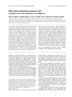
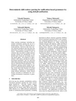
The chelation of cadmium by the thiol-containing pep-
tides GSH, a-GSH and c-Glu-Cys (chemical structures
are given in Fig. 1) in aqueous solution and physiolog-
ical pH can visually be observed and characterized by
simple 1D NMR experiments. Indeed, after the addi-
tion of cadmium to the GSH sample in a 1 : 2
Cd ⁄ GSH stoichiometry (0.5–5 mm solutions in our
experiments), a white precipitate instantaneously
formed and the solution became acidic. Integration of
NMR signals relative to a reference peak (CH
2
of
l-glycine) indicated that the precipitate corresponded
to 10% at pH 6.4 to 20% at pH 7.2 of the total
amount of GSH. Notably, the precipitate was resolubi-
lized after restoring the pH to a neutral value and
shaking the sample. These results were confirmed using
radioactive
35
S-GSH to quantify both precipitate and
soluble forms of complexes (Table S1). Similarly, the
addition of cadmium to the a-GSH sample resulted in
O. Delalande et al. Cadmium–glutathione complex in solution
FEBS Journal 277 (2010) 5086–5096 ª 2010 The Authors Journal compilation ª 2010 FEBS 5087
the formation of a precipitate, but in this case, its dis-
solution was impossible, even after changing the pH,
vigorous shaking or sonication. Furthermore, analysis
of NMR spectra indicated the precipitation of a 1 : 1
stoichiometric complex. A simple pH-dependent analy-
sis of the electric charges indicated that because the
precipitated complex is necessarily neutral at this pH,
the cysteine residue should be in its thiolate S
)
form.
Indeed, at neutral pH, a-GSH bears two carboxylate
groups (COO
)
) and the amino group of the glutamate
is protonated (NH
þ
3
).
From these simple observations, information about
the co-ordination modes of a-GSH and GSH can be
deduced. The transient precipitate observed in the case
of GSH should have a very similar complexation mode
to that of a-GSH, with a 1 : 1 stoichiometry and a glo-
bal charge of zero. Restoring the pH to its initial value
allows this precipitated form to be transformed to the
more stable 1 : 2 complex [13,14,31,32]. In both cases,
a-GSH and GSH peptides form a bidentate complex
with cadmium where the sulfur of the cysteine and the
carboxylate group of the glutamate or the glycine resi-
due are implicated. These two carboxylate groups are
fully equivalent in terms of metal co-ordination struc-
tures. Consequently, the difference observed in cadmium
chelation with a-GSH and GSH is due to the different
location of the amino group of the glutamate residue
in both peptides. The Cd(a-GSH) complex in 1 : 1
stoichiometry is structurally stable, whereas intermo-
lecular interactions between GSH chains are necessary
to stabilize the 2 : 1 complex of GSH with cadmium.
Analyses of
1
H 1D NMR spectra of GSH and
c-Glu-Cys in the presence of cadmium are also very
informative (Fig. 1) and show that both Cd(GS)
2
and
Cd(c-Glu-Cys)
2
complexes have common properties.
First, the broadening of both cysteine a and b proton
resonances after cadmium addition to the sample sug-
gests the existence of an exchange process involving
the metal ion and the cysteine residue. Second, we
observed that the two cysteine b protons, which are
equivalent in the absence of cadmium (only one chemi-
cal shift in NMR spectra), are well differentiated (two
chemical shifts) after metal addition [see 1D spectra
for GSH (Fig. S1) or 2D spectra for c-Glu-Cys
(Fig. S2)]. This observation clearly indicates an asym-
metry of the final complex due to metal co-ordination
and represents a direct probe to follow cadmium chela-
tion. Finally, the dependence of NMR spectra on pH
values is a way to probe the chemical structure of the
complex. In acidic conditions (pH = 5.6), the addition
GSH
GSH + Cd
GSSG
α-GSH
γ-EC + Cd
γ-EC
NH
2E
NH
G
NH
C
** * #
α
C
α
G
α
E
γ
E
β
E
β′
C
/β′′
C
9.0 8.5 4.5 4.0 3.5 2.53.0
p
.
p
.m.
p
.
p
.m.
Fig. 1. 1D
1
H NMR spectra of the different glutathione species. Spectra were recorded in H
2
O at 280K and pH 7.2. From bottom to top,
GSH, Cd(GS)
2
, GSSG, c-Glu-Cys, Cd(c-Glu-Cys)
2
and a-GSH. The chemical structures from bottom to top of GSH, c-Glu-Cys and a-GSH are
indicated on the right. *Corresponds to impurities present in the aGSH sample.
#
Indicates the resonance of the CH
2
group of L-glycine,
which was used as a reference signal for peak integrations. Arrows indicate characteristic resonances of cadmium chelation by GSH or
c-Glu-Cys and of the oxidized form of glutathione. Spectra were aligned with respect to
L-glycine CH
2
resonance.
Cadmium–glutathione complex in solution O. Delalande et al.
5088 FEBS Journal 277 (2010) 5086–5096 ª 2010 The Authors Journal compilation ª 2010 FEBS
of cadmium to the sample did not affect the amino
group resonance at 7.6 p.p.m. of the GSH peptide
(Fig. S3), indicating that this potential ligand is not
involved in metal chelation. At neutral pH (pH = 6.4
and pH = 7.2), the exchange rate with the solvent of
amide protons (NH) of both cysteine and glycine as a
function of temperature was of the same order
(Fig. S4), demonstrating that no amide deprotonation
occurs after cadmium chelation to GSH or c-Glu-Cys
peptides, as has been previously suggested for the
cysteine amide nitrogen [15].
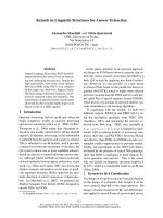
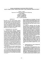
The lability of cadmium bound to the GSH mole-
cules was assessed by
14
N versus
15
N glutathione
competition reactions (Fig. 2). Chelation of cadmium
to the
15
N-labelled GSH pool led to the formation of
the Cd(
15
N-GS)
2
complex, which induced (despite a
partial signal overlap with the cysteinyl proton) the
disappearance of the glycine amide cross-peak on the
spectra. After the addition of
14
N-GSH in the same
proportion to the
15
N-enriched GSH (1 : 1 stoichiom-
etry), the glycine
15
N-
1
H cross-peak was restored.
This indicates the presence of free
15
N-GSH peptide.
The observed reappearance consequently resulted
from a chemical exchange process, in the 0.1–10 ms
range, between bound and unbound GSH molecules
to the cadmium.
The relative affinity of cadmium to GSH and other
peptides was explored by competitive complexation
experiments, as shown in part of the TOCSY spectrum
in Fig. S2. The quantification of NMR data allowed
the evaluation of the chelation fraction: 42.5 ± 5.5
and 57.5 ± 5.5% for GSH and c-Glu-Cys, respec-
tively. These values clearly indicate the similar affinities
of both natural metabolites for cadmium.
NMR structural models for Cd(GS)
2
and
comparison with the GSSG model
Because of the small molecular mass of GSH (307.5 Da),
NOESY experiments are not appropriate to determine
internuclear distances and ROESY-type experiments are
also known to lead to quantification problems [33]. To
circumvent this major problem, we decided to use an
alternative approach based on the off-resonance ROESY
pulse sequence [34]. This method allows the determina-
tion of absolute internuclear distances and of local corre-
lation times. These parameters were used to build initial
structural models of both GSSG and Cd(GS)
2
com-
plexes. A refinement protocol with an explicit solvent
was first performed on GSSG structures and then applied
to the Cd(GS)
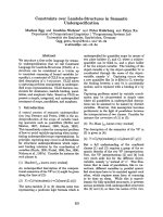
2
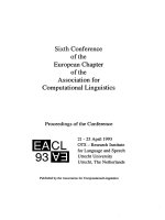
complex. The best structures (shown in
Fig. 3) were obtained, in agreement with the co-ordina-
tion study, using the protonated N-terminal c-glutamate
residue. A total of 26 and 25 NMR constraints per
monomer (GSH unit) were respectively used for GSSG
and Cd(GS)
2
structure determinations (Table 1). Sur-
prisingly, strong differences were observed for the major-
ity of the distances recorded for both molecules, despite
their similar topological arrangement. Because the exis-
tence of a disulfide bond between the two glutathione
units in the oxidized form cannot induce those striking
variations, this suggests that the tridimensional organiza-
tion of the Cd(GS)
2
complex is very different from
GSSG. When the pH was varied between 6.4 and 7.2, sig-
nificant differences for several distances were observed
for the Cd(GS)
2
complex (distances marked in Table 1).
Some of them were characterized as effects of inter-GSH
interactions. This interpretation resulted from sampling
of numerous conformations in molecular dynamic trajec-
tories, which indicated that these NMR data could not
be due to interproton distances in a unique GSH unit of
the dimer (Table S2). This was confirmed by bad refine-
ment convergence of simulated annealing calculations
parametrized only for intra-GSH distances, in agreement
with the fact that we did not observe the 1 : 1 stoichiome-
tric complex in the experimental conditions.
The pairwise local correlation times (s
c
) extracted
from off-resonance ROESY experiments were longer for
Cd(GS)
2
complexes ( 1.5 · 10
)9
s) than for oxidized
p.p.m.
p.p.m.
116
117
118
119
120
121
*
GSH
15
N
+
Cd
+
14
N-GSH
8.18.28.38.4
Fig. 2. Expanded contour plot of three superimposed
15
N-
1
H
heteronuclear single quantum coherence spectra:
15
N-GSH without
cadmium (red), after the addition of cadmium and the formation of
the Cd(
15
N-GS)
2
complex (green), after the addition of
14
N-GSH in a
1 : 1 stoichiometry with respect to
15
N-GSH (purple). Spectra were
recorded in H
2
O at 275 K and pH = 7.0. Because of the overlap of
the cross-peaks, the 2D spectra (green and purple) were shifted
towards the
15
N dimension (vertical axis).
O. Delalande et al. Cadmium–glutathione complex in solution
FEBS Journal 277 (2010) 5086–5096 ª 2010 The Authors Journal compilation ª 2010 FEBS 5089
glutathione ( 0.4 · 10
)9
s). A detailed analysis of the
local correlation times revealed that pH changes mainly
affected inter-GSH unit glutamic acid ⁄ glycine chain
interactions with an increase in correlation times for
higher pH revealing a greater complex rigidity.
Cd(GS)
2
structures determined by simulated anneal-
ing calculations were clustered into six families based on
minimal penalties induced from NMR distance
restraints. 3D models representative of each conforma-
tional family and calculated for pH = 6.4 or pH = 7.2
are presented in Fig. 3 and coordinates are given in
Tables S3–S4. Those of GSSG model are given in Table
S5. Our model clearly indicates that between zero and
two water molecules are present and complete the cad-
mium co-ordination sphere. This number depends on
the pH value and the cadmium charge, which varies
from 0 to +2.0 in our calculations. Despite these varia-
tions, the final models are very similar. The pH value
mainly influences the interactions between the two GSH
units, whereas the presence of a positively charged
cadmium clearly favours the bonding of the glycine
carboxylate group. It should be noted that symmetric
structural models are severely penalized because of their
deviation from imposed NMR restraints.
Discussion
Cadmium glutathione complexes
When cadmium is complexed by glutathione, different
species exist in solution in equilibrium: a mixture of the
Cd(GS)
2
1 : 2 complex and the Cd(GS) 1 : 1 complex.
A recent study [7] suggested that the 1 : 1 monochelate
is one of the major complexes formed at low glutathi-
one concentration. On the other hand, at physiological
pH and higher glutathione concentration, which are the
relevant in cellulo conditions, the 1 : 2 complex is pre-
dominant, as shown by speciation studies [7,14]. As a
consequence, in this work, we focused on the Cd(GS)
2
complex, as apart from its biological relevance, it is the
transport-active complex [15]. To this end, we per-
formed NMR experiments at the optimal conditions
for the formation of the Cd(GS)
2
complex, considering
that the 1 : 1 complex that precipitates is almost totally
transformed into the water-soluble Cd(GS)
2
1 : 2 com-
plex after restoring the pH and shaking. Consequently,
as shown by NMR, we observed only the major and
the most stable soluble Cd(GS)
2
complexes formed
under the conditions of the study (pH 6.4 and 7.2, a
temperature of 17 °C, concentration over 1 mm and at
a favourable 1 : 2 stoichiometry). Eventual disturbing
effects on spectra arising from additional minor species
could not be excluded, but the absence of such NMR
spectral signatures substantiates the assumption that
these forms are negligible in our experimental condi-
tions. Finally, the characterization of unambiguous
inter-GSH unit cross-peaks was also in accordance with
the hypothesis of the predominance of the dimeric form
in solution. On the other hand, interproton distance
variations observed after pH changes may result from
local conformational changes in the dimeric Cd(GS)
2
complex.
88%
12%
66%
pH 6.4 pH 7.2
Cd charge = 1.0
Cd charge = 0.0
71%
25%
59%
Fig. 3. Representation of the best 3D con-
formational families obtained for Cd(GS)
2
NMR-refined structures at pH = 6.4 and
pH = 7.2. The first two rows present two
different rotated views of the major confor-
mations (population in %) obtained for the
Cd(GS)
2
complex model refinement at both
pH 6.4 and 7.2 and using noncharged
cadmium. The bottom row presents two
views of the major conformation of refine-
ments carried out with a +1 charged
cadmium. In these structures, the sphere of
co-ordination of cadmium (green sphere) is
completed with two water molecules in the
case of the Cd(GS)
2
models with
noncharged metal. For models with charged
cadmium there is one water molecule in
the case of pH = 6.4 (bottom left) and no
water molecule for pH = 7.2 (bottom right).
Cadmium–glutathione complex in solution O. Delalande et al.
5090 FEBS Journal 277 (2010) 5086–5096 ª 2010 The Authors Journal compilation ª 2010 FEBS
Other cadmium sulfur–metabolite complexes
NMR experiments performed on complexation of cad-
mium with other sulfur metabolites revealed some
characteristics of their chelation with glutathione.
First, at biologically relevant concentrations, GSSG
does not chelate cadmium, demonstrating that the
thiol group is essential. Second, a-GSH and c-GSH
have different co-ordination modes with cadmium
(1 : 1 stoichiometry for Cd ⁄ a-GSH with implication of
glutamate and 1 : 2 stoichiometry for Cd ⁄ c-GSH)
showing that c-glutamate is not implicated in the che-
lation of GSH. Third, GSH and c-Glu-Cys have the
same co-ordination mode for cadmium with nearly the
same affinity, indicating that the carboxylate group of
glycine is not a key cadmium ligand. Finally, NMR
experiments demonstrated unambiguously that the
chelation is a spontaneous and rapid (millisecond time
scale) phenomenon.
Dynamic co-ordination effects
In the complex formed in aqueous solution with both
glutathione and c-Glu-Cys at biologically relevant con-
centrations and pH, cadmium is mainly linked by thiols
from two distinct GSH or c-Glu-Cys units. Cysteine
sulfur affinity for cadmium provides a strong anchoring
site for glutathione derivatives [8–10,13]. Nevertheless,
we observed during our
14
N-GSH versus
15
N-GSH com-
petition experiments, a significant lability of cadmium,
suggesting that we need to reconsider the strength of
cadmium chelation by glutathione in a biological con-
text. Based on the disappearance of the
1
H-
15
N cysteine
resonance, this exchange rate typically occurs in the
millisecond time range. This phenomenon could explain
our inability to directly observe the
113
Cd resonance at
high magnetic field (data not shown). An obvious con-
clusion of the in vitro part of the present study is
that the formation of Cd(GS)
2
complexes is spontane-
ous and rapid in the tested conditions, which were close
to physiological (ambient temperature, pH tested from
5.6 to 7.2, GSH concentration in the millimolar range).
Consequently, it is unlikely that glutathione S-transfer-
ases are required to catalyse complex formation [24–26].
In our structural models, cadmium always has a tet-
rahedral co-ordination sphere in which two ligands are
thiolate groups. Precipitation observations and NMR
hydrogen exchange data between amide protons of
GSH and bulk water show that cadmium ligation by
nitrogen (amino group of glutamic acid or amide of
cysteine) does not seem to occur, conversely to what
has been previously proposed for cadmium [15] as
derived from zinc studies [35]. This was confirmed by
numerical simulations, which never led to a structure
where nitrogen was involved in cadmium complexation.
Moreover, it is in agreement with the pKa measured
for the amino group of the N-terminal c-glutamate resi-
due (9.42–9.48 from references [14,16]). In most of our
structural models, metal completes its co-ordination
with two water molecules. Furthermore, at neutral pH,
for the best calculated NMR structures, no carboxylate
group is involved. Our results demonstrate that the
complexation of cadmium by glutathione primarily
involves the deprotonated sulfhydryl groups from
cysteine residues and two water molecules.
Dimerization effects
Although cadmium is only complexed through the thio-
late groups, the relevance of glutathione for cadmium
Table 1. NMR interproton distances. Distances were calculated
from build-up curves measured at different h angles for oxidized
glutathione GSSG (pH = 7.0) and Cd(c-GS)
2
complexes at pH = 6.4
and 7.2, respectively.
Glutathione state
GSSG
pH = 7.0
Cd(c-GS)
2
pH = 6.4
Cd(c-GS)
2
pH = 7.2
Interproton distances r (A
˚
) ± 0.7 r (A
˚
) ± 0.2 r (A
˚
) ± 0.3
NH (CYS) – b (GLU) 5.2 4.2 4.3
NH (CYS) – c (GLU) 5.0 2.5 2.5
NH (CYS) – b¢¢ (CYS) 4.6 2.3 2.3
NH (CYS) – b¢ (CYS) 5.7 2.6 2.7
NH (CYS) – a (GLU) 5.0 4.1
a
4.5
a
NH (GLY) – b (GLU) 5.9 4.7
a
5.0
a
NH (GLY) – c (GLU) 4.7 3.6 3.7
NH (GLY) – b¢¢ (CYS) 3.8 2.8
b
2.8
b
NH (GLY) – b¢ (CYS) 3.7 2.8
b
2.8
b
NH (GLY) – a (GLY) 2.9 2.8 3.0
NH (GLY) – a (CYS) 2.6 2.6 2.7
a (CYS) – b (GLU) 6.8 3.6 3.5
a (CYS) – c (GLU) 4.6 3.6
a
3.3
a
a (CYS) – b¢¢ (CYS) 3.1 2.1 2.1
a (CYS) – b¢ (CYS) 3.1 2.0 2.1
a (CYS) – a (GLY) 3.5 4.0
a
3.6
a
a (GLU) – b (GLU) 2.8 2.7 2.7
a (GLU) – c (GLU) 3.2 3.0 2.9
a (GLY) – b¢¢ (CYS) 3.1 Not
observed
a
3.2
a
a (GLY) – b¢ (CYS) 3.4 Not
observed
Not
observed
b¢¢ (CYS) – b (GLU) 3.7 3.4
b
3.3
b
b¢¢ (CYS) – c (GLU) 3.6 4.6
b
4.5
b
b¢ (CYS) – b (GLU) 3.3 3.6
a,b
4.2
a,b
b¢ (CYS) – c (GLU) 3.1 Not
observed
a
4.0
a,b
b¢ (CYS) – b¢¢ (CYS) 1.7 1.8 1.8
b (GLU) – c (GLU) 2.5 2.6 2.5
a
Significant distance differences when pH varied from 6.4 to 7.2
for Cd(GS)
2
.
b
Inter-GSH unit distance for the Cd(c-GS)
2
complexes.
O. Delalande et al. Cadmium–glutathione complex in solution
FEBS Journal 277 (2010) 5086–5096 ª 2010 The Authors Journal compilation ª 2010 FEBS 5091
detoxification clearly depends on the whole structure
and not just the co-ordination modes. Indeed, strong
interactions between two GSH units are directly
observed on the off-resonance ROESY spectra, leading
to clear structural constraints. Based on most struc-
tural models of the dimer complex, the driving force
for these peptide interactions seems to involve gluta-
mate side chains of both GSH units, which form a
hydrophobic core and electrostatic interactions
between glycine and glutamate side chains. Under
these conditions, the atypical c-configuration of the
N-terminal glutamate seems to lock the structure of
the Cd(GS)
2
complex and decrease the accessibility to
the metal on one side. This breaks the symmetry of the
complex, explaining 1D NMR spectral modifications
after metal addition.
Off-resonance ROESY experiments also provided a
reliable description of flexibility occurring in the com-
plex. Pairwise correlation times collected for Cd(GS)
2
complexes are much longer (about 1.5 ns) than those
derived from GSSG (0.4 ns). Even if this is partially
expected due to the increase in the relative molecular
mass, the observed difference, almost a factor of 4,
cannot be ascribed to this sole effect. This result thus
reveals the appearance of a significant structural rigidi-
fication after metal co-ordination. Furthermore, local
correlation times also confirmed that the side chain of
glutamate is more rigid in Cd(GS)
2
compared with GSSG,
substantiating the previous comment on the importance
of the GSH side chains in dimer stabilization.
Cadmium–glutathione complex and the
detoxification process
The protonation state of the cadmium–glutathione com-
plex is strongly dependent on the pH, which can signifi-
cantly differ in the different subcellular compartments.
Intracellular pH values are in the range from 6.5 to 7.2
in the cytosol and from 6.0 to 6.5 in the vacuole [36–38].
The complex should thus be stabilized in the cytosol,
favouring specific recognition and efficient transport by
Ycf1p. In vacuolar acidic conditions, the equilibria
should be displaced to protonated forms with enhance-
ment of inter-GSH interactions and destabilization of
Cd(GS)
2
leading to possible ligand substitution. In this
schema, thiolate reprotonation could be the first chemi-
cal event in the cadmium releasing process by glutathi-
one and so a key step in the detoxification process.
The competition experiment showing similar effi-
ciency in the formation of Cd(GS)
2
and Cd(c-Glu-
Cys)
2
complexes suggests that the latter complex can
also be formed in vivo,asc-Glu-Cys pools can reach
high concentrations in the range of GSH levels
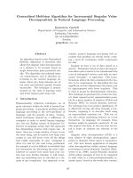
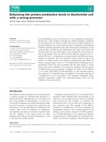
following cadmium exposure [3]. In addition, the het-
erologous complex involving the two metabolites
Cd(GS)(c-Glu-Cys) is also expected. Interestingly the
mutant strain Dgsh2, devoid of glutathione synthase
activity and unable to produce glutathione, accumu-
lates c-Glu-Cys at high intracellular levels (Fig. 4).
This strain has a high chelating capacity, demon-
strated by a global level of free thiols (GSH +
c-Glu-Cys) higher than the wild-type (Fig. 4A). In
addition, although our data indicate that Cd(c-Glu-
Cys)
2
complexes are efficiently formed, this strain
was shown to be hypersensitive to cadmium [39].
This phenotype suggests an impaired detoxification of
cadmium in this strain due to a decreased rate of
transport of Cd(c -Glu-Cys)
2
compared with Cd(GS)
2
complexes. This defect may concern the transport
into the vacuole through Ycf1 or the export outside
the cell through Yor1 or both. Using wild-type cells
labelled with
35
S-GSH, we observed that the total
export of glutathione [including free GSH and
Cd(GS)
2
complexes] outside the cells is not significant
(5.9–8.7% in cells treated for 3 h with 0.1 mm cad-
mium compared with 4.4% in untreated cells;
Table 2). This very low level of Cd(GS)
2
export is
consistent with the very slight cadmium-sensitive phe-
notype of the yor1D strain [27]. Thus, considering the
low contribution of Cd(GS)
2
complex export to cad-
mium resistance, we assume that the gsh2D pheno-
type is caused by a low efficiency transport into the
vacuole of Cd(c-Glu-Cys)
2
compared with Cd(GS)
2
complexes. Consistent with this interpretation is the
observation that, even under standard conditions,
c-Glu-Cys is far less efficiently transported than GSH
into purified vacu oles overexpressing YCF1 (M. Lazard
γ
-Glu-Cys
0
5
10
15
20
Concentration (mM)
WT
Dgsh2
Concentration (mM)
GSH
0
5
10
15
WT
Dgsh2
Dgsh2
Cysteine
γ
-glutamyl-cysteine
Glutathione
Gsh1 Gsh2
Glutamate Glycine
A
B
Fig. 4. c-Glu-Cys concentration is strongly increased in Dgsh2 cells.
(A) Wild-type and Dgsh2 cells grown in minimum medium supple-
mented with 400 l
M glutathione were treated with 200 lM cad-
mium for 3 h. The intracellular metabolites were extracted and
analysed by LC ⁄ MS as previously described [3]. (B) Representation
of the glutathione biosynthesis pathway. Gsh1, c-glutamyl-cysteine
synthetase; Gsh2, glutathione synthetase.
Cadmium–glutathione complex in solution O. Delalande et al.
5092 FEBS Journal 277 (2010) 5086–5096 ª 2010 The Authors Journal compilation ª 2010 FEBS
& P. Plateau, personal communication). The absence
of cadmium co-o rdination vi a c arboxy late gr oups of
glycine residues in structur al models supports the idea
that detoxification differences observed for GSH and
c-Glu-Cys only occur at the recognition step of cad-
mium–metabolite c omplexes by the t ra nsporter. We
thus sugg est that the gl yci ne residue may b e involved
in metal-complex recognition by the Ycf1p transporter
and no t in direct interaction with cadmium.
The transporter Ycf1 is a key element in cadmium
detoxification, as shown by the cadmium-sensitive
phenotype of the ycf1D mutant strain and the cad-
mium-resistant phenotype of strains overexpressing
YCF1 [40]. Our data suggest that under physiological
conditions, the formation of Cd(GS)
2
complexes is
spontaneous and should not constitute a bottleneck in
the detoxification process. The next steps, transport
into the vacuole and the metabolism of the complexes
in the vacuole, remain to be fully understood.
Experimental procedures
Strains and culture conditions
The Saccharomyces cerevisiae strain used for the production
of
15
N-GSH was S288C (Mata SUC2 mal mel gal2 CUP1).
The strain used for the production of
15
N-c-Glu-Cys was
Dgsh2 previously constructed in the BY4742 genetic back-
ground (MATa, ura3D0, his3D1, lys2D0, leu2D0) [41] by
EUROSCARF (Intitute of Molecular Biosciences, Johann
Wolfgang Goethe-University Frankfurt, Germany). This mutant
has the KanMX4 marker inserted into the GSH2 locus.
Cells were grown at 30 °C in minimal yeast nitrogen base
medium (0.67%) supplemented with 2% glucose as a
carbon source and with auxotrophic requirements (uracil,
histidine, lysine, leucine and 30 lm glutathione) when nec-
essary (strain Dgsh2). The standard yeast nitrogen base
medium contains 30 m m
14
N-ammonium sulfate. For
15
N
labelling, 30 mm
14
N-ammonium sulfate was replaced by
10 mm
15
N-ammonium sulfate (Eurisotop, Gif-sur-Yvette,
France) as the sole source of nitrogen.
Preparation of
15
N-enriched metabolites
After growth for at least 25 generations in
15
N-ammonium
sulfate, the cell culture (400 mL corresponding to
10
10
cells) was treated with 50 lm Cd
2+
to induce an
overproduction of
15
N-GSH [3]. After 4 h of treatment, the
cells were collected by centrifugation, washed quickly with
cold water and resuspended in 3 mL of 0.1% perchloric
acid. Cells were transferred to boiling water for 5 min, cen-
trifuged and the supernatant was collected. This extract
contained soluble yeast metabolites, including
15
N-GSH
(S288C strain) and
15
N- c-Glu-Cys (Dgsh2 strain).
The extracts containing
15
N-GSH and
15
N-c-Glu-Cys
were purified on a carbohydrate analysis column
(4.6 · 250 mm, 5 lm) from Waters (Saint-Quentin en
Yvelines, France). Chromatographic separations were
performed using a Surveyor pump and a Surveyor auto-
sampler (ThermoFisher Scientifics, Les Ulis, France), under
isocratic conditions with a flow rate of 0.8 mLÆmin
)1
. The
mobile phase consisted of water containing acetic acid at
0.4%. The effluent from the liquid chromatography was
split by a factor of 1 ⁄ 20 before its introduction into the
MS. ESI MS was performed using an LCQ-Duo ion trap
MS fitted with an electrospray source (ThermoFisher
Scientifics) operated in the positive mode. The mass spec-
trometer was operated with the capillary temperature at
250 °C, sheath gas at 80 (arbitrary units) and the auxiliary
gas at 20 (arbitrary units). The target was fixed at 2 · 10
7
ions and the automatic gain control was turned on. The
electrospray voltage was 4.5 kV, the capillary voltage
10.6 V and the tube lens offset )6 V. The injection time
was 50 ms. MS were recorded at unit mass resolution with-
out in-source fragmentation using the single ion recording
detection mode. The signals for
15
N-GSH and
15
N- c-Glu-
Cys were monitored at m ⁄ z 311 and 253, with retention
times of 23 and 40 min, respectively. The fractions corre-
sponding to these retention time ranges were collected and
finally lyophilized before NMR experiments.
Sample preparation
In the case of samples used for distance extraction, chelation
or competition reaction experiments, GSH or GSSG were
dissolved in 500 mL (90% H
2
O 10% D
2
O) resulting in 1 mm
minimal ionic strength samples, with a final 1 : 2 Cd ⁄ GSH
stoichiometry. To decrease oxidation processes, all samples
were sealed after bubbling with dry nitrogen gas for a few
minutes. Classical peptides were purchased from Sigma-Aldrich
(St Louis, MO, USA) and a-GSH was synthesized and
purified for NMR quality by Eurogentec (Seraing, Belgium).
NMR spectroscopy
All NMR experiments were performed on Bruker Advance
DRX spectrometers (Bruker, Ettlingen, Germany). 1D and
2D
1
H spectra in H
2
O were recorded at 500 MHz by using
a Watergate [42] or an excitation sculpting sequence [43] to
suppress the water signal. Peak assignments were carried
out using classical techniques, in particular for proton
Table 2. Total export of
35
S-glutathione by wild-type cells. The
values reported in the Table are the ratio S ⁄ T (see Radioactive
experiments section in Experimental procedures).
Strain No treatment 100 l
M cadmium
S288C 4.4 ± 0.3% 5.9 ± 0.8%
BY4742 4.4 ± 0.8% 8.7 ± 0.3%
O. Delalande et al. Cadmium–glutathione complex in solution
FEBS Journal 277 (2010) 5086–5096 ª 2010 The Authors Journal compilation ª 2010 FEBS 5093
resonances through TOCSY and off-resonance ROESY
experiments. Our attribution agreed with those obtained in
other studies [16,44]. l-glycine at a final concentration of
2.5 mm was added to the sample before recording 1D
NMR spectra because the CH
2
resonance was not affected
by the addition of cadmium and this resonance did not
overlap with the other signals (see Fig. 1). It was used as
an internal reference for peak integration and species
quantification.
Proton–proton distances were extracted from off-reso-
nance ROESY build-up curves using a procedure already
described [45]. The off-resonance ROESY pulse sequence
[46] was adapted for the excitation sculpting water suppres-
sion method. Seven h angles between the effective and static
magnetic field directions (5, 15, 25, 35, 45 and 54.7°) and
six mixing times (25, 50, 75, 100, 150 and 200 ms) were
used. For metal-reduced glutathione, the spectra were
recorded at three different pH values: 5.6, 6.4 and 7.2. For
the samples with c-Glu-Cys and metal-free oxidized gluta-
thione, the experiments were performed at pH = 7.0. All
off-resonance ROESY spectra were collected at 500 MHz
at 274.3K with TXI or BBI probes.
1
H,
15
N-heteronuclear single quantum coherence experi-
ments were carried out using gradient coherence selection
and sensitivity enhancement. Backbone
15
N amide reso-
nances were observed on a 600 MHz spectrometer equipped
with a TCI cryoprobe (Bruker). Natural abundance
13
C
NMR experiments were also performed, using heteronucle-
ar multiple bond correlation sequences and a TCI cryo-
probe. All chemical shifts for
1
H were referenced to an
internal TSP signal.
Molecular modelling
Molecular mechanics calculations and molecular dynamic
simulation methods were used for model construction using
the amber 9 suite programs. Parameters for the c-gluta-
mate residue were developed from the Gaussian03 DFT
charge calculations method and adapted to the Parm99
force-field [47] using the Resp module and a standard
charge fitting protocol. Both protonated and nonprotonat-
ed states for the amino group of the c-glutamate residue
were implemented and simulated. Cadmium was considered
as a hard sphere with a modulated charge varying between
0.0 and +2.0 and a Cd-S distance of 2.46 A
˚
[8]. Other
main parameters for angles and dihedrals were adapted
from Amber force-field data previously depicted for zinc
ion in the four-cysteine tetrahedral environment [8,48]. An
explicit solvation model (TIP3P water model and +1.0
dielectric constant) was used in all simulations. Structure
refinements were performed using NMR interproton dis-
tances as restraints implemented as harmonic functions into
a simulated annealing protocol with 5000 final structures
collected. Free and restraint molecular dynamic simulations
were used to analyse characteristic key distances for
co-ordination-type discrimination. Cd-N (2.3 ± 0.1 A
˚
[8])
or C-O (2.2 ± 0.1 A
˚
[49]) restraint distances were imposed
between cadmium and potential ligand atoms during the
production period. Structures obtained from simulated
annealing calculations were sorted and divided into homog-
enous conformational groups leading to the best NMR
refined models.
Radioactive experiments
Cells (3 ml at D = 0.4) grown in minimum medium were
labelled with 2 lCi of
35
S-GSH (PerkinElmer) for 40 min
at 30 °C. Cells were washed and re-suspended in the same
medium (with or without 100 lm cadmium). Total
35
S-GSH pools were counted (T). After 3h incubation, the
cultures were centrifuged and the amount of radioactivity
present in the supernatant was measured (S).
Acknowledgements
This work was supported by the Commissariat a
`
l’Energie Atomique (grant from the Programme de
Toxicologie Nucle
´
aire Environnementale). We thank
R. Genet for kindly providing
15
N-ammonium sulfate.
We thank Dr Carl Mann for careful reading and
helpful comments on the manuscript.
References
1 Tamas M, Labarre J, Toledano MB & Wysocki R
(2006) Mechanisms of Toxic Metal Tolerance in Yeast.
Springer, Berlin.
2 Lu SC (1999) Regulation of hepatic glutathione synthe-
sis: current concepts and controversies. FASEB J 13,
1169–1183.
3 Lafaye A, Junot C, Pereira Y, Lagniel G, Tabet JC,
Ezan E & Labarre J (2005) Combined proteome and
metabolite-profiling analyses reveal surprising insights
into yeast sulfur metabolism. J Biol Chem 280, 24723–
24730.
4 Singhal RK, Anderson ME & Meister A (1987) Gluta-
thione, a first line of defense against cadmium toxicity.
FASEB J 1, 220–223.
5 Muller EG (1996) A glutathione reductase mutant of
yeast accumulates high levels of oxidized glutathione
and requires thioredoxin for growth. Mol Biol Cell 7,
1805–1813.
6 Grant CM, Perrone G & Dawes IW (1998) Glutathione
and catalase provide overlapping defenses for protection
against hydrogen peroxide in the yeast Saccharomyces
cerevisiae. Biochem Biophys Res Commun 253, 893–898.
7 Leverrier P, Montigny C, Garrigos M & Champeil P
(2007) Metal binding to ligands: cadmium complexes
with glutathione revisited. Anal Biochem 371, 215–228.
Cadmium–glutathione complex in solution O. Delalande et al.
5094 FEBS Journal 277 (2010) 5086–5096 ª 2010 The Authors Journal compilation ª 2010 FEBS
8 Zaima H, Ueyama N, Adachi H & Nakamura A (1995)
1H-, 13C-, and 113Cd-NMR study of the Cd(II)
complex of a blocked peptide, Z-Cys-Ala-Pro-His-OMe,
in organic solvents. Biopolymers 35 , 319–329.
9 Cherifi K, Decock-Le Reverend B, Varnagy K, Kiss T,
Sovago I, Locheux C & Kozlowski H (1990) Transition
metal complexes of L-cysteine containing di- and tripep-
tides. J Inorg Biochem 38, 69–80.
10 Kozlowski H, Urbanska J, Sovago I, Varnagy K,
Kiss A, Spychala J & Cherifi K (1990) Cadmium ion
interaction with sulphur containing amino acid and
peptide ligands. Polyhedron 9, 831–837.
11 Li NC & Manning RA (1955) Some metal complexes of
sulfur-containing amino acids. J Am Chem Soc 77 ,
5225–5228.
12 Birgerssonn B, Carter RE & Drakenberg T (1977) A
cadmium-113 NMR study of cadmium-glutathione
complexes. J Magn Reson 28, 299–302.
13 Fuhr BJ & Rabenstein DL (1973) Nuclear magnetic
resonance studies of the solution chemistry of metal
complexes. IX. The binding of cadmium, zinc, lead,
and mercury by glutathione. J Am Chem Soc 95, 6944–
6950.
14 Perrin DD & Watt AE (1971) Complex formation of
zinc and cadmium with glutathione. Biochim Biophys
Acta 230, 96–104.
15 Li ZS, Lu YP, Zhen RG, Szczypka M, Thiele DJ &
Rea PA (1997) A new pathway for vacuolar cadmium
sequestration in Saccharomyces cerevisiae: YCF1-cata-
lyzed transport of bis(glutathionato)cadmium. Proc Natl
Acad Sci USA 94, 42–47.
16 Kadima W & Rabenstein DL (1990) Nuclear magnetic
resonance studies of the solution chemistry of metal
complexes. 26. Mixed ligand complexes of cadmium,
nitrilotriacetic acid, glutathione, and related ligands.
J Inorg Biochem 38, 277–288.
17 Hemmingsen L, Damblon C, Antony J, Jensen M,
Adolph HW, Wommer S, Roberts GC & Bauer R
(2001) Dynamics of mononuclear cadmium beta-lac-
tamase revealed by the combination of NMR
and PAC spectroscopy. J Am Chem Soc 123,
10329–10335.
18 Goodfellow BJ, Rusnak F, Moura I, Domke T &
Moura JJ (1998) NMR determination of the global
structure of the 113Cd derivative of desulforedoxin:
investigation of the hydrogen bonding pattern at the
metal center. Protein Sci 7, 928–937.
19 Pan T, Freedman LP & Coleman JE (1990) Cadmium-
113 NMR studies of the DNA binding domain of the
mammalian glucocorticoid receptor. Biochemistry 29,
9218–9225.
20 Hemmingsen L, Olsen L, Antony J & Sauer SP (2004)
First principle calculations of (113)Cd chemical shifts
for proteins and model systems. J Biol Inorg Chem 9,
591–599.
21 Kidambi SS & Ramamoorthy A (2003) Characteriza-
tion of metal centers in bioinorganic complexes using
ab initio calculations of 113Cd chemical shifts. Inorg
Chem 42, 2200–2202.
22 Abrahams IL & Garner CD (1985) Nature of the cad-
mium sites in rat liver metallothionein 1 from Cd K-
Edge EXAFS. J Am Chem Soc 107, 4596–4597.
23 Kadima W & Rabenstein DL (1990) A quantitative
study of the complexation of cadmium in hemolyzed
human erythrocytes by 1H NMR spectroscopy. J Inorg
Biochem 40, 141–149.
24 Rai R, Tate JJ & Cooper TG (2003) Ure2, a prion precur-
sor with homology to glutathione S-transferase, protects
Saccharomyces cerevisiae cells from heavy metal ion and
oxidant toxicity.
J Biol Chem 278, 12826–12833.
25 Adamis PD, Gomes DS, Pinto ML, Panek AD &
Eleutherio EC (2004) The role of glutathione transfer-
ases in cadmium stress. Toxicol Lett 154, 81–88.
26 Lewinska A & Bartosz G (2007) Protection of yeast
lacking the Ure2 protein against the toxicity of heavy
metals and hydroperoxides by antioxidants. Free Radic
Res 41, 580–590.
27 Nagy Z, Montigny C, Leverrier P, Yeh S, Goffeau A,
Garrigos M & Falson P (2006) Role of the yeast ABC
transporter Yor1p in cadmium detoxification. Biochimie
88, 1665–1671.
28 Mendoza-Cozatl D, Loza-Tavera H, Hernandez-Navar-
ro A & Moreno-Sanchez R (2005) Sulfur assimilation
and glutathione metabolism under cadmium stress in
yeast, protists and plants. FEMS Microbiol Rev 29,
653–671.
29 Adle DJ, Sinani D, Kim H & Lee J (2007) A cadmium-
transporting P1B-type ATPase in yeast Saccharomyces
cerevisiae. J Biol Chem 282, 947–955.
30 Dameron CT, Smith BR & Winge DR (1989) Glutathi-
one-coated cadmium-sulfide crystallites in Candida glab-
rata. J Biol Chem 264, 17355–17360.
31 Williams DR, Corrie AM & Walker MD (1976) Ther-
modynamic considerations in co-ordination, Part XXII.
Sequestering ligands for improving the treatment of
plumbism and cadmiumism. J Chem Soc Dalton Trans,
1012–1015.
32 Krezel A & Bal W (1999) Coordination chemistry of
glutathione. Acta Biochim Pol 46, 567–580.
33 Desvaux H, Berthault P, Birlirakis N & Goldman M
(1994) Off-resonance ROESY for the study of dynamic
processes. J Magn Reson A 108, 219–229.
34 Esposito V, Das R & Melacini G (2005) Mapping poly-
peptide self-recognition through (1)H off-resonance
relaxation. J Am Chem Soc 127, 9358–9359.
35 Martin RB & Edsall JT (1959) The association of
divalent cations with glutathione. J Am Chem Soc 81,
4044–4047.
36 Gillies RJ, Ugurbil K, den Hollander JA & Shulman
RG (1981) 31P NMR studies of intracellular pH and
O. Delalande et al. Cadmium–glutathione complex in solution
FEBS Journal 277 (2010) 5086–5096 ª 2010 The Authors Journal compilation ª 2010 FEBS 5095
phosphate metabolism during cell division cycle of
Saccharomyces cerevisiae. Proc Natl Acad Sci USA 78,
2125–2129.
37 Preston RA, Murphy RF & Jones EW (1989) Assay of
vacuolar pH in yeast and identification of acidification-
defective mutants. Proc Natl Acad Sci USA 86, 7027–
7031.
38 Chattopadhyay S, Muzaffar NE, Sherman F &
Pearce DA (2000) The yeast model for batten disease:
mutations in BTN1, BTN2, and HSP30 alter pH
homeostasis. J Bacteriol 182, 6418–6423.
39 Jin YH, Dunlap PE, McBride SJ, Al-Refai H, Bushel
PR & Freedman JH (2008) Global transcriptome and
deletome profiles of yeast exposed to transition metals.
PLoS Genet 4, e1000053.
40 Wemmie JA, Szczypka MS, Thiele DJ & Moye-Rowley
WS (1994) Cadmium tolerance mediated by the yeast
AP-1 protein requires the presence of an ATP-binding
cassette transporter encoding gene, YCF1. J Biol Chem
269, 32592–32597.
41 Brachmann CB, Davies A, Cost GJ, Caputo E, Li J,
Hieter P & Boeke JD (1998) Designer deletion strains
derived from Saccharomyces cerevisiae S288C: a useful
set of strains and plasmids for PCR-mediated gene dis-
ruption and other applications. Yeast 14, 115–132.
42 Piotto M, Saudek V & Sklenar V (1992) Gradient-tai-
lored excitation for single-quantum NMR spectroscopy
of aqueous solutions. J Biomol NMR 2, 661–665.
43 Hwang TL & Shaka AJ (1995) Water suppression that
works. Excitation sculpting using arbitrary wave-forms
and pulsed-field gradients. J Magn Reson 112, 275–
279.
44 Rabenstein DL, Isab AA, Kadima W & Mohanakrish-
nan P (1983) A proton nuclear magnetic resonance
study of the interaction of cadmium with human
erythrocytes. Biochim Biophys Acta 762, 531–541.
45 Desvaux H, Berthault P & Birlirakis N (1995) Dipolar
spectral densities from off-resonance 1H NMR relaxa-
tion measurements. Chem Phys Lett 233, 545–549.
46 Desvaux H, Berthault P, Birlirakis N, Goldman M &
Piotto M (1995) Improved versions of off-resonance
ROESY. J Magn Reson 113, 47–52.
47 Wang J, Cieplak P & Kollman PA (2000) How well
does a restrained electrostatic potential (RESP) model
perform in calculating conformational energies of
organic and biological molecules. J Comput Chem 21,
1049–1074.
48 Stote RH & Karplus M (1995) Zinc binding in proteins
and solution: a simple but accurate nonbonded repre-
sentation. Proteins 23, 12–31.
49 Siripornadulsil S, Traina S, Verma DP & Sayre RT
(2002) Molecular mechanisms of proline-mediated toler-
ance to toxic heavy metals in transgenic microalgae.
Plant Cell 14, 2837–2847.
Supporting information
The following supplementary material is available:
Fig. S1. 1D NMR spectra of GSH in the presence of
increased quantities of cadmium.
Fig. S2. An expanded contour plot of the 2D TOCSY
spectrum.
Fig. S3. 1D NMR spectra of GSH in the presence or
not of cadmium at different pH levels.
Fig. S4. Proton exchange of the Cd(GS)
2
complex in
H
2
O.
Table S1. Proportion of GSH precipitated during Cd-
GSH
2
complex preparation.
Table S2. Inter- and intraproton–proton distances
whether dimeric or monomeric GSH models are con-
sidered.
Table S3. Coordinates (in PDB format) of the best
NMR structure models for the Cd(GS)
2
complex at
pH = 7.2.
Table S4. Coordinates (in PDB format) of the best
NMR structure models for the Cd(GS)
2
complex at
pH = 6.4.
Table S5. Coordinates (in PDB format) of the NMR
structure model for the GSSG molecule at pH = 7.0.
This supplementary material can be found in the
online version of this article.
Please note: As a service to our authors and readers,
this journal provides supporting information supplied
by the authors. Such materials are peer-reviewed and
may be re-organized for online delivery, but are not
copy-edited or typeset. Technical support issues arising
from supporting information (other than missing files)
should be addressed to the authors.
Cadmium–glutathione complex in solution O. Delalande et al.
5096 FEBS Journal 277 (2010) 5086–5096 ª 2010 The Authors Journal compilation ª 2010 FEBS