Prevalence and Its Predictors of Extrapulmonary Involvement in Patients with Pulmonary Tuberculosis pdf
Bạn đang xem bản rút gọn của tài liệu. Xem và tải ngay bản đầy đủ của tài liệu tại đây (116.4 KB, 5 trang )
INTRODUCTION
Extrapulmonary tuberculosis (EPTB) comprises 9.7-46%
of all cases of tuberculosis (TB) (1-3). Although tuberculous
bacilli could spread to any organs, the common organs in-
volved with EPTB include lymph nodes, pleura, bones and
joints, brain and meninges, gastrointestinal organs, liver,
genitourinary organs, peritoneum, and pericardium. Although
TB lymphadenitis or TB pleuritis respond relatively well to
anti-TB treatment, some forms of EPTB (e.g., TB meningi-
tis) are notorious for their association with high morbidity
and mortality (4, 5). Furthermore, miliary TB, the extreme
form of EPTB, presents a great challenge to human health
because of its high mortality rate of 18-24%, even in recent
reports (6-9).
Extrapulmonary organ involvement (10) in human immun-
odeficiency virus (HIV)-infected patients with pulmonary
TB is reported to be 26%, however, the clinical characteris-
tics of patients with pulmonary TB at risk of simultaneous
extrapulmonary organ involvement have not been studied
in detail, although the initiation of treatment following early
identification of extrapulmonary involvement is crucial. The
aim of this study was to determine the prevalence and clini-
cal predictors of the presence of extrapulmonary involvement
in patients with pulmonary TB.
MATERIALS AND METHODS
Study settings, subjects, and data collection
All adult patients with culture-proven pulmonary TB diag-
nosed between January 1, 2004 and July 31, 2006 at Seoul
National University Hospital, a tertiary referral hospital were
included for this study. We retrospectively reviewed the med-
ical records of these patients, which included demographic
data, results of laboratory tests, and so on. We also reviewed
the radiographic examinations of the patients. The protocol
of this study was approved by the institutional review board
of Seoul National University Hospital.
Definition of extra-pulmonary involvement of TB
The presence of extra-pulmonary involvement in patients
with pulmonary TB was based on the following criteria: 1)
demonstration of acid-fast bacilli or the growth of Mycobac-
Extrapulmonary organ involvement in human immunodefiaency virus (HIV)-infect-
ed patients with pulmonary tuberculosis (TB) is reported to be 26%, however, the
clinical predictors of extrapulmonary involvement in pulmonary TB patients has not
been reported yet. We tried to determine the clinical predictors of presence of extra-
pulmonary involvement in patients with pulmonary TB. Cross-sectional study was
performed including all adult patients with culture-proven pulmonary TB diagnosed
between January 1, 2004 and July 30, 2006, at a tertiary referral hospital in South
Korea. The presence of extra-pulmonary TB involvement was diagnosed based on
bacteriological, pathological, or clinical evidence. Among 320 patients with a culture-
proven pulmonary TB, 40 had extrapulmonary involvement. Patients with bilateral
lung involvement were more likely to have extrapulmonary involvement, with an
adjusted odds ratio (OR) of 4.21 (95% confidence interval [CI], 1.82-9.72), while
patients older than 60 yr (adjusted OR, 0.27; 95% CI, 0.08-0.89), patients with cavi-
tary lesions (adjusted OR, 0.37; 95% CI, 0.16-0.84), and with higher levels of serum
albumin (adjusted OR, 0.45; 95% CI, 0.25-0.78) had less frequent involvement.
Clinicians should be aware of the possibility of extrapulmonary involvement in TB
patients with bilateral lung involvement without cavity formation or lower levels of
serum albumin.
Key Words : Tuberculosis; Tuberculosis, Miliary; Diagnosis
237
Min Jae Kim, Hye-Ryoun Kim,
Seung Sik Hwang
*
, Young Whan Kim,
Sung Koo Han, Young-Soo Shim,
and Jae-Joon Yim
Division of Pulmonary and Critical Care Medicine,
Department of Internal Medicine and Lung Institute,
Seoul National University College of Medicine, Seoul;
Department of Social and Preventive Medicine
*
,
College of Medicine, Inha University, Incheon, Korea
Address for correspondence
Jae-Joon Yim, M.D.
Division of Pulmonary and Critical Care Medicine,
Department of Internal Medicine and Lung Institute,
103 Daehak-ro, Jongno-gu, Seoul 110-744, Korea
Tel : +82.2-2072-2059, Fax : +82.2-762-9662
E-mail :
J Korean Med Sci 2009; 24: 237-41
ISSN 1011-8934
DOI: 10.3346/jkms.2009.24.2.237
Copyright
�
The Korean Academy
of Medical Sciences
Prevalence and Its Predictors of Extrapulmonary Involvement in
Patients with Pulmonary Tuberculosis
Received : 1 December 2007
Accepted : 24 June 2008
terium tuberculosis from tissue; 2) presence of granulomas with
or without caseation necrosis in tissue; 3) positive polymerase
chain reaction (PCR) results for the DNA of M. tuberculosis
from tissues; or 4) a clinical diagnosis by duty physicians based
on symptoms, laboratory, radiographic findings, and treatment
response to anti-TB medications. Tuberculous pleuritis was
not classified as EPTB because pleura is believed to be involved
by direct invasion from frequently accompanying pulmonary
parenchymal TB or hypersensitivity reaction by M. tubercu-
losis rather than blood stream dissemination (11-13).
Statistical analyses
Univariate comparisons between the group with pulmonary
TB and extrapulmonary involvement and the group with
pulmonary TB without extrapulmonary involvement were
performed using Pearson’s chi-square test or Fisher’s exact
test for categorical variables and Student’s t-test for continu-
ous variables. Variables analyzed included demographic cha-
racteristics, laboratory results, and radiographic findings.
Using variables with p values of <0.20 from the univariate
comparisons, multiple logistic regression models were con-
structed to identify predictors of the presence of extrapul-
monary involvement. In logistic regression, backward elim-
ination was used to select variables to be maintained in the
final model, using a p value of <0.10 as the criterion for sta-
tistical significance of associations. The area under the receiv-
er operator characteristic (ROC) curve was used to evaluate
the performance of the models. To successfully split patients
into more homogeneous subgroups, classification and regres-
sion trees (CART) were used to build a binary classification
tree through recursive partitioning. All tests of significance
were two sided and p<0.05 was considered statistically sig-
nificant. We used statistical software Stata 9.0 (Stata Corpo-
ration, College Station, TX, U.S.A.) to perform the multi-
ple logistic regression and R 2.4.1 (The R foundation for sta-
tistical computing) to construct the CART.
RESULTS
Three hundred and twenty patients were diagnosed with
culture-proven pulmonary TB at Seoul National University
Hospital between January 1, 2004 and July 31, 2006. Their
median age was 45 yr and 198 (62%) were male: 85 patients
(26.6%) had underlying diseases including HIV infection,
diabetes, chronic liver diseases, and so on; 83 patients (25.9%)
had previously diagnosed and treated TB (Table 1).
Forty (12.5%) of the 320 patients with pulmonary TB had
extrapulmonary involvement. Miliary involvement of the
lung was the most common manifestation of EPTB (12 pa-
tients, 30%). TB lymphadenitis (8 patients), intestinal TB
(8 patients), and TB laryngitis (8 patients) followed. The
tuberculous involvement of extrapulmonary organs was con-
firmed bacteriologically in 11 patients (27.5%) and diagnosed
based on positive PCR for M. tuberculosis DNA in 7 patients
(Table 2).
We compared the clinical characteristics and laboratory
results between the 40 pulmonary TB patients with extra-
pulmonary involvement and the 280 patients without. There
was no difference between the two groups in terms of age,
underlying diseases, history of previous TB, and drug suscep-
tibility pattern. However, bilateral lung involvement was
more common in patients with extrapulmonary involvement
(77.5% vs. 46.4%, p<0.001). In addition, the mean hemat-
ocrit, albumin, and cholesterol values were lower in the pa-
238 M.J. Kim, H R. Kim, S.S. Hwang, et al.
320 patients
Age, yr, median (range) 45 (20-74)
Male/female 198 (62%)/122 (38%)
Underlying diseases 85 (26.6%)
HIV infection 5 (1.6%)
Diabetes 38 (11.9%)
Chronic liver disease 9 (2.8%)
Connective tissue disease 13 (4.1%)
Chronic renal failure 1 (0.3%)
Asthma 6 (1.9%)
COPD 2 (0.6%)
Cancer 20 (6.3%)
Post-transplantation state 5 (1.6%)
On immunosuppressant 19 (5.9%)
Previous history of TB 83 (25.9%)
Diagnosis of pulmonary TB
Negative AFB smear but positive culture 167 (52.2%)
of M. tuberculosis
Positive AFB smear and positive culture 153 (47.8%)
of M. tuberculosis
Drug susceptibility tests
Sensitive to all drug 221 (69.1%)
Resistant but not MDR 30 (9.4%)
MDR 69 (21.6%)
Presence of extrapulmonary involvement 40 (12.5%)
Radiographic characteristics
Presence of cavitary lesion 126 (39.4%)
Extent of radiographic lesion
Confined to unilateral lung 159 (49.7%)
Extended to bilateral lung 161 (50.3%)
Laboratory tests (mean
±
standard deviation)
Leukocytes (×1,000/
μ
L) 7.60
±
3.33
Neutrophil (×1,000/
μ
L) 5.08
±
2.75
Lymphocyte (×1,000/
μ
L) 1.64
±
0.95
Hemoglobin (g/dL) 12.92
±
2.16
Hematocrit (%) 39.37
±
5.78
Total protein (g/dL) 7.16
±
0.89
Albumin (g/dL) 3.84
±
0.61
Cholesterol (mg/dL) 162.96
±
40.41
Creatinine (mg/dL) 0.97
±
0.42
Table 1. Demographic and clinical characteristics of enrolled
patients
HIV, human immunodefiaency virus; COPD, chronic obstructive pulm-
mary disease; TB, tuberculosis; AFB, acid-fast bacilli; MDR, multi-drug
resistance.
tients with extrapulmonary involvement (Table 3).
The final multiple logistic regression model showed that
after adjustment only the presence of cavitary lesions, absence
of bilateral lung involvement, and lower albumin levels were
associated with extrapulmonary involvement in patients with
pulmonary TB. Patients with bilateral lung involvement were
more likely to have extrapulmonary involvement, with an
adjusted odds ratio (OR) of 4.21 (95% confidence interval
[
CI
]
, 1.82-9.72), while patients older than 60 yr (adjusted
OR, 0.27; 95% CI, 0.08-0.89) and patients with cavitary
lesions were less likely to have extrapulmonary involvement
(adjusted OR, 0.37; 95% CI, 0.16-0.84). In addition, patients
with higher levels of albumin had less frequent extrapulmo-
nary involvement (adjusted OR, 0.45; 95% CI, 0.25-0.78)
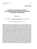
(Table 4). The fitness of the final model was good in terms
of multiple logistic regression (area under the ROC curve,
0.76; 95% CI, 0.68-0.84) as well as CART analysis (area
under the ROC curve, 0.73; 95% CI, 0.65-0.82) (Fig. 1).
DISCUSSION
The presence of cavities in patients with pulmonary TB is
Pulmonary
TB without
extrapulmonary
involvement (%)
Pulmonary TB
with extrapul-
monary
involvement (%)
p
value
Table 3. Comparison of demographic and clinical characteris-
tics between pulmonary tuberculosis (TB) patients with extra-
pulmonary involvement and without extrapulmonary involvement
(univariate analysis)
DM, diabetes mellitus; COPD, chronic obstructive pulmmary disease;
MDR, Multi-drug resistance.
Number of patients 280 40
Sex
Male 177 (63.2) 21 (52.5)
Female 103 (36.8) 19 (47.5) 0.192
Age (yr) 0.499
20-39 109 (39.9) 16 (40.0)
40-59 115 (41.1) 19 (47.5)
+60 56 (20.0) 5 (12.5)
Underlying diseases
HIV infection 3 (1.1) 2 (5.0) 0.119
DM 34 (12.1) 4 (10.0) 1.000
Chronic liver disease 9 (3.2) 0 (0) 0.609
Connective tissue disease 9 (3.2) 4 (10.0) 0.065
Chronic renal failure 0 1 (2.5) 0.215
Asthma 6 (2.1) 0 (0) 1.000
COPD 2 (0.7) 0 (0) 1.000
Cancer 18 (6.4) 2 (5.0) 1.000
Post-transplantation state 4 (1.4) 1 (2.5) 0.489
On Immunosuppressant 12 (4.3) 7 (17.5) 0.005
History of TB 72 (25.7) 11 (27.5) 0.810
Drug susceptibility tests
Sensitive to all 192 (68.6) 29 (72.5)
Resistant but not MDR 25 (8.9) 5 (12.5) 0.413
MDR 63 (22.5) 6 (15.0)
Radiographic characteristics
Presence of cavitary lesion 115 (41.1) 11 (27.5) 0.100
Extent of radiographic lesion
Confined to unilateral lung 150 (53.6) 9 (22.5) <0.001
Extended to bilateral lung 130 (46.4) 31 (77.5)
Results of laboratory tests
(mean
±
standard deviation) 7.45
±
2.86 8.72
±
5.78 0.171
Leukocytes (×1,000/
μ
L)
Neutrophil (×1,000/
μ
L) 4.97
±
2.64 5.85
±
3.41 0.061
Lymphocyte (×1,000/
μ
L) 1.63
±
0.66 1.71
±
2.09 0.799
Hematocrit (%) 39.72
±
5.72 36.91
±
5.69 0.004
Total protein (g/dL) 7.19
±
0.86 6.96
±
1.04 0.200
Albumin (g/dL) 3.88
±
0.58 3.54
±
0.75 0.008
Cholesterol (mg/dL) 164.93
±
40.99 148.90
±
33.16 0.020
Cr (mg/dL) 0.96
±
0.45 0.94
±
0.47 0.699
EPTB in Patients with Pulmonary TB 239
Involved organ* 40 patients (100%)
Miliary involvement 12 (30%)
Lymph node 8 (20%)
Intestine 8 (20%)
Larynx 6 (15%)
Soft tissue 5 (12.5%)
Bone and joint 4 (10%)
Peritoneum 1 (2.5%)
Meninges 1 (2.5%)
Method of diagnosis
Bacteriologically confirmed 11 (27.5%)
Positive PCR for M. tuberculosis DNA in tissue 7 (17.5%)
Pathologically diagnosed 9 (22.5%)
Clinically diagnosed 13 (32.5%)
Disseminated Miliary nodules in chest radiographs 9 (22.5%)
Others
�
4 (10%)
Table 2. Sites and methods of diagnois of extrapulmonary in-
volvement in 40 patients
*, When a patient had more than one organ involved, all of them were
counted independently;
�
, 2 patients with intestinal TB diagnosed based
on typical colonosopic findings and the other 2 patients with TB laryn-
gitis without AFB bacilli and caseating granuloma in pathologic exami-
nations.
PCR, polymerase chain reaction; TB, tuberculosis; AFB, acid-fast bacilli.
TB, tuberculosis; CI, confidence interval.
Variable Odds ratio 95% CI p value
Age (yr)
20-39 1.0 (ref.)
-
40-59 0.71 0.62-1.59 0.411
+60 0.27 0.08-0.89 0.031
On immunosuppressant 2.86 0.87-9.41 0.084
Radiographic characteristics
Presence of cavitary lesion 0.37 0.16-0.84 0.018
Extended to bilateral lung 4.21 1.82-9.72 0.001
Results of laboratory tests
Albumin per increase of 1 g/dL 0.45 0.25 - 0.78 0.005
Table 4. Risk factors for combined extra-pulmonary involvement
in patients with pulmonary TB (multiple logistic regression-final
model)
regarded as a marker for high bacillary burden and is reported
to be associated with relapse after completion of treatment
(14). Our observation that the extrapulmonary involvement
was less frequently observed in cavitary pulmonary TB pati-
ents suggests that the higher bacillary burden per se does not
make the host prone to extrapulmonary involvement. On the
contrary, the presence of cavities was associated with a lower
possibility of the spread of tuberculous bacilli to extrapulmo-
nary organs in this study. Given that pulmonary cavities have
been reported to be rare in TB patients with immune com-
promise (15, 16), the presence of cavities could be a hallmark
of a certain level of intact immunity against tuberculous ba-
cilli, guaranteeing protection from further dissemination to
other organs. This hypothesis could be tested through future
study comparing systemic as well as local immunity against
M. tuberculosis between TB patients with or without pulmo-
nary cavity should be performed through future studies. In
fact, differences were already reported in expression of vari-
ous genes between pulmonary TB patients and extrapulmo-
nary TB patients (17).
In contrast to the presence of pulmonary cavities, bilateral
lung involvement might better reflect attenuated host immu-
nity than bacillary burden (18). Considering that various
types of impaired cell-mediated immunity have been con-
sidered to play an important role in the development of EPTB
(10, 19-22), the decreased host immunity suggested by the
presence of bilateral lung involvement could be crucial in
the dissemination of tuberculous bacilli to extrapulmonary
organs. In fact, pulmonary TB patients on immunosuppres-
sants were prone to have extrapulmonary involvement (p=
0.08) in this study, although we failed to get statistical signif-
icance because of the small numbers of patients on immuno-
suppressants.
Hypoalbuminemia is generally regarded as a marker of poor
nutritional status in patients with TB (23, 24). In addition,
hypoalbuminemia/protein malnutrition itself could impair
host immunity against M. tuberculosis through decreased pro-
duction of cytokines including interferon-
γ
(25) or the reduc-
tion of CD4 and CD8 T cell numbers observed in animal
models (26). Hypoalbuminemia as a predictor for the pres-
ence of extrapulmonary organ involvement as observed in
this study could be explained by probable immune dysfunc-
tion against tuberculous bacilli and matches previous reports
showing lower albumin levels in patients with disseminated
TB (27).
Results from our study that older patients with pulmonary
TB have a lower risk of having a extrapulmonary involve-
ment (adjusted OR, 0.27; 95% CI, 0.08-0.89) disagrees with
previous reports that show that EPTB was higher in the elder-
ly (28). In addition, the lower risk of EPTB in the elderly
does not support immunity as a determinant of the spread
of tuberculous bacilli to other organs because of the higher
incidence of TB in the aged group (29, 30) and decreased
immunity to tuberculous bacilli in older mice (31). This ob-
servation could be interpreted in two ways. First, the decre-
ased risk for extrapulmonary involvement in the elderly could
result from the small number of patients older than 60 yr (61
patients, 19.1%) in this study. In this setting, a small change
in the number of patients with extrapulmonary involvement
could make significant changes in the OR. Second, extrapul-
monary dissemination with bilateral lung involvement but
without cavity formation could be understood as a character-
istic of TB bacilli rather than host immune status. The clin-
ical manifestations might differ among TB patients infected
with different strains of M. tuberculosis. For example, the ‘Beijing
strain’ was reported to cause more severe pathology in mice
(32) as well as more advanced radiographic lesions in humans
(33). In this context, infection by specific strains of M. tuber-
culosis might cause intra- and extrapulmonary dissemination
rather than cavity formation.
In conclusion, the extrapulmonary organ involvement in
patients with pulmonary TB was more common in patients
with bilateral lung involvement but without cavity forma-
tion or low levels of serum albumin. Clinicians should keep
in mind the possibility of extrapulmonary involvement in
these patients.
REFERENCES
1. Rieder HL, Snider DE Jr, Cauthen GM. Extrapulmonary tuberculo-
sis in the United States. Am Rev Respir Dis 1990; 141: 347-51.
240 M.J. Kim, H R. Kim, S.S. Hwang, et al.
EPTB=40
No EPTB=280
Total=320
EPTB=9*
No EPTB=150*
(5.7%)
EPTB=31*
No EPTB=130*
(19.3%)
EPTB=25*
No EPTB=124*
(16.8%)
EPTB=13*
No EPTB=27*
(32.5%)
EPTB=12*
No EPTB=97*
(11.0%)
EPTB=3*
No EPTB=18*
(14.3%)
EPTB=10*
No EPTB=9*
(52.6%)
EPTB=6*
No EPTB=6*
(50.0%)
Fig. 1. Classification and regression trees (CART) analysis for pre-
dicting combined extra-pulmonary involvement in patients with
pulmonary TB
EPTB, pulmonary TB with extra-pulmonary involvement; sAlbumin,
serum level of albumin.
Unilateral lung lesion Extended to bilateral lung
Not on immunosuppressant On immunosuppressant
sAlbumin <4 g/dL
Cavitary lesion Non-cavitary lesion
sAlbumin
≥
4 g/dL
2. Cowie RL, Sharpe JW. Extra-pulmonary tuberculosis: a high fre-
quency in the absence of HIV infection. Int J Tuberc Lung Dis 1997;
1: 159-62.
3. Huang J, Shen M, Sun Y. Epidemiological analysis of extrapulmo-
nary tuberculosis in Shanghai. Zhonghua Jie He He Hu Xi Za Zhi
2000; 23: 606-8.
4. Kent SJ, Crowe SM, Yung A, Lucas CR, Mijch AM. Tuberculous
meningitis: a 30-year review. Clin Infect Dis 1993; 17: 987-94.
5. Ramachandran P, Duraipandian M, Reetha AM, Mahalakshmi SM,
Prabhakar R. Long-term status of children treated for tuberculous
meningitis in south India. Tubercle 1989; 70: 235-9.
6. Kim JH, Langston AA, Gallis HA. Miliary tuberculosis: epidemiol-
ogy, clinical manifestations, diagnosis, and outcome. Rev Infect Dis
1990; 12: 583-90.
7. Maartens G, Willcox PA, Benatar SR. Miliary tuberculosis: rapid
diagnosis, hematologic abnormalities, and outcome in 109 treated
adults. Am J Med 1990; 89: 291-6.
8. Mert A, Bilir M, Tabak F, Ozaras R, Ozturk R, Senturk H, Aki H,
Seyhan N, Karayel T, Aktuglu Y. Miliary tuberculosis: clinical man-
ifestations, diagnosis and outcome in 38 adults. Respirology 2001;
6: 217-24.
9. Kim JY, Park YB, Kim YS, Kang SB, Shin JW, Park IW, Choi BW.
Miliary tuberculosis and acute respiratory distress syndrome. Int J
Tuberc Lung Dis 2003; 7: 359-64.
10. Lado Lado FL, Barrio Gomez E, Carballo Arceo E, Cabarcos Ortiz
de Barron A. Clinical presentation of tuberculosis and the degree of
immunodeficiency in patients with HIV infection. Scand J Infect Dis
1999; 31: 389-91.
11. Kim HJ, Lee HJ, Kwon SY, Yoon HI, Lee CT, Han SK, Shim YS,
Yim JJ. The prevalence of pulmonary parenchymal tuberculosis in
patients with tuberculous pleuritis. Chest 2006; 129: 1253-8.
12. Moudgil H, Sridhar G, Leitch AG. Reactivation disease: the com-
monest form of tuberculous pleural effusion in Edinburgh, 1980-
1991. Respir Med 1994; 88: 301-4.
13. Antoniskis D, Amin K, Barnes PF. Pleuritis as a manifestation of
reactivation tuberculosis. Am J Med 1990; 89: 447-50.
14. Benator D, Bhattacharya M, Bozeman L, Burman W, Cantazaro A,
Chaisson R, Gordin F, Horsburgh CR, Horton J, Khan A, Lahart C,
Metchock B, Pachucki C, Stanton L, Vernon A, Villarino ME, Wang
YC, Weiner M, Weis S. Rifapentine and isoniazid once a week ver-
sus rifampicin and isoniazid twice a week for treatment of drug-sus-
ceptible pulmonary tuberculosis in HIV-negative patients: a ran-
domised clinical trial. Lancet 2002; 360: 528-34.
15. Aderaye G, Bruchfeld J, Assefa G, Feleke D, Kallenius G, Baat M,
Lindquist L. The relationship between disease pattern and disease
burden by chest radiography, M. tuberculosis Load, and HIV status
in patients with pulmonary tuberculosis in Addis Ababa. Infection
2004; 32: 333-8.
16. Batungwanayo J, Taelman H, Dhote R, Bogaerts J, Allen S, Van de
Perre P. Pulmonary tuberculosis in Kigali, Rwanda. Impact of human
immunodeficiency virus infection on clinical and radiographic pre-
sentation. Am Rev Respir Dis 1992; 146: 53-6.
17. Kim DK, Park GM, Hwang YI, Kim HJ, Han SK, Shim YS, Yim JJ.
Microarray analysis of gene expression associated with extrapul-
monary dissemination of tuberculosis. Respirology 2006; 11: 557-65.
18. Mabiala Babela JR, Makosso E, Senga P. Radiological specifities of
pulmonary tuberculosis in Congolese children: effect of HIV infection.
Med Trop (Mars) 2006; 66: 255-9.
19. Ergun I, Ekmekci Y, Sengul S, Kutlay S, Dede F, Canbakan B, Erbay
B. Mycobacterium tuberculosis infection in renal transplant recipi-
ents. Transplant Proc 2006; 38: 1344-5.
20. Hussein MM, Mooij JM, Roujouleh H. Tuberculosis and chronic
renal disease. Semin Dial 2003; 16: 38-44.
21. Keiper MD, Beumont M, Elshami A, Langlotz CP, Miller WT Jr.
CD4 T lymphocyte count and the radiographic presentation of pul-
monary tuberculosis. A study of the relationship between these fac-
tors in patients with human immunodeficiency virus infection. Chest
1995; 107: 74-80.
22. Jones BE, Young SM, Antoniskis D, Davidson PT, Kramer F, Barnes
PF. Relationship of the manifestations of tuberculosis to CD4 cell
counts in patients with human immunodeficiency virus infection. Am
Rev Respir Dis 1993; 148: 1292-7.
23. Karyadi E, Schultink W, Nelwan RH, Gross R, Amin Z, Dolmans
WM, van der Meer JW, Hautvast JG, West CE. Poor micronutrient
status of active pulmonary tuberculosis patients in Indonesia. J Nutr
2000; 130: 2953-8.
24. Onwubalili JK. Malnutrition among tuberculosis patients in Harrow,
England. Eur J Clin Nutr 1988; 42: 363-6.
25. Dai G, McMurray DN. Altered cytokine production and impaired
antimycobacterial immunity in protein-malnourished guinea pigs.
Infect Immun 1998; 66: 3562-8.
26. Mainali ES, McMurray DN. Protein deficiency induces alterations
in the distribution of T-cell subsets in experimental pulmonary tuber-
culosis. Infect Immun 1998; 66: 927-31.
27. Crump JA, Reller LB. Two decades of disseminated tuberculosis at
a university medical center: the expanding role of mycobacterial
blood culture. Clin Infect Dis 2003; 37: 1037-43.
28. Cailhol J, Decludt B, Che D. Sociodemographic factors that con-
tribute to the development of extrapulmonary tuberculosis were iden-
tified. J Clin Epidemiol 2005; 58: 1066-71.
29. Hong YP, Kim SJ, Lew WJ, Lee EK, Han YC. The seventh nation-
wide tuberculosis prevalence survey in Korea, 1995. Int J Tuberc
Lung Dis 1998; 2: 27-36.
30. Stead WW, Dutt AK. Tuberculosis in elderly persons. Annu Rev
Med 1991; 42: 267-76.
31. Vesosky B, Turner J. The influence of age on immunity to infection
with Mycobacterium tuberculosis. Immunol Rev 2005; 205: 229-43.
32. Lopez B, Aguilar D, Orozco H, Burger M, Espitia C, Ritacco V, Bar-
rera L, Kremer K, Hernandez-Pando R, Huygen K, van Soolingen D.
A marked difference in pathogenesis and immune response induced
by different Mycobacterium tuberculosis genotypes. Clin Exp Immunol
2003; 133: 30-7.
33. Drobniewski F, Balabanova Y, Nikolayevsky V, Ruddy M, Kuznet-
zov S, Zakharova S, Melentyev A, Fedorin I. Drug-resistant tuber-
culosis, clinical virulence, and the dominance of the Beijing strain
family in Russia. JAMA 2005; 293: 2726-31.
EPTB in Patients with Pulmonary TB 241