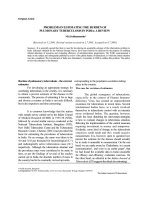
FREQUENCY OF SPUTUM POSITIVE AFB CASES AMONG PATIENTS OF PULMONARY TUBERCULOSIS IN TERTIARY CARE HOSPITALS OF NORTHERN PAKISTAN pot
Bạn đang xem bản rút gọn của tài liệu. Xem và tải ngay bản đầy đủ của tài liệu tại đây (186 KB, 5 trang )
J Ayub Med Coll Abbottabad 2010;22(2)
56
FREQUENCY OF SPUTUM POSITIVE AFB CASES AMONG PATIENTS
OF PULMONARY TUBERCULOSIS IN TERTIARY CARE HOSPITALS
OF NORTHERN PAKISTAN
Mohammad Ishaq Khattak, Ihsanullah*, Amir Muhammad**, Nisar Khan***,
Munawar Zaman
Department of Medicine, *Department of Pathology, Khyber Teaching Hospital, Peshawar, **Department of Pathology, Kohat Institute
of Medical Sciences, Kohat, ***Department of Pulmonology, Ayub Teaching Hospital, Abbottabad, Pakistan
Objective: This study was aimed to find out the frequency of sputum positive Acid Fast Bacilli
(AFB) cases among pulmonary tuberculosis patients and to determine those patients who are the
potential source of transmitting infection. Methods: This study was conducted in four medical units
of Khyber Teaching Hospital, Peshawar and Chest Unit, Ayub Teaching Hospital, Abbottabad in
collaboration with the pathology unit of Khyber Teaching Hospital, Peshawar, and Kohat Institute of
Medical Sciences, Kohat. Three specimens of sputum were collected for three consecutive days in
the morning and were transported immediately to the laboratory along with full details of the
patients. Results: Out of two hundred total patients studied, 104 patients (52%) were sputum AFB
positive. Among the 104 patients 60 patients (57.4%) were females. Sixty-four (61.52%) individuals
were between 20–50 years. Majority of the patients were from poor, deprived and lower social class.
Fifty-two (50%) patients had monthly income of less than Rs. 4,000; only 8 patients (7.67%) had
monthly income of more than Rs. 12,000). Forty-eight patients <46.12% were house wives, 10
patients (9.61%) were unemployed. Most of the patients were under weight for their age and height.
24 patients (23.06%) were below 42 kg. The maximum (53.84%) number of patients was in weight
range of 43–50 kg. Conclusion: Sputum AFB positive pulmonary tuberculosis is more in individuals
of low socioeconomic group and in females. The patients put their children and family members at
risk of tuberculosis infection. For the control of this disease early diagnosis of active disease and
their treatment under supervision is important.
Keyword: Sputum, AFB-Positive, Pulmonary tuberculosis, Low socioeconomic group
INTRODUCTION
Tuberculosis has been with us from the beginning of
civilisation and it likely will be with us until the end.
1
It is defined as a disease caused by bacteria
belonging to Mycobacterium tuberculosis complex.
2
It can affect any organ of the body but in two third of
the cases it involves the lung parenchyma.
2,3
This
form of tuberculosis is called pulmonary tuberculosis.
Pulmonary tuberculosis may be primary or post
primary (secondary) depending upon prior exposure.
Commonly, pulmonary tuberculosis is
infectious, transmitted between individuals by droplet
infection,
4
with greater spread of infection from
patients having sputum smear positive for acid fast
bacilli
5,6
. Transmission is also influenced by features
of the potential recipient of the organism (contact)
especially the immune status, and by the environment
in which they live.
Examination of the sputum smear for acid
fast bacilli by direct microscopy is by far the most
important investigation for the diagnosis of
pulmonary tuberculosis.
7
Early detection and
effective treatment of smear positive tuberculosis
patients has been found to be the most cost effective
strategy for the control of the disease.
8
Among other investigations, culture of sputum
for isolation of organism is the only definitive way of
making a diagnosis, however it is time consuming and
the facility is not widely available. Serological
techniques lack reliability and the newly developed
molecular techniques, though sensitive and rapid, are
expensive and sparsely available in Pakistan, making
them impractical for use in most cases.
9
When anti-tuberculosis drugs were
introduced, it was widely assumed that eradication of
tuberculosis; one of mankind’s most ancient and
deadly diseases, was within easy reach. Unfortunately,
such optimism was not well founded. Worldwide, the
number of tuberculous cases has continued to increase.
The disease is thought to cause at least 3 million deaths
each year and the annual number of new cases is
approximately 9 million.
10
Of these new cases, perhaps
50% of the patients have Mycobacterium tuberculosis
identified in sputum smears and the other 50% would
have TB proved by isolation of the organism in
culture, if facilities for culture were available.
4
In Pakistan, tuberculosis is a leading cause
of morbidity where 80% of the disease is present in
persons who are in their reproductive age. Around
1.5 million persons are suffering from active
tuberculosis along with more than 0.2 million new
cases each year.
11
With the migration of Afghan
refugees to Pakistan, especially to NWFP, the
problem has worsened. Most of them live in poor
J Ayub Med Coll Abbottabad 2010;22(2)
57
hygienic conditions in camps and are the source of
TB among themselves and in the native population.
12
With proper treatment the disease is curable
in virtually all cases caused by drug susceptible
strains but without treatment it may kill the patient
within five years, in more than half of the cases.
2
The development of multi-drug-resistant
(MDR) tuberculosis has emerged as a public health
concern in Pakistan in the last decade. Lack of public
awareness, easy availability of the anti-tuberculosis
drugs, poverty, no proper follow up, relatively
complex and prolonged treatment regimen are all
potential sources for default, treatment failure and
development of MDR tuberculosis. National
tuberculosis programme, if managed in accordance
with WHO’s recommendations, will help in
combating MDR tuberculosis.
In a developing country like ours where
poverty, illiteracy, overcrowding, poor hygienic
conditions, social deprivation and lack of proper
medical care is present, presence of persons who are
coughing up tuberculous bacilli is forming a chain of
transmission of infection and identification of smear
positive cases is a major detrimental factor in finding
or predicting the magnitude of disease, and by their
effective treatment the spread of mycobacterium
tuberculosis will be prevented, which is the basis of
anti-tuberculosis campaign in a developing country.
MATERIAL AND METHODS
This study was conducted in four medical units of
Khyber Teaching Hospital Peshawar and Chest Unit
of Ayub Teaching Hospital, Abbottabad in
collaboration with the Pathology Department, Khyber
and Ayub Teaching Hospitals. The study period was
from Jun 2005 to Dec 2006. Two hundred patients
were included in the study.
All patients with age of 16 years and above,
patients having clinical features suggestive of
pulmonary tuberculosis as evening pyrexia, weight loss,
productive cough, haemoptysis, night sweats, malaise,
tiredness, anorexia, chest pain, and patients with raised
ESR and X-ray chest finding suggestive of pulmonary
tuberculosis like abnormal shadows, cavitation, and/or
abnormality in the lymph nodes were included.
Patients having tuberculosis other than
pulmonary, diagnosed case of malignancy and a
diagnosed case of HIV were excluded.
A thorough clinical assessment was carried
out after admission with emphasis on clinical history,
physical examination, and necessary investigations. A
printed proforma containing a comprehensive record
of all patients was completed from each patient. Blood
complete picture with ESR, Urine R/E, Chest X-ray,
Blood Urea, Blood Sugar, and Sputum smear
examination by direct microscopy after Ziehl Neelsen
staining were done for all patients.
A wide mouthed, leak proof, clean bottle
was given to each patient on day of admission and
were advised to collect sputum as soon as he/she
wakes up in the morning. Patients were instructed to
collect at least 3–5 ml of sputum by coughing
vigorously after deep inspiration, repeatedly if
necessary. This produces sputum specimen from deep
in the lungs. The specimen was transported
immediately to the laboratory, along with a request
form, having full details, written on it about the
patient. Sputum was sent on 3 consecutive days.
Sputum smears were prepared by selecting the solid
or most dense particles of sputum and smearing it on
a microscopy slide using a wire loop. The slide then
dried, fixed and was stained with Ziehl Neelsen
Carbol Fuchsin. Slides were examined with 100× oil
immersion and 100 fields were examined before the
smear was reported as negative.
RESULTS
Out of a total 200 patients, 104 patients (52%) were
sputum AFB positive (Table-1). Among these 104
cases, 60 patients (57.4%) were females. Sixty-four
individuals (61.52%) were between 20–50 years of
age (Table-2). Majority of the patients were members
of poor, deprived and lower local class.
Fifty-two patients (50%) had monthly
income of less than Rs: 400 only, 8 patients (7.67%)
had monthly income of more than Rs. 12,000; 48
patients (46.12%) were housewives, and 10 patients
(9.61%) were unemployed (Table-3). Most of the
patients were under weight for their age and height;
24 patients (23.06%) were below 42 kg. The
maximum (53.84%) number of patients was in
weight range 43–50 kg. (Table-4). The chief
presenting symptoms were evening pyrexia
(90.38%), productive cough (84.61%), and weight
loss (53.84%). Chest pain (5.76%) and anorexia
(7.69%) were less common symptoms, (Table-5).
Common signs at presentation were anaemia, i.e., 78
patients (75%) were anaemia, 16 patients (15.38%)
had bronchial breathing, 30 patients (28.84%) had
cracles and 3 patients (5.76%) had ronchi, (Table-6).
During investigation most of the patients especially
female were having Hb level of 8–10 gm/dl
(40.38%). Total leucocyte count was in normal range.
High ESR was observed in many patients. Only 14
patients (13.46%) had an ESR of <20 mm/1
st
hour
(Table-7). Common X-ray (chest) features were
unilateral upper lung field involvement seen in 24
patients (46.15%), (Table-8).
J Ayub Med Coll Abbottabad 2010;22(2)
58
Table-1: Patients with sputum AFB positive result
out of 200 patients studied
Sputum smear result Number %
AFB positive
104 52.0
AFB negative
96 48.0
Table-2: Age distribution of 104 sputum AFB
positive patients
Age (years) Number %age
16–25
16 15.38
26–50
64 61.53
51 and above
24 23.07
Table-3: Occupational status of 104 sputum AFB
positive
Occupation Number %
House wife
48 46.15
Labourer
24 23.07
Government servant
6 5.76
School teacher
6 5.76
Shop keeper
4 3.84
Student
4 3.84
Tailor
2 1.92
Unemployed
10 9.61
Table-4: Weight distribution of 104 sputum AFB
positive patients
Weight (kg) Number %
<40
24 23.07
41–50
56 53.84
51–60
18 17.30
>61
6 5.76
Table-5: Symptoms at presentation in 104 sputum
AFB positive patients
Symptoms Number %
Evening pyrexia
94 90.38
Tiredness
46 44.23
Malaise
52 50
Weight loss
56 50
Night sweats
34 53.84
Productive cough
88 84.61
Haemoptysis
28 26.92
Anorexia
8 7.69
Chest pain
6 5.76
Palpitations
16 15.38
Table 6: Signs at presentation in 104 sputum AFB
positive patients
Sings Number %age
Temperature 98.4F
12 11.53
100 ºF
82 78.84
101 ºF
4 3.84
102 ºF
4 3.84
> 102 ºF
2 1.92
Anaemia
78 75
Bronchial breathing
16 15.38
Crackles
30 28.84
Ronchi
6 5.76
Table-7: ESR of 104 sputum AFB positive patients
ESR (mm/1
st
hour) Number %
<20
14 13.46
20–60
34 32.69
60–100
14 13.46
>100
42 40.38
Table-8: Chest radiological pattern in 104 sputum
AFB positive patients
X- ray pattern Number %
Unilateral upper lung field
48 46.15
Bilateral upper lung fields
22 21.15
Cavitary disease
26 25
Diffuse pulmonary TB
2 1.92
Lower lung field
6 5.76
DISCUSSION
Tuberculosis has caused more deaths than any other
infectious disease and 95% of these deaths are in the
developing world.
12
It is the fourth major cause of
death in Pakistan. Early diagnosis and effective
treatment of active cases particularly pulmonary who
are infectious to the community is the best way of
controlling TB in our country. The delay in diagnosis
and inability to cure a high proportion of pulmonary
smear positive cases are the main reasons of
increased risk of infection, high death rate and MDR
cases in Pakistan.
Currently for diagnosis, developing
countries rely on AFB stains and culture (where
available) and radiographic changes. ZN-staining is a
rapid, simple and cheap way of diagnosing
pulmonary tuberculosis but it lacks sensitivity, still it
is the most rewarding method if performed by an
experienced microbiologist.
In this study, 104 out of 200 patients (52%)
were sputum AFB positive. Though the validity of
the AFB positivity on sputum specimens may be
questioned, because they were not confirmed by
culture for AFB, this was thought not be the case as
patients with the possible diagnosis of tuberculosis
only on clinical, laboratory and radiological grounds
were included in the study.
In another study by Asch S and colleagues
(Los Angeles 1998)
13
, 56% patients had positive
sputum AFB results. This study was done on
homeless patients and the increased frequency as
compared to my study could be because of the
selection of high-risk patients.
Of the 104 patients, 60 patients (57.6%)
were females and 44 patients (43.30%) were males
that match with the sex distribution of TB patients
noted by Akhtar T and colleagues (1994)
8
and
Ahmed M and colleagues
14
. These findings are in
agreement with earlier findings that tendency to
disease and mortality from TB is higher in females as
compared to males.
15
Females from illiterate families
in general are treated lower than men and so have a
poor nutritional status. In addition, early marriages
and multiple pregnancies put extra burden on the
defence leaving them more vulnerable to develop TB.
TB in women puts their children and family members
at risk of tuberculosis infection, disease and death.
J Ayub Med Coll Abbottabad 2010;22(2)
59
This triple threat makes detection and treatment of
TB in a woman absolutely vital.
As compared to a developed country, where
TB is common among elderly, it is a disease of young
in a developing country. Seventy five percent (75%) of
tuberculosis cases occur in age group of 15–59 years
16
,
the most economically productive sector of society. It
was true in this study, as 61.52% of patients were in
the age group of 20–50 years and 15.38% of patients
were in the age group of 16–25 years.
The monthly income of 50% of patients was
below Rs. 4,000, and only 7.6% had more than Rs.
10,000. Similarly in a study by Iqbal ZH and
colleages
17
, majority of tuberculous patients were
from lower socioeconomic group. As regard
occupation, 46.15% were housewives, and 9.61% of
patients were unemployed. Poverty, unemployment
and homelessness are all linked and increases the risk
of developing TB.
18
The commonest symptom at presentation of
which 90.38% of patients complained was of evening
pyrexia. Another common symptom was productive
cough; present in 84.61% of patients; Phuc LT and
colleagues
19
reported similar results.
Weight loss was the presenting symptom in
53.84% of patients, tiredness in 44.23% of patients,
and malaise in 50% of patients. It is said that in
pulmonary TB the frequency of weight loss and
malaise are less common and very difficult to
quantify.
4
The difference may be because of the setup
to which the patients belong, as many of the patients
in my study were under weight and malnourished
Night sweats was noticed in 32.69% of the
patients, although it has been described as a classic
symptom of pulmonary TB
4
, Kumar and Clark
20
described drenching night sweats as a less common
feature and attributed it to the anxiety associated with
the disease. The reason for night sweats in my
patients was high grade fever in some patients and
anxiety in others.
Haemoptysis was the presentation in 26.92%
of patients. About 75% of patients were clinically
anaemic. It is said that nutritional status of patients
with active pulmonary TB is poor as compared to the
healthy subjects. This could be one of the reasons for
anaemia, similarly haemoptysis and anaemia of
chronic disease could be the additional factors
responsible.
Examination of the chest revealed no
positive findings, except that on auscultation 15.38%
of the patients had bronchial breathing, 28.84% of
patients had crackles and only 5.76% of patients had
ronchi. In general, the examination of chest
contributes relatively little to the diagnosis or
assessment of post-primary TB.
21
Low haemoglobin of 10–12 gm/dl was
reported in 51.92% of patients, 40.38% of patients
had Hb levels of 8–10 gm/dl. In many patients, total
leukocyte count was within normal range. Hafiz S
22
described low haemoglobin, normal or high TLC and
raised ESR as the haematological findings in
tuberculosis patients. ESR was elevated in majority
of patients; only 13.46% of patients had an ESR of
less than 20 mm/1
st
hour.
On chest x-ray examination, the commonest
presentation was typical pattern of upper lung field
infiltrates and or nodules with or without cavitation
seen in 47 patients (90.38%). Wilcke JT et al
23
reported typical presentation in 92% of patients. Of
the typical pattern, unilateral upper lung field
involvement was noticed in 46.15% of patients,
21.15% had bilateral upper lung field involvement,
while 25% of patients had cavitary disease. In an
earlier study
24
, unilateral lung field involvement was
seen in 37.89% of patients, bilateral upper field
involvement in 62.2% of patients and cavitary
disease in 39.6% of patients. Lower lung field
tuberculosis (with lesion confined to area below the
hilum) was noticed in 5.76% of patients.
CONCLUSIONS
Sputum AFB positive pulmonary TB is more in
females, in young age individuals, and in people of
low socio economic group.
For the control of tuberculosis, early diagnosis of
active cases and their treatment under supervision is
important.
Acid fast staining of sputum is the best method, if
performed by experienced microbiologist, as it is
reliable and economical. Its diagnostic yield can be
increased by liquefaction and centrifugation of sputum
and by examining more than one sample.
REFERENCES
1. Rubin SA. Tuberculosis, captain of all these men of death. In:
Radiol North Am 1995;33:619–39.
2. Raviglione MC, O’ Brien RJ. Tuberculosis. In: Fauci AS,
Braunwald E, Isselbacher KJ, et al (eds). Harrison’s principles of
internal medicine. 14
th
edition. USA: McGraw-Hill Companies;
1998.1000–14.
3. Hussain R. Pakistan journal of chest medicine (editorial). Pak J
Chest Med 1999;5(1):1–2.
4. Hope Well PC, Bloom BR. Tuberculosis and other mycobacterial
diseases. In: Murray JF, Nadel JA (eds). Textbook of respiratory
medicine. Philadelphia: WB Saunders Co. 1994: p. 1096–1100.
5. Cayla JA. Factors associated with secondary TB cases.
Symposium. Investigation of contacts to tuberculosis cases. New
York city department of public health. Bureau TB Control. New
York, June 7–8, 1996.
6. Kritski A. Management of TB contacts in Brazil: is it a priority?
Symposium. Investigation of contacts of TB cases. New York
City Department of Pubic Health. Bureau of TB control. New
York City. June 7–8, 1996.
J Ayub Med Coll Abbottabad 2010;22(2)
60
7. Samad A, sadiq M, Yousaf M, Javaid A. Role of Fiber optic
bronchoscopy in sputum smear negative pulmonary TB. Pak J
Chest Med 2000;6(1):5–9.
8. Akhtar T, Imran M. Management of TB by practitioners of
Peshawar. J Pak Med Assoc 1994;44(12):280–2.
9. Farida N. Histological and cytological diagnosis of TB. Inf Dis J
1998;5:35–6.
10. Ahsan N. Multi drug resistant tuberculosis: present and future
(editorial). Bang Med Int (Khulna) 1997;30:41–2.
11. Idrees M, Batti AH, Khan FM, Khan SN, Riazuddin S. Detection
of Rifampicin resistant mutants in mycobacterium TB by
polymerase chain reaction –single strand confirmation
polymorphism. J Coll Physicians Surg Pak 2000;10(5):166–9.
12. Javaid A. Over view of tuberculosis problem in Pakistan. Pak J
Chest Med 1997;(special suppl):23–33.
13. Asch S, Leake B, Knowles L, Gelberg L. TB in homeless
patients: potential for case finding in public emergency
departments. Ann Emerg Med 1998;32(2):144–7.
14. Ahmed M, Aziz S. Pattern of TB in general practice. J Pak Med
Assoc 1998;48(6):183–4.
15. Habibullah S, Rizvi N. Who is at risk of TB. Specialist, Pak J
Med Sci 1999;16(1):17–23.
16. Ansari A. TB is a disease of young people in province of Sindh.
Analysis of 5023 TB cases. Proceedings of 1
st
Biennial
conference on chest diseases and TB. Peshawar, Pakistan. March
1994:49.
17. Iqbal ZH, Mohammad SK. The epidemiological factors
responsible for the high prevalence of TB in Pakistan. Pak J
Chest Med 2000;6(1):15–7.
18. Spence DPS, Hotchkiss J, William CSD, Davies PDO. TB and
poverty. BMJ 1993;307:759–61.
19. Phuc LT, Sang TV. Remarks on the diagnosis of 183 smear
positive pulmonary TB cases and circumstances of patients on
beginning hospitalization in NITRD in 1995. Int J Tubercul Lung
Dis 1998;2(11):S243.
20. Kumar P, Clark M. Respiratory disease. In: clinical medicine 4
th
edition. Philadelphia: Saunders WB Co. Ltd.;1998.p. 745–828.
21. Leitch AG. Tuberculosis. In: Seaton A, Seaton D, Leitch AG
(eds). Crofton and Douglas Respiratory disease. Blackwell;
2000.p. 476–564.
22. Hafiz S. Diagnosis of TB – Where we stand (editorial). J Pak
Med Assoc 1998;48(6):156–7.
23. Wilcke JT, Askgaard DS, Nybo Jensen B, Dossing M.
Radiographic spectrum of adult pulmonary TB in a developed
country. Respir Med 1998;92(3):493–7.
24. Khan SU, Cheema TM, Tariq M, Siddiqui MAJ. A comparative
study of radiological pattern of pulmonary TB in diabetics versus
non diabetics. Pak J Med Res 1997;36(2):55–6.
Address for Correspondence:
Dr. Mohammad Ishaq Khattak, Department of Medicine, Khyber Teaching Hospital, Peshawar, Pakistan. Cell:
+92-3