Báo cáo khoa học: Heme oxygenase-1 ⁄p21WAF1 mediates peroxisome proliferator-activated receptor-c signaling inhibition of proliferation of rat pulmonary artery smooth muscle cells pot
Bạn đang xem bản rút gọn của tài liệu. Xem và tải ngay bản đầy đủ của tài liệu tại đây (257.38 KB, 8 trang )
Heme oxygenase-1
⁄
p21
WAF1
mediates peroxisome
proliferator-activated receptor-c signaling inhibition of
proliferation of rat pulmonary artery smooth muscle cells
Manxiang Li
1
, Zongfang Li
2
, Xiuzhen Sun
1
, Lan Yang
3
, Ping Fang
1
, Yun Liu
1
, Wei Li
1
, Jing Xu
1
,
Jiamei Lu
1
, Minxing Xie
1
and Dexin Zhang
1
1 Department of Respiratory Medicine, The Second Affiliated Hospital of Medical College, Xi’an Jiaotong University, China
2 Department of General Surgery, The Second Affiliated Hospital of Medical College, Xi’an Jiaotong University, China
3 Department of Respiratory Medicine, The First Affiliated Hospital of Medical College, Xi’an Jiaotong University, China
Introduction
Peroxisome proliferator-activated receptors (PPARs)
are a group of ligand-activated transcription factors
belonging to the nuclear receptor superfamily. PPARs
form heterodimers with retinoid X receptors, binding
to specific PPAR-responsive elements and governing
the expression of relevant genes [1]. Three subclasses
of PPARs have been identified: PPARa, PPARb ⁄ d,
and PPARc [1]. PPARc is expressed predominantly in
adipocytes, activated macrophages, vascular smooth
muscle cells, and vascular endothelial cells [2]. PPARc
is activated by several natural ligands, such as
15-deoxy-D12,14-prostaglandin J
2
, 9-hydroxyoctade-
cadienoic acid, 3-hydroxyoctadecadienoic acid, 12-hy-
droxyeicosatetaenoic acid, 15-hydroxyeicosatetaenoic
acid, and nitro lipids [3]. It is also activated by syn-
thetic ligands such as thiazolidinediones, e.g. troglitazone
Keywords
heme oxygenase-1 (HO-1); p21
WAF1
;
proliferator-activated receptor-c (PPARc);
pulmonary artery smooth muscle cells;
rosiglitazone
Correspondence
M. Li or Z. Li, The Second Affiliated Hospital
of Medical College, Xi’an Jiaotong
University, No. 157, West 5th Road, Xi’an,
Shaanxi, China 710004
Fax: +86 29 87679463
Tel: +86 29 85520128
E-mail: or
(Received 13 November 2009, revised
22 December 2009, accepted 15 January
2010)
doi:10.1111/j.1742-4658.2010.07581.x
Activation of peroxisome proliferator-activated receptor (PPAR)-c sup-
presses proliferation of rat pulmonary artery smooth muscle cells
(PASMCs), and therefore ameliorates the development of pulmonary
hypertension in animal models. However, the molecular mechanisms under-
lying this effect remain largely unknown. This study addressed this issue.
The PPARc agonist rosiglitazone dose-dependently stimulated heme
oxygenase (HO)-1 expression in PASMCs, 5 lm rosiglitazone inducing a
12.1-fold increase in the HO-1 protein level. Cells pre-exposed to rosiglitaz-
one showed a dose-dependent reduction in proliferation in response to
serotonin; this was abolished by pretransfection of cells with sequence-
specific small interfering RNA against HO-1. In addition, rosiglitazone
stimulated p21
WAF1
expression in PASMCs, a 2.34-fold increase in the
p21
WAF1
protein level being achieved with 5 lm rosiglitazone; again, this
effect was blocked by knockdown of HO-1. Like loss of HO-1, loss of
p21
WAF1
through siRNA transfection also reversed the inhibitory effect of
rosiglitazone on PASMC proliferation triggered by serotonin. Taken
together, our findings suggest that activation of PPARc induces HO-1
expression, and that this in turn stimulates p21
WAF1
expression to suppress
PASMC proliferation. Our study also indicates that rosiglitazone, a medi-
cine widely used in the treatment of type 2 diabetes mellitus, has potential
benefits for patients with pulmonary hypertension.
Abbreviations
5-HT, 5-hydroxytryptamine; CDK, cyclin-dependent kinase; GAPDH, glyceraldehyde-3-phosphate dehydrogenase; HO, heme oxygenase;
PASMC, pulmonary artery smooth muscle cell; PPAR, peroxisome proliferator-activated receptor; siRNA, small interfering RNA.
FEBS Journal 277 (2010) 1543–1550 ª 2010 The Authors Journal compilation ª 2010 FEBS 1543
and rosiglitazone, which have been commonly used in
the treatment of type 2 diabetes mellitus [2,3]. Reduced
expression of PPARc has been recently reported to be
associated with the development of pulmonary hyper-
tension [4,5]. On the other hand, activation of PPARc
has been shown to inhibit the proliferation of pulmo-
nary vascular smooth muscle cells [1,4] and the devel-
opment of pulmonary hypertension in animal models
[1,6]. However, the mechanisms by which activation of
PPARc inhibits the proliferation of pulmonary vascu-
lar smooth muscle cells, a pivotal point in pulmonary
vascular remodeling and consequent development of
pulmonary hypertension, are still largely unknown.
Heme oxygenase (HO) is the rate-limiting enzyme of
heme catabolism. Three isoforms of HO have been
identified: HO-1, HO-2, and HO-3 [7]. HO-1 is an
inducible isoform of HO, and its induction has been
shown to be cytoprotective [8]. HO catalyzes the
breakdown of heme into iron, biliverdin, and carbon
monoxide [9]. Both biliverdin and carbon monoxide
have been found to dilate the vasculature and to inhi-
bit the proliferation of vascular smooth muscle cells
[10]. Induction of HO-1 by either genetic approaches
or pharmacological intervention has been shown to be
effective in preventing or treating pulmonary hyperten-
sion in animal models [11,12]. A recent study has sug-
gested that activation of PPARc induces expression of
HO-1 in human umbilical vein endothelial cells and
human umbilical artery or vein smooth muscle cells
[13]. However, it is still unknown whether activation of
PPARc also stimulates HO-1 expression in pulmonary
artery smooth muscle cells (PASMCs) (vascular
smooth muscle cells showing some differences from
systemic vascular smooth muscle cells). If so, whether
and how HO-1 induction further inhibits proliferation
of PASMCs are still unclear, especially stimulated with
several mitogenic agonists involved in the pathogenesis
of pulmonary hypertension, such as serotonin
[5-hydroxytryptamine (5-HT)] and endothelin-1 [14,15].
Vascular smooth muscle cells are normally quies-
cent, and remain in the G
1
⁄ G
0
phase of the cell cycle.
However, upon stimulation, cells exit the G
1
⁄ G
0
phase
and start to divide [16]. Cell cycle progression is
precisely controlled by the activity of a series of cyclin-
dependent kinases (CDKs), which are activated by
cyclin binding and negatively regulated by CDK
inhibitors. P21
WAF1
is one of several important CDK
inhibitors [17]. We hypothesized that activation of
PPARc could induce the expression of HO-1, and that
this in turn could upregulate the expression of
p21
WAF1
, leading to suppression of PASMC prolife-
ration. To test our hypothesis, we isolated and
cultured primary PASMCs, and determined the
impact of activation of PPARc on the expression of
HO-1 and p21
WAF1
. We also explored whether these
responses modulate cell proliferation induced by 5-HT.
Results
Effect of PPARc agonist on HO-1 expression
Activation of PPARc by rosiglitazone has been shown
to induce the expression of HO-1 in several types of
mammalian cells, including non-PASMCs; however,
this effect has not been reported in PASMCs to date. To
examine this effect in pulmonary vascular smooth mus-
cle cells, we treated PASMCs with various concentra-
tions of rosiglitazone for 24 h, and analyzed the
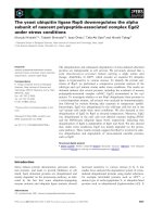
expression of HO-1 using western blotting. As shown in
Fig. 1, cells treated with rosiglitazone displayed a dose-
dependent increase in HO-1 expression. As compared
with control cells, 5 lm rosiglitazone caused a 12.1-fold
increase in protein expression of HO-1 (P < 0.01), sug-
gesting that activation of PPARc specifically and effec-
tively mediates HO-1 induction in PASMCs.
Role of HO-1 in PPARc agonist suppression of
proliferation of PASMCs
HO-1 has been found to be highly effective against
pulmonary hypertension, through vasodilating, inhibit-
ing the inflammatory response, and suppressing the
proliferation of PASMCs. At the same time, activation
HO-1
GAPDH
Con
Rosiglitazone
0
5
10
15
20
**
**
**
HO-1/GAPDH
Fold over control (AU)
Fig. 1. The PPARc agonist rosiglitazone induces HO-1 expression.
Primary cultured PASMCs were stimulated with different concen-
trations of rosiglitazone for 24 h. The expression of HO-1 was
determined using immune blotting. GAPDH was used as loading
control. Representative western blotting and quantification of bands
are shown (n = 3 in each group). **P < 0.01 versus control (Con).
PPARc inhibition of cell proliferation M. Li et al.
1544 FEBS Journal 277 (2010) 1543–1550 ª 2010 The Authors Journal compilation ª 2010 FEBS
of PPARc has also been shown to inhibit the prolifera-
tion of PASMCs and thus to ameliorate the develop-
ment of pulmonary hypertension. It is therefore
interesting to know whether induction of HO-1 medi-
ates the protective effect of PPARc against PASMC
proliferation. To test this, we applied serotonin to
stimulate PASMC proliferation, and then examined
whether knockdown of HO-1 by small interfering
RNA (siRNA) attenuated the effect of PPARc agonist
on cell proliferation. Figure 2A shows that PASMCs
stimulated with 5-HT (1 lm for 24 h) exhibited 4.21-
fold increase in DNA synthesis as assessed by [
3
H]thy-
midine incorporation assay (P < 0.01 as compared
with control), and pretreatment of cells with the
PPARc agonist rosiglitazone for 12 h dose-depen-
dently suppressed 5-HT-induced cell proliferation. At
5 lm, rosiglitazone fully inhibited 5-HT-triggered
DNA synthesis in cells (Fig. 2A). Figure 2B shows that
sequence-specific HO-1 siRNA transfection for 72 h
reduced basal HO-1 expression by 91% (P < 0.01 ver-
sus control), whereas nontargeting siRNA transfection
did not change the HO-1 level. More importantly, we
found that prior HO-1 knockdown by siRNA abol-
ished the inhibitory effect of rosiglitazone on the pro-
liferation of PASMCs induced by 5-HT (Fig. 2C),
whereas HO-1 knockdown alone did not impact on
basal or 5-HT-stimulated DNA synthesis in cells. Our
study indicates that induction of HO-1 mediates the
suppressive effect of PPARc agonist on PASMC
proliferation.
Role of p21
WAF1
in HO-1-mediated suppression of
proliferation of PASMCs
Recent studies have revealed that an antiproliferative
effect of HO-1 on non-PASMC pulmonary vascular
smooth muscle cells and other types of cells is associ-
ated with upregulation of the CDK inhibitor p21
WAF1
,
which is involved in negative regulation of cellular pro-
liferation [18,19]. We thus determined whether
increased HO-1 expression induced by PPARc agonist
could, in turn, trigger upregulation of p21
WAF1
, lead-
ing to an increase in its activity against PASMC prolif-
eration stimulated with 5-HT. Figure 3 shows that
PASMCs treated with rosiglitazone (5 lm for 24 h)
displayed a 2.34-fold increase in expression of p21
WAF1
(P < 0.01 as compared with control), whereas this
increase was dramatically blocked by prior knockdown
of HO-1, suggesting that HO-1 induction caused by
PPARc agonist is apparently involved in the upregula-
tion of p21
WAF1
in PASMCs. To further confirm this
observation functionally, we examined whether knock-
down of p21
WAF1
by siRNA transfection could reverse
the effect of PPARc agonist on suppression of
PASMC proliferation. We first applied sequence-spe-
cific siRNA to knock down expression of p21
WAF1
.As
shown in Fig. 4A, transfection of p21
WAF1
siRNA for
72 h produced an 82% reduction in p21
WAF1
protein
0
100
200
300
400
500
5-HT
0 0 0.5 1.5 5 µM
0 + + + + 1 µM
rosiglitazone
DNA synthesis
(% of control)
**
**
#
**
##
##
DNA synthesis
(% of control)
Con 5-HT HO-1 siRNA
Rosi
5-HT
Rosi
5-HT
HO-1
siRNA
HO-1 siRNA
5-HT
HO-1
GAPDH
0
100
200
300
400
500
600
**
##
**
##
**
††
0
0.5
1
1.5
HO-1/GAPDH
fold over control (AU)
**
Con HO-1 siRNA
Non-targeting
siRNA
C
B
A
Fig. 2. HO-1 mediates the inhibitory effect of the PPARc agonist
rosiglitazone (Rosi) on PASMC proliferation. (A) Primary cultured
PASMCs were stimulated 5-HT (1 l
M for 24 h), and this was
followed by labeling with [
3
H]thymidine (1 lCiÆmL
)1
for 12 h).
Rosiglitazone was added 12 h before stimulation of cells with 5-HT.
Cells were lysed, and cell-associated radioactivity was measured by
liquid scintillation counting. Summary data show that rosiglitazone
dose-dependently suppressed 5-HT-induced DNA synthesis (n =4
in each group). (B) Primary cultured PASMCs were transfected with
HO-1 sequence-specific siRNA (HO-1 siRNA) or nontargeting con-
trol siRNA for 72 h. Equal amounts of protein were loaded and
probed using specific HO-1 and GAPDH (loading control) antibodies.
Representative western blotting and quantification of HO-1 bands
are shown. (C) Prior knockdown of HO-1 by siRNA significantly
reversed the inhibitory effect of rosiglitazone on DNA synthesis in
5-HT-treated cells (n = 4 in each group). **P < 0.01 versus control;
#P < 0.05, ##P < 0.01 versus 5-HT-treated cells; P < 0.01 versus
rosiglitazone and 5-HT-treated cells.
M. Li et al. PPARc inhibition of cell proliferation
FEBS Journal 277 (2010) 1543–1550 ª 2010 The Authors Journal compilation ª 2010 FEBS 1545
level (P < 0.01 versus control), whereas nontargeting
control siRNA transfection did not change the
p21
WAF1
level in cells. Next, we investigated the influ-
ence of the loss of p21
WAF1
on the effect of PPARc
activation on suppression of cell proliferation. Fig-
ure 4B indicates that knockdown of p21
WAF1
by
siRNA transfection significantly reversed the inhibitory
effect of PPARc agonist on PASMC proliferation
induced by 5-HT. The DNA synthesis rate was
increased again from a 1.4-fold increase over control
in cells treated with PPARc agonist and 5-HT to a
4.04-fold increase over control in cells with p21
WAF1
siRNA silencing (despite the presence of PPARc ago-
nist and 5-HT), which is similar to that in cells stimu-
lated with 5-HT alone or cells treated with the
combination of HO-1 siRNA transfection, PPARc
agonist, and 5-HT. These results suggest that upregu-
lation of p21
WAF1
by HO-1 mediates the effect
of PPARc agonist in suppression of PASMC prolife-
ration.
Discussion
In this study, we demonstrate that activation of
PPARc by rosiglitazone induces significant HO-1
expression in primary cultured PASMCs, and this sub-
sequently upregulates the expression of p21
WAF1
, lead-
ing to inhibition of proliferation of PASMCs
stimulated with 5-HT. The present study provides a
P21
GAPDH
HO-1 siRNA
rosiglitazone
Con Rosiglitazone
0
1
2
3
p21/GAPDH
fold over control (AU)
**
##
Fig. 3. HO-1 mediates the effect of the PPARc agonist rosiglitaz-
one in upregulating p21
WAF1
expression. Primary cultured PASMCs
were treated with rosiglitazone (5 l
M), with or without prior knock-
down of HO-1, for 24 h. Expression of p21
WAF1
was determined
using immune blotting. GAPDH was used as loading control (Con).
Representative western blotting and quantification of bands are
shown (n = 4 in each group). **P < 0.01 versus control;
##P < 0.01 versus rosiglitazone-treated cells.
P21
GAPDH
0
0.2
0.4
0.6
0.8
1
1.2
**
p21/GAPDH
fold over control (AU)
p21 siRNACon
Non-targeting
siRNA
Con 5-HT Rosiglitazone
5-HT
HO-1 siRNA
rosiglitazone
5-HT
p21 siRNA
rosiglitazone
5-HT
DNA synthesis
(% of control)
0
100
200
300
400
500
600
**
**
††
##
**
††
B
A
Fig. 4. p21
WAF1
mediates the inhibitory
effect of HO-1 on PASMC proliferation.
(A) Primary cultured PASMCs were
transfected with p21
WAF1
sequence-specific
siRNA (p21 siRNA) or nontargeting control
siRNA for 72 h. Equal amounts of protein
were loaded and probed using specific
p21
WAF1
and GAPDH (loading control)
antibodies. Representative western blotting
and quantification of p21
WAF1
bands are
shown. (B) Primary cultured PASMCs with
or without prior p21
WAF1
or HO-1 siRNA
transfection were stimulated with 5-HT
(1 l
M for 24 h), and this was followed by
labeling with [
3
H]thymidine (1 lCiÆmL
)1
for
12 h). Rosiglitazone (5 l
M) was added 12 h
before stimulation of cells with 5-HT. Cells
were lysed, and cell-associated radioactivity
was measured by liquid scintillation counting
(n = 4 in each group). **P < 0.01 versus
control; ##P < 0.01 versus 5-HT-treated
cells; P < 0.01 versus cells treated with
rosiglitazone and 5-HT.
PPARc inhibition of cell proliferation M. Li et al.
1546 FEBS Journal 277 (2010) 1543–1550 ª 2010 The Authors Journal compilation ª 2010 FEBS
novel molecular mechanism by which PPARc activa-
tion suppresses PASMC proliferation and therefore
ameliorates the development of pulmonary hyperten-
sion. It also indicates that rosiglitazone might be useful
in the treatment of pulmonary hypertension.
Activation of PPARc by pharmacological ligands
has been shown to exert anti-inflammatory and anti-
proliferative effects on a variety of cell types, and thus
has potential value in the treatment of multiple
diseases [2,20–22]. Recent evidence from studies with
animal models indicates that the enhancing activity of
PPARc attenuates the development of pulmonary
hypertension [4,6,23]. Further studies suggest that acti-
vation of PPARc confers protection against pulmo-
nary hypertension by suppressing PASMC
proliferation. Proliferation of PASMCs is a hallmark
of pathogenesis of pulmonary hypertension [1,4]. How-
ever, the mechanisms responsible for inhibition of
PASMC proliferation by activation of PPARc are still
largely unknown. Recent studies have suggested that
induction of HO-1 mediates the effect of activation of
PPARc against proliferation of non-PASMCs and
endothelial cells [13]. In the present study, we show
that the synthetic PPARc agonist rosiglitazone dose-
dependently inhibited 5-HT-stimulated proliferation of
PASMCs, and that this was accompanied by a dose-
dependent increase in expression of HO-1. Knockdown
of HO-1 abolished the inhibitory effect of PPARc
agonist on PASMC proliferation, suggesting that
induction of HO-1 fully mediates this effect. Our study
not only confirms previous findings, but also extends
this notion to the pulmonary system.
Mammalian cell proliferation is controlled by a
group of cell cycle protein complexes consisting of two
key regulatory molecules: CDKs and cyclins [17,24].
A CDK molecule is activated by association with a
cyclin, forming a CDK complex. CDKs are constitu-
tively expressed in cells, whereas cyclins are synthesized
at specific stages of the cell cycle [25]. The expression
of a cyclin is regulated at the transcriptional and
degradation level to influence CDK activity [26]. In
addition, CDK activity is modulated by a group of
CDK inhibitors comprising two families of proteins:
inhibitor of kinase 4 ⁄ alternative reading frame and
CDK inhibitor protein ⁄ kinase inhibitor protein. The
inhibitor of kinase 4 ⁄ alternative reading frame family
includes p16INK4a and p14arf, which bind to CDK4
and arrest the cell cycle in the G
1
phase or prevent p53
degradation, respectively [27,28]. The CDK inhibitor
protein ⁄ kinase inhibitor protein family includes
p21
WAF1
, p27
Kip1
, and p57
Kip2
. They halt the cell cycle
in the G
1
phase by binding to, and inactivating,
cyclin–CDK complexes [29,30]. The results of our
study reveal that activation of PPARc increases
p21
WAF1
expression, and that this effect is significantly
blocked by prior knockdown of HO-1. This indicates
that PPARc agonist-induced HO-1 expression mediates
p21
WAF1
upregulation. We further confirmed this
observation functionally by using p21
WAF1
siRNA
silencing, when loss of p21
WAF1
significantly reversed
the inhibitory effect of PPARc agonist on cell proli-
feration. Our result is consistent with that of Pae [31],
showing that curcumin-induced HO-1 expression regu-
lates p21 expression in aortic smooth muscle cells. The
mechanisms underlying HO-1 induction of p21
WAF1
expression may be explained by accumulation of iron
and carbon monoxide, two key products of HO-1 [32].
Pulmonary hypertension and consequent cor pulmo-
nale, particularly secondary to chronic obstructive pul-
monary disease, are common clinical conditions and
some of the major causes of hospitalization and death
in patients with chronic obstructive pulmonary disease
[33,34]. Increased pulmonary vascular resistance caused
by pulmonary vasoconstriction and vascular remodel-
ing (prominent with vascular smooth muscle cell pro-
liferation) is the major basis for the development of
pulmonary hypertension [35,36]. Most drugs currently
used in the treatment of pulmonary hypertension are
vasodilators; few are aimed effectively against pulmo-
nary vascular remodeling [37,38], which is considered
to be a more critical mechanism for chronic pulmonary
hypertension [39]. Therefore, putative candidates to
modulate vascular remodeling have important poten-
tial applications in the treatment of pulmonary hyper-
tension. Rosiglitazone is a wildly used medicine with
beneficial effects in the long-term treatment of diabetic
mellitus [40]. Accumulated clinical experience and the
safety record of rosiglitazone suggest that this may be
an important chronic therapeutic approach for human
pulmonary hypertensive disease.
Experimental procedures
Cell preparation and culture
Primary smooth muscle cells from pulmonary arteries were
prepared from Sprague–Dawley rats (125–250 g) by the
method reported by Golovina et al. [41]. Isolated arterial
rings were incubated in Hanks’ balanced salt solution con-
taining 1.5 mg ÆmL
)1
collagenase II (Worthington, Lake-
wood, NJ, USA) for 20 min. After incubation, a thin layer
of the adventitia was carefully stripped off with fine
forceps, and the endothelium was removed by gently
scratching the intima surface with a surgical blade. The
remaining smooth muscle was then digested with
2.0 mgÆmL
)1
collagenase II and 0.5 mgÆmL
)1
elastase IV
M. Li et al. PPARc inhibition of cell proliferation
FEBS Journal 277 (2010) 1543–1550 ª 2010 The Authors Journal compilation ª 2010 FEBS 1547
(Sigma, St Louis, MO, USA) for 45 min at 37 °C. The cells
were plated onto 10 cm Petri dishes containing DMEM
(Invitrogen, Carlsbad, CA, USA) with 10% fetal bovine
serum, 100 UÆmL
)1
penicillin, and 100 lgÆmL
)1
strepto-
mycin, and cultured in a 37 °C ⁄ 5% CO
2
humidified incuba-
tor. Cells were passaged by trypsinization, using 0.05%
trypsin ⁄ EDTA (Invitrogen). All experiments were per-
formed using cells between passages 4 and 8. To test the
purity of smooth muscle cells, cells were stained with
4¢,6¢-diamidino-2-phenylindole (Invitrogen) and fluorescein
isothiocyanate-labeled antibody against smooth muscle
a-actin (Sigma), for nucleus and smooth muscle actin,
respectively. Fluorescence microscope images indicated that
cells contained more than 93% smooth muscle cells (data
not shown here). Before each experiment, cells were
incubated in 0.5% fetal bovine serum ⁄ DMEM for 24 h to
minimize serum-induced effects on the cells. 5-HT (Sigma)
was used to stimulate the proliferation of PASMCs. Rosig-
litazone (Cayman Chemical Co., Ann Arbor, MI, USA)
was used to stimulate PPARc activation.
siRNA transfection
To silence the expression of HO-1 and p21
WAF1
protein,
PASMCs were transfected with sequence-specific or nontar-
geting control siRNA (Dharmacon, Lafayette, CO, USA),
using Lipofectamine 2000 reagent (Invitrogen). Briefly, cells
were cultured up to 30–40% confluence, and siRNA and
Lipofectamine were diluted in serum-free DMEM sepa-
rately and incubated for 5 min at room temperature.
siRNA was mixed with Lipofectamine and incubated at
room temperature for 20 min. Then, the complex of siRNA
and Lipofectamine was added to cells, and culture was
maintained for 72 h at 37 °C and 5% CO
2
in a humidified
incubator. Cells were transfected for 24 h before the prepa-
ration of the [
3
H]thymidine incorporation assay. The effect
of protein silencing was analyzed using western blot.
Immunoblotting
Cells were lysed in 50 mm Tris ⁄ HCl (pH 7.4), 1% Nonidet
P-40, 0.1% SDS, 150 mm NaCl, 0.5% sodium deoxycho-
late, 1 mm EDTA, 1 mm phenylmethanesulfonyl fluoride,
1mm Na
3
VO
4
,1mm NaF, and proteinase inhibitors.
Lysates were centrifuged at 15 700 g at 4 °C for 15 min,
and the supernatant was collected as total protein. The pro-
tein concentration was determined with a bicinchoninic acid
protein assay kit (Pierce, Rockford, IL, USA). Protein was
separated on an SDS ⁄ PAGE gel, and transferred to a
Trans-Blot nitrocellulose membrane (Bio-Rad, Hercules,
CA, USA). Monoclonal antibodies against p21
WAF1
and
glyceraldehyde-3-phosphate dehydrogenase (GAPDH) and
polyclonal antibody against HO-1 (Millipore, Bedford,
MA, USA) were used according to the manufacturer’s
instructions. Horseradish peroxidase-conjugated goat anti-
(mouse IgG) and goat anti-(rabbit IgG) were used as sec-
ondary antibodies (Sigma). Reactions were developed with
SuperSignal West Pico Chemiluminescent Substrate (Pierce)
and exposure to autoradiographic film. Signaling was quan-
tified from scanned films using scion nih image software
(Scion, Frederick, MD, USA).
[
3
H]Thymidine incorporation assay
PASMCs were grown to 50–60% confluence in 24-well
plates, and serum starved for 24 h (0.5% fetal bovine serum
in DMEM) before the start of experiments. Cells were
treated with 1 lm 5-HT or vehicle for 24 h, and this was
followed by labeling with [
3
H]thymidine (1 lCiÆmL
)1
) for
12 h. PPARc agonist was added 12 h before the stimulation
of cells with serotonin. After labeling, cells were washed
twice with ice-cold NaCl ⁄ P
i
and incubated in 5% trichloro-
acetic acid for 30 min at 4 °C. Cell lysates were then
washed with ice-cold NaCl ⁄ P
i
and solubilized by adding
0.5 mL of 0.5 m NaOH ⁄ 0.5% SDS. Cell-associated radio-
activity was measured by liquid scintillation counting.
Statistics
Values are presented as mean ± standard deviation. Data
were analyzed using one-way ANOVA with Tukey post hoc
test. P < 0.05 was considered to represent significant
differences between groups.
Acknowledgements
This work was supported by the Chinese National
Science Foundation (30871116), the Tengfei Talent
Project of Xi’an Jiaotong University and the start-up
package to M. Li from the Second Affiliated Hospital
of Medical College of Xi’an Jiaotong University, PR
China.
References
1 Nisbet RE, Sutliff RL & Hart CM (2007) The role of
peroxisome proliferator-activated receptors in pulmo-
nary vascular disease. PPAR Res 2007, 18797.
2 Duan SZ, Usher MG & Mortensen RM (2008) Peroxi-
some proliferator-activated receptor-gamma-mediated
effects in the vasculature. Circ Res 102, 283–294.
3 Touyz RM & Schiffrin EL (2006) Peroxisome prolifera-
tor-activated receptors in vascular biology – molecular
mechanisms and clinical implications. Vascul Pharmacol
45, 19–28.
4 Hansmann G, de Jesus Perez VA, Alastalo TP, Alvira
CM, Guignabert C, Bekker JM, Schellong S, Urashima
T, Wang L, Morrell NW et al. (2008) An antiprolifera-
tive BMP-2 ⁄ PPARgamma ⁄ apoE axis in human and
PPARc inhibition of cell proliferation M. Li et al.
1548 FEBS Journal 277 (2010) 1543–1550 ª 2010 The Authors Journal compilation ª 2010 FEBS
murine SMCs and its role in pulmonary hypertension.
J Clin Invest 118, 1846–1857.
5 Ameshima S, Golpon H, Cool CD, Chan D, Vandivier
RW, Gardai SJ, Wick M, Nemenoff RA, Geraci MW
& Voelkel NF (2003) Peroxisome proliferator-activated
receptor gamma (PPARgamma) expression is decreased
in pulmonary hypertension and affects endothelial cell
growth. Circ Res 92, 1162–1169.
6 Hansmann G, Wagner RA, Schellong S, Perez VA,
Urashima T, Wang L, Sheikh AY, Suen RS, Stewart
DJ & Rabinovitch M (2007) Pulmonary arterial
hypertension is linked to insulin resistance and
reversed by peroxisome proliferator-activated
receptor-gamma activation. Circulation 115,
1275–1284.
7 Siow RC, Sato H & Mann GE (1999) Heme
oxygenase–carbon monoxide signalling pathway in
atherosclerosis: anti-atherogenic actions of bilirubin
and carbon monoxide? Cardiovasc Res 41, 385–
394.
8 Zhou H, Liu H, Porvasnik SL, Terada N, Agarwal A,
Cheng Y & Visner GA (2006) Heme oxygenase-1 medi-
ates the protective effects of rapamycin in monocrota-
line-induced pulmonary hypertension. Lab Invest 86,
62–71.
9 Idriss NK, Blann AD & Lip GY (2008) Hemoxygenase-
1 in cardiovascular disease. J Am Coll Cardiol 52,
971–978.
10 Stanford SJ, Walters MJ, Hislop AA, Haworth SG,
Evans TW, Mann BE, Motterlini R & Mitchell JA
(2003) Heme oxygenase is expressed in human pulmo-
nary artery smooth muscle where carbon monoxide has
an anti-proliferative role. Eur J Pharmacol 473,
135–141.
11 Christou H, Morita T, Hsieh CM, Koike H, Arkonac
B, Perrella MA & Kourembanas S (2000) Prevention of
hypoxia-induced pulmonary hypertension by enhance-
ment of endogenous heme oxygenase-1 in the rat. Circ
Res 86, 1224–1229.
12 Minamino T, Christou H, Hsieh CM, Liu Y, Dhawan
V, Abraham NG, Perrella MA, Mitsialis SA &
Kourembanas S (2001) Targeted expression of heme
oxygenase-1 prevents the pulmonary inflammatory and
vascular responses to hypoxia. Proc Natl Acad Sci USA
98, 8798–8803.
13 Kronke G, Kadl A, Ikonomu E, Bluml S, Furnkranz
A, Sarembock IJ, Bochkov VN, Exner M, Binder BR &
Leitinger N (2007) Expression of heme oxygenase-1 in
human vascular cells is regulated by peroxisome prolif-
erator-activated receptors. Arterioscler Thromb Vasc
Biol 27, 1276–1282.
14 Li M, Sun X, Li Z & Liu Y (2009) Inhibition of cGMP
phosphodiesterase 5 suppresses serotonin signalling in
pulmonary artery smooth muscle cells. Pharmacol Res
59, 312–318.
15 Bohm F & Pernow J (2007) The importance of endo-
thelin-1 for vascular dysfunction in cardiovascular dis-
ease. Cardiovasc Res 76, 8–18.
16 Gordon D, Reidy MA, Benditt EP & Schwartz SM
(1990) Cell proliferation in human coronary arteries.
Proc Natl Acad Sci USA 87, 4600–4604.
17 Vermeulen K, Van Bockstaele DR & Berneman ZN
(2003) The cell cycle: a review of regulation, deregula-
tion and therapeutic targets in cancer. Cell Prolif 36,
131–149.
18 Chang T, Wu L & Wang R (2008) Inhibition of vascu-
lar smooth muscle cell proliferation by chronic hemin
treatment. Am J Physiol Heart Circ Physiol 295, H999–
H1007.
19 Song R, Mahidhara RS, Zhou Z, Hoffman RA, Seol
DW, Flavell RA, Billiar TR, Otterbein LE & Choi AM
(2004) Carbon monoxide inhibits T lymphocyte prolifer-
ation via caspase-dependent pathway. J Immunol 172,
1220–1226.
20 Wayman NS, Hattori Y, McDonald MC, Mota-Filipe
H, Cuzzocrea S, Pisano B, Chatterjee PK & Thiemer-
mann C (2002) Ligands of the peroxisome proliferator-
activated receptors (PPAR-gamma and PPAR-alpha)
reduce myocardial infarct size. FASEB J 16, 1027–1040.
21 Zhao X, Strong R, Zhang J, Sun G, Tsien JZ, Cui Z,
Grotta JC & Aronowski J (2009) Neuronal PPAR-
gamma deficiency increases susceptibility to brain dam-
age after cerebral ischemia. J Neurosci 29, 6186–6195.
22 Li MY, Hsin MK, Yip J, Mok TS, Underwood MJ &
Chen GG (2010) PPAR{gamma} activation extinguishes
smoking carcinogen by inhibiting NNK-mediated prolif-
eration. Am J Respir Cell Mol Biol 42, 113–122.
23 Crossno JT Jr, Garat CV, Reusch JE, Morris KG,
Dempsey EC, McMurtry IF, Stenmark KR & Klemm
DJ (2007) Rosiglitazone attenuates hypoxia-induced
pulmonary arterial remodeling. Am J Physiol Lung Cell
Mol Physiol 292, L885–897.
24 Karpurapu M, Wang D, Singh NK, Li Q & Rao GN
(2008) NFATc1 targets cyclin A in the regulation of
vascular smooth muscle cell multiplication during reste-
nosis. J Biol Chem 283, 26577–26590.
25 Oredsson SM (2003) Polyamine dependence of normal
cell-cycle progression. Biochem Soc Trans 31, 366–370.
26 Welcker M, Singer J, Loeb KR, Grim J, Bloecher A,
Gurien-West M, Clurman BE & Roberts JM (2003)
Multisite phosphorylation by Cdk2 and GSK3 controls
cyclin E degradation. Mol Cell 12, 381–392.
27 Kim WY & Sharpless NE (2006) The regulation of
INK4 ⁄ ARF in cancer and aging. Cell 127, 265–275.
28 Gallagher SJ, Kefford RF & Rizos H (2006) The ARF
tumour suppressor. Int J Biochem Cell Biol 38,
1637–1641.
29 Besson A, Dowdy SF & Roberts JM (2008) CDK
inhibitors: cell cycle regulators and beyond. Dev Cell
14, 159–169.
M. Li et al. PPARc inhibition of cell proliferation
FEBS Journal 277 (2010) 1543–1550 ª 2010 The Authors Journal compilation ª 2010 FEBS 1549
30 Abbas T & Dutta A (2009) p21 in cancer: intricate net-
works and multiple activities. Nat Rev Cancer 9, 400–414.
31 Pae HO, Jeong GS, Jeong SO, Kim HS, Kim SA, Kim
YC, Yoo SJ, Kim HD & Chung HT (2007) Roles of
heme oxygenase-1 in curcumin-induced growth inhibition
in rat smooth muscle cells. Exp Mol Med 39, 267–277.
32 Gonzalez-Michaca L, Farrugia G, Croatt AJ, Alam J &
Nath KA (2004) Heme: a determinant of life and death
in renal tubular epithelial cells. Am J Physiol Renal
Physiol 286, F370–377.
33 Han MK, McLaughlin VV, Criner GJ & Martinez FJ
(2007) Pulmonary diseases and the heart. Circulation
116, 2992–3005.
34 Zielinski J, MacNee W, Wedzicha J, Ambrosino N,
Braghiroli A, Dolensky J, Howard P, Gorzelak K,
Lahdensuo A, Strom K et al. (1997) Causes of death in
patients with COPD and chronic respiratory failure.
Monaldi Arch Chest Dis 52, 43–47.
35 Stenmark KR, Fagan KA & Frid MG (2006) Hypoxia-
induced pulmonary vascular remodeling: cellular and
molecular mechanisms. Circ Res 99, 675–691.
36 Rabinovitch M (2007) Pathobiology of pulmonary
hypertension. Annu Rev Pathol 2, 369–399.
37 Dandel M, Lehmkuhl HB & Hetzer R (2005) Advances
in the medical treatment of pulmonary hypertension.
Kidney Blood Press Res 28, 311–324.
38 Benedict N, Seybert A & Mathier MA (2007) Evidence-
based pharmacologic management of pulmonary
arterial hypertension. Clin Ther 29, 2134–2153.
39 Tuder RM, Marecki JC, Richter A, Fijalkowska I &
Flores S (2007) Pathology of pulmonary hypertension.
Clin Chest Med 28, 23–42.
40 Balkrishnan R, Arondekar BV, Camacho FT,
Shenolikar RA, Horblyuk R & Anderson RT (2007)
Comparisons of rosiglitazone versus pioglitazone
monotherapy introduction and associated health care
utilization in medicaid-enrolled patients with type 2
diabetes mellitus. Clin Ther 29, 1306–1315.
41 Golovina VA & Blaustein MP (2006) Preparation of
primary cultured mesenteric artery smooth muscle
cells for fluorescent imaging and physiological studies.
Nat Protoc 1, 2681–2687.
PPARc inhibition of cell proliferation M. Li et al.
1550 FEBS Journal 277 (2010) 1543–1550 ª 2010 The Authors Journal compilation ª 2010 FEBS