Báo cáo khoa học: Effects of thymoquinone on isolated and cellular proteasomes pot
Bạn đang xem bản rút gọn của tài liệu. Xem và tải ngay bản đầy đủ của tài liệu tại đây (752.98 KB, 14 trang )
Effects of thymoquinone on isolated and cellular
proteasomes
Valentina Cecarini, Luana Quassinti, Alessia Di Blasio, Laura Bonfili, Massimo Bramucci,
Giulio Lupidi, Massimiliano Cuccioloni, Matteo Mozzicafreddo, Mauro Angeletti and
Anna Maria Eleuteri
School of Biosciences and Biotechnology, University of Camerino, Italy
Introduction
Black cumin seed (Nigella sativa) oil extracts have been
used for many centuries in the treatment of several
human diseases, and thymoquinone (TQ), its active
component, has recently been tested for its efficacy
against several diseases, including cancer [1–3].
In this regard, TQ was found to inhibit proliferation
in a concentration-dependent manner in numerous cell
lines [4,5]. It has shown significant antineoplastic activ-
ity against multidrug-resistant human pancreatic ade-
nocarcinoma, uterine sarcoma and leukemic cell lines,
with minimal toxicity for normal cells [6].
In a mouse model, the injection of the essential oil
into the tumor site significantly inhibited solid tumor
development as well as the incidence of liver metasta-
sis, thus improving mouse survival [5]. These results
indicate that the antitumor activity or cell growth inhi-
bition could in part be due to the effect of TQ on the
cell cycle [5]. Furthermore, it has been demonstrated
that the growth of prostate cancer cells is highly
sensitive to the inhibitory effect of TQ, and that this
inhibitory action is extremely selective, showing very
little effect on the growth of noncancerous prostate
epithelial cells in culture, and preventing the growth of
human prostate tumors in nude mice [7].
Despite awareness of these potential antineoplastic
effects, the molecular pathways involved are not
Keywords
apoptosis; glioblastoma; p53; thymoquinone;
ubiquitin proteasome system
Correspondence
V. Cecarini, School of Biosciences and
Biotechnology, University of Camerino, Via
Gentile III da Varano, 62032 Camerino (MC),
Italy
Fax: +39 0737 403290
Tel: +39 0737 403247
E-mail:
(Received 18 November 2009, revised
24 February 2010, accepted 26 February
2010)
doi:10.1111/j.1742-4658.2010.07629.x
Thymoquinone, a naturally derived agent, has been shown to possess anti-
oxidant, antiproliferative and proapoptotic activities. In the present study,
we explored thymoquinone effects on the proteasomal complex, the major
system involved in the removal of damaged, oxidized and misfolded pro-
teins. In purified 20S complexes, subunit-dependent and composition-depen-
dent inhibition was observed, and the chymotrypsin-like and trypsin-like
activities were the most susceptible to thymoquinone treatment. U87 MG
and T98G malignant glioma cells were treated with thymoquinone, and 20S
and 26S proteasome activity was measured. Inhibition of the complex was
evident in both cell lines, but predominantly in U87 MG cells, and was
accompanied by accumulation of ubiquitin conjugates. Accumulation of
p53 and Bax, two proteasome substrates with proapoptotic activity, was
observed in both cell lines. Our results demonstrate that thymoquinone
induces selective and time-dependent proteasome inhibition, both in isolated
enzymes and in glioblastoma cells, and suggest that this mechanism could
be implicated in the induction of apoptosis in cancer cells.
Abbreviations
AMC, 7-amino-4-methyl-coumarin; BrAAP, branched chain amino acid-preferring; ChT-L, chymotrypsin-like; ECL, enhanced
chemiluminescence; GAPDH, glyceraldehyde-3-phosphate dehydrogenase; MTT, 3-(4,5-dimethylthiazol-2-yl)-2,5-diphenyl-tetrazolium bromide;
pAB, 4-aminobenzoate; PGPH, peptidyl-glutamyl peptide-hydrolyzing; PVDF, poly(vinylidene difluoride);
Suc, succinyl; T-L, trypsin-like; TQ, thymoquinone; Ub, ubiquitin.
2128 FEBS Journal 277 (2010) 2128–2141 ª 2010 The Authors Journal compilation ª 2010 FEBS
completely clear. Recent findings suggest that TQ has
a strong chemopreventive potential for the inhibition
of carcinogenesis by modulating lipid peroxidation and
the cellular antioxidant milieu [8,9]. In fact, TQ is
reported to possess strong antioxidant properties,
inhibiting free radical generation [10]. Interestingly,
according to Gali-Muhtasib et al., TQ is able to trigger
apoptosis in several cell lines in a p53-independent or
a p53-dependent manner [11,12], and, as recently
shown, its proapoptotic effects are linked to its
pro-oxidant activity [13].
Among the different mechanisms involved in the
induction of apoptotic pathways, the tumor suppressor
protein p53 plays a pivotal role [14]. Under physiologi-
cal conditions, p53 is maintained at low steady-state
levels by the MDM2 protein, an E3 ubiquitin (Ub)
ligase, which ubiquitinates and targets p53 for protea-
some-mediated degradation [15]. Specific stress agents
make p53 and MDM2 undergo different post-transla-
tional modifications, including phosphorylation, thus
disrupting the interaction and leading to activation of
p53 [16]. At this point, p53 induces a series of down-
stream events that regulate the transcription of a sub-
set of genes involved in apoptosis, such as that
encoding Bax, a member of the Bcl-2 family [17].
The Ub–proteasome pathway is a nonlysosomal pro-
tein degradation system responsible for degrading both
damaged/unfolded proteins dangerous for normal cell
growth and metabolism [18], and critical regulatory
proteins involved in apoptosis [19], cell cycle regulation
[20], gene expression [21], carcinogenesis and DNA
repair [22–24]. Because of this, studies on the discovery
of molecules that are able to modulate proteasome
activity have recently been gaining great attention. The
central core of this system is the 20S proteasome. This
is a cylindrical structure with an internal cavity, com-
posed of four rings, each containing seven different
a subunits and b subunits, resulting in the following
arrangement: a
1–7
b
1–7
b
1–7
a
1–7
[19]. Only three of the
seven b subunits, b1, b2, and b5, located inside the
main chamber, show proteolytic activity. Specifically,
b1 is associated with the peptidyl-glutamyl peptide-
hydrolyzing (PGPH) activity and possesses limited
branched chain amino acid-preferring (BrAAP) activ-
ity, b2 is associated with the trypsin-like (T-L) activity,
and b5 is associated with the chymotrypsin-like (ChT-
L) activity. However, mutational analyses have shown
that b5 also has a tendency to cleave after small neu-
tral and branched side chains; therefore, two other
activities, BrAAP and small neutral amino acid-prefer-
ring (SNAAP), can be assigned to this subunit [25]. In
certain conditions, such as in the presence of c-inter-
feron, these three b subunits can be replaced by
homologous subunits, b1i, b2i, and b5i, resulting in a
de novo synthesized proteasomal form, the immuno-
proteasome, which produces mainly immunogenic pep-
tides in association with major histocompatibility
complex class I [19].
Malignant gliomas are the most common and lethal
tumors of the central nervous system [26]. Treatment
outcomes, even with an aggressive approach including
surgery, radiation therapy, and chemotherapy, are dis-
mal. The median survival of treated patients with glio-
blastoma multiforme is less than 1 year, with fewer
than 20% surviving for 2 years [27]. There is therefore
an urgent need to devise alternative therapeutic strate-
gies with which to fight gliomas.
In the present work, the effects of TQ on protea-
some functionality were investigated both in isolated
and in cellular complexes. For this purpose, constitu-
tive and immune-isolated proteasomes and two human
glioblastoma cell lines, U87 MG and T98G, differing
in their p53 gene status, were used. Specifically,
U87 MG cells present the wild-type form of p53,
whereas T98G cells harbor a single p53 mutation [28].
Results
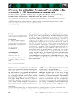
Nucleophilic susceptibility analysis
TQ was examined for sites of electrophilic and nucleo-
philic susceptibility. Computational analysis revealed
that TQ possessed two carbons (C
1
and C
4
) with simi-
lar nucleophilic susceptibility (Fig. 1) that are likely to
be the target of a nucleophilic attack [29].
TQ effects on isolated 20S proteasomes
To test TQ effects on isolated 20S constitutive
and immunoproteasome functionality, we incubated
purified enzymes with different concentrations of TQ
(0.0–100 lm). In particular, the ChT-L, T-L, PGPH
and BrAAP activities of the isolated complexes were
tested using specific substrates, as described in Experi-
mental procedures.
As shown in Fig. 2, it is possible to highlight that
TQ is able to modulate proteasome functionality indu-
cing a subunit and composition-dependent inhibition.
Of the two complexes, the immunoproteasome was the
most susceptible to the presence of TQ, and the ChT-
L and T-L activities were the components with the
highest degree of inhibition. The PGPH component
was not particularly altered in the presence of TQ;
only 16% inhibition was evident at 30 lm. Finally, the
BrAAP activity was not significantly influenced by the
presence of TQ (data not shown).
V. Cecarini et al. Thymoquinone inhibits proteasome functionality
FEBS Journal 277 (2010) 2128–2141 ª 2010 The Authors Journal compilation ª 2010 FEBS 2129
Interestingly, the inhibition showed concentration-
dependent behavior only up to 20 lm, when the maxi-
mum detectable rates of inhibition were 30% and 40%
for the ChT-L and T-L components, respectively, of
the immunoproteasome. Thereafter, an increase in TQ
concentrations did not lead to enhanced inhibition.
Supported by the literature [30], we propose that this
U-shaped inhibition depends on the presence of an
additional binding site on the proteasomal complex to
which TQ binds with a lower affinity than it does to
the active site. Our model assumes that TQ preferen-
tially binds to the active site at low concentrations,
resulting in the observed inhibition, whereas at higher
concentrations the binding to the additional site
becomes significant, allosterically restoring the activity.
The fraction of TQ bound to the active site is now
released, allowing the substrate to enter it and be suc-
cessfully degraded, resulting in the activity recovery
observed at TQ concentrations higher than 20 lm.To
verify this hypothesis, we performed an experiment
using the peptide aldehyde Z-LLF-CHO, a selective
and reversible proteasome inhibitor, with the aim of
blocking part of the proteasome active sites [31]. After
1 h of incubation of the 20S immunoproteasome with
Z-LLF-CHO, TQ at different concentrations was
added and the T-L activity was measured (Fig. 3). In
agreement with the mechanism described above, we
observed a recovery in the proteasome activity.
The Nitro Blue tetrazolium assay, which monitors
the formation of quinone adducts, shows the existence
of additional TQ-binding sites on the proteasome.
Figure 4 indicates that the formation of b-subunit–TQ
adducts increases at TQ concentrations of 5 and
20 lm, whereas it decreases at a concentration of
100 lm (corresponding to the recovery of proteasome
activity). At the same time, increases in TQ concentra-
tion resulted in clear enhancement in the levels of
a-subunit–TQ adducts, confirming our model of the
presence of two different TQ-binding sites on the pro-
teasome complex.
TQ inhibits cell proliferation
Two cell lines, U87 MG and T98G, derived from
human glioblastomas were used as a model. They
Fig. 2. Effects of increasing TQ concentrations (0–100 lM) on iso-
lated 20S complexes. The ChT-L, T-L and PGPH activities were
assayed. r, constitutive proteasome;
, immunoproteasome.
ABC
Lowest
susceptibility
Highest
susceptibility
Fig. 1. Chemical structure and nucleophilic susceptibility of TQ. Chemical structure (A), nucleophilic susceptibility (B) and electrophilic
susceptibility (C) of TQ. Isosurfaces were calculated with
WEBMO.C
1
and C
4
carbonyl were found to be nucleophilically attacked by the OH
group of Thr1.
Thymoquinone inhibits proteasome functionality V. Cecarini et al.
2130 FEBS Journal 277 (2010) 2128–2141 ª 2010 The Authors Journal compilation ª 2010 FEBS
carry, respectively, the wild-type and a mutant p53
gene. This mutation consists of a single G fi A transi-
tion in codon 237, resulting in a missense mutation of
methionine to isoleucine [32,33]. Interestingly, a study
conducted by Van Meir et al. on different glioblastoma
lines and their p53 status revealed that this mutation
in the T98G line results in a transcriptionally inactive
form of p53 [34].
A set of dose–response experiments was performed
to compare the effects of TQ on cell viability in
U87 MG and T98G cells. Cells were incubated in the
presence of TQ at concentrations ranging from 0.0 lm
to 200 lm. Analysis by light microscopy showed that
treatment of glioblastoma cells with increasing
amounts of TQ resulted in significant alterations in
cell morphology and impaired the ability of the cells
to become confluent (Fig. 5A). Data obtained with
the 3-(4,5-dimethylthiazol-2-yl)-2,5-diphenyl-tetrazo-
lium bromide (MTT) assay indicated that cell viability
was significantly reduced in a dose-dependent and
exposure time-dependent manner in both cell lines
(Fig. 5B). In both cell lines, almost complete loss of
viability was seen after exposure to 200 lm TQ. At
lower concentrations, TQ exerted a stronger inhibitory
effect on U87 MG cells than on T98G cells. A com-
parison of IC
50
values, reported in Table 1, showed
that, after 48 h of treatment with TQ, IC
50
values
were 38.82 lm for U87 MG cells and 62.48 lm for
T98G cells.
TQ effects on the proteasome functionality of
glioblastoma cells
Considering the major role of the proteasome in medi-
ating numerous cellular pathways, including apoptosis,
we wanted to determine whether TQ was able to mod-
ulate its functionality in the two glioblastoma cell lines.
Cells were treated with TQ at 20 lm, the concentration
with the greatest effects on isolated proteasomes, for
12, 24, 48 and 72 h. Control cells were cultured in par-
allel in the presence of dimethylsulfoxide. Both cell
lines had a high level of responsiveness to TQ treat-
ment, showing compromised activities as compared
with controls (Figs 6 and 7). Parallel assays run in the
presence of specific proteasome inhibitors, Z-GPFL-
CHO and lactacystin, demonstrated that the contribu-
tion to the proteolysis was effectively due to the 20S
proteasome (data not shown).
Figures 5 and 6 illustrate the presence of time-
dependent proteasome inhibition, which assumes par-
ticular significance after 48 and 72 h of treatment.
Interestingly, U87 MG cells showed a higher extent of
proteasome inhibition, with relevant differences also at
24 h, as evident for the T-L and BrAAP activities.
Generally, in this cell line, TQ induced a global and
stronger decrease in proteasome functionality than that
observed in T98G cells.
We also measured the ChT-L component of the 26S
proteasome, whose proteolytic activity is ATP-depen-
dent, and obtained, at 72 h, similar percentages of inhi-
bition in the two lines. However, at 48 h, a significant
Fig. 3. TQ binding to a secondary site of the proteasome complex.
After Z-LLF-CHO and 20S immunoproteasome preincubation, in
order to partially inhibit the enzyme, the effects of increasing con-
centrations of TQ on the T-L activity were tested. Data are reported
as percentages relative to proteasome activity in the presence of Z-
LLF-CHO (mean values ± standard deviations of five independent
determinations).
A
B
Fig. 4. Detection of quinone adducts. 20S isolated immunoprotea-
somes were treated with different concentrations of TQ and lacta-
cystin (see Experimental procedures), resolved by SDS ⁄ PAGE, and
electroblotted onto PVDF membranes. Adducts were visualized
after 45 min of incubation with Nitro Blue tetrazolium. Lane C rep-
resents 20S proteasome loaded without pretreatment with TQ and
lactacystin. (A) Densitometry related to three different experiments.
(B) A representative membrane after the Nitro Blue tetrazolium
staining.
V. Cecarini et al. Thymoquinone inhibits proteasome functionality
FEBS Journal 277 (2010) 2128–2141 ª 2010 The Authors Journal compilation ª 2010 FEBS 2131
difference after TQ exposure was evident for U87 MG
cells.
To verify the above-mentioned proteasome inhibi-
tion, we conducted western blot analyses, using anti-
bodies against Ub. In fact, the abnormal presence of
Ub conjugates is a clear marker of impaired protea-
some activity. Our findings demonstrate time-depen-
dent accumulation of Ub–protein aggregates,
confirming the data on proteasome inhibition (Fig. 8).
Furthermore, western blot assays performed with an
antibody against 20S suggested that the observed inhi-
bition was really due to compromised complex func-
tionality and not to downregulation of its synthesis
(Fig. 9). These results support the findings regarding
the ability of TQ to act directly on the proteasome
activity, and remove the possibility of decreased syn-
thesis of the enzyme.
TQ effects on p53 and Bax levels
In order to strengthen the data on proteasome inhibi-
tion, we measured the levels of two proteasome
substrates, p53 and Bax, that play an important role in
the onset of apoptotic events.
In both cell lines, time-dependent accumulation of
p53 was observed. In T98G cells, this increase was sig-
nificant even after 24 h of treatment, but was particu-
larly evident at 48 h and 72 h (levels that are 2.3-fold
and a 2.8-fold higher, respectively, than in controls).
In U87 MG cells, instead, the enhancement in protein
levels was delayed, and became consistent only after
48 h of TQ exposure (Fig. 10). Bax accumulation was
more evident in T98G cells than in U87 MG cells. Spe-
cifically, the former responded in a shorter time, with
significant increases at 48 h and 72 h (1.21-fold and
1.42-fold, respectively, that seen in controls), whereas
the latter presented significant enhancement only at
72 h, with a 1.44-fold increase as compared with the
respective control (Fig. 11).
Discussion
The debate on the use of naturally derived drugs as
coadjuvants in the treatment of cancer is of growing
interest. In fact, owing to concerns about the possible
A
B
Fig. 5. TQ effects on U87 MG and T98G
cells. (A) Morphology of U87 MG and T98G
cells grown under standard conditions and
treated with 50 l
M or 100 lM TQ dissolved
in dimethylsulfoxide. Dimethylsulfoxide con-
centrations in treated and control cells did
not exceed 0.25% per well. Cells were
observed by using an inverted microscope
24 h post-treatment. (B) Dose–response
curve for the effect of TQ on cell viability
after 24, 48 and 72 h of exposure. Cell via-
bility was determined by the MTT assay,
and is reported as the percentage of viable
cells. Each value is the mean ± standard
deviation of three separate experiments
performed in triplicate.
Table 1. Thymoquinone IC
50
values for glioma cell lines after incu-
bation periods of 24, 48 and 72 h. CI, confidence interval.
Incubation
period (h)
IC
50
(lM) (95% CI)
T98G U87 MG
24 77.73 (71.93–83.99) 47.08 (41.84–52.97)
48 62.48 (57.81–67.53) 38.82 (35.37–41.26)
72 61.46 (58.58–64.48) 35.83 (31.64–38.35)
Thymoquinone inhibits proteasome functionality V. Cecarini et al.
2132 FEBS Journal 277 (2010) 2128–2141 ª 2010 The Authors Journal compilation ª 2010 FEBS
toxic side effects of conventional medicine, the use of
natural products as alternatives to such treatments has
been increasing. TQ is the most abundant constituent
of N. sativa, and has pivotal roles in several biological
processes. Numerous studies have demonstrated the
antioxidant, antiproliferative and proapoptotic
activities of TQ. Most notably, TQ is able to induce
selective apoptosis, discriminating between tumor and
normal cells, in a p53-dependent or p53-independent
way. For example, previous published data established
that osteosarcoma cells [4] and neoplastic keratino-
cytes [35] are susceptible to TQ treatment, whereas
normal cells and mouse primary keratinocytes do not
exhibit morphological and ⁄ or proliferative alterations
[4,35].
The observation that proteasome inhibitors are able
to induce apoptosis in tumor cells opened the possibil-
ity of their use as potential drugs, and numerous stud-
ies have been conducted with the aim of finding
natural, nontoxic and inexpensive compounds [36–38].
Fig. 6. 20S and 26S proteasome functionality in U87 MG cells treated with 20 lM TQ. Activities were assayed as reported in Experimental
procedures. Data are expressed as percentage of activity relative to control cells in each set (mean values ± standard deviations of five inde-
pendent determinations). Fluorescence due to nonproteasomal degradation was subtracted. The asterisks indicate data points that are statis-
tically significant as compared with the respective untreated control cells (*P < 0.05, **P < 0.01).
V. Cecarini et al. Thymoquinone inhibits proteasome functionality
FEBS Journal 277 (2010) 2128–2141 ª 2010 The Authors Journal compilation ª 2010 FEBS 2133
In this scenario, we decided to investigate the possible
interaction between TQ and proteasomes in order to
determine whether TQ could modulate the enzyme
functionality.
Considering the data obtained from computational
analysis, it is reasonable to think that TQ could
behave as a nucleophilic target, resulting in inhibition
of proteasome activity. To confirm this hypothesis, we
tested proteasome functionality after TQ treatment of
both isolated and cellular complexes. Interestingly, we
observed subunit-dependent and composition-depen-
dent inhibition of both the purified enzymes, with the
immunoproteasome being the most sensitive and the
ChT-L and T-L components being the most influenced
activities. We also demonstrated that TQ induces a
U-shaped inhibition in proteasome complexes through
the binding of two distinct sites with different degrees
of affinity.
Exposure of two human glioblastoma cell lines,
U87 MG and T98G, to TQ was able to significantly
Fig. 7. 20S and 26S proteasome functionality in T98G cells treated with 20 lM TQ. Activities were assayed as reported in Experimental pro-
cedures. Data are expressed as percentage of activity remaining relative to control cells in each set (mean values ± standard deviations of
five independent determinations). Fluorescence due to nonproteasomal degradation was subtracted. The asterisks indicate data points that
are statistically significant as compared with the respective untreated control cells (*P < 0.05, **P < 0.01).
Thymoquinone inhibits proteasome functionality V. Cecarini et al.
2134 FEBS Journal 277 (2010) 2128–2141 ª 2010 The Authors Journal compilation ª 2010 FEBS
compromise proteasome activity. The two cell lines are
different with respect to a single mutation in the p53
gene; this characterizes the T98G line, whereas the
U87 MG line maintains the wild-type form of the pro-
tein. As previously shown by other authors, this muta-
tion results in a transcriptionally inactive p53 gene
[34].
Assaying TQ effects on cell viability, we found that
both cell lines showed clear changes in cell morphol-
ogy, although with different degrees of sensitivity. In
fact, U87 MG cells were more susceptible to the
treatment, as shown by the different IC
50
values
obtained after the treatments.
Cells were then treated with TQ at 20 lm, the con-
centration with the highest effect according to the
in vitro data, and both 20S and 26S proteasomes
showed changes in their functionality. In particular,
our assays showed significant, time-dependent but dif-
ferential sensitivities of U87 MG and T98G cells to
TQ treatment. T-L, BrAAP and PGPH activities were
significantly more affected in U87 MG cells than in
T98G cells, with the former showing altered protea-
some functionality at 24 h. This inhibition was also
confirmed by accumulation of Ub–protein conjugates.
Furthermore, when we tested the 20S expression levels
with specific antibodies, we could not detect any differ-
ences between control and treated cells, demonstrating
the ability of TQ to directly alter proteasome activity
without affecting its synthesis.
Considering our data, it is clear that TQ is able to
modulate proteasome activity, inducing global inhibi-
tion in the studied models, although to different
extents. These results are in line with previously pub-
lished data from our laboratory and others reporting
on the ability of small, naturally derived ligands, e.g.
flavonoids, to inhibit proteasome functionality and
selectively modulate its activity, depending on the
subunit composition [37,39,40].
It has been widely reported that the proteasome,
being responsible for the removal of proapoptotic
A
B
C
Fig. 8. Detection of Ub–protein conjugates in U87 MG and T98G cells. The densitometric analysis from five separate blots, shown as mean
values ± standard deviations, and a representative western blot are shown (A, B). Membranes were reprobed with GAPDH antibody to
ensure equal protein loading (C). Detection was performed with an ECL western blotting analysis system. The asterisks indicate data points
that are statistically significant as compared with the respective untreated control cells (*P < 0.05, **P < 0.01).
V. Cecarini et al. Thymoquinone inhibits proteasome functionality
FEBS Journal 277 (2010) 2128–2141 ª 2010 The Authors Journal compilation ª 2010 FEBS 2135
proteins, is involved in the induction of programmed
cell death [19]. Its inhibition, in fact, triggers the accu-
mulation of proteins such as p53 and Bax [41–43]. For
this reason, numerous compounds with the ability to
modulate proteasome activity have been used in the
treatment of malignancies.
A
B
C
Fig. 9. Detection of the 20S ‘core’ in U87 MG and T98G cells. The densitometric analysis from five separate blots, shown as mean val-
ues ± standard deviations, and a representative western blot are shown (A, B). Membranes were reprobed with GAPDH antibody to ensure
equal protein loading (C). Detection was performed with an ECL western blotting analysis system.
A
B
C
Fig. 10. Detection of p53 in U87 MG and T98G cells. The densitometric analysis from five separate blots, shown as mean values ± standard
deviations, and a representative western blot are shown (A, B). Membranes were reprobed with GAPDH antibody to ensure equal protein
loading (C). Detection was performed with an ECL western blotting analysis system. The asterisks indicate data points that are statistically
significant as compared with the respective untreated control cells (*P < 0.05, **P < 0.01).
Thymoquinone inhibits proteasome functionality V. Cecarini et al.
2136 FEBS Journal 277 (2010) 2128–2141 ª 2010 The Authors Journal compilation ª 2010 FEBS
Our results on the accumulation of both p53 and
Bax are in line with the data describing the ability of
TQ to inhibit proteasome activity. These two proapop-
totic proteins are proteasome substrates, and their
intracellular levels increase together with proteasome
malfunctions. It is therefore likely that one of the
mechanisms through which TQ triggers apoptosis in
cancer cells is the induction of proteasome inhibition.
In summary, our data demonstrate that TQ is able
to modulate proteasome functionality, inducing compo-
sition-dependent inhibition both in isolated complexes
and in glioblastoma cells. This inhibition leads to intra-
cellular increases in the levels of apoptotic proteins
such as p53 and Bax, and may be linked to the onset of
apoptotic events. Such findings represent evidence that
this compound, characterized by very low toxicity,
deserves further clinical analysis and investigation,
mostly for its potential application as an adjuvant in
the treatment of cancer and other diseases.
Experimental procedures
Reagents and chemicals
Thymoquinone, substrates for assaying the ChT-L, T-L
and PGPH activities [succinyl (Suc)-Leu-Leu-Val-Tyr-7-
amino-4-methyl-coumarin (AMC), Z-Leu-Ser-Thr-Arg-
AMC, and Z-Leu-Leu-Glu-AMC], proteasome inhibitors
(Z-Gly-Pro-Phe-Leu-CHO and lactacystin), Nitro Blue Tet-
razolium and MTT were purchased from Sigma-Aldrich
S.r.L. (Milan, Italy). The substrate Z-Gly-Pro-Ala-Phe-
Gly-4-aminobenzoate (pAB), for testing BrAAP activity,
and the proteasome inhibitor Z-LLF-CHO (Cbz-Leu-
Leu-Phe-CHO) were kind gifts from M. Orlowski (Depart-
ment of Pharmacology, Mount Sinai School of Medicine,
New York, NY, USA). Aminopeptidase N (EC 3.4.11.2)
for the coupled assay utilized to detect BrAAP activity
[44] was purified from pig kidney as reported elsewhere
[45,46]. TQ was dissolved in dimethylsulfoxide (Sigma
Aldrich S.r.l.). U87 MG and T98G human glioblastoma
cell lines were purchased from the American Type Culture
Collection (ATCC, Manassas, VA, USA). All of the
reagents for cell cultures were obtained from Euroclone
(Milan, Italy). Rabbit anti-(human 20S proteasome)
serum, rabbit anti-(human 20S proteasome b5 subunit)
serum and mouse anti-[human 20S a(1, 2, 3, 5, 6, and 7)
subunits] serum were purchased from BIOMOL Interna-
tional, L.P. The mouse monoclonal antibodies against Ub,
p53 and Bax were obtained from Santa Cruz Biotechnol-
ogy, Inc. (Heidelberg, Germany). Membranes for western
blot analyses were purchased from Millipore (Milan,
Italy). Proteins immobilized on films were detected with
the enhanced chemiluminescence (ECL) system (Amersham
Pharmacia Biotech, Milan, Italy). All chemicals and sol-
vents were of the highest analytical grade available.
A
B
C
Fig. 11. Detection of Bax in U87 MG and T98G cells. The densitometric analysis from five separate blots, shown as mean values ± standard
deviations, and a representative western blot are shown (A, B). Membranes were reprobed with GAPDH antibody to ensure equal protein
loading (C). Detection was performed with an ECL western blotting analysis system. The asterisks indicate data points that are statistically
significant as compared with the respective untreated control cells (*P < 0.05, **P < 0.01).
V. Cecarini et al. Thymoquinone inhibits proteasome functionality
FEBS Journal 277 (2010) 2128–2141 ª 2010 The Authors Journal compilation ª 2010 FEBS 2137
Nucleophilic susceptibility analysis
The frontier electron density isosurfaces of TQ were cre-
ated using webmo [47], by performing a Gaussian ab initio
and semiempirical calculation of nuclear susceptibility
analysis using the pm3 wavefunction. Electrophilic
(HOMO) and nucleophilic (LUMO) frontier density sur-
faces were computed from the magnitudes of molecular
orbitals available for attack by an electrophile or a nucle-
ophile. The results are represented as a ‘bull’s eye’ pattern,
with blue representing the highest probability of an
attack.
Measurements of isolated 20S proteasome activity
To evaluate the effects of TQ on the 20S constitutive and
immunoproteasome peptidase activities, in vitro assays were
performed with fluorogenic peptides. Suc-Leu-Leu-Val-Tyr-
AMC was used for ChT-L activity, Z-Leu-Ser-Thr-Arg-
AMC for T-L activity, Z-Leu-Leu-Glu-AMC for PGPH
activity, and Z-Gly-Pro-Ala-Phe-Gly-pAB for BrAAP activ-
ity [48–50]. Isolation and purification of the 20S protea-
some from bovine brain and thymus were performed as
previously reported [50,51]. The incubation mixture con-
tained TQ at concentrations ranging from 0.0 to 100.0 lm,
1 lg of the isolated 20S proteasomes, the appropriate sub-
strate, and 50 mm Tris ⁄ HCl (pH 8.0), up to a final volume
of 100 lL. Incubation was performed at 37 °C, and after
60 min the fluorescence of the hydrolyzed 7-amino-
4-methyl-coumarin (AMC) and 4-aminobenzoic acid (pAB)
was detected (AMC, k
exc
= 365 nm, k
em
= 449 nm; pAB,
k
exc
= 304 nm, k
em
= 664 nm) on a SpectraMax Gemini
XPS microplate reader.
To test the presence of a TQ secondary binding site on
the proteasome complex, 1 lg of isolated 20S immunopro-
teasome was preincubated with 3 lm Z-LLF-CHO for 1 h
at 37 °C. Then, TQ at different concentrations (0.0–
200 lm) and the appropriate substrate for testing the T-L
activity were added. After 60 min, the hydrolyzed AMC
was detected on a SpectraMax Gemini XPS microplate
reader.
Detection of proteasome–quinone adducts
Detection of the TQ-mediated formation of quinone
adducts in isolated 20S immunoproteasomes was performed
as described by Gallop et al. [52]. Twenty micrograms of
purified complex was preincubated for 1 h at 37 °C with
different concentrations of lactacystin (0, 2.5, 5 and 10 lm).
Then, TQ (5, 20 and 100 lm) was added to the mixtures
and incubated for 1 h at 37 °C. Samples were then resolved
by 12% SDS ⁄ PAGE and electroblotted onto poly(vinyli-
dene difluoride) (PVDF) membranes. The detection was per-
formed by staining the membrane with Nitro Blue
tetrazolium (0.24 mm in 2 m potassium glycinate, pH 10)
for 45 min in the dark. As internal control, 20 lg of iso-
lated immunoproteasome was loaded and stained without
treatment with either TQ or lactacystin. Proteasome
a subunits and b subunits were identified by staining the
same membrane with a primary antibody specific for the
20S a1, a2, a3, a5, a6 and a7 subunits, and with a primary
antibody specific for the b5 subunit, respectively.
Cell culture
T98G and U87 MG cells were maintained in EMEM
with 2 mml-glutamine, 0.1 mm nonessential amino
acids, 1 mm sodium pyruvate, 100 IUÆmL
)1
penicillin G,
and 100 lgÆmL
)1
streptomycin, supplemented with 10%
heat-inactivated fetal bovine serum. Cells were maintained
in a 5% CO
2
atmosphere at 37 °C.
Cell viability assay
Cell viability was determined by the standard MTT assay
[53]. Cells were seeded at an initial density of 2 · 10
4
cellsÆmL
)1
in 96-well microtiter plates (Iwaki, Tokyo,
Japan) in 100 lL of growth medium. After incubation for
24 h at 37 °C, cells were exposed to different concentrations
of TQ (0.0–200 lm) containing 0.25% dimethylsulfoxide,
which was applied as a control, for 24, 48 and 72 h in a
humidified atmosphere at 37 °C in the presence of 5%
CO
2
. Cell viability was then quantified by the ability of
living cells to reduce the yellow dye MTT to a purple
formazan product. Cells were incubated with MTT for 4 h,
the medium was replaced with 100 lL of dimethylsulfoxide,
and the attenuance was measured with a Titertek Multiscan
microElisa microplate spectrophotometer reader (Labsys-
tems, Helsinki, Finland) at 540 nm. The IC
50
values were
determined using graphpad prism 4 (GraphPad Software,
San Diego, CA, USA).
TQ treatment
Cells were grown in 100 mm tissue culture dishes at an ini-
tial concentration of 2 · 10
4
cells per dish, and were then
exposed to 20 lm TQ for 12, 24, 48 and 72 h. Control
treatments were performed in the presence of dimethylsulf-
oxide for each time point. After removal of the medium
and washing with cold NaCl ⁄ P
i
, cells were harvested in
4 mL of NaCl ⁄ P
i
and centrifuged at 1600 g for 5 min. The
pellet was resuspended in lysis buffer (20 mm Tris, pH 7.4,
250 mm sucrose, 1 mm EDTA, and 5 mm b-mercaptoetha-
nol), and passed through a 25-gauge needle at least 10
times. Lysates were centrifuged at 12 000 g for 15 min, and
the supernatants were stored at )80 °C. The protein
concentration in cell lysates was determined by the method
of Bradford [54], using BSA as standard.
Thymoquinone inhibits proteasome functionality V. Cecarini et al.
2138 FEBS Journal 277 (2010) 2128–2141 ª 2010 The Authors Journal compilation ª 2010 FEBS
Measurements of proteasome activities in cell
lysates
Proteasome peptidase activities in cell lysates (1 lg in the
mixture) were determined with fluorogenic peptides, as previ-
ously described. The 26S proteasome ChT-L activity was
tested using Suc-Leu-Leu-Val-Tyr-AMC as substrate, and a
50 mm Tris ⁄ HCl (pH 8.0) buffer containing 10 mm MgCl
2
,
1mm dithiothreitol, and 2 m m ATP. In order to evaluate
the effective 20S proteasome contribution to the short pep-
tide cleavage, control experiments were performed using spe-
cific proteasome inhibitors, Z-Gly-Pro-Phe-Leu-CHO and
lactacystin (5 lm in the reaction mixture). Fluorescence val-
ues obtained by analyzing the lysates were then subtracted
from the values of control assays in the presence of the two
inhibitors to find the effective proteasome contribution.
BrAAP activity was determined in a coupled test in the pres-
ence of aminopeptidase N [49]. Incubation was performed at
37 °C for 60 min. The fluorescence of hydrolyzed AMC and
pAB was then measured (AMC, k
exc
= 365 nm,
k
em
= 449 nm; pAB, k
exc
= 304 nm, k
em
= 664 nm) on a
SpectraMax Gemini XPS microplate reader.
Western blot analysis
Cell lysates were resolved by 12% SDS ⁄ PAGE and elec-
troblotted onto PVDF membranes. Membranes with trans-
ferred proteins were incubated with the mouse monoclonal
antibodies against p53 and Bax, and with the polyclonal
rabbit anti-(human 20S proteasome) serum. Cell lysates
resolved by 10% SDS ⁄ PAGE were electroblotted and then
incubated with mouse monoclonal antibody against Ub.
The immunoblot detection was performed with an ECL
western blotting analysis system, using peroxidase-conju-
gated secondary antibodies (Santa Cruz Biotechnology).
Each gel was loaded with molecular mass markers, includ-
ing proteins with molecular masses from 6.5 kDa to
205 kDa (SigmaMarker – Wide Molecular Weight Range;
Sigma-Aldrich S.r.l.). Glyceraldehyde-3-phosphate dehydro-
genase (GAPDH) was utilized as a control for equal pro-
tein loading: membranes were stripped and reprobed for
GAPDH using a monoclonal antibody diluted 1 : 500
(Santa Cruz Biotechnology Inc.). The bands were quantified
as reported elsewhere [55,56].
Statistical analysis
Values are expressed as mean values and standard deviation
of results obtained from separate experiments. Student’s
t-test was used to compare differences between the means
of control and treated groups. Statistical tests were
performed with sigma-stat 3.1 software (SPSS, Chicago,
IL, USA). P-values < 0.05 and < 0.01 were considered to
be significant.
References
1 Edris AE (2009) Anti-cancer properties of Nigella spp.
essential oils and their major constituents, thymoqui-
none and beta-elemene. Curr Clin Pharmacol 4, 43–46.
2 Yi T, Cho SG, Yi Z, Pang X, Rodriguez M, Wang Y,
Sethi G, Aggarwal BB & Liu M (2008) Thymoquinone
inhibits tumor angiogenesis and tumor growth through
suppressing AKT and extracellular signal-regulated
kinase signaling pathways. Mol Cancer Ther 7 , 1789–
1796.
3 Padhye S, Banerjee S, Ahmad A, Mohammad R &
Sarkar FH (2008) From here to eternity – the secret of
Pharaohs: therapeutic potential of black cumin seeds
and beyond. Cancer Ther 6, 495–510.
4 Shoieb AM, Elgayyar M, Dudrick PS, Bell JL & Tithof
PK (2003) In vitro inhibition of growth and induction
of apoptosis in cancer cell lines by thymoquinone. Int J
Oncol 22, 107–113.
5 Ait Mbarek L, Ait Mouse H, Elabbadi N, Bensalah M,
Gamouh A, Aboufatima R, Benharref A, Chait A,
Kamal M, Dalal A et al. (2007) Anti-tumor properties
of blackseed (Nigella sativa L.) extracts. Braz J Med
Biol Res 40, 839–847.
6 Worthen DR, Ghosheh OA & Crooks PA (1998) The
in vitro anti-tumor activity of some crude and purified
components of blackseed, Nigella sativa L. Anticancer
Res 18, 1527–1532.
7 Kaseb AO, Chinnakannu K, Chen D, Sivanandam A,
Tejwani S, Menon M, Dou QP & Reddy GP (2007)
Androgen receptor and E2F-1 targeted thymoquinone
therapy for hormone-refractory prostate cancer. Cancer
Res 67, 7782–7788.
8 Badary OA, Al-Shabanah OA, Nagi MN, Al-Rikabi
AC & Elmazar MM (1999) Inhibition of benzo(a)py-
rene-induced forestomach carcinogenesis in mice by
thymoquinone. Eur J Cancer Prev 8, 435–440.
9 Badary OA, Abd-Ellah MF, El-Mahdy MA, Salama
SA & Hamada FM (2007) Anticlastogenic activity of
thymoquinone against benzo(a)pyrene in mice. Food
Chem Toxicol 45, 88–92.
10 Mansour MA, Nagi MN, El-Khatib AS & Al-Bekairi
AM (2002) Effects of thymoquinone on antioxidant
enzyme activities, lipid peroxidation and DT-diaphorase
in different tissues of mice: a possible mechanism of
action. Cell Biochem Funct 20, 143–151.
11 Gali-Muhtasib H, Diab-Assaf M, Boltze C, Al-Hmaira
J, Hartig R, Roessner A & Schneider-Stock R (2004)
Thymoquinone extracted from black seed triggers apop-
totic cell death in human colorectal cancer cells via a
p53-dependent mechanism. Int J Oncol 25, 857–866.
12 Roepke M, Diestel A, Bajbouj K, Walluscheck D,
Schonfeld P, Roessner A, Schneider-Stock R &
Gali-Muhtasib H (2007) Lack of p53 augments
thymoquinone-induced apoptosis and caspase activation
V. Cecarini et al. Thymoquinone inhibits proteasome functionality
FEBS Journal 277 (2010) 2128–2141 ª 2010 The Authors Journal compilation ª 2010 FEBS 2139
in human osteosarcoma cells. Cancer Biol Ther 6, 160–
169.
13 El-Najjar N, Chatila M, Moukadem H, Vuorela H,
Ocker M, Gandesiri M, Schneider-Stock R & Gali-
Muhtasib H (2009) Reactive oxygen species mediate
thymoquinone-induced apoptosis and activate ERK and
JNK signaling. Apoptosis 15, 183–195.
14 Vogelstein B, Lane D & Levine AJ (2000) Surfing the
p53 network. Nature 408, 307–310.
15 Haupt S, Louria-Hayon I & Haupt Y (2003) P53
licensed to kill? Operating the assassin. J Cell Biochem
88, 76–82.
16 Saito S, Yamaguchi H, Higashimoto Y, Chao C, Xu Y,
Fornace AJ Jr, Appella E & Anderson CW (2003)
Phosphorylation site interdependence of human p53
post-translational modifications in response to stress.
J Biol Chem 278, 37536–37544.
17 Miyashita T & Reed JC (1995) Tumor suppressor p53
is a direct transcriptional activator of the human bax
gene. Cell 80, 293–299.
18 Jung T & Grune T (2008) The proteasome and its role
in the degradation of oxidized proteins. IUBMB Life
60, 743–752.
19 Jung T, Catalgol B & Grune T (2009) The proteasomal
system. Mol Aspects Med 30, 191–296.
20 Takeuchi J & Toh-e A (1997) [Regulation of cell cycle
by proteasome in yeast]. Tanpakushitsu Kakusan Koso
42, 2247–2254.
21 Balantinou E, Trougakos IP, Chondrogianni N, Marga-
ritis LH & Gonos ES (2009) Transcriptional and post-
translational regulation of clusterin by the two main
cellular proteolytic pathways. Free Radic Biol Med 46,
1267–1274.
22 Voutsadakis IA (2008) The ubiquitin–proteasome sys-
tem in colorectal cancer. Biochim Biophys Acta 1782,
800–808.
23 Vlachostergios PJ, Patrikidou A, Daliani DD &
Papandreou CN (2009) The ubiquitin–proteasome
system in cancer, a major player in DNA repair. Part 1:
Post-translational regulation. J Cell Mol Med 13,
3006–3018.
24 Vlachostergios PJ, Patrikidou A, Daliani DD &
Papandreou CN (2009) The ubiquitin–proteasome
system in cancer, a major player in DNA repair. Part 2:
Transcriptional regulation. J Cell Mol Med 13,
3019–3031.
25 Groll M, Bochtler M, Brandstetter H, Clausen T & Hu-
ber R (2005) Molecular machines for protein degrada-
tion. Chembiochem 6, 222–256.
26 Burton EC & Prados MD (2000) Malignant gliomas.
Curr Treat Options Oncol 1, 459–468.
27 Stupp R, Mason WP, van den Bent MJ, Weller M,
Fisher B, Taphoorn MJ, Belanger K, Brandes AA,
Marosi C, Bogdahn U et al. (2005) Radiotherapy plus
concomitant and adjuvant temozolomide for glioblas-
toma. N Engl J Med 352, 987–996.
28 Zhao S, Zhang J, Zhang X, Dong X & Sun X (2008)
Arsenic trioxide induces different gene expression pro-
files of genes related to growth and apoptosis in glioma
cells dependent on the p53 status. Mol Biol Rep 35,
421–429.
29 Baumeister W, Walz J, Zuhl F & Seemuller E (1998)
The proteasome: paradigm of a self-compartmentalizing
protease. Cell 92
, 367–380.
30 Bieth J, Aubry M & Travis J (1974) The interaction of
human cationic trypsin and chymotrypsin II with
human serum inhibitors. In Proteinase Inhibitors (Fritz
H, Tschesche H, Greene LJ & Truscheit E, eds),
pp. 53–62. Springer-Verlag, Berlin.
31 Vinitsky A, Michaud C, Powers JC & Orlowski M
(1992) Inhibition of the chymotrypsin-like activity of
the pituitary multicatalytic proteinase complex. Bio-
chemistry 31 , 9421–9428.
32 Ullrich SJ, Mercer WE & Appella E (1992) Human
wild-type p53 adopts a unique conformational and
phosphorylation state in vivo during growth arrest of
glioblastoma cells. Oncogene 7, 1635–1643.
33 Godbout R, Miyakoshi J, Dobler KD, Andison R,
Matsuo K, Allalunis-Turner MJ, Takebe H & Day RS
III (1992) Lack of expression of tumor-suppressor genes
in human malignant glioma cell lines. Oncogene 7,
1879–1884.
34 Van Meir EG, Kikuchi T, Tada M, Li H, Diserens AC,
Wojcik BE, Huang HJ, Friedmann T, de Tribolet N &
Cavenee WK (1994) Analysis of the p53 gene and its
expression in human glioblastoma cells. Cancer Res 54,
649–652.
35 Gali-Muhtasib HU, Abou Kheir WG, Kheir LA,
Darwiche N & Crooks PA (2004) Molecular pathway
for thymoquinone-induced cell-cycle arrest and apopto-
sis in neoplastic keratinocytes. Anticancer Drugs 15,
389–399.
36 Bonfili L, Amici M, Cecarini V, Cuccioloni M, Tacconi
R, Angeletti M, Fioretti E, Keller JN & Eleuteri AM
(2009) Wheat sprout extract-induced apoptosis in
human cancer cells by proteasome modulation. Biochi-
mie 91, 1131–1144.
37 Bonfili L, Cecarini V, Amici M, Cuccioloni M,
Angeletti M, Keller JN & Eleuteri AM (2008) Natural
polyphenols as proteasome modulators and their role
as anti-cancer compounds. FEBS J 275, 5512–5526.
38 Yang H, Landis-Piwowar KR, Chen D, Milacic V &
Dou QP (2008) Natural compounds with proteasome
inhibitory activity for cancer prevention and treatment.
Curr Protein Pept Sci 9, 227–239.
39 Pettinari A, Amici M, Cuccioloni M, Angeletti M,
Fioretti E & Eleuteri AM (2006) Effect of polyphenolic
compounds on the proteolytic activities of constitutive
Thymoquinone inhibits proteasome functionality V. Cecarini et al.
2140 FEBS Journal 277 (2010) 2128–2141 ª 2010 The Authors Journal compilation ª 2010 FEBS
and immuno-proteasomes. Antioxid Redox Signal 8,
121–129.
40 Chen D, Daniel KG, Chen MS, Kuhn DJ,
Landis-Piwowar KR & Dou QP (2005) Dietary
flavonoids as proteasome inhibitors and apoptosis
inducers in human leukemia cells. Biochem Pharmacol
69, 1421–1432.
41 Rajkumar SV, Richardson PG, Hideshima T &
Anderson KC (2005) Proteasome inhibition as a novel
therapeutic target in human cancer. J Clin Oncol 23,
630–639.
42 Landis-Piwowar KR, Huo C, Chen D, Milacic V,
Shi G, Chan TH & Dou QP (2007) A novel prodrug of
the green tea polyphenol (–)-epigallocatechin-3-gallate
as a potential anticancer agent. Cancer Res 67, 4303–
4310.
43 Kazi A, Daniel KG, Smith DM, Kumar NB & Dou QP
(2003) Inhibition of the proteasome activity, a novel
mechanism associated with the tumor cell apoptosis-
inducing ability of genistein. Biochem Pharmacol 66,
965–976.
44 Orlowski M & Michaud C (1989) Pituitary multicatalyt-
ic proteinase complex. Specificity of components and
aspects of proteolytic activity. Biochemistry 28, 9270–
9278.
45 Almenoff J & Orlowski M (1983) Membrane-bound
kidney neutral metalloendopeptidase: interaction with
synthetic substrates, natural peptides, and inhibitors.
Biochemistry 22, 590–599.
46 Pfleiderer G (1970) Isolation of an aminopetidase from
kidney particles. In Methods in Enzymology (Perlman
GE & Lorand L, eds), pp. 514–521. Academic Press,
New York, NY.
47 Polik WF & Schmidt JR (2001) WebMO: visualizing
computational chemistry on the World Wide Web.
Abstr Papers Am Chem Soc 222, U181.
48 Wilk S & Orlowski M (1983) Evidence that pituitary
cation-sensitive neutral endopeptidase is a multicatalytic
protease complex. J Neurochem 40, 842–849.
49 Orlowski M, Cardozo C & Michaud C (1993) Evidence
for the presence of five distinct proteolytic components
in the pituitary multicatalytic proteinase complex. Prop-
erties of two components cleaving bonds on the car-
boxyl side of branched chain and small neutral amino
acids. Biochemistry 32, 1563–1572.
50 Eleuteri AM, Angeletti M, Lupidi G, Tacconi R, Bini L
& Fioretti E (2000) Isolation and characterization of
bovine thymus multicatalytic proteinase complex. Pro-
tein Expr Purif 18, 160–168.
51 Amici M, Lupidi G, Angeletti M, Fioretti E & Eleuteri
AM (2003) Peroxynitrite-induced oxidation and its
effects on isolated proteasomal systems. Free Radic Biol
Med 34, 987–996.
52 Paz MA, Fluckiger R, Boak A, Kagan HM & Gallop
PM (1991) Specific detection of quinoproteins by redox-
cycling staining. J Biol Chem 266, 689–692.
53 Mosmann T (1983) Rapid colorimetric assay for cellu-
lar growth and survival: application to proliferation
and cytotoxicity assays. J Immunol Methods 65, 55–
63.
54 Bradford MM (1976) A rapid and sensitive method for
the quantitation of microgram quantities of protein uti-
lizing the principle of protein-dye binding. Anal Bio-
chem 72, 248–254.
55 Marchini C, Angeletti M, Eleuteri AM, Fedeli A &
Fioretti E (2005) Aspirin modulates LPS-induced
nitric oxide release in rat glial cells. Neurosci Lett
381, 86–91.
56 Cecarini V, Bonfili L, Amici M, Angeletti M, Keller JN
& Eleuteri AM (2008) Amyloid peptides in different
assembly states and related effects on isolated and cellu-
lar proteasomes. Brain Res 1209, 8–18.
V. Cecarini et al. Thymoquinone inhibits proteasome functionality
FEBS Journal 277 (2010) 2128–2141 ª 2010 The Authors Journal compilation ª 2010 FEBS 2141