MASTER DENTISTRY: vol 1 Oral and Maxillofacial Pathology and Oral Medicine pot
Bạn đang xem bản rút gọn của tài liệu. Xem và tải ngay bản đầy đủ của tài liệu tại đây (38.23 MB, 277 trang )
MASTER
DENTISTRY
Oral and Maxillofacial
Surgery,Radilolgy,
Pathology and Oral Medicine
Dedication
Our
partners
and
Matthew,
Francesca
and
Imogen
Commissioning Editor:
Michael Parkinson
Project
Development Manager:
Barbara
Simmons
Project
Manager:
Frances
Affleck
Designers:
George
Ajayi
Paul
Coulthard
BBS
MFGDP
MDS
FDSRCS
PHD
Senior Lecturer
in
Oral
and
Maxillofacial Surgery
University
of
Manchester;
Honorary Consultant
Central Manchester
and
Manchester Children's University
Hospitals
NHS
Trust
Keith Horner
BchD
MSC
PhD
FDSRCPS
FRCR
DDR
Professor
of
Oral
and
Maxillofacial Imaging
University
of
Manchester;
Honorary
Consultant
Central Manchester
and
Manchester Children's University
Hospitals
NHS
Trust
Philip Sloan
BDS
PhD
FRCPath FRSRCS
Professor
of
Oral Pathology
University
of
Manchester;
Honorary Consultant
Central Manchester
and
Manchester Children's University
Hospitals
NHS
Trust
Elizabeth
D.
Theaker
BDS
BS
C
MS
C
MPWI
Lecturer
in
Oral
Medicine
and
Senior
Tutor
for
Undergraduate
Dental Studies
University
of
Manchester
CHURCHILL
LIVINGSTONE
EDINBURGH
LONDON
NEW
YORK
OXFORD
PHILADELPHIA
ST
LOUIS
SYDNEY
TORONTO
2003
MASTER
DENTISTRY
Oral and Maxillofacial Surgery
Oral and Maxillofacial Surgery,
Radiology, Pathology
and Oral Medicine
CHURCHILL
LIVINGSTONE
An
imprint
of
Elsevier Science Limited
©
2003, Elsevier Science Limited.
All
rights reserved.
The
rights
of Dr
Paul Coulthard, Professor
Keith
Horner, Professor Philip
Sloan
and Ms
Elizabeth
D.
Theaker
to be
identified
as
authors
of
this work
has
been asserted
by
them
in
accordance with
the
Copyright, Designs
and
Patents
Act
1988.
No
part
of
this publication
may be
reproduced, stored
in a
retrieval
system,
or
transmitted
in any
form
or by any
means, electronic,
mechanical, photocopying, recording
or
otherwise, without either
the
prior permission
of the
publishers
or a
licence permitting restricted
copying
in the
United Kingdom issued
by the
Copyright Licensing
Agency,
90
Tottenham Court Road, London
WIT
4LP. Permissions
may
be
sought directly
from
Elsevier's Health Sciences Rights Department
in
Philadelphia, USA: phone: (+1)
215 238
7869, fax: (+1)
215 238
2239,
e-mail:
You may
also complete your
request on-line
via the
Elsevier Science homepage
(),
by
selecting 'Customer Support'
and
then
'Obtaining
Permissions'.
First
edition 2003
ISBN
0443
061920
British
Library
Cataloguing
in
Publication
Data
A
catalogue record
for
this book
is
available
from
the
British
Library
Library
of
Congress Cataloging
in
Publication
Data
A
catalog record
for
this book
is
available
from
the
Library
of
Congress
Notice
Medical
knowledge
is
constantly changing. Standard
safety
precautions
must
be
followed,
but as new
research
and
clinical experience broaden
our
knowledge, changes
in
treatment
and
drug
therapy
may
become
necessary
or
appropriate. Readers
are
advised
to
check
the
most current
product information provided
by the
manufacturer
of
each drug
to be
administered
to
verify
the
recommended dose,
the
method
and
duration
of
administration,
and
contraindications.
It is
responsibility
of the
practitioner, relying
on
experience
and
knowledge
of the
patient,
to
determine dosages
and the
best treatment
for
each individual patient.
Neither
the
Publisher
nor the
author assumes
any
liability
for any
injury
and/or
damage
to
persons
or
property arising
from
this publication.
The
Publisher
The
publisher's
policy
is to use
paper manufactured
from
sustainable forests
Printed
in
Spain
This
book
is
written
for
clinical
students,
undergraduate
Dentistry
2:
Restorative
Dentistry,
Paediatric
Dentistry
and
and
postgraduate,
as an aid to
understanding clinical
Orthodontics,
edited
by
Peter Heasman.
We
hope that
dentistry.
Our
purpose
in
producing
yet
another dental
the
format
is
fresh
and
stimulating with ample opportu-
textbook
is to
present
our
specialties
in an
integrated nity
for
readers
to
test their knowledge,
patient-focussed
way.
The
disciplines
of
oral
and
max- Whilst this book will
act as a
core
text
for
undergrad-
illofacial
surgery, oral
and
maxillofacial radiology, oral uates approaching
final
examinations,
it
will also
be
and
maxillofacial
pathology
and
oral medicine have
useful
for
dental students
at any
stage
of the
course
who
been brought together
to
provide
an
understanding
of
want
to
expand their knowledge. Postgraduates
clinical
problems.
We
have therefore worked together
to
approaching professional examinations such
as
MFDS
compile chapters although
we
have each taken
a
lead
in
should
find
the
book particularly appropriate,
coordinating particular chapters (Paul Coulthard chap-
We
would like
to
thank
Dr
Catherine Teale,
ters 2,3,5,7,8; Keith Horner chapters
1,4,6,14,15;
Philip Consultant Anaesthetist,
Salford
Royal
Hospital
NHS
Sloan
chapters
9,10,11,12;
and
Elizabeth Theaker chap- Trust,
who
reviewed chapters
2 and 3 for us.
ter
13). This book deals primarily
with
those clinical
problems that would traditionally come under
the
'sur- Manchester 2003
Paul
Coulthard
gical
and
medical umbrella'.
We did not
presume
to
Keith
Horner
trespass into other areas
of
dentistry;
these
are
dealt Philip Sloan
with
in the
accompanying volume
of
this series
-
Master
Elizabeth
Theaker
v
Preface
This page intentionally left blank
Using
this book
1
1.
Assessing
patients
3
2.
Medical
aspects
of
patient
care
15
3.
Control
of
pain
and
anxiety
37
4.
Infection
and
inflammation
of the
teeth
and
jaws
59
5.
Removal
of
teeth
and
surgical
implantology
79
6.
Diseases
of
bone
and the
maxillary sinus
101
7.
Oral
and
maxillofacial injuries
727
8.
Dentofacial
and
craniofacial
anomalies
737
9.
Cysts
749
10.
Mucosal
disease
165
11.
Premalignancy
and
malignancy
185
12.
Salivary gland
disease
799
13.
Facial
pain
277
14.
Disorders
of the
temporomandibular
joint
229
15.
Radiation
protection
247
Index
257
vii
Contents
This page intentionally left blank
Introduction
Using this book
Philosophy
of the
book
This
book brings together core text
from
the
traditional
subject
areas
of
oral surgery, oral medicine, oral pathol-
ogy
and
radiology
to
help readers
to
organise their
knowledge
in a
useful
way to
solve clinical problems.
We
believe that this core
text
of
knowledge
is
essential
reading
for
university
final
examination success
and
will also
be of
help
to
graduates undertaking vocational
training, their trainers
and
those preparing
for
post-
graduate professional examinations such
as
MFDS.
During your professional education,
you
will
be
gain-
ing
knowledge
of
oral surgery, oral medicine, oral pathol-
ogy
and
radiology
and
also developing your clinical
experience
in
these areas
of
dentistry.
You
may, however,
be
anxious
to
know
how
much
you
should know
to
answer examination questions
successfully.
The aim of
this book
is to
help
you to
understand
how
much
you
should know. However,
we
also believe that learning
is
for
the
purpose
of
solving clinical problems rather than
just
to
pass examinations
and we,
therefore,
hope
to
help
you to
develop understanding.
To
ensure examination
success,
you
will need
to
integrate knowledge
and
expe-
rience
from
different
clinical areas
so
that
you can
solve
real
clinical problems.
If you aim to do
this, then
you
will
be
able
to
cope with
the
simulated ones
in
examinations.
You
are
required
to be
competent
to
practise dentistry
on
graduation
and
this requirement
is
directly related
to
how to be
successful
in the
Finals examinations.
Your
examiners will wish
you to
demonstrate
to
them that
you
will make sensible
and
safe
decisions concerning
the
management
of
your patients.
So
demonstrate that
to
them!
Your
clinical judgement
may not be
based
on a
lot of
experience
but it
will
be
sound
if you
stick
to
basic
principles. Ensure that
you can
take
a
logical,
efficient
history
from
a
patient
and
that
you are
confident
in
your
clinical
examination.
You
will
be
required
to use
your
findings
together with your knowledge
and the
results
of
appropriate investigations
to
reach
a
diagnosis
and
suggested
treatment
plan.
Various aspects
of
this
process
are
examined
in
different
ways
but to be
suc-
cessful
in
final
university
and
postgraduate examina-
tions
you
must appreciate that there
is a
difference
between learning
and
understanding. Being able
to
regurgitate
facts
is not the
same
as
applying knowledge
and
will
not
help your patients.
It
is
important that
you
understand what
you
would
be
expected
to
know
and
manage
for
your particular
working situation.
We
have,
therefore,
been explicit
about
the
knowledge
and
skills required
of
those gradu-
ates
working
in
primary care
and the
areas that
you
need
to
know about
but do not
need
to
understand
to
the
same degree. There
is
often
confusion
about
the
role-
play
in an
examination,
and
candidates attempt
to
avoid
further
questioning
by
stating that they would
refer
the
patient
to a
specialist rather
than
manage them them-
selves!
In
reality, there
are
clearly some things that
you
must know
and
others that
you
need only
to be
aware
of;
it is
important
to
know when
to
refer.
However, even
if
you are not
working
in a
hospital environment
you
need
to be
able
to
explain
to
your patient what
is
likely
to
happen
to
them.
For
instance,
if a
patient experiences
intermittent swelling associated with
a
salivary gland,
then
you
will need
to
refer
the
patient
to
hospital
for
investigation
but you
also need
to be
able
to
give your
patient
an
idea about
the
most likely pathosis
and
man-
agement. Also, when deciding that your patient requires
general anaesthesia
for
their treatment,
you
need
suffi-
cient
knowledge
to
make
an
appropriate sensible
refer-
ral and to
provide
the
relevant information
for
your
patient even though
you
will
not be
providing
the
anaesthesia.
Layout
and
contents
We
have presented
the
text
in a
logical
and
concise
way
and
used illustrations where appropriate
to
help under-
standing. Principles
of
diagnosis
and
management
are
explained
rather than stated
and
where there
is
contro-
versy, this
is
described.
The
contents cover
the
broad
areas
of
subjects
of
relevance
to
oral surgery, oral medi-
cine,
oral pathology
and
radiology
but are
approached
by
subject
area rather than
by
clinical discipline.
We
deliberately present
an
integrated approach,
as
this
is
more
helpful
when learning
to
solve clinical problems.
The
artificial
boundaries
of
specialities
do not
assist
the
clinician
learning
to
deal with
a
patient's problems.
Many
of the
answers
to the
questions
in the
self-
assessment sections present
new
information
not
found
in the
text
of the
chapter
so to get the
most
out of
this
book,
it is
important
to
include these assessment
sec-
tions. While
it may be
tempting
to go
straight
to the
answers,
it
would
be
more
beneficial
to
attempt
to
write
1
down
the
answers
before
turning
to
them,
or at
least
think about
the
answers
first.
Approaching
the
examinations
The
discipline
of
learning
is
closely linked
to
prepara-
tion
for
examinations. Give yourself
sufficient
time.
Superficial
memorising
of
facts
may be
adequate
for
some multiple choice examinations
but
will
not be
ade-
quate when understanding
is
required. Spending time
to
acquire
a
deeper knowledge
and
understanding will
not
only
get you
through
the
examination
but
will have
long-term
use
solving real problems
in
clinical practice.
It
is
useful
to
discuss
topics
with
colleagues
and
your
teachers. Talking through
an
issue will
let you
know
very
quickly whether
or not you
understand
it,
just
as it
will
in an
oral examination!
This
book alone will
not get you
through
an
exami-
nation.
It is
designed
to
complement your lecture notes,
your
recommended textbooks, past examination papers
and
your clinical experience. Large reference textbooks
are of
little
use
when preparing
for
examinations
and
should have been used
to
supplement your notes
and
answer particular questions during
the
course. Short
revision guides
may
have lists
of
facts
for
cramming
but
will
not
provide
sufficient
information
to
facilitate
any
understanding
and
will
not be
enough
for
finals
and
postgraduate
examinations. Medium-sized textbooks
recommended
by
your teachers will, therefore,
be the
most
useful.
This book will help
to
direct your learning
and
enable
you to
organise your knowledge
in a
useful
way.
The
main types
of
examination
Make
sure that
you are
familiar
with
the
exam-
ination
style
and
look
at
past
examination
papers
if
possible.
Multiple
choice questions
Multiple choice questions
are
usually marked
by
com-
puter
and are
seen
to be a
good method
of
examining
because they
are
objective,
but
they
do not
often check
understanding. They
do
require detailed knowledge
about
the
subject.
Be
sure
to
read
the
stem statements
carefully
as it is
possible
to
know
the
answer
but not
score
a
point because
you
misunderstand
the
question.
Calculate
in
advance
how
much time
you
have
for
each
question
and
check that
you are on
schedule
at
time
intervals
during
the
examination. Find
out if a
negative
marking
system
is to be
used, such that marks
are
lost
for
incorrect answers,
as
this will determine whether
it
is
worth
a
guess
or not
when
you do not
know
the
answer.
Short
notes
Do
not
waste time writing irrelevant text. Short note
questions
are
marked
by
awarding points
for key
facts.
While
layout
is
always important
to
allow
the
examiner
to
identify
these
facts
easily,
a
logical approach
is
less
important than
for an
essay. Give each section
of the
question
the
correct proportion
of
time rather than
spending
too
long
on one
part
in an
attempt
to get
every
point.
It is
more
efficient
to get the
easiest
points
down
for
every question rather than
all for one
part
and
none
for
another.
Essays
Answer
the
number
of
essays requested.
It is
danger-
ous not to
answer
a
question
at all and
many marking
systems will mean that
you
cannot pass even
if you
answered
another
question
rather well. Quickly
plan
your
answer
so
that
you can
present
a
logical
approach.
The use of
subheadings
will
guide
your
examiner through
the
essay, indicating that
you
have
an
understanding
of the
breadth
of the
question
and
score
you
points
on the
way.
A
brief
introduction
to set
the
scene will produce
a
good impression. Describe
common factors
first
and
rare
things
later.
Try to
devote
a
similar amount
of
text
to
each aspect
of the
answer.
Maintain
a
concise
approach
even
for an
essay.
Finish
the
essay with
a
conclusion
or
summary
to
draw
together
the
threads
of the
text
or
describe
the
clinical
importance.
Vivas
The
viva
is
probably
the
most anxiety inducing
of all
types
of
examinations.
It can be
very
difficult
to
know
how
well
or not you are
doing,
depending
on the
atti-
tude
of the
examiners.
The
examiners usually begin
with general questions
and
then move
on to
requests
for
more detailed information
and
continue until
you
reach
the
limit
of
your knowledge.
It is
useful
to
have pre-
prepared
initial
statements
on key
subjects, which might
include
a
definition
and a
list
of
causes
or
types
of
pathology. This
can
help
you to be
articulate
at the
start
of
the
viva until
you
settle into things.
There
is
frequently more than
one
answer
to a
ques-
tion
of
patient management
and it is not
wrong
to
state
this
in an
examination.
To
explain that
a
particular area
is not
well supported
by
scientific
evidence
and
describe
the
alternative views will
be
respected
and
appreciated.
Students
are
often
advised
to
lead
the
direction
of the
viva,
but in
practice this
may be
difficult
to do. In
reality,
the
examiner
may
insist that
you
follow
rather than
lead. Remain calm
and
polite
and do not
hold back
on
showing
off
what
you
know.
1.1
History
1.2
Extra-oral examination
1.3
Intra-oral examination
1.4
Special
investigations
1.5
Writing
a
referral letter
Overview
3
4
6
8
12
This
chapter
describes
the
basic
principles
of
assessing
a
dental
patient.
A
history
should
include
significant
medical
and
social
facts
as
well
as the
dental
problem.
An
initial
extra-oral
examination
covers
both
the
visual
appearance
of the
patient
and
features
such
as
swellings
and
nerve
dysfunction.
Once
these
aspects
are
completed,
the
intra-oral
examination
will
attempt
to
identify
any
lumps
or
swellings
and to
differentiate
these
into
dental
and
non-dental
origins.
Features
such
as
ulcers
and
motor
or
sensory nerve
dysfunction
will
also
be
noted
before
the
detailed
examination
of the
troublesome
tooth
or
teeth.
The
physical
examination
of the
teeth
is
described.
Specific
investigations
must
be
chosen
for
their
suitability
both
in
terms
of the
usefulness
of the
results
and
the
medicolegal aspects
of
their use.
For
example,
both
HIV
testing
and the use of
X-rays
have implications beyond
the
results that they provide.
The
relative merits
of the
various
investigations
are
described.
1.1
History
Learning
objectives
You
should:
•
understand
what
information
should
be
elicited
in
history
taking
•
develop
a
questioning
style
that
is
consistent,
thorough
and
obtains
the
most
information.
A
full
and
accurate history
is of
paramount importance
in
assessment
of a
patient.
In
some cases,
the
history
may
provide
the
diagnosis while
in the
remainder
it
will
give essential clues
to the
nature
of the
problem.
The
approach
to
history taking needs
to be
tailored
to the
type
of
complaint being investigated.
It is
important
to
have
a
systematic approach
to
tak-
ing a
history.
A
consistent series
of
questions will avoid
inadvertently
missing
an
important clue.
Use
'open'
rather
than 'closed' (those usually eliciting
a
yes/no
response) questions wherever possible
to
avoid leading
the
patient.
Record
the
patient's
own
responses rather
than paraphrasing.
The
history will cover:
the
complaint
the
history
of the
complaint
past dental history
social
and
family
history
medical history.
The
complaint
'What
is the
problem?' Record
the
patient's symptoms.
If
there
are
several symptoms make
a
list,
but
with
the
principal problem
first.
History
of the
complaint
'When
did the
problem(s) start?'
Identify
the
duration
of
the
problem. Also remember
to ask
whether this
is the
first
incidence
of the
problem
or the
latest
of a
series
of
recurrences.
Past
dental history
'Do you see
your dentist regularly?' Establish whether
the
patient
is a
regular
or
irregular attender. Obtain
a
general picture
of
their treatment experience
(fillings,
dentures,
local
and
general anaesthetic experience).
Social
and
family
history
'Just
a few
questions about yourself.'
The
importance
of
recording such basic details
as the age of the
patient
is
self-evident.
Other
factors
such
as
marital status
and job
help
to
gain
a
picture
of the
patient
as a
person rather
than
a
mere collection
of
symptoms. Occupation
can
have direct relevance
to
some clinical conditions
but
may
also reveal aggravating
factors
such
as
physical
or
psychological stress. Record alcohol consumption (units
per
week)
and
smoking. Family history
may be
relevant
3
Assessing patients
in
some instances,
for
example
in
some genetic disor-
ders such
as
amelogenesis imperfecta.
Medical
history
'Now some questions about your general
health/
This
is
obviously important. Some medical conditions
may
have
oral manifestations while others will
affect
the
manner
in
which dental treatment
is
delivered. Even
if
the
patient
volunteers
that they
are
'fit
and
healthy'
when
you say you are
going
to ask
them
a few
medical
questions,
you
must persist
and
enquire
specifically
about
key
systems
of the
body:
cardiovascular
(heart
or
chest problems)
respiratory (chest trouble)
central
nervous system
(fits,
faints
or
epilepsy)
allergies
current
medical treatment:
a
negative response
should
be
further
confirmed
by
asking whether
the
patient
has
visited their general practitioner recently
•
current
and
recent drug therapy
•
past medical history: previous occurrences
of
hospitalisation
or
medical care
•
bleeding disorders
•
history
of
rheumatic
fever
•
history
of
jaundice
or
hepatitis
• any
other current health problems:
a
negative
response
can be
confirmed, with
a
final
'so you are fit
and
well?'.
See
Chapter
2 for a
more detailed discussion
of the
med-
ical
aspects
of
dental care.
1.2
Extra-oral examination
Learning
objectives
You
should:
•
know
how
to
palpate lymph nodes
• be
able
to
identify
and
assess
swellings,
sensory
disturbance
and
motor
disturbances
•
understand
what
to
look
for
based
on the
history.
Like
history taking, examination necessitates
a
system-
atic
approach.
As a
general rule,
use
your eyes
first,
then
your
hands
to
examine
a
patient. Start with
the
extra-
oral examination
before
proceeding
to
examine
the
oral
cavity.
Take
time
to
look
at the
patient. This
may
seem obvi-
ous but
will
identify
swellings, skin lesions
and
facial
palsies.
Facial pallor
may
indicate
anaemia,
or
that
the
patient
may be
about
to
faint.
This process
of
observa-
tion will start while
you are
taking
the
history.
Visual
areas would cover:
•
general patient condition
•
symmetry
•
swellings
•
lips/perioral tissues.
Palpation would cover:
•
lymph nodes
•
temporomandibular joint
(TMJ)
•
salivary glands
•
problem-specific examination.
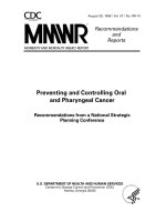
Lymph
node examination
The
major
lymph
nodes
of the
maxillofacial region
and
neck
are
shown
Figure
1. The
submental, submandibu-
lar
and the
internal jugular nodes (jugulo-digastric
and
jugulo-omohyoid node being
the
largest)
are of
particu-
lar
importance because these receive lymph drainage
from
the
oral cavity. Examination
of the
nodes should
be
systematic, although
the
order
of
examination
is not
crit-
ically
important.
To
palpate
the
nodes,
the
examiner
should stand behind
the
patient while
he/she
is
seated
in an
upright position.
Use
both
hands
(left
hand
for the
left
side
of the
patient etc.).
A
common sequence would
be
to
start
in the
submental
region,
working back
to the
submandibular nodes then
further
back
to the
jugulo-
digastric node
(Fig.
1).
Then continue
by
palpation
of
the
parotid region downwards
to the
retromandibular
area
and
down
the
cervical chain
of
nodes. When
a
node
is
perceived
as
enlarged, record
the
texture:
a
hard node
of
a
metastasising malignancy contrasts well with
a
ten-
der,
softer
node
in an
inflammatory
process.
Fig.
1
Principal
lymph
nodes
in the
head
and
neck.
The
dotted
lines
indicate
the
outline
of the
sternocleidomastoid
muscle.
Temporomandibular
joint
A
detailed examination
of the TMJ is
probably only
needed
when
a
specific
problem
is
suspected
from
the
history.
Details
of
examination
of
this
joint
and the
asso-
ciated
musculature
is
given
in
Chapter
14.
Salivary
glands
As
with
the
TMJ, examination
of the
salivary glands
is
only
required when
the
history suggests this
is
relevant.
Chapter
12
describes
the
examination
of the
major
sali-
vary
glands.
Problem-specific
examination
The
examination will
be
made
in the
light
of the
symp-
toms reported
by the
patient
but the
examiner
may
detect
swelling, sensory
or
motor disturbance that
the
patient
has not
noticed.
Swelling/lump
The
procedure
for
examination
of a
swelling
or a
lump
must
encompass
a
range
of
observations:
anatomical
site
shape
and
size
colour
single
or
multiple
surface
texture/warmth
tenderness
fluctuation
sensation/pulsation.
Consistency
can be
informative, ranging
from
the
soft
swelling
of a
lipoma, through 'cartilage
hard'
pleomor-
phic
adenomas
and
'rubbery
hard'
nodes
in
Hodgkin's
disease
to the
'rock
hard'
nodes
of
metastatic malig-
nancy.
Tenderness
and
warmth
on
palpation usually
indicates
an
inflammatory process, while neoplasms
are
commonly
painless
unless
secondarily infected.
Fluctuation
indicates
the
presence
of
fluid.
To
assess
fluctuation,
place
two
fingers
on the
swelling
and
press
down
with
one
finger.
If
fluid
is
present
the
other fin-
ger
will record
an
upward pressure. Pulsation
in a
swelling will indicate direct (i.e.
it is a
vascular lesion)
or
indirect involvement (i.e.
in
immediate contact)
of an
artery.
Paraesthesia/anaesthesia
The
presence
of
sensory disturbance
is
usually identi-
fied
initially
by the
patient
in the
history.
It is
important
to
identify
the
extent
of the
affected
area
and the
degree
of
alteration
in
sensation.
It is
best
to use a
fairly
fine,
but
blunt-ended,
instrument
for
this
at
first,
for
example
the
handle
of a
dental mirror. First,
run the
instrument gen-
tly
over what
is
assumed
to be a
normal area
of
skin
so
that
the
patient knows what
to
expect. Then repeat this
over
the
symptomatic area, asking
the
patient
to say
whether they
can
feel
anything.
Record
the
area
of
altered
sensation
in the
notes using
a
drawing.
The
degree
of
alteration
in
sensation
can be
assessed
by
using
different
'probes'.
A
teased-out piece
of
cotton
wool
can be
used
or,
where anaesthesia appears
to be
profound,
a
sharp
probe
can be
(carefully)
tried.
The
extent
of the
area
of
paraesthesia
or
anaesthesia
will tell
you the
particular nerve,
or
branch
of a
nerve,
involved (Fig.
2).
This will,
in
turn, inform
you
about
the
possible location
of the
underlying lesion.
For
example,
a
patient with disturbed sensation
of the
upper
lip has a
lesion
affecting
the
maxillary
division
of the
trigeminal
nerve.
If
this
is the
sole site
of
sensory
deficit,
it
suggests
a
lesion closer
to the
terminal branches
of
this cranial
nerve (e.g.
in the
maxillary sinus).
In
contrast,
if
sensory
deficiencies
are
simultaneously present
in
other
branches
of the
nerve,
it
suggests that
the
lesion
is
more
centrally located.
Paralysis/motor
disturbance
While paralysis
or
motor disturbance
may be
reported
as a
symptom
by the
patient,
it may
initially
be
identi-
fied
during
an
examination.
In the
maxillofacial
region,
the
motor
nerves
that
are
likely
to be
under
considera-
tion
are the
facial
nerve,
the
hypoglossal nerve (see
below)
and the
nerves controlling
the
muscles that move
the
eyes.
Disturbance
in
function
of the
facial
nerve will result
in
effects
on the
muscles
of
facial
expression. Paralysis
of
the
lower
face
indicates
an
upper motor neurone
lesion
(stroke,
cerebral tumour
or
trauma). Paralysis
of all the
facial
muscles
(on the
affected
side) indicates
a
lower
motor neurone
lesion.
The
latter
is
seen
in a
large num-
ber of
conditions but,
for the
dentist, important causes
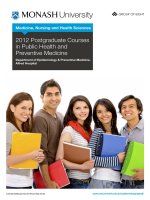
include Bell's palsy (Fig.
3),
parotid tumours,
a
mis-
placed inferior dental local anaesthetic
and
trauma.
Fig.
2
Cutaneous sensory innervation
of the
head
and
neck
by
the
trigeminal
and
cervical
nerves.
5
Fig.
3
Patient
with
Bell's palsy.
1.3
Intra-oral
examination
Learning
objectives
You
should:
• be
able
to
differentiate
dental
and
non-dental
sources
of
symptoms
•
understand
the
significance
of
features
of
ulcers
such
as
form,
site
and
pain
• be
able
to
examine
for
motor
and
sensory
nerve
dysfunction
•
know
how
to
examine
a
tooth.
Swelling/lump
The
examination
of an
intra-oral swelling
or
lump
is
essentially
the
same
as
that described above
as
part
of the
extra-oral
examination. Most oral swellings
are
inflam-
matory,
caused
by
periapical
or
periodontal infections.
However,
the
minority
of
oral swellings
and
lumps that
are
non-dental encompasses
a
wide range
of
conditions,
the
details
of
which
form
a
significant part
of
this book.
Ulcer
Examination
of an
ulcer should include assessment
of
eight
important characteristics:
site
single /multiple
size
shape
base
of the
ulcer
edge
pain
time
period.
Visual
inspection
is
essential
but
palpation
is
also
an
important part
of the
examination
of an
ulcer. Gloves
must
be
worn
for
palpation
and the
texture
of the
ulcer
base,
margin
and
surrounding tissues should
be
ascer-
tained
by
gentle pressure. Malignant neoplasms tend
to
ulcerate,
and
these
often
feel
firm,
hard
or
even
fixed
to
deeper tissues.
A
raised margin
is a
suspicious finding,
as is the
presence
of
necrotic,
friable
tissue
in the
ulcer
base
and
bleeding
on
lightly pressing (Fig.
4).
Healing
traumatic
ulcers tend
to be
painful
on
palpation
and
they
feel
soft
and
gelatinous.
The
finding
of an
ulcer
on
examination
may
necessi-
tate
taking additional history,
for
example,
if a
traumatic
ulcer
is
suspected, direct questioning
may
prompt
the
patient
to
recall
the
injury
(Fig.
5). If
multiple ulcers
are
detected, this
may
lead
to
further
enquiries about
any
Again,
a
systematic approach
is
essential
to
avoid
being
distracted
by the
first
unusual
finding
you
encounter.
The
examination must include
lips,
cheeks,
parotid gland orifices, buccal gingivae, lingual gingi-
vae
and
alveolar ridges
in
edentulous areas, hard
palate,
soft
palate, dorsal
surface
of the
tongue, ventral
surface
of the
tongue,
floor
of
mouth, submandibular
gland
orifices
and,
finally,
the
teeth.
Different
clinicians
will
have their
own
sequence
of
examination,
but it is
the
thoroughness
of the
examination that
is
important,
not the
order
in
which
the
regions
of the
mouth
are
examined.
Once
the
general intra-oral examination
is
complete,
a
problem-specific examination
can
proceed. This
is
tailored
to the
clinical problem.
Fig.
4
Clinical
photograph
of a
squamous
cell
carcinoma
of
the
tongue.
Note
the
raised
edges
and
necrotic
centre.
6
Fig.
5
Clinical
photograph
of a
traumatic ulcer
of the
lingual
mucosa.
Note
the
superficial
nature
of the
ulcer.
Its
base
is
covered
by
fibrous exudates
and the
surrounding
area
is
inflamed.
previous history
of
recurrent oral ulceration
or
specific
gastrointestinal diseases.
It is
surprising
how
often
ulceration
is
discovered that
the
patient
is not
aware
of.
When
an
ulcer
is
found,
it is
vital that
a
detailed record
of
the
history
and
examination findings
is
made.
Any
oral
mucosal ulcer that does
not
heal within
3
weeks
should
be
considered
as
possibly malignant
and
urgent
referral
must
be
arranged.
Certain
ulcers have
a
tendency
to
occur
in
particular
oral sites,
for
example squamous
cell
carcinomas
are
most common
on the
lower lip,
in the
floor
of
mouth
and the
lateral border
of the
tongue.
On the
other hand
traumatic ulcers
are
most common
on the
lateral border
of
the
tongue
and
buccal mucosa
in the
occlusal plane.
Ulceration
on the
lower
lip is
also
a
common site
for
traumatic
ulceration, particularly following administra-
tion
of an
inferior
dental block
or
after
a
sports
injury.
Site
is
also important
in
diagnosis,
for
example, minor
aphthae
are
restricted
to
lining mucosa
and can be
ruled
out
if
ulceration
is
occurring
on the
hard palate
or
gingivae.
Size
and
shape
can
also
be
helpful,
for
example linear
fissure-type
ulcers
may be
seen
in
Crohn's disease,
though aphthae
are
more usual.
The
shape
of a
trau-
matic
ulcer
may
reveal
the
cause,
for
example semicircu-
lar
ulcers
are
sometimes caused
by the
patient's
fingernail.
Bizarre persistent ulceration
is
sometimes
a
result
of
deliberate self-harm, unusual habits
or
taking
recreational
drugs;
in
such cases, diagnosis
can be
diffi-
cult
as the
patient
may
deny knowledge
of the
causa-
tion. Minor aphthae have characteristic size
and
site
features,
which
can
distinguish them
from
major
and
herpetiform
aphthae (see
Ch.
10).
Pain,
as
mentioned above,
is a
feature
of
inflamma-
tory
and
traumatic ulcers, while
in the
early stages
a
malignant ulcer
is
often
painless. Advanced malignant
ulcers eventually tend
to
become
painful
as a
result
of
infection
and
involvement
of
adjacent nerves.
Presentation with
a
painful traumatic ulcer
is
common
in
dentistry.
The
cause should
be
eliminated
if
possible
(e.g.
smoothing
or
replacement
of an
adjacent
frac-
tured restoration), symptomatic treatment such
as
analgesic mouthwash prescribed
and
most impor-
tantly,
review arranged
to
ensure that healing
has
occurred.
Paraesthesia/'anaesthesia
The
principles
of
examination
are
those
described above
for
extra-oral examination. Once again,
you
need good
anatomical
knowledge
of the
nerves supplying
different
parts
of the
oral cavity
to
interpret
the
possible site
of the
underlying pathological process (Fig.
6).
Paralysis/motor
disturbance
Within
the
oral cavity, motor disturbance
is
seen
in the
tongue (owing
to
damage
to the
function
of the
hypoglossal nerve)
and the
soft
palate (owing
to
lesions
affecting
the
vagus nerve). With hypoglossal nerve
lesions, there
is
deviation
of the
tongue towards
the
affected
side when attempting protrusion. There
is
also
a
problem with speech, with 'lingual' sounds such
as T,
't' and 'd'
affected.
ASA
=
Anterior superior alveolar nerve
PSA
=
Posterior superior alveolar nerve
Fig.
6
Sensory
innervation
of the
oral
cavity
is
principally
from
the
trigeminal
nerve
(V)
while
the
glossopharyngeal nerve (IX)
supplies
the
posterior
third
of the
tongue.
NB
Taste sensation
in the
anterior two-thirds
of the
tongue
is
provided
by
fibres
of
VII
nerve
origin
passing
through
the
lingual
nerve.
7
Tooth
problems
Tooth
problems
are,
of
course,
the
commonest problems
facing
the
dentist.
The
context
is
usually pain
or
swelling.
A
standard method
of
examination helps
in
reaching
a
diagnosis.
You
should
not
simply hammer
the
suspect
tooth with
the
mirror
handle
and
take
a
radiograph
as
your method
of
assessment! Indeed,
careful
examination
may
establish
a
diagnosis
and
thus
avoid
any
need
for
radiography
or
other special tests. Examination will
involve:
visual
probe restorations
assess
mobility
periodontal probing
thermal tests
pressure tests.
Visual
examination will reveal gross caries,
the
presence
of
restorations, signs
of
tooth wear
and
gingivitis.
A
probe will allow tactile assessment
of
restoration
margins.
Mobility
should
be
assessed manually. Periodontal
probing should
be
carried
out to
assess pocketing,
the
presence
of
calculus/overhangs
and,
ultimately, bone
loss.
A
basic test
of
vitality
should
always
be
performed,
using
a
cotton wool pledget soaked with ethyl chloride
(cold
stimulus)
and
sometimes heated gutta-percha
(hot
stimulus). While
these
are
usually
sufficient
to
reveal
a
hypersensitive tooth with pulpitis,
an
electri-
cal
pulp
test
can be
used
to
assess
vitality
in
some
cases.
Pressure sensitivity should
be
assessed
using direct
finger
pressure
and,
when
this does
not
evoke
a
response,
can be
supplemented
by
percussion using
a
dental mirror handle. This will assess whether peri-
odontitis
is
present
or
not.
However,
if a
single cusp
is
tender
to
percussion, this
may be
indicative
of
cracked
cusp syndrome.
1.4
Special investigations
Learning
objectives
You
should:
•
understand what samples
can be
taken
for
tests,
how
to
take
and
treat
these materials
and
what tests
are
available
•
know
how
to
interpret
the
results that
are
returned
•
know when imaging techniques would
be
informative
and
which type
of
imaging
to
choose.
Chairside
laboratory investigations
Evidence-based
laboratory
medicine
Whenever
special tests
are
undertaken,
it is
important
to
consider medicolegal
issues,
informed consent, appro-
priateness
of the
test
and the
evidence base
for the use of
any
particular laboratory investigation.
It is
always
nec-
essary
to
have
a
differential
clinical diagnosis
in
mind
when requesting
an
investigation. Certain tests, such
as
those
for
human immunodeficiency virus (HIV)
infec-
tion, require pre-test counselling
and
informed consent;
such
tests should
be
undertaken only
by
specialists
in
the
field.
When requesting
a
test,
it is
vital
to
possess
the
knowledge
and
skills
so
that
the
result
can be
acted
upon appropriately.
In
some situations,
for
example
sus-
pected oral cancer,
it may be
wise
to
refer
the
patient
directly
to a
specialist
for a
biopsy. Other important
con-
siderations when considering laboratory testing
are:
•
obtaining
a
representative/appropriate sample
•
collecting
in the
right specimen container
and
fluid
if
appropriate
•
completing
the
information required
by the
laboratory
correctly
•
having systems that avoid mixing
up
specimens;
labelling
the
specimen container with patient details
•
organising
the
correct packaging
and
transport
to the
laboratory
•
reading reports
and
acting
on
them;
filling
in
patient
records
•
interpretation: sensitivity
and
specificity.
Most
laboratories
can
advise
on
current codes
of
practice
relating
to the
above
issues
and may
give
reference
ranges
and
advice,
for
example about
a
particular biopsy result.
Sending pathological material through
the
post
is
poten-
tially
hazardous
and
current regulations must
be
followed.
It
should
be
remembered that laboratory
tests
require
considered interpretation
in
conjunction
with
the
patient's history. Some tests have
low
sensitivities,
for
example
certain cytology tests,
and a
negative result
can-
not
be
relied upon
to
exclude disease.
The
test
may
need
to be
repeated,
or an
alternative test with
a
higher sensi-
tivity
used. Other tests have
low
specificity
and a
positive
result
does
not
necessarily indicate that disease
is
present.
Examples
include low-titre autoantibodies, which
may be
detected
in the
serum
but
which
can be of no
clinical
sig-
nificance.
The
receiver-operator curve (ROC)
for any
lab-
oratory
test
can be
plotted
to
guide clinical
use.
Use of
resources
is
also important, particularly when expensive
reagents
or
complex procedures
are
required.
Microbiology
Diagnosis
of
infection
and
determination
of
sensitivity
of
the
infectious agent
to
pharmacotherapeutic agents
8
are
the
principal requirements
for
microbiology tests
in
dentistry.
Viruses.
Most
often
a
clinical diagnosis
is
adequate
for
acute
or
recurrent viral oral infections such
as
herpes
simplex.
A
viral swab
can be
used
to
collect virus
from
fresh
vesicles
and
must
be
forwarded
in
special transport
medium
to the
virology laboratory. Other virus
infec-
tions such
as
glandular
fever
can be
detected
by
looking
for
a
rising titre
of
antibodies
in the
patient's serum.
Bacteria.
Bacterial
infections
in the
oral cavity, jaws
and
salivary glands
may be
identified
by
forwarding
a
swab
or
specimen
of pus to the
laboratory, with
a
request
for
culture
and
antibiotic sensitivity.
Fungi.
Candida
sp. is the
most common organism
to
cause oral fungal infection. Often clinical
diagnosis
is
adequate;
for
example
in
denture-related stomatitis,
the
clinical history
and
appearance
of the
mucosa
may be
sufficient.
Direct smears
from
the
infected mucosa
and
the
denture-fitting
surface
can be
stained
by the
periodic
acid-Schiff
or
Gram's method.
The
presence
of
typical
pseudohyphae indicates candidal proliferation consis-
tent with
infection.
Swabs
or
oral rinses
can be
used
to
discriminate
the
various
Candida
species
and
heavy
growth suggests infection rather than carriage.
Aspiration
biopsy
Fluid
from
suspected cysts
can be
collected
with
a
stan-
dard gauge needle
and
syringe: radicular cysts contain
brown shimmering
fluid
because
of the
presence
of the
cholesterol crystals, whereas odontogenic keratocysts
contain pale greasy fluid,
which
may
include keratotic
squames. Infection
after
aspiration biopsy
can be a
prob-
lem
and
indeed
the
technique tends
to be
restricted
to
atypical cystic lesions where neoplasia
is
suspected.
Fine needle aspiration biopsy
(FNAB)
can be
used
to
obtain
a
sample
of
cells
from
a
solid tumour
and is a
hos-
pital
procedure.
Incisional/excisional
biopsy
Mucosal
biopsy
is one of the
more common investiga-
tions used
by
dentists
in
primary
and
secondary care.
Tissue
is
removed under local
or
general anaesthesia
using
sharp
dissection
to
avoid
crushing
the
specimen.
It
is
fixed
in at
least
10
times
its
volume
of 10%
neutral
buffered
formalin
or
similar fixative.
It is
then
for-
warded
to the
histopathology
or
specialist oral
and
max-
illofacial
pathology laboratory.
Excisional
biopsy.
The
entire lesion
is
removed
and
submitted
for
diagnosis.
It is
suitable
for
benign polyps,
papillomas, mucocoeles, epulides
and
other small reac-
tive lesions.
Incisional
biopsy.
A
representative sample
of a
larger
lesion
is
taken
for
diagnosis prior
to
treatment. This
is a
specialist procedure requiring some expertise
and
expe-
rience.
It is
used
for
generalised
mucosal
disorders
such
as
lichen planus
or for the
diagnosis
of
other
red and
white patches.
An
important consideration
is
obtaining
a
sample
from
an
appropriate area. Non-healing ulcers
are
often
investigated
by
incisional biopsy; here
it is
important
to
include
the
margin
of the
ulcer with some
normal tissue
and to
obtain
a
sufficiently
large sample
(normally
10 mm x 10 mm) to
identify
or
exclude cancer.
Sometimes
fresh
tissue
is
required
for
diagnosis,
for
instance
in the
vesiculo-bullous diseases where
immunofluorescence
is
needed. Special arrangements
must
be
made with
the
laboratory when such tests
are
planned.
Haematology
Patients presenting with oral manifestations
of
haema-
tological disease
are
normally
referred
for
specialist
opinion. Full blood count
and
assay
of
haematinics
is an
important investigation
for
patients presenting with lin-
gual papillary atrophy
or
recurrent oral ulceration,
for
example. Coagulation studies
and
platelet counts
may
be
required when excessive bleeding
is
encountered.
Patients
on
anticoagulant therapy should have their
INR
(international normalised ratio) checked
before
any
sur-
gical
procedure
is
undertaken.
The
Sickledex test
may be
used
to
screen
for
sickle
cell
anaemia prior
to
giving general anaesthesia
in
situ-
ations
of
urgency.
The
blood sample
should
be
subjected
to
haemoglobin electrophoresis.
Haematological parameters
of
importance
in
den-
tistry
are
described
in
Table
1.
Biochemistry
Biochemical
investigations
are
used principally
in
spe-
cialist
clinics
to
investigate patients presenting with oral
manifestations
of
systemic disease,
for
example estima-
tion
of
alkaline phosphatase
in
Paget's disease
of
bone,
and
serum calcium
to
exclude hyperparathyroidism
when
a
giant cell granuloma
is
diagnosed. Biochemical
estimation
of
cyst fluid
for
protein content
is
some-
times undertaken
as
part
of
diagnosis
of
odontogenic
keratocyst.
Immunology
Advances
in
knowledge
and
methods
in
immunology
have resulted
in a
large number
of
laboratory immuno-
logical
investigations, available
in
specialist laborato-
ries.
Sometimes diagnostic arrays
of
tests
are
offered
by
the
laboratory. Examples
of
tests
in
dentistry include
detection
of
antibodies against extractable nuclear anti-
gens, including SS-A
and
SS-B,
for the
diagnosis
of
9
Table
1
Important haematological values
in
dentistry
Conventional
units
SI
units
Haemoglobin (Hb)
Male
Female
Red
cell count (RBC)
Male
Female
Haematocrit (HCT)
Male
Female
Mean
cell volume, adults (MCV)
Mean cell haemoglobin, adults (MCH)
Mean
cell haemoglobin concentration, adults (MCHC)
White cell count, adults (leucocytes; WBC)
Neutrophils
Lymphocytes
Monocytes
Eosinophils
Platelets,
adults (PLT)
Erythrocyte
sedimentation rate, adults (ESR)
13.0-1
8.0
g/dl
11.5-16.5g/dl
4.5-6.5
million/mm
3
3.8-5.8
million/mm
3
40-54
ml/dl
37-47
ml/dl
80-90
urn
3
27-32
pg/cell
31-36.5
g/dl
4500-11
000
/mm
3
2000-7500/mm
3
1500-4000/mm
3
200-1
200/mm
3
40-400/mm
3
0-8
mm/h
8.1-11.2mmol/l
7.4-9.9
mmol/l
4.5-6.5
x10
12
/l
3.8-5.8
x10
12
/l
0.40-0.54
0.37-0.47
80-97
fl
27-32
pg/cell
31-36.5g/l
4.0-1
1.0
x10
9
/l
2.0-7.5
x10
9
/l
1.
5-4.0 x10
9
/l
0.2-1.2x10
9
/l
0.04-0.40 x10
9
/l
1
50-400
x10
9
/l
0-8
mm/h
Every
laboratory
has its own
reference range which should
be
consulted
when
laboratory test results
are
received.
The
values
are
typical
for
adults;
the
ranges
for
full-term infants
and
children
vary
considerably
.
Sjogren's syndrome
and
autoantibodies
in
vesiculo-
bullous diseases.
HIV
testing should only
be
undertaken
by
specialists
and
does
not
fall
directly into
the
remit
of
dentistry.
It
requires informed patient consent
and
counselling.
Dentists must
be
able
to
recognise
the
oral manifestations
of
immunodeficiency states
and
arrange proper
referral.
Imaging
Imaging
is an
important special test
in
dentistry
and
oral
and
maxillofacial
surgery. Because
X-ray
exposure carries
a
quantifiable risk
(see
Ch.
15),
X-ray
examinations
should
be
selected according
to
specific
selection
(refer-
ral)
criteria. Other imaging
investigations
not
using
ionis-
ing
radiations (ultrasound
and
magnetic resonance
imaging) have their place
and
should
be
used
in
prefer-
ence
to
X-ray
techniques (radiography
and
computed
tomography) when they
can
provide
the
same
or
better
diagnostic information. Selection criteria should
be
based
upon
the
diagnostic
efficacy
of the
technique
for the
dis-
ease process being examined.
For
example, approximal
caries
diagnosis
is
best
aided
by
bitewing rather than
other radiographs. There
are a
large number
of
imaging
techniques available
and
these
are
summarised below.
Details
of the
specific
uses
of
these techniques
are
given
where appropriate
in
subsequent chapters.
Conventional
radiography
This
is
familiar
to
every dentist
and
student
in the
forms
of
bitewing, periapical, occlusal
and
panoramic radiog-
raphy
and
these techniques
are
covered
in
more detail
in
the
companion volume
to
this book (Dentistry II).
Other
maxillofacial
radiographs should
be
used
in
addition
to the
traditional
'dental'
techniques when
appropriate. While detailed prescription
of
radiographs
depends
on the
particular needs
of
each patient, some
general guidelines
are
useful
and are
given
in
Table
2.
Contrast
investigations
Some
radiological techniques
use
radio-opaque contrast
media injected into parts
of the
body.
In the
maxillo-
facial
region, they
can be
used
to
demonstrate fistulae
and
sinuses
and in
vascular studies (angiograms). How-
ever,
they
are
most commonly used
for
sialography
(Ch.
12) and
arthrography
of the TMJ
(Ch. 14).
Computed tomography
Computed tomography (CT)
is
also known
as CAT
scanning
(Fig.
7). It
provides primarily
axial
cross-
sectional images
and
uses
X-rays.
The
computer
cal-
culates
the
X-ray
absorption
(and
thus indirectly
the
density)
of
each unit volume
(voxel)
of
tissue
and
then
assembles
the
information into
an
image made
up of
many pixels (picture elements). Each pixel
is
given
a
grey-scale
value according
to its
density (Hounsfield
scale).
Dense bone
is
white, most
soft
tissues
are
mid-grey,
fat is
dark grey
and air is
black. Metals
are
beyond
the
comprehension
of the
computer software,
so
dental fillings cause
artefacts.
10
Table
2
Guidelines
of
radiographic projections
Anatomical
site
to be
examined
Radiographic projections
Anterior
mandible
Body
of
mandible
Third molar region, angle
and
ramus
of
mandible
Condyle
temporomandibular joint
Anterior
maxilla
Posterior maxilla
Maxillary
sinus
Parotid
gland (for calculi)
Submandibular gland (for calculi)
Periapical, oblique
and
true occlusal views
Periapical, true occlusal, panoramic
(or
lateral oblique) views
Periapical
and
true occlusal
(third
molar region only)
Panoramic
(or
lateral oblique) view
Postero-anterior
(PA) view
of
mandible
Panoramic
(or
lateral oblique) view
Transpharyngeal view
Transcranial views (open/closed)
Reverse
Towne's view (modified
PA
projection)
Periapical
and
oblique occlusal views
Periapical,
oblique occlusal,
panoramic
(or
lateral oblique)
views
Periapical, oblique occlusal, panoramic
(or
lateral oblique) views
Occipitomental view
Intra-oral soft tissue view
of
parotid
papilla
region
Localised PA/antero-posterior
of
face with cheek blown
out
True
occlusal
of
floor
of
mouth
Modified oblique occlusal
for
submandibular gland
Fig.
7 A
typical computed tomographic scan.
Clinical
maxillofacial
applications
include:
•
large maxillary cysts/benign tumours
•
malignancy arising
in the
antrum
•
soft
tissue masses
•
oral carcinoma.
Images
can be
reconstructed
in two or
three dimensions.
In
maxillofacial
work, reconstructions
are
invaluable
for
implantology
and
useful
in
major facial
trauma
and
orthognathic surgical
treatment
planning.
CT
is
associated with
a
relatively high dose
of
radia-
tion.
Generally,
the
thinner
the
sections (and
the
better
the
fine
detail),
the
higher
the
dose.
Diagnostic ultrasound
Ultrasound uses
the
principle that high
frequency
(3.5-10
MHz) sound waves
can
pass through
soft
tissue
but
will
be
reflected
back
from
tissue
interfaces.
The
echoes
can be
detected
to
produce
an
image.
The
sound
is
transmitted
and
detected
by the
same hand-held
transducer.
Imaging
is
'real-time'.
Clinical
maxillofacial
applications
include: soft
tissue
lumps
in the
neck
and the
salivary glands.
Radioisotope imaging
Radioisotope
imaging
is
also known
as
nuclear medicine
(Fig.
8). The
technique uses radioisotopes (usually
gamma
ray
emitters) tagged
on to
pharmaceuticals,
which
are
usually
injected
into
the
bloodstream.
By
choosing
the
radiopharmaceutical appropriately, partic-
ular
organs
or
types
of
tissues will become radioactive.
The
patient
is
placed
in
front
of a
gamma camera, which
detects
the
emitted radiation
to
give
an
image
of
physio-
logical
activity.
It is not an
anatomical imaging modality.
Clinical
maxillofacial
applications
include:
•
salivary scanning (particularly
in
Sjogren's
syndrome):
uses sodium pertechnetate-99m
•
bone scanning
(for
bone tumours, metastatic disease,
Paget's disease, arthritis
and
condylar hyperplasia):
uses
technetium-99m-labelled
methylene
bisphosphonate.
Magnetic resonance imaging
Magnetic
resonance imaging
is
also known
as MR, MRI
or
NMR.
In
this technique, patients
are
placed into
an
intense magnetic
field, forcing
their hydrogen nuclei
11
Fig.
8
Radioisotope scan
of the
salivary glands. Frontal view.
Foci
of
activity
are
visible
in the
four major salivary glands,
in
the
mouth and,
at the
bottom
of the
image,
the
thyroid gland.
(principally
in
water molecules)
to
align
in the
field.
Radiofrequency
waves
are
pulsed into
the
patient,
the
hydrogen nuclei
'wobble',
producing
an
alteration
in the
magnetic
field.
This induces
an
electric current
in
coils
placed around
the
patient.
The
computer
is
capable
of
reading this and, because
different
tissues contain dif-
ferent
amounts
of
hydrogen
(in
water),
of
producing
an
image
that,
superficially,
is
like
a CT
scan. However,
imaging
can be in any
plane (axial, sagittal
or
coronal).
Clinical
maxillofacial
applications
include:
•
anything
CT can do
(but
no
ionising radiation)
•
imaging
of
the
TMJ.
Problems
are
twofold:
the
immense cost
of MR
means
that
waiting lists
in NHS
hospitals
in the UK are
very
long
and, second, patients with some metallic implants
(intracranial
vascular clips, cardiac pacemakers)
are not
eligible
for the
technique.
1.5
Writing
a
referral
letter
Learning
objectives
You
should:
•
know
when
to
refer
a
patient
• be
able
to
write
a
competent
referral
letter
•
know
now
to
keep
good
records
of the
referral.
However good your diagnostic abilities
are and
how-
ever
skilled
you are as a
clinician, there will come
a
time
when
you
need
to
refer
a
patient
on to a
colleague.
The
letter
should
be
thorough, providing
the
second clini-
cian
with
a
detailed history
and the
results
of
your
examination.
It is
reprehensible
to
write
a
'Dear Sir,
please
see and
treat, yours sincerely' letter.
The
referral
must
include:
name, address, date
of
birth
of the
patient
description
of the
patient's
problem/symptoms
a
history
of the
problem
the
results
of
your examination
the
results
of any
special tests
you
have performed
your provisional diagnosis,
if any
the
medical history
any
special
factors,
such
as
difficulty
in
attending
all
relevant radiographs
or
investigations.
The
letter should
be
word-processed wherever possible,
rather
than hand-written,
to
ensure
accuracy.
A
model
letter
is
shown
in
Figure
9. It is
important
to
remember
that patients tend
to
open
and
read
referral
letters
and
that they become ultimately part
of the
hospital medical
record.
Such records
are
available
to
patients
and
their
legal advisers.
The
example
in
Figure
9
demonstrates
that
the
dentist acted promptly
and
exercised
a
high
standard
of
care
and
consideration
for the
patient.
A
copy
of the
referral
letter should
be
kept with
the
patient's
records.
It
is
good practice
to
establish
a
working relation-
ship between primary
and
secondary carers.
In the
sit-
uation described
in
Figure
9,
when
an
oral cancer
is
suspected,
it can be
helpful
for the
primary care
dentist
to
telephone
the
oral
and
maxillofacial
department
for
advice.
Sometimes
an
early appointment
can be
offered.
A
letter should still
be
forwarded,
for the
rea-
sons given above. However,
it is not
helpful
to
tele-
phone
or
send patients with non-urgent conditions
to
hospital with
an
expectation
of
being seen immedi-
ately.
It is
better
for all
concerned
to
write
a
letter
and
advise
the
patient
of
likely waiting times,
often
obtain-
able
from
hospital
intranet
links.
Guidelines
for
refer-
ral
have been produced
by
national
and
local
authorities, such
as the
National Institute
for
Clinical
Excellence
(NICE)
and the
Royal Colleges. These
should
be
consulted whenever
possible,
as
inappropri-
ate
referral
should
be
avoided.
12
The
Dental Practice
1,
High
Street
Anytown
Dr A
Smith
Consultant Oral
and
Maxillofacial Surgeon
Anytown
General Hospital
Anytown
2
January 2001
Dear
Dr
Smith,
Re:
Mr
John Doe,
24
Green Lane, Anytown. Date
of
birth:
25.12.40
Tel: 0123 456789
I
would
be
grateful
if you
would
see
this 60-year-old man.
He
presented today complaining
of
a
'growth' from
a
recent extraction socket
in his
upper jaw.
He
said that
this
had
appeared
after
an
extraction
I
carried
out two
weeks
ago and was
getting slowly bigger.
He
also
complains
of a
numb feeling
on the
left
cheek.
I had
extracted
/6 two
weeks
ago at the
request
of the
patient because
it was
loose.
Examination revealed
a
palpable
left
cervical lymph node. There
was
reduced sensation
to
touch
on the
left
upper
lip and
cheek. Intra-orally there
was a
mass
on the
left
maxillary
alveolus
in /6
region, about
2 by 1 cm. The
mass
has an
irregular surface, feels indurated,
bleeds
easily
on
palpation
and
looks necrotic
in
places.
I
have taken
a
periapical radiograph,
which shows some bone destruction
at the
site
of the
socket.
I am
worried that this might
be
maxillary sinus malignancy
and I
would appreciate your
urgent opinion
and
management.
Mr
Doe has a
history
of
mild
hypertension
for
which
he
takes
a
bendrofluazide tablet (2.5mg)
in
the
morning. Otherwise there
is no
other
medical
history
of
note.
He is a
nervous patient
generally
and
will
probably
be
accompanied
by his
wife.
Mr Doe is a
non-smoker
and
drinks
7-8
units
of
alcohol
per
week.
He can
attend
at any
time.
Yours
sincerely,
Mrs
B
Jones
BDS
Fig.
9 An
example
of a
referral
letter.
13
This page intentionally left blank
2.1
Medical
assessment
2.2
Dental
relevance
of the
medical
condition
2.3
Medical
emergencies
2.4
Drug
delivery
Self-assessment:
questions
Self-assessment:
answers
Overview
15
16
21
30
33
34
This
chapter
discusses
the
assessment
of a
patient
with
a
pre-existing
medical
condition
that
might
affect
dental
treatment.
Particular
aspects
are the
effects that
anaesthetic
drugs
might
have
on
these
conditions
and the
potential
for
drug
interactions. Medical emergencies
are
described
in
terms
of
their
signs
and
symptoms.
The
immediate
first-line
treatment
is
listed
and
subsequent management steps
outlined.
The
technique
for
resuscitation
of a
patient
is
clearly
described. Finally
the
methods
of
administration
of
drugs
are
described
and
their
relative
merits
in
dentistry.
2.1
Medical
assessment
Learning
objectives
You
should:
•
know
how
to
obtain information
on
relevant medical
problems
• be
able
to
assess
a
patient's
fitness
for
treatment
•
know when
a
patient should
be
referred
for
treatment
in a
hospital
setting.
Today,
many patients with life-threatening
disease
sur-
vive
as a
result
of
advances
in
medical
and
surgical
treatment
and may
present
for
dental treatment looking
deceptively
fit and
well.
The
medical assessment:
• is
important
to
establish
the
suitability
of the
patient
to
undergo dental treatment
and may
significantly
affect
the
dental management
• may
prompt examination
for
particular oral
manifestations
• may be
particularly relevant when
a
sedation
technique
or
general anaesthesia (GA)
is
being
considered
• may
give prior warning
of a
possible medical
emergency.
Medical
history
As a
full
medical examination
of the
patient
is
generally
not
feasible
or
appropriate,
the
medical history should
be
comprehensive. This will include questions about
previous serious illness
and
operations, present drug
history
and
known allergies,
and the
possibility
of
preg-
nancy.
Information
may
then
be
obtained concerning
the
individual systems
by
relevant questions depending
on the age of the
patient,
the
dental treatment necessary
and the
anticipated type
of
anaesthesia.
Questions
should
refer
to
known medical problems,
past history
and
present general fitness.
Cardiovascular
system
Questions should
refer
to
known medical problems,
past history
and
present general
fitness.
• Is
there
a
history
of
heart valve surgery, rheumatic
fever
or
murmurs, which might necessitate
prophylactic antibiotic cover
• Is the
patient aware
of any
heart disease
or
hypertension?
•
Does
the
patient
suffer
from
palpitations, swelling
of
the
ankles
and
dizziness?
• Can the
patient
lie
flat
without breathlessness?
•
What
is the
patient's general
fitness?
For
example,
can
the
patient climb stairs without breathlessness
or
chest
pain?
Respiratory
system
•
Does
the
patient have
a
cough
or
cold?
If
there
is a
cough,
is
this continuous
or
intermittent
and is it
productive?
•
Does
the
patient
suffer
from
bronchitis, emphysema
or
asthma?
• Is
there shortness
of
breath
or
symptoms
of
wheeze
or
chest pain?
• If the
patient
is a
smoker,
how
many cigarettes
are
smoked
on
average each
day?
15
Medical aspects of patient care