Musculoskeletal Diseases Diagnostic Imaging and Interventional Techniques pot
Bạn đang xem bản rút gọn của tài liệu. Xem và tải ngay bản đầy đủ của tài liệu tại đây (5.42 MB, 197 trang )
Musculoskeletal Diseases
Diagnostic Imaging and Interventional Techniques
G.K. von Schulthess • Ch.L. Zollikofer (Eds)
MUSCULOSKELETAL
DISEASES
DIAGNOSTIC IMAGING AND INTERVENTIONAL
TECHNIQUES
37th International Diagnostic Course
in Davos (IDKD)
Davos, April 2-8, 2005
including the
Pediatric Satellite Course “Kangaroo”
Davos, April 2-3, 2005
presented by the Foundation for the
Advancement of Education in Medical Radiology, Zurich
III
J. H
ODLER G. K. VON SCHULTHESS
Department of Radiology Universitätsspital
University Hospital Balgrist Nuklearmedizin
Zurich, Switzerland Zurich, Switzerland
C
H. L. ZOLLIKOFER
Kantonsspital
Institut für Radiologie
Winterthur, Switzerland
Library of Congress Control Number: 2005922183
ISBN 88-470-0 8- Springer Milan Berlin Heidelberg New York
This work is subject to copyright. All rights are reserved, whether the whole or part of the material is
concerned, specifically the rights of translation, reprinting, re-use of illustrations, recitation,
broadcasting, reproduction on microfilms or in other ways, and storage in data banks. Duplication of
this publication or parts thereof is only permitted under the provisions of the Italian Copyright Law
in its current version, and permission for use must always be obtained from Springer. Violations are
liable for prosecution under the Italian Copyright Law.
Springer is a part of Springer Science+Business Media
springeronline.com
© Springer-Verlag Italia 2005
Printed in Italy
The use of general descriptive names, registered names, trademarks, etc., in this publication does not
imply, even in the absence of a specific statement, that such names are exempt from the relevant
protective laws and regulations and therefore free for general use.
Product liability: The publisher cannot guarantee the accuracy of any information about dosage and
application contained in this book. In every individual case the user must check such information by
consulting the relevant literature.
Typesetting: Kley & Sebastianelli Srl, Milan
Printing and binding: Grafiche Porpora, Cernusco sul Naviglio (Milan)
Cover design: Simona Colombo
Printed in Italy
IV
1
3
0
Preface
The International Diagnostic Course in Davos (IDKD) offers a unique learning
experience for imaging specialists in training as well as for experienced radi-
ologists and clinicians wishing to be updated on the current state of the art and
the latest developments in the fields of imaging and image-guided interventions.
This annual course is focused on organ systems and diseases rather than on
modalities. This year’s program deals with diseases of the musculoskeletal sys-
tem. During the course, the topics are discussed in group seminars and in plenary
sessions with lectures by world-renowned experts and teachers. While the semi-
nars present state-of-the-art summaries, the lectures are oriented towards future
developments.
This syllabus represents a condensed version of the contents presented under
the 20 topics dealing with imaging and interventional therapies in the muscu-
loskeletal radiology. The topics encompass all the relevant imaging modalities in-
cluding conventional x-rays, computed tomography, nuclear medicine, ultrasound
and magnetic resonance angiography, as well as image-guided interventional tech-
niques.
The volume is designed to be an “aide-mémoire” for the course participants so
that they can fully concentrate on the lectures and participate in the discussions
without the need of taking notes. Additional information is found on the web page
of the IDKD (http//:www
.idkd.ch).
J. Hodler00ii00
G.K. von Schulthess
Ch.L. Zollikofer00
V
IDKD 2005
Table of Contents
Seminars
Shoulder
J. Beltran, M. Recht . . . . . . . . . . . . . . . . . . . . . . . . . . . . . . . . . . . . . . . . . . 3
Magnetic Resonance Imaging of the Elbow
C. Chung, L. Steinbach . . . . . . . . . . . . . . . . . . . . . . . . . . . . . . . . . . . . . . . 7
Radiology of Hand and Wrist Injuries
A.J. Wilson . . . . . . . . . . . . . . . . . . . . . . . . . . . . . . . . . . . . . . . . . . . . . . . . 13
Wrist and Hand
L.A. Gilula . . . . . . . . . . . . . . . . . . . . . . . . . . . . . . . . . . . . . . . . . . . . . . . . 17
Imaging of the Painful Hip and Pelvis
C.W.A. Pfirrmann, C.A. Petersilge . . . . . . . . . . . . . . . . . . . . . . . . . . . . . . . 21
Imaging of the Knee
D.A. Rubin, W.E. Palmer . . . . . . . . . . . . . . . . . . . . . . . . . . . . . . . . . . . . . . 26
Imaging of the Foot and Ankle
Z.S. Rosemberg, M. Zanetti . . . . . . . . . . . . . . . . . . . . . . . . . . . . . . . . . . . . 39
Magnetic Resonance Imaging of Muscle
M.N. Pathria, R.D. Boutin . . . . . . . . . . . . . . . . . . . . . . . . . . . . . . . . . . . . . 48
Soft Tissue Tumors and Tumo-Like Masses: A Systematic Approach
to Diagnosis
M.J. Kransdorf, M.D. Murphey . . . . . . . . . . . . . . . . . . . . . . . . . . . . . . . . . . 54
Tumors and Tumor-Like Lesions of Bone
M. Sundaram, D. Vanel . . . . . . . . . . . . . . . . . . . . . . . . . . . . . . . . . . . . . . . 62
Imaging of Bone Marrow Disorders
B. Vande Berg, J. Malghem, F. Lecouvet, B. Maldagu . . . . . . . . . . . . . . . . . 68
Bone Marrow Disorders
A. Stäbler . . . . . . . . . . . . . . . . . . . . . . . . . . . . . . . . . . . . . . . . . . . . . . . . . 73
Metabolic and Systemic Bone Diseases
J. Freyschmidt . . . . . . . . . . . . . . . . . . . . . . . . . . . . . . . . . . . . . . . . . . . . . . 83
Metabolic Bone Disease
J.E. Adams . . . . . . . . . . . . . . . . . . . . . . . . . . . . . . . . . . . . . . . . . . . . . . . . 89
VII
IDKD 2005
The Radiology of Hip and Knee Joint Prostheses
I. Watt, B.N. Weissman . . . . . . . . . . . . . . . . . . . . . . . . . . . . . . . . . . . . . . . 106
Traumas of the Axial Skeleton
H. Imhof, G.Y. El-Khoury . . . . . . . . . . . . . . . . . . . . . . . . . . . . . . . . . . . . . 112
Trauma of the Appendicular Skeleton
J.J. Kaye, M.K. Dalinka . . . . . . . . . . . . . . . . . . . . . . . . . . . . . . . . . . . . . . . 121
Inflammatory Diseases of the Spine
V. Jevtic, V. Pullicino . . . . . . . . . . . . . . . . . . . . . . . . . . . . . . . . . . . . . . . . . 127
Degenerative Diseases of the Spine
D. Weishaupt, I. McCall . . . . . . . . . . . . . . . . . . . . . . . . . . . . . . . . . . . . . . . 132
Osteomyelitis and Septic Arthritis
D. Forrester, R.F. Kilcoyne . . . . . . . . . . . . . . . . . . . . . . . . . . . . . . . . . . . . . 138
Peripheral Arthritis
L.F. Rogers, C.S. Resnik . . . . . . . . . . . . . . . . . . . . . . . . . . . . . . . . . . . . . . 143
Special Aspects of Musculoskeletal Imaging in Children
D. Jaramillo, G. Sebag . . . . . . . . . . . . . . . . . . . . . . . . . . . . . . . . . . . . . . . . 148
Musculoskeletal Sonography
S. Bianchi, S. Marcelis . . . . . . . . . . . . . . . . . . . . . . . . . . . . . . . . . . . . . . . . 156
Pediatric Satellite Course “Kangaroo”
The Spectrum of Non-Accidental Injury and Its Imitators in Children
P.K. Kleinman . . . . . . . . . . . . . . . . . . . . . . . . . . . . . . . . . . . . . . . . . . . . . . 169
Contrast Enhancement of the Growing Skeleton: Rationale and
Optimization in Pediatric MRI
G. Sebag . . . . . . . . . . . . . . . . . . . . . . . . . . . . . . . . . . . . . . . . . . . . . . . . . . 175
Imaging the Osseous and Soft Tissue Tumors in the Child
A. Geoffray . . . . . . . . . . . . . . . . . . . . . . . . . . . . . . . . . . . . . . . . . . . . . . . . 180
Imaging the Child’s Inflammatory and Infectious Musculoskeletal
Pathology
S.G.F. Robben . . . . . . . . . . . . . . . . . . . . . . . . . . . . . . . . . . . . . . . . . . . . . . 185
VIII Table of Contents
SEMINARS
1
Introduction
This seminar places special emphasis on the MRI mani-
festations of shoulder pathology. The discussion includes
the following topics:
1. Rotator cuff pathology and impingement lesions.
2. Glenohumeral instability and related lesions.
3. Miscellaneous shoulder conditions.
Rotator Cuff Pathology and Impingement
Lesions
Impingement syndrome is a clinical entity produced by
compression of the supraspinatus tendon under the region
of the acromial arch, and it can be related to abnormal
morphology of the acromion process, thickening of the
coracoacromial ligament, subacromial spurring, or de-
generative arthritis of the acromioclavicular joint.
Alternatively, it can be related to degeneration, repeated
trauma or overuse during overhead exercise, such as
swimming. Normal anatomical variants, such as type III
undersurface of the acromion with a hooked configura-
tion and os acromiale, have been described associated
with rotator cuff impingement and tears.
There are two types of impingement syndrome: pri-
mary, associated with abnormalities in the coracoacromi-
al arch; and secondary to rotator cuff dysfunction. The
secondary form of rotator cuff impingement may be fur-
ther subdivided into two types: internal and external. The
internal type refers to the articular surface side of the ro-
tator cuff and it is often termed posterosuperior impinge-
ment syndrome. The external variety occurs as a result of
external compression of the anterior aspect of the cuff in
the bursal side and includes the coracoid impingement
syndrome. Posterosuperior impingement syndrome oc-
curs in the throwing athlete as a result of continuous
strain of the anterior capsular mechanism, which leads to
laxity and anterior subluxation of the glenohumeral joint
with the arm in abduction and external rotation. This sit-
uation produces impingement of the supraspinatus ten-
don at the level of its insertion in the greater tuberosity
of the humerus as well as small impaction fractures and
posterosuperior labral lesions. The coracoid impingement
syndrome may occur when the distance between the pos-
terior aspect of the coracoid process and the humerus is
decreased, producing compression of the rotator cuff,
mainly the subscapularis tendon.
Inflammatory changes within the supraspinatus tendon
can be seen during the early phases of the disease, along
with subacromial bursitis, but this can progress into rota-
tor cuff tear. Three histological stages of impingement
syndrome have been described. In stage I, edema and he-
morrhage of the subacromial soft tissues are present. In
stage II, there is fibrosis and thickening, while in stage
III, partial or complete rotator cuff tears are seen.
Full-thickness rotator cuff tears involve most often the
supraspinatus tendon, but they can also extend to the in-
fraspinatus and subscapularis tendons. Tear of the teres
minor is very rare. Partial-thickness rotator cuff tears may
involve the articular or the bursal surfaces, or they may
be located within the substance of the tendon.
Delaminating tears of the rotator cuff can be partial or
full thickness. They extend in the longitudinal direction
of the tendon fibers, and there may be different degrees
of retraction of the various layers. Delaminating tears
may be associated with fluid collections extending from
the tear into the muscle (sentinel cyst). Full-thickness
tears allow communication between the articular space of
the glenohumeral joint and the subacromial-subdeltoid
bursa, unless the tear is covered by granulation or scar tis-
sue. On rare occasions, tears may involve the rotator cuff
interval, with capsular disruption. Tears of the rotator cuff
interval may be associated with lesions of the structures
present within this anatomical space, namely, the long
head of the biceps tendon, the coracoacromial ligament,
the superior glenohumeral ligament and also the superior
labrum.
Glenohumeral Instability and Related Lesions
Restraints to anterior translation of the humeral head are
provided by the capsule and the glenohumeral ligaments
IDKD 2005
Shoulder
J. Beltran
1
, M. Recht
2
1
Department of Radiology, Maimonides Medical Center, Brooklin, NY, USA
2
Department of E-Radiology, The Cleveland Clinic Foundation, Cleveland, OH, USA
(GHL). The labrum is torn as part of the avulsion forces
produced by the GHL at the time of the injury.
Anteroinferior dislocation is the most frequent cause of
anterior glenohumeral instability. A single event origi-
nates a constellation of lesions leading to other episodes
of dislocation or subluxation. The lesions that may take
place during an anteroinferior dislocation include an-
teroinferior labral tear, tear of the inferior GHL (IGHL)
and/or capsular-periosteal stripping, fracture of the an-
teroinferior glenoid margin and compression fracture of
the superior lateral aspect of the humeral head (Hill-
Sachs lesion).
The classic Bankart lesion is the combination of ante-
rior labral tear and capsuloperiosteal stripping. On
arthroscopy, the Bankart lesion is seen as a fragment of
labrum attached to the anterior band of the IGHL and to
the ruptured scapular periosteum, “floating” in the ante-
rior-inferior aspect of the glenohumeral joint. Extensive
bone and soft-tissue damage and persistent instability
may lead to multidirectional instability, resulting in
episodes of posterior dislocation.
A number of variants of anterior labral tears have
been described. The Perthes lesion is similar to the
Bankart lesion, but without the tear of the capsule.
Anterior labroligamentous periosteal sleeve avulsion
(ALPSA) refers to a tear of the anteroinferior labrum,
with associated capsuloperiosteal stripping. The torn
labrum is rotated medially, and a small cleft or separa-
tion can be seen between the glenoid margin and the
labrum. In contrast to the Bankart lesion, the ALPSA le-
sion can heal, leaving a deformed and patulous labrum.
The glenoid labral articular disruption (GLAD) repre-
sents a tear of the anteroinferior labrum, attached to a
fragment of articular cartilage, without associated cap-
suloperiosteal stripping.
Posterior shoulder dislocation more often occurs as a
result of a violent muscle contraction, e.g., by electrical
shock or seizures. After the acute episode of dislocation,
the arm frequently remains locked in adduction and in-
ternal rotation. Posterior instability caused by repeated
micro-trauma, without frank dislocation, may cause per-
sistent shoulder pain in young athletes. Abduction, flex-
ion and internal rotation are the mechanism involved in
these cases (swimming, throwing, and punching). This
may be also associated with posterior capsular laxity.
Lesions that may occur during posterior dislocation or in
cases of repeated micro-trauma include posterior labral
tear, posterior capsular stripping or laxity, fracture, ero-
sion, or sclerosis and ectopic bone formation of the pos-
terior glenoid, and vertical impacted fracture of the ante-
rior aspect of the humeral head (reverse Hill-Sachs,
McLaughlin fracture).
Superior labral anterior and superior lesions (SLAP
lesions) are not as rare as originally thought. These le-
sions involve the superior part of the labrum with vary-
ing degrees of biceps tendon involvement. Pain, click-
ing, and occasional instability in a young patient are the
typical clinical manifestations. Four types of SLAP le-
4
sions were originally described based on arthroscopic
findings. Type I is a partial tear of the superior part of
the labrum with fibrillation of the LHBT. Type II is an
avulsion of the LHBT with tear of the anterior and pos-
terior labrum. Type III is a bucket-handle tear of the
labrum and type IV is a bucket-handle tear of the labrum
with longitudinal tear to the LHBT. More recently, up to
ten types of SLAP lesions have been described, repre-
senting a combination of superior labral tears with ex-
tension into different areas of the labrum and gleno-
humeral ligaments.
Miscellaneous Lesions
The following lesions are discussed:
a. Biceps tendon
b. Compressive neuropathies
d. Inflammatory and other miscellaneous lesions
Biceps Tendon
Tendinosis or tenosynovitis of the LBT may occur in as-
sociation with shoulder impingement syndrome and rota-
tor cuff tears, where the intracapsular portion of the LBT
is compressed between the humeral head, the acromion,
and the coracoacromial ligament during abduction and
rotation of the arm. Attritional tendinosis is associated
with a narrow bicipital groove and hence it affects the ex-
tracapsular portion of the tendon. Magnetic resonance
imaging (MRI) may demonstrate fluid in the joint ex-
tending into the bicipital grove, although this a non-spe-
cific sign unless the fluid completely surrounds the ten-
don, in the absence of a joint effusion. Trauma and de-
generation may involve the LBT, producing swelling and
increased signal intensity (SI) on T2 and T2* pulse se-
quences.
Complete rupture of the LBT more often occurs prox-
imally, at the level of the proximal portion of the extra-
capsular segment, within the groove. MRI demonstrates
the absence of the LBT in the groove and its distal dis-
placement. Intracapsular tears of the LBT are seen more
often in patients with rotator cuff tears. Attritional tendi-
nosis affecting the intertubercular portion of the LBT can
progress to longitudinal splits within the tendon, result-
ing in thickening of the LBT with increased intrasub-
stance SI on T2-weighted images. A bifid LBT (normal
variant) should not be confused with a partial longitudi-
nal tear.
Biceps tendon dislocation occurs with tears of the sub-
scapularis tendon and coracohumeral ligament. Two types
of dislocation of the LBT have been described, depend-
ing on whether the tendon is located in front or behind
the subscapularis tendon. In the first type, the insertional
fibers of the subscapularis tendon are intact . In the sec-
ond type, the subscapularis tendon is detached and the
LBT is medially displaced, becoming entrapped intra-ar-
ticularly.
J. Beltran, M. Recht
Compressive Neuropathies
The suprascapular nerve and its branches can become
compressed or entrapped by stretching due to repetitive
scapular motion, or they can be damaged by scapular
fractures, overhead activities, soft-tissue masses or direct
trauma. T2-weighted images can show hyperintensity of
the involved muscle. Nerve thickening and muscle atro-
phy due to denervation may be noted in advanced cases.
Ganglion cysts at the scapular incisura typically associat-
ed with posterior labral tears can be easily detected by
MRI of the shoulder.
The quadrilateral space syndrome is caused by com-
pression of the axillary nerve at the quadrilateral space.
The teres minor and deltoid muscles and the posterolat-
eral cutaneous region of the shoulder and upper arm are
innervated by the axillary nerve. Proximal humeral and
scapular fractures, shoulder dislocations, or axillary mass
lesions can result in damage or compression of the axil-
lary nerve. Entrapment of this nerve can also be produced
by extreme abduction of the arm during sleep, hypertro-
phy of the teres minor muscle in paraplegic patients or by
a fibrous band within the quadrilateral space. Patients
may have shoulder pain and paresthesia. In advanced cas-
es, atrophy of the deltoid and teres minor muscles can oc-
cur, but more often there is selective atrophy of the teres
minor muscle.
Parsonage-Turner syndrome, also referred to as acute
brachial neuritis, is clinically characterized by sudden on-
set of severe atraumatic pain in the shoulder girdle. The
pain typically decreases spontaneously in 1-3 weeks, and
is followed by weakness of at least one of the muscles
about the shoulder. The exact etiology has not been es-
tablished but viral and immunological causes have been
considered. MRI findings in the acute stage include dif-
fuse increased SI on T2-weighted images consistent with
interstitial muscle edema associated with denervation.
The most commonly affected muscles are those innervat-
ed by the suprascapular nerve, including the supra- and
infraspinatus. The deltoid muscle can also be compro-
mised in cases of axillary nerve involvement. Later in the
course of the disease, there may be muscle atrophy, man-
ifested by decreased muscle bulk.
Inflammatory and Other Miscellaneous Lesions
The manifestations of idiopathic synovial osteochondro-
matosis on MRI depend on the degree of calcification or
ossification of the cartilaginous bodies. If no calcifica-
tion is present, it may simulate a joint effusion, with low
SI on T1-weighted images and high SI on T2-weighted
images. However, high-resolution MRI may be able to
demonstrate a signal that is more inhomogeneous than
fluid. If calcifications are present, these will manifest
themselves as multiple small foci of decreased SI on both
T1- and T2-weighted pulse sequences, surrounded by
high SI haloes on T2-weighted images, which represent
the cartilaginous coverage. The presence of low-SI mate-
rial mixed with hyperintense cartilage may mimic pig-
mented villonodular synovitis, especially if bone erosions
are present. Other differential diagnostic considerations
include entities that can produce multiple intra-articular
bodies, such as osteocartilaginous loose bodies related to
osteoarthritis or osteochondral trauma, and “rice bodies”,
such as those seen in rheumatoid arthritis and tuberculo-
sis (see below).
The appearance of PVNS on MRI is quite distinct due
to the paramagnetic effect of the hemosiderin deposits,
which produces characteristic foci of low SI on T1- and
T2-weighted sequences. An heterogeneous pattern is also
frequently observed, due to the presence of areas of low
hemosiderin deposition and associated joint effusion. The
paramagnetic effect of hemosiderin is enhanced on gra-
dient-echo pulse sequences. Associated ancillary find-
ings, such as bone erosions and capsular distension, are
often seen in the diffuse form of PVNS. The differential
diagnosis of hypointense intra-articular material includes
urate crystals of gout, synovial osteochondromatosis, and
amyloid deposition.
MRI of rheumatoid arthritis shows joint effusion, sub-
acromial-subdeltoid bursitis, rotator cuff tendinosis and
tears secondary to the effect of the inflamed synovium on
the undersurface of the tendons, and “rice bodies”.
Chronic articular inflammation evolves into proliferation
of elongated synovial villi that become fibrotic and even-
tually detach, producing grains similar to polished rice.
On MRI, these “rice bodies” manifest themselves as nu-
merous rounded nodules of intermediate SI occupying
the joint space and/or the subacromial bursa. Similar
findings can be seen in tuberculous arthritis and even
synovial chondromatosis.
Suggested Readings
Basset RW, Cofield RH (1983) Acute tears of the rotator cuff: the
timing of surgical repair. Clin Orthop 175:18-24
Beltran J, Bencardino J, Mellado J, Rosenberg ZS, Irish RD (1997)
MR arthrography of the shoulder: Variations and pitfalls.
Radiographics 17:1403-1412
Beltran J, Rosenberg ZS, Chandanani VP, Cuomo F, Beltran S,
Rokito A (1997) Glenohumeral instability: evaluation with
MR arthrography. Radiographics 3:657-673
Blacksin MF, Ghelman B, Freiberger RH, Salvati E (1990)
Synovial chondromatosis of the hip: evaluation with air com-
puted arthrotomography. Clin Imaging 14:315-318
Bureau NJ, Dussault RG, Keats TE (1996) Imaging of bursae
around the shoulder joint. Skeletal Radiol 25:513-517
Burkhead WZ Jr (1990) The biceps tendon. In: Rockwood CA Jr,
Matsen III FA (eds): The shoulder. WB Saunders, Philadelphia,
p 791
Campeau NG, Lewis BD (1998) Ultrasound appearance of syn-
ovial osteochondromatosis of the shoulder. Mayo Clin Proc
73:1079-1081
Cervilla V, Schweitzer ME, Ho C, Motta A, Kerr R, Resnick D
(1991) Medial dislocation of the biceps brachii tendon: ap-
pearance at MR imaging. Radiology 180(2):523-526
Chung C, Coley BD, Martin LC (1998) Rice bodies in juvenile
rheumatoid arthritis. Am J Roentgenol 170:698-700
Chung CB, Dwek JR, Feng S, Resnick D (2001) MR arthrography of
the glenohumeral joint: a tailored approach. AJR 177:217-219
Shoulder
5
Crotty JM, Monu JU, Pope TL Jr (1996) Synovial osteochondro-
matosis. Radiol Clin North Am 34:327-342
Deutsch A, Altchek DW, Veltri DM, Potter HG, Warren RF (1997)
Traumatic tears of the subscapularis tendon. Clinical diagno-
sis, magnetic resonance imaging findings, and operative treat-
ment. Am J Sports Med 25:13-22
Dzioba RB, Quinlan WJ (1984) Avascular necrosis of the glenoid.
J Trauma 24:448-451
Erickson SJ, Fitzgerald SW, Quinn SF, Carrera GF, Black KP,
Lawson TL (1992) Long bicipital tendon of the shoulder: nor-
mal anatomy and pathologic findings on MR imaging. AJR
158:1091-1096
Farber JM, Buckwalter KA. (2002) Sports-related injuries of the
shoulder: instability. Radiol Clin N Am; 235-249
Fleckenstein JL, Watumull D, Conner KE et al (1993) Denervated
human skeletal muscle: MR imaging evaluation. Radiology
187:213-218
Greenan TJ, Zlatkin MB, Dalinka MK, Estehai JL (1993)
Posttraumatic changes in the posterior glenoid and labrum in
a handball player. Am J Sports Med 21:153-156
Fritz RC, Helms CA, Steinbach LS, Genant HK. (1992)
Suprascapular nerve entrapment: evaluation with MR imaging.
Radiology 182:437-444
Helms CA, Martinez S, Speer KP (1999) Acute brachial neuritis
(Parsonage-Turner syndrome): MR imaging appearance-report
of three cases. Radiology 207:255-259
Jee WH, McCauley TR, Katz LD, Matheny JM, Ruwe PA,
Daigneault JP (2001) Superior labral anterior posterior
(SLAP) lesions of the glenoid labrum: reliability and accura-
cy of MR arthrography for diagnosis. Radiology 218:127-132
Kramer J, Recht M, Deely DM, Schweitzer M, Pathria MN et al
(1993) MR appearance of idiopathic synovial osteochondro-
matosis. J Comput Assist Tomogr 17:772-776
Lin J, Jacobson JA, Jamadar DA, Ellis JH (1999) Pigmented vil-
lonodular synovitis and related lesions: the spectrum of imag-
ing findings. Am J Roentgenol 172:191-197
Linker CS, Helms CA, Fritz RC (1993) Quadrilateral space syn-
drome: evaluation of median nerve circulation with dynamic
contrast-enhanced MR imaging. Radiology 188:675-676
McCarty DJ, Halverson PB, Carrera GF et al (1981) “Milwaukee
shoulder” – association of microspheroids containing hydrox-
6
yapatite crystals, active collagenase, and neutral protease with
rotator cuff defects. I Clinical aspects. Arthritis Rheum
24:464-473
Musgrave DS, Rodosky MW. (2001) SLAP Lesions: Current
Concepts. Am J Orthop 1:29-38
Neviaser TJ (1993) The anterior labroligamentous periosteal sleeve
avulsion lesion: A cause of anterior Instability of the shoulder.
Arthroscopy 9:17-21
Parsonage MJ, Turner JWA (1948) Neuralgic amyotrophy. The
shoulder-girdle syndrome. Lancet 1:973-8
Pecina MM, Krmpotic-Nemanic J, Markiewitz AD. (1991) Tunnel
syndromes in the upper extremities. In: Pecina MM, Krmpotic-
Nemanic J, Markiewitz AD (eds) Tunnel syndromes. CRC,
New York, 29-53
Rokito AS, Bilgen OF, Zuckerman JD, Cuomo F (1996) Medial
dislocation of the long head of the biceps tendon. Magnetic
resonance imaging evaluation. Am J Orthop 25:314, 318-323
Shanley DJ, Mulligan ME (1990) Osteochondrosis dissecans of the
glenoid. Skeletal Radiol 19:419-421
Snyder SJ: Shoulder Arthroscopy. MacGraw-Hill, New York, 1994,
pp 121-124
Tirman PF, Feller JF, Janzen DL, Peterfy CG, Bergman AG (1994)
Association of glenoid labral cysts with labral tears and gleno-
humeral instability: radiologic findings and clinical signifi-
cance. Radiology 190:653-658
Tuckman GA (1994) Abnormalities of the long head of the biceps
tendon of the shoulder: MR imaging findings. Am J
Roentgenol 163:1183-1188
Uetani M, Kuniaki H, Matsunaga N, Imamura K, Ito N (1983)
Denervated skeletal muscle: MR imaging. Radiology 189:511-
515
Walch G, Nove-Josserand L, Boileau P, Levigne C (1998)
Subluxations and dislocations of the tendon of the long head
of the biceps. J Shoulder Elbow Surg 7:100-108
Yu JS, Greenway G, Resnick D (1998) Osteochondral defect of the
glenoid fossa: Cross-sectional imaging features. Radiology
206:35-40
Zanetti M, Weishaupt D, Jost B, Gerber C, Hodler J (1999) MR
imaging for traumatic tears of the rotator cuff: High prevalence
of greater tuberosity fractures and subscapularis tendon tears.
Am J Roentgenol 172:463-467
J. Beltran, M. Recht
Elbow injuries are common, especially in the athlete, and
can be basically classified into acute or chronic injuries.
The following discussion of magnetic resonance imaging
(MRI) of the elbow will address variations in normal
anatomy that represent pitfalls in imaging diagnosis, and
commonly encountered osseous and soft-tissue pathology.
Osseous Anatomic Considerations and Pathology
The lateral articulating surface of the humerus is formed
by the capitellum, a smooth, rounded prominence that
arises from its anterior and inferior surfaces. As it does
so, its width decreases from anterior to posterior. This
morphology of the capitellum (smooth surface), in con-
junction with the knowledge that the adjacent lateral epi-
condyle (rough surface) is a posteriorly oriented osseous
projection of the distal humerus, explains the pseudode-
fect of the capitellum which must be distinguished from
a post-traumatic osteochondral lesion [1].
The articular surface of the proximal ulna is formed by
the combination of the posterior olecranon and the ante-
rior coronoid processes, with the articular surfaces taking
the configuration of a figure of eight. At the waist of the
eight, or junction between anterior and posterior aspects
of the ulna, the articular surface is traversed by a carti-
lage-free bony ridge. This trochlear ridge is 2 to 3 mm
wide and is at the same height as the adjacent cartilagi-
nous surface. It should not be mistaken for a central os-
teophyte. The waist of the figure of eight is formed by the
tapered central surfaces of the coronoid and olecranon
processes both medially and laterally, forming small cor-
tical notches devoid of cartilage. On sagittal MRI, these
focal regions devoid of cartilage could be mistaken for a
focal chondral lesion [2].
Osteochondral Lesions
In the case of acute medial elbow injury, the involvement
of a valgus force is usually described as one of the most
common mechanisms of injury [3]. Subchondral bone
and cartilage injuries that occur in this setting result from
impaction and shearing forces applied to the articular sur-
faces. The overall configuration of the humeroradial ar-
ticulation, in this case, can be likened to a mortar and
pestle, with the capitellar articular surface impacting that
of the radius to result in a chondral or osteochondral le-
sion of the capitellar surface. These acute post-traumatic
lesions are manifested on MRI as irregularity of the
chondral surface, disruption or irregularity of the sub-
chondral bone plate, and or the presence of a fracture
line. The acuity of the lesion and a post-traumatic etiolo-
gy are implied by the presence of marrow edema and
joint effusion. Close inspection of the location of the le-
sion on coronal and sagittal MRI is of the utmost impor-
tance in order to distinguish a true osteochondral lesion
from the pseudodefect of the capitellum. Correlation with
presenting clinical history is also helpful in determining
the etiology of imaging findings.
The entity of osteochondritis dissecans remains contro-
versial, primarily due to debate over its etiology. The pre-
cise relationship of osteochondritis dissecans and an os-
teochondral fracture is unclear, but many investigators re-
gard the former as a post-traumatic abnormality that may
lead to osteonecrosis. Osteochondritis dissecans is thought
to occur in immature athletes between 11 and 15 years of
age, rarely in adults [4]. Osteochondritis dissecans of the
elbow involves primarily the capitellum, but reports have
described this process in the radius and trochlea [5].
Regardless of the etiology of the osteochondral injury,
the role of imaging is to provide information regarding the
integrity of the overlying articular cartilage, the viability
of the separated fragment, and the presence of associated
intra-articular bodies. Both computed tomography (CT)
and MRI with and without arthrography can provide this
information to varying degrees, although no scientific in-
vestigation has been performed to date that establishes
specific indications for each study. MRI, with its excellent
soft-tissue contrast, allows direct visualization of the ar-
ticular cartilage, as well as of the character of the interface
of the osteochondral lesion with native bone (Fig. 1). The
presence of joint fluid or granulation tissue at this inter-
face, manifested as increased signal intensity on fluid-sen-
sitive MRI, generally indicates an unstable lesion. The in-
IDKD 2005
Magnetic Resonance Imaging of the Elbow
C. Chung
1
, L. Steinbach
2
1
University of California, San Diego, and VAHCS, CA, USA
2
Musculoskeletal Imaging, University of California San Francisco, San Francisco, CA, USA
8
troduction of contrast into the articulation in conjunction
with MRI can be helpful in two ways: (1) to facilitate the
identification of intra-articular bodies, and (2) to establish
communication of the bone-fragment interface with the
articulation by following the route of contrast, providing
even stronger evidence for an unstable fragment [6, 7].
Ligament Pathology
Valgus Instability
The principle function of the ulnar collateral ligament com-
plex is to maintain medial joint stability to valgus stress. The
anterior bundle is the most important component of the lig-
amentous complex to this end, as it serves as the primary me-
dial stabilizer of the elbow from 30 to 120 degrees of flex-
ion. The most common mechanisms of ulnar collateral liga-
ment insufficiency are chronic attenuation, as seen in over-
head or throwing athletes, and post-traumatic, usually after a
fall on the outstretched arm. In the case of the latter, an acute
tear of the ulnar collateral ligament may be encountered.
With throwing sports, high valgus stresses are placed
on the medial aspect of the elbow. The maximum stress
on the ulnar collateral ligament occurs during the late
cocking and acceleration phases of throwing [8].
Repetitive insults to the ligament allow microscopic tears
that progress to significant attenuation or frank tearing
within its substance (Fig. 2). While MRI facilitates direct
C. Chung, L. Steinbach
a
c
b
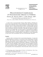
Fig. 1. A Conventional radiograph demonstrates a lytic osteochon-
dral lesion in the capitellum (arrow). B This lesion is low signal in-
tensity on a T1-weighted image and has a high signal intensity rim
on a T2-weighted axial image, C suggesting instability (arrow)
Fig. 2. Coronal FSE T2-weighted image with fat suppression
shows a full-thickness tear of the anterior band of the ulnar collat-
eral ligament at the attachment to the sublime tubercle (arrow)
visualization of the ligament complex, in chronic cases,
the development of heterotopic calcification along the
course of the ligament has been described [9].
Varus Instability
Lateral elbow instability related to isolated abnormalities
of the lateral collateral ligament complex is not as well
described as that on the medial side of the elbow. If it
were to occur, the mechanism would be a stress or force
applied to the medial side of the articulation, resulting in
compression on that side, with opening of the lateral ar-
ticulation and subsequent insufficiency of the radial col-
lateral ligament. As the radial collateral ligament attach-
es on and is intimately associated with the annular liga-
ment, an abnormality discovered in one of the structures
obligates careful inspection of the other.
Varus stress applied to the elbow may occur as an acute
injury, but rarely as a repetitive stress, as encountered on
the medial side. While lateral collateral ligament injuries
rarely occur as the result of an isolated varus stress, other
causes can commonly lead to this injury, including dislo-
cation, subluxation and overly aggressive surgery (release
of the common extensor tendon or radial head resection).
Varus instability is also tested with the elbow in full
extension and 30 degrees of flexion to unlock the olecra-
non. A varus stress is applied to the elbow while palpat-
ing the lateral joint line.
Posterolateral Rotary Instability and Elbow Dislocation
The subject of elbow instability is complex and has been
a challenge due to the difficulty in establishing the mech-
anism of injury and reliable clinical tests for diagnosis.
With the realization that elbow instability is more com-
mon than previously thought, marked advances in the un-
derstanding of this entity are occurring.
For recurrent instability, posterolateral rotary instabil-
ity is the most common pattern. This type of instability
represents a spectrum of pathology consisting of three
stages, according to the degree of soft-tissue disruption.
In stage 1, there is posterolateral subluxation of the ulna
on the humerus that results in insufficiency of the lateral
ulnar collateral ligament (Fig. 3) [10, 11, 12]. In stage 2,
the elbow dislocates incompletely so that the coronoid is
perched under the trochlea. In this stage, the radial col-
lateral ligament, and anterior and posterior portions of the
capsule are disrupted, in addition to the lateral ulnar col-
lateral ligament. Finally, in stage 3, the elbow dislocates
fully so that the coronoid rests behind the humerus. Stage
3 is subclassified into three further categories. In stage
3A, the anterior band of the medial collateral ligament is
intact and the elbow is stable to valgus stress after reduc-
tion. In stage 3B, the anterior band of the medial collat-
eral ligament is disrupted so that the elbow is unstable
with valgus stress. In stage 3C, the entire distal humerus
is stripped of soft tissues, rendering the elbow grossly un-
stable even when a splint or cast is applied with the el-
bow in a semi-flexed position. This classification system
is helpful, as each stage has specific clinical, radiograph-
ic and pathologic features that are predictable and have
implications for treatment [10].
Subluxation or dislocation of the elbow can be associ-
ated with fractures. Fracture-dislocations most common-
ly involve the coronoid and radial head, a constellation of
findings referred to as the “terrible triad” of the elbow, as
the injury complex is difficult to treat and prone to un-
satisfactory results [10]. Radial-head fractures do not
cause clinically significant instability unless the medial
collateral ligament is disrupted. An important feature of
elbow injuries to recognize is that the small flake fracture
of the coronoid, commonly seen in elbow dislocations, is
not an avulsion fracture. Nothing attaches to the very tip
of the coronoid; rather, the capsule attaches on the down-
ward slope of the coronoid, the brachialis even more dis-
tally. This fracture is a shear fracture and is likely pathog-
nomonic of an episode of elbow subluxation or disloca-
tion. A second consideration with respect to elbow dislo-
cation is that, as the ring of soft tissues is disrupted from
posterolateral to medial, the capsule is torn and insuffi-
cient. In the absence of an intact capsule, joint fluid dis-
sects through the soft-tissue planes of the forearm, negat-
ing an indirect radiographic sign of trauma in the elbow,
that of joint effusion.
Tendon Pathology
The many muscles about the elbow can be divided into
four groups: posterior, anterior, medial and lateral. The
Magnetic Resonance Imaging of the Elbow
9
Fig. 3. Coronal-fat-suppressed T1-weighted image reveals full-
thickness tears of the proximal aspects of the lateral ulnar collater-
al ligament and extensor tendon at the lateral epicondyle (arrow)
muscles of the posterior group are the triceps and an-
coneus. The muscles of the anterior group are the biceps
brachii and brachialis. The muscles in the medial group
are the pronator teres, the palmaris longus and the flex-
ors of the hand and wrist. The muscles in the lateral
group include the supinator, brachioradialis and extensor
muscles of the hand and wrist. The vast majority of
pathology encountered in the flexor and extensor groups
will be isolated to the common flexor and common ex-
tensor tendons.
The classification of tendon injuries about the elbow
can be organized by location, acuity and degree of injury.
Tendon injury related to a single isolated event is un-
common, although exceptions to this rule do occur. More
commonly, tendinous injuries in this location relate to
chronic repetitive micro-trauma. MRI is particularly well
suited, with its excellent soft-tissue contrast, to diagnose
tendon pathology. This is done primarily by close inspec-
tion of signal intensity and morphology of the tendons.
As elsewhere in the body, the tendons about the elbow
should be smooth, linear structures of low signal intensi-
ty. Abnormal morphology (attenuation or thickening) can
be seen in tendinosis or tear. If signal intensity becomes
bright or increased on fluid-sensitive sequences within
the substance of a tendon, a tear is present. Tears can be
further characterized as partial or complete. A complete
tear is diagnosed by a focal area of discontinuity (Fig. 3).
Epicondylitis and Overuse Syndromes
Chronic stress applied to the elbow is the most frequent in-
jury in athletes, and a spectrum of pathology can exist with
varying degrees of severity. The frequency of involvement
of the common flexor and extensor tendons to the medial
and lateral epicondyles, respectively, has led to the desig-
nation of “epicondylitis” as a general term applied to these
overuse syndromes. Anatomically, they are classified by
location and are further associated with sports that incite
the pathology. The injury is believed to result from extrin-
sic tensile overload of the tendon, which, over time, pro-
duces microscopic tears that do not heal appropriately.
Although these overuse entities about the elbow have
been termed “epicondylitis” for the purpose of clinical
diagnosis, inflammatory osseous changes rarely occur.
The imaging findings are those reflecting chronic change
in the tendon, as evidenced by tendinosis alone, or in con-
junction with partial or complete tear. As previously men-
tioned, the distinction between types of pathology is
made by consideration of both morphology and signal in-
tensity changes.
Medial epicondylitis involves pathology of the com-
mon flexor tendon and is associated primarily with the
sport of golfing. It has also been reported with javelin
throwers, racquetball and squash players, swimmers and
bowlers. The pronator teres and flexor carpi radialis ten-
dons are involved most frequently, resulting in pain and
tenderness to palpation over the anterior aspect of the me-
dial epicondyle of the humerus and origin of the common
10
flexor tendon. The mechanism of injury includes repeti-
tive valgus strain with pain resulting from resisting
pronation of the forearm or flexion of the wrist [13]. The
imaging findings encountered can include tendinosis, or
tendinosis with superimposed partial- or full-thickness
tear. When assessing the tendon, it is necessary to close-
ly scrutinize the underlying ulnar collateral ligament
complex to ensure integrity.
Lateral epicondylitis is the most common problem in
the elbow in athletes, and has been termed tennis elbow.
This term may be somewhat inappropriate as 95% of cas-
es of the clinical entity of lateral epicondylitis occur in
non-tennis players [14]. Moreover, it has been estimated
that 50% of people partaking in any sport with overhead
arm motion will develop this process [15].
It is associated with repetitive and excessive use of the
wrist extensors. The pathology most commonly affects the
extensor carpi radialis brevis at the common extensor ten-
don. A number of investigators have described the pathol-
ogy encountered in the degenerated tendon of this disease
process. Histologically, necrosis, round-cell infiltration,
focal calcification and scar formation have been shown
[16]. In addition, invasion of blood vessels, fibroblastic
proliferation, and lymphatic infiltration, the combination
of which are referred to as angiofibroblastic hyperplasia,
occur and ultimately lead to mucoid degeneration as the
process continues [17, 18]. The absence of a significant
inflammatory response has been emphasized repeatedly,
and may explain the inadequacy of the healing process.
The imaging findings in this process are exactly those
encountered in the clinical entity of medial epicondylitis
(Fig. 4). As on the medial side, when pathology is en-
countered in the tendon, close scrutiny of the underlying
ligamentous complex is necessary to exclude concomi-
tant injury. In particular, thickening and tears of the lat-
eral ulnar collateral ligament have been encountered with
lateral epicondylitis [13].
C. Chung, L. Steinbach
Fig. 4. Coronal T1-weighted (left) and fat-suppressed FSE T2-
weighted images show thickening and intermediate signal intensi-
ty in the common extensor tendon (arrows), consistent with tendi-
nosis (lateral epicondylitis)
Biceps Tendon
Rupture of the tendon of the biceps brachii muscle at the
elbow is rare and constitutes less than 5% of all biceps
tendon injuries [19]. It usually occurs in the dominant
arm of males. Injuries to the musculotendinous junction
have been reported, but the most common injury is com-
plete avulsion of the tendon from the radial tuberosity.
Although the injury often occurs acutely after a single
traumatic event, the failure is thought to be due to pre-ex-
isting changes in the distal biceps tendon, due to intrinsic
tendon degeneration, enthesopathy at the radial tuberosi-
ty, or cubital bursal changes. The typical mechanism of
injury relates to forceful hyperextension applied to a
flexed and supinated forearm. Athletes involved in
strength sports, such as competitive weightlifting, foot-
ball and rugby, often sustain this injury.
Clinically the patient describes a history of feeling a
“pop” or sudden sharp pain in the antecubital fossa. The
classic presentation of a complete distal biceps rupture is
that of a mass in the antecubital fossa due to proximal mi-
gration of the biceps muscle belly. Accurate diagnosis is
more difficult in cases of the rare partial tear of the ten-
don, or more common complete tear of the tendon with-
out retraction. The latter can occur with an intact bicipi-
tal aponeurosis, which serves to tether the ruptured ten-
don to the pronator flexor muscle group.
MRI diagnosis of biceps tendon pathology becomes
important in patients who do not present with the classic
history or mass in the antecubital fossa, or for evaluation
of the integrity of the lacertus fibrosus. MRI diagnosis of
tendon pathology, as previously mentioned, is largely de-
pendent on morphology, signal intensity and the identifi-
cation of areas of tendon discontinuity (Fig. 5). In the
case of the biceps tendon, an important indirect sign of
tendon pathology is the presence of cubital bursitis.
Triceps Tendon
Rupture of the triceps tendon is quite rare. The mecha-
nism of injury has been reported to result from a direct
blow to the triceps insertion, or a deceleration force ap-
plied to the extended arm with contraction of the triceps,
as in a fall. Similar to the pathology encountered in the
distal biceps tendon, most ruptures occur at the insertion
site, although musculotendinous junction and muscle bel-
ly injuries have been reported. Complete ruptures are
more common than partial tears. Associated findings
may include olecranon bursitis, subluxation of the ulnar
nerve, or fracture of the radial head. Accurate clinical di-
agnosis relies on the presence of local pain, swelling, ec-
chymosis, a palpable defect, and partial or complete loss
of the ability to extend the elbow. With more than 2 cm
of retraction between the origin and the insertion, a 40%
loss of extension strength can result [19].
For MRI diagnosis of triceps tendon pathology, it is
imperative to be aware that the triceps tendon appearance
is largely dependent on arm position. The tendon will ap-
pear lax and redundant when imaged in full extension,
whereas it is taut in flexion. The MRI features of a tear
are similar to those associated with any other tendon.
Entrapment Neuropathy
The ulnar, median and radial nerves may become com-
pressed at the elbow, leading to symptoms of entrapment
neuropathy. Abnormal nerves may have increased signal
intensity on T2-weighted images, focal changes in girth,
and deviation that may result from subluxation or dis-
placement by an adjacent mass.
Ulnar nerve entrapment most commonly occurs in the
cubital tunnel. Nerve compression may be caused by a
medial trochlear osteophyte or incongruity between the
trochlea and olecranon process [20]. Anatomic variations
also contribute. The absence of the triangular reticulum,
the anatomic roof of the cubital tunnel, occurs in about
10% of cases, permitting subluxation of the nerve with
flexion. It is necessary, therefore, to include axial images
of the flexed elbow in patients suspected of this disorder.
The presence of the anomalous anconeous
epitrochlearis muscle over the cubital tunnel causes sta-
tic compression of the nerve. In addition, there are many
other causes of ulnar neuritis, including thickening of the
overlying ulnar collateral ligament, medial epicondylitis,
adhesions, muscle hypertrophy, direct trauma, and callus
from a fracture of the medial epicondyle. MRI can be
used to identify these abnormalities and to assess the ul-
nar nerve itself. When compressed, the nerve may be-
come enlarged and edematous. If conservative treatment
fails, the nerve can be transposed anteriorly, deep to the
flexor muscle group, or more superficially, in the subcu-
taneous tissue. One can follow these patients with MRI
Magnetic Resonance Imaging of the Elbow
11
Fig. 5. Axial-fat-suppressed T2-weighted image shows complete
disruption of the distal biceps at the radial tuberosity (arrow)
postoperatively if they become symptomatic to deter-
mine whether symptoms are secondary to scarring or in-
fection around the area of nerve transposition.
Compression of the median nerve may be seen with
osseous or muscular variants and anomalies, soft-tissue
masses and dynamic forces. In the pronator syndrome,
compression occurs as the median nerve passes between
the two heads of the pronator teres and under the fibrous
arch of the flexor digitorum profundus.
The radial nerve can become entrapped following di-
rect trauma, mechanical compression by a cast or overly-
ing space-occupying mass, or a dynamic compression as
a result of repeated pronation, forearm extension, and
wrist flexion, as is seen in violinists and swimmers.
Motor neuropathy of the hand extensors is a dominant
feature when the posterior interosseous nerve is en-
trapped [21].
References
1. Rosenberg ZS, Beltran J, Cheung YY (1994) Pseudodefect of
the capitellum: potential MR imaging pitfall. Radiology
191(3):821-823
2. Rosenberg ZS, Beltran J, Cheung Y, Broker M (1995) MR
imaging of the elbow: normal variant and potential diagnostic
pitfalls of the trochlear groove and cubital tunnel. Am J
Roentgenol164(2):415-418
3. Pincivero DM, Heinrichs K, Perrin DH (1994) Medial elbow
stability. Clinical implications. Sports Med 18(2):141-148
4. Bradley JP, Petrie RS (2001) Osteochondritis dissecans of the
humeral capitellum. Diagnosis and treatment. Clin Sports Med
20(3):565-590
5. Patel N, Weiner SD (2002) Osteochondritis dissecans involv-
ing the trochlea: report of two patients (three elbows) and re-
view of the literature. J Pediatr Orthop 22(1):48-51
6. Steinbach LS, Palmer WE, Schweitzer ME (2002) Special fo-
cus session. MR arthrography. Radiographics 22(5):1223-1246
7. Carrino JA, Smith DK, Schweitzer ME (1998) MR arthrogra-
12
phy of the elbow and wrist. Semin Musculoskelet Radiol
2(4):397-414
8. Phillips CS, Segalman KA (2002) Diagnosis and treatment of
post-traumatic medial and lateral elbow ligament incompe-
tence. Hand Clin 18(1):149-159
9. Mulligan SA, Schwartz ML, Broussard MF, Andrews JR
(2000) Heterotopic calcification and tears of the ulnar collat-
eral ligament: radiographic and MR imaging findings. Am J
Roentgenol 175(4):1099-1102
10. O’Driscoll SW (2000) Classification and evaluation of recur-
rent instability of the elbow. Clin Orthop 370:34-43
11. Potter HG, Weiland AJ, Schatz JA, Paletta GA, Hotchkiss RN
(1997) Posterolateral rotatory instability of the elbow: useful-
ness of MR imaging in diagnosis. Radiology 204(1):185-189
12. Dunning CE, Zarzour ZD, Patterson SD, Johnson JA, King GJ
(2001) Ligamentous stabilizers against posterolateral rotatory in-
stability of the elbow. J Bone Joint Surg Am 83-A(12):1823-1828
13. Bredella MA, Tirman PF, Fritz RC, Feller JF, Wischer TK,
Genant HK (1999) MR imaging findings of lateral ulnar col-
lateral ligament abnormalities in patients with lateral epi-
condylitis. Am J Roentgenol 173(5):1379-1382
14. Frostick SP, Mohammad M, Ritchie DA. Sport injuries of the
elbow. Br J Sports Med 199933(5):301-311
15. Field LD, Savoie FH (1998) Common elbow injuries in sport.
Sports Med 26(3):193-205
16. Nirschl RP, Pettrone FA (1979) Tennis elbow. The surgical
treatment of lateral epicondylitis. J Bone Joint Surg Am
61(6A):832-839
17. Regan W, Wold LE, Coonrad R, Morrey BF (1992)
Microscopic histopathology of chronic refractory lateral epi-
condylitis. Am J Sports Med 20(6):746-749
18. Nirschl RP (1992) Elbow tendinosis/tennis elbow. Clin Sports
Med 11(4):851-870
19. Rettig AC (2002) Traumatic elbow injuries in the athlete.
Orthop Clin North Am 33(3):509-522
20. Kim YS, Yeh LR, Trudell D, Resnick D (1998) MR imaging of
the major nerves about the elbow: Cadaveric study examining
the effect of flexion and extension of the elbow and pronation
and supination of the forearm. Skeletal Radiol 27:419-426
21. Yanagisawa H, Okada K, Sashi R (2001) Posterior in-
terosseous nerve palsy caused by synovial chondromatosis of
the elbow joint. Clin Radiol 6(6):510-514
C. Chung, L. Steinbach
Introduction
Musculoskeletal trauma is common and the distal upper
extremity is one of the most frequent sites of injury.
Imaging of hand and wrist injuries should always begin
with conventional radiographs. While computed tomog-
raphy (CT) and magnetic resonance imaging (MRI) are
very helpful in some cases, their overall impact on trau-
ma imaging in the hand and wrist is small. Radiographs
remain the primary diagnostic modality. It is therefore es-
sential for radiologists who work in a trauma and emer-
gency setting to be familiar not only with the normal ra-
diographic anatomy of the hand and wrist but also with
the range of injuries that can occur. Our learned col-
league, Lee F. Rogers, put it all quite simply in a few
statements that can be called “Rogers’ Rules”: Rule #1,
make the diagnosis; Rule #2, avoid embarrassment; Rule
#3, stay out of court. In order to meet these objectives, we
must get adequate radiographs and we must interpret
them correctly. Thus, not only should we know where to
look when there is nothing obvious at first glance but we
must also know where else to look when there are obvi-
ous findings.
Normal Anatomy
Before considering injury patterns and mechanisms, it es-
sential to have a working knowledge of the normal radi-
ographic anatomy. The standard trauma series for the hand
includes three views, which should cover the anatomy
from the radiocarpal joint to the finger tips. These views
are a pronated frontal view (PA), a pronated oblique view
and a lateral view. For wrist injuries, these same three pro-
jections are used but are centered and collimated to cover
the wrist area, from the metadiaphyses of the distal radius
and ulna to the proximal metacarpal diaphyses. A fourth
view, the so-called scaphoid view, should always be in-
cluded in the wrist trauma series. This is a PA view, more
tightly collimated than the other three, that is centered on
the scaphoid, with the wrist in maximum ulnar deviation.
This view rotates the scaphoid about its short axis, pre-
senting the waist of the bone in profile.
When evaluating radiographs of the wrist, several
anatomic points are important to observe. First, look at
the soft tissues. On the lateral view, convexity of the dor-
sal soft-tissue margin represents soft-tissue swelling
around the carpus and distal radius. It is often a sign of
subtle underlying bone or joint injury. Also on the lateral
view is the pronator fat pad, which lies parallel to the pal-
mar cortex of the distal radius in most normal individu-
als. When the distal radius is fractured, the pronator fat
pad will be deformed and displaced, becoming convex in
a palmar direction. A second but less frequently present
fat pad is the scaphoid fat pad. When present, it should
be relatively straight and lateral and parallel to the
scaphoid bone. If the scaphoid fat pad is convex lateral-
ly, a scaphoid fracture should be suspected.
There are several lines and angles that can be drawn in
and around the carpus that are helpful in detecting in-
juries which may otherwise be overlooked. On the PA
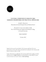
view, the three carpal arcs (of Gilula) are smooth curves
that will be disrupted in injuries to the intercarpal joints.
Arc I is drawn across the proximal surfaces of the proxi-
mal carpal row. Arc II is drawn across the distal surfaces
of the proximal carpal row. Arc III is drawn across the
proximal surfaces of the distal carpal row (Fig. 1). The
long axis of the capitate, drawn on the PA view, should
bisect the third metacarpal shaft regardless of the degree
of ulnar or radial deviation (Fig. 1).
The second through fifth carpometacarpal joints
should be seen in profile on a good-quality PA view,
forming a “lazy M” shape on the radiograph (Fig. 1).
While it may not always be possible to see the entire lazy
M, most of it should be visible if the wrist is positioned
correctly. The key to the carpometacarpal joints is to look
at those joint surfaces that have been profiled by the X-
ray beam. If one side of a joint (carpal or metacarpal) is
seen in profile, the other side of that same joint should be
seen in profile and parallel to its mate. When only one
side is profiled or the articular surfaces are overlapping
or not parallel, the joint is either subluxed or dislocated.
On the lateral view, the distal radial articular surface
and proximal lunate articular surface should form paral-
lel curves. Similarly, the distal lunate and proximal capi-
IDKD 2005
Radiology of Hand and Wrist Injuries
A.J. Wilson
University of Washington, Harborview Medical Center, WA, USA
tate should form parallel curves (Fig. 2). If one or more of
these articulations are not parallel, the carpus has been
dislocated or subluxed. By determining the long axes of
the scaphoid, lunate and capitate on the lateral view and
measuring the angles between them, the presence of vari-
ous carpal instabilities and/or ligament injuries can be
predicted. The normal scapholunate angle lies between 30
and 60°. The normal capitolunate angle is ±30° (Fig. 3).
14
An increase in the scapholunate angle indicates a dorsal
intercalated segment instability (DISI). A decrease in the
scapholunate angle indicates a palmar intercalated seg-
ment instability (PISI). In both DISI and PISI, the capi-
tolunate angle will usually be increased.
The articular cartilage has approximately the same
thickness throughout the carpus. If the apparent space be-
tween any two carpal bones appears wider than the ap-
parent space between the others, a ligament disruption
has probably occurred. The joints most commonly affect-
ed by ligament injuries are the scapholunate and lunotri-
quetral joints. Therefore, the apparent space between the
lunate and scaphoid and the lunate and triquetrum should
always be carefully evaluated.
Injury Patterns and Mechanisms
The majority of upper-extremity injuries are the result of a
fall onto the out-stretched hand (FOOSH). Many of these
FOOSH injuries are concentrated around the wrist and some
involve the hand. Those around the wrist are somewhat age-
dependent. In very small children, whose bones are rela-
tively soft, buckle or torus fractures of the distal radius are
the most common injuries. While most of these are obvious,
the findings may be limited to very subtle angulation of the
cortex, seen only on the lateral view. These injuries are of-
ten associated with similar fractures of the distal ulna.
As adolescents enter the growth spurt associated with
puberty, their physes become weaker and subject to frac-
ture. The commonest FOOSH injuries in this age group
are physeal fractures of the distal radius, which may or
may not be associated with ulnar fractures, particularly of
the styloid process. These physeal fractures are described
in the Salter-Harris classification as follows: type 1, phy-
seal shear injury; type 2, physeal shear with marginal
metaphyseal fracture; type 3, physeal shear with epiphy-
seal fracture; type 4, epiphyseal, physeal and metaphyseal
fractures; type 5, physeal crush injury. In general, these
injuries are displaced and easy to recognize, with excep-
tion of type 5 injuries. However, in some patients, partial
auto-reduction may make a type 1 or 2 fracture difficult
to find on the radiographs. Secondary signs, such as dis-
placement the pronator fat pad, may be helpful.
In young adults, the bones are at their strongest. This
puts the ligaments at increased risk. The center of most
frequent injury moves to the carpus, where fractures and
dislocations are most likely to occur in the so-called zone
of vulnerability (Fig. 4). This zone runs in a curved man-
ner across the radial styloid, scaphoid, capitate, triquetrum
and ulnar styloid. The commonest injury within the zone
of vulnerability is a scaphoid fracture. The second com-
monest is an avulsion fracture of the dorsal triquetrum.
Next in frequency are various dislocations and fracture
dislocations, involving predominantly the midcarpal joint.
Scaphoid fractures are important to consider in all injured
wrists for two reasons. First, they have a high incidence of
nonunion and ischemic necrosis. Second, they tend to be
truly nondisplaced and may be difficult to see on radi-
A.J. Wilson
Fig. 1. The arcs of Gilula, lazy M and capitate axis
Fig. 2. The radial, lunate and
capitate articulations
Fig. 3. The scapholunate and
capitolunate angles
ographs taken on the day of injury. Follow up radiographs,
after 2 weeks, will often show these occult fractures. If
prompt diagnosis is needed, MRI is much more sensitive
in revealing nondisplaced fractures than radiography.
In older adults, as osteoporosis sets in and the bones be-
come weaker, the distal radius once again becomes the
commonest site for FOOSH injuries. The most common va-
riety of distal radial fracture is one in which the distal frac-
ture fragment is displaced and angulated in a dorsal direc-
tion. This fracture was first described by Abraham Colles,
in 1814, and now bears his name. Since Colles described
this fracture 81 years before the discovery of X-rays, he did
not know the detail or radiographic manifestations of this
injury. His real contribution was to point out that these are
fractures, not dislocations. He showed that they could be re-
duced and splinted and could heal with excellent results.
When the deformity is in the opposite direction (palmar) we
refer to the injury as a Smith’s fracture. When there is no
deformity, the injury should be described simply as a
nondisplaced, distal, radial fracture. Fractures of the ulnar
styloid commonly occur in association with distal radial
fractures but are not always present. Their presence does not
change the designation as a Colles’, Smith’s or nondis-
placed fracture. One of the most important findings to ob-
serve in these fractures is extension into the distal radial ar-
ticular surface. Intra-articular fractures often require surgi-
cal repair and should be further evaluated with CT.
When fractures of the distal radius are associated with ra-
diocarpal dislocations, they are referred to as “Barton’s frac-
tures”. If the dorsal lip is fractured, the carpus will be dis-
placed dorsally. This is referred to as a “dorsal Barton’s frac-
ture”. Conversely, if the palmar lip of the radius is fractured,
the carpus will be displaced palmarly. This is referred to as
a “palmar Barton’s fracture”. While pure dislocations of the
radiocarpal joint can occur without radial lip fractures, they
are much less frequent than Barton’s fracture-dislocations.
Carpal dislocations
Most carpal dislocations involve the midcarpal joint,
which is between the proximal and distal carpal rows. On
the lateral view, these injuries show disruption of the nor-
mal relationship between lunate and capitate, usually with
dorsal displacement of the capitate. The distal articular
surface of the lunate is “empty”. On the PA projection, the
lunate takes on a triangular shape as it rotates about its
horizontal axis. Arcs I and II are disrupted, while arc III is
normally intact. These dislocations usually occur around
the lunate and are therefore called “perilunate” disloca-
tions. The majority of perilunate dislocations are associat-
ed with fractures through the scaphoid waist but any frac-
ture within the zone of vulnerability is possible. Perilunate
dislocation without an associated fracture is not uncom-
mon. The description of the injury includes the fractures
and the words “perilunate dislocation”. For example: a
trans-radial, trans-scaphoid, trans-capitate, perilunate dis-
location would be one of these dislocations with fractures
through the radial styloid, scaphoid waist and capitate
neck. Ulnar styloid fractures are frequently present but are
usually not included in the descriptive classification.
When the lunate is displaced from the radial articular sur-
face in a midcarpal joint disruption, it is called a “lunate
dislocation”. “Midcarpal dislocation” is the term used to
describe the intermediate position, when the capitate is
dislocated from the lunate and the lunate is subluxed from
the radius. This term is confusing, since all of these pat-
terns are dislocations of the midcarpal joint.
Other, less-common, carpal dislocations include the
longitudinal variety. These are the result of high-energy
trauma and separate the carpus into medial and lateral
portions. They are usually obvious radiographically and
frequently require surgical repair.
Carpometacarpal dislocations
Perhaps the most commonly missed serious injury to the
hand and wrist is dislocation along the carpometacarpal
joint. These injuries can be surprisingly subtle on initial ra-
diographs. In spite of this, they are serious injuries that usu-
ally require surgical repair. There are two keys to finding
them:. (1) they are frequently associated with avulsion frac-
tures of the distal carpals or proximal metacarpals; (2) on at
least one of the standard views, the affected car-
pometacarpal joints will show loss of parallelism. On the lat-
eral radiograph, dorsal displacement of the metacarpal bases
may be apparent. So, the important point to remember is:
any time a fracture at the carpometacarpal junction is seen,
a dislocation must be assumed, until proven otherwise.
CT or fluoroscopy may be required to resolve this issue.
Radiology of Hand and Wrist Injuries
15
Fig. 4. The zone of vulnerability
Metacarpal Injuries
While metacarpal fractures may occur in FOOSH, they are
more frequent when the fist is closed. In other words, they
are most commonly associated with punching, usually dur-
ing a fist fight. A well placed punch will line up the sec-
ond metacarpal with the radius, often resulting in a frac-
ture of the second metacarpal neck. However, most bare-
fisted fighters have not been trained to punch correctly and
strike glancing blows with the ulnar aspect of the fist.
These blows frequently result in fractures of the fifth
metacarpal neck. This has been called the “boxers fracture”
but would be more accurately defined as the “amateur
street-fighter’s fracture”. The head of the metacarpal is typ-
ically displaced and angulated in a palmar direction. If the
fracture is allowed to heal in this position, the next time the
individual participates in a fist fight, a fracture of the
fourth metacarpal neck is likely, as the fifth is now de-
pressed and allows the fourth to receive the maximum
force of the punch. In indirect trauma from FOOSH or oth-
er mechanisms, twisting injuries to the metacarpal may oc-
cur, resulting in spiral, diaphyseal, fractures.
Finger Injuries
Finger fractures can occur from FOOSH but are more
commonly the result of direct trauma to the fingers. As in
the metacarpals, twisting injuries will result in spiral, dia-
physeal, phalangeal, fractures. Direct dorsal blows to the
finger tip, such as hitting with a hammer, result in burst
fractures of the terminal tuft. These are typically commin-
uted but minimally displaced. Injuries in which the finger
is bent backward may result in dislocation of the interpha-
langeal joint or avulsion of the volar plate. The volar plate
is a fibrocartilaginous structure at the insertion of the short
flexor tendon, at the palmar base of the middle phalanx.
When the finger is acutely bent backwards, this plate may
be avulsed and often takes a small fragment of bone with
it. These injuries can be subtle and may be visible only on
the lateral view. When the finger is stuck directly on its tip,
as in a failed attempt to catch a hard ball, the tip of the fin-
ger is forced palmarly against tensed flexor and extensor
tendons. This results in avulsion of the extensor tendon in-
sertion, at the dorsal base of the distal phalanx, sometimes
with a small avulsed fragment of bone. Detachment of the
extensor tendon produces a characteristic finger deformity
in which there is persistent slight flexion of the distal in-
terphalangeal joint. This deformity has been variously de-
scribed as “mallet finger” or “baseball finger”. It is readi-
ly diagnosed, both clinically and on the lateral radiograph,
with or without an avulsion fracture.
Penetrating injuries
Penetrating injuries to the hand and wrist result from stab
wounds, gunshot injuries and explosions with the grasp.
16
The latter are most commonly seen around times of cel-
ebration with fireworks. In the United States, these in-
juries most frequently occur around July Fourth and New
Year’s Eve. Penetrating injuries are very variable, de-
pending on the location and force of penetration. They
are often devastating, resulting in multiple fractures, se-
vere soft-tissue loss and a hand beyond repair. The radi-
ologist’s job is simple: describe what is broken and what
is missing. Penetrating trauma rarely presents the same
challenges as blunt trauma.
Advanced Imaging
As stated earlier, CT and MRI have a limited role in di-
agnosing hand and wrist trauma. However, in certain sit-
uations, they can prove invaluable.
CT often provides the best method for characterizing
complex injuries. It is far more reliable than radiography
for the assessment of fracture healing. CT is the most re-
liable method for evaluating alignment of the distal ra-
dioulnar joints in suspected instability, dislocation or sub-
luxation. In pre-operative planning, CT gives the most re-
liable assessment of comminution, displacement or in-
volvement of articular surfaces. It is also helpful in cal-
culating the volume of bone graft that is needed for sur-
gical repair.
MRI remains the most sensitive and accurate method
for excluding occult fractures. With radiography, 2
weeks of immobilization may be required before an oc-
cult fracture can be reliably excluded. By contrast, with
MRI, a definitive decision can usually be made on the
day of injury. In professional athletes and others, whose
occupations do not lend themselves to prolonged or un-
necessary immobilization, such prompt diagnosis is im-
portant.
Suggested Reading
Fisher MR, Rogers LF, Hendrix RW (1983) Systematic approach
to identifying fourth and fifth carpometacarpal dislocations.
AJR 140:319
Gilula LA (1990) The traumatized hand and wrist. WB Saunders,
Philadelphia, pp 94-97
Gilula LA (1990) The traumatized hand and wrist. WB Saunders,
Philadelphia, pp 287-314
Gilula LA, Yin YM (1996) Imaging of the wrist and hand. WB
Saunders, Philadelphia, pp 43-224
Gilula LA, Yin YM (1996) Imaging of the wrist and hand. WB
Saunders, Philadelphia, pp 311-318
Hill N (1970) Fractures and dislocations of the carpus. Orthop Clin
North 1:275
Rawles JG (1988) Dislocations and fracture at the carpometacarpal
joints of the fingers. Hand Clin 4:103
Rogers LF (2001) Radiology of skeletal trauma, 3rd Edition.
Churchill Livingstone, Philadelphia, pp 813-855
Rogers LF (2001) Radiology of skeletal trauma, 3rd Edition.
Churchill Livingstone, Philadelphia, pp 904-929
Wagner CJ (1959) Fracture-dislocations of the wrist. Clin Orthop
15:181
A.J. Wilson
Introduction
This chapter will emphasize general principles when as-
sessing a variety of lesions of the hand and wrist. An ap-
proach to analyzing the wrist and hand bones will be pro-
vided, followed by a discussion of applications of these
principles with respect to trauma, infection, neoplasia,
arthritis, and metabolic bone disease. Obviously it is im-
possible to cover all of musculoskeletal imaging and
pathology in a short article; however, some major points
will be emphasized in each of these different areas, with
the most emphasis placed on complex carpal trauma.
Overview of Analysis
As described by D. Forrester [1], looking at the muscu-
loskeletal system anywhere can be evaluated by the “A,
B, C, D, ‘S” system. “A” stands for alignment, “B” for
bone mineralization, “C” for cortex, cartilage and joint
space abnormalities, “D” for distribution of abnormali-
ties, and “S” for soft tissues. Utilizing these principles
will help keep one from missing major observations.
Starting with “S” for soft tissues will keep one from for-
getting to evaluate soft tissues. Recognizing soft-tissue
(“S”) abnormalities will point to an area of major abnor-
mality and should trigger a second or third look at the
center of the area of soft-tissue swelling to see whether
there is an underlying abnormality. The soft tissues dor-
sally over the carpal bones are normally concave. When
the soft tissues over the dorsum of the wrist are straight
or convex, swelling should be suspected. The pronator fat
line volar to the distal radius suggests deep swelling when
it is convex outward, as normally it should be straight or
concave [2]. Soft-tissue swelling along the radial and ul-
nar styloids may be seen in synovitis or trauma. Swelling
along the radial or ulnar side of a finger joint can indi-
cate collateral ligament injury. Exceptions to this state-
ment exist along the radial side of the index finger and
the ulnar side of the small finger. Focal swelling circum-
ferentially around one interphalangeal or metacarpopha-
langeal joint is highly suggestive of capsular or joint
swelling. Another cause for diffuse swelling along one
side of the wrist or finger can be tenosynovitis.
The evaluation of alignment (“A”) allows deviations
from normal to be recognized. Angular deformities are
commonly seen in arthritis. Dislocations and carpal in-
stabilities manifest as abnormalities in alignment.
In evaluating bone mineralization (“B”), different pat-
terns are evident. Acute bone demineralization presents
as subcortical bone loss in the metaphyseal areas and at
the ends of bones, in regions of increased vascularity of
bones. A typical example is the young person who has
an injured part of the body placed in a cast with subse-
quent development of rapid demineralization. Diffuse
even demineralization commonly develops over longer
periods of time and may be seen in older people with dif-
fuse osteopenia of age and also from prolonged disuse.
Focal osteopenia, especially associated with cortical
loss, should raise the question of infection or a more
acute inflammatory process in that area of local bone
demineralization.
“C” reminds us to look at all the joint spaces as well as
the margins of these joints and bones for cartilage space
narrowing, erosions, and other cortical abnormalities.
“D” refers to the distribution of abnormalities. It is
most vividly exemplified by the distribution of erosions,
as may be seen distally in psoriasis and more proximally
in rheumatoid arthritis.
Three major concepts relate to alignment: (1) paral-
lelism, (2) overlapping articular surfaces, and (3) three
carpal arcs [3-5]. All three can be especially applied to the
carpal bones., while the first two can be applied throughout
the body. Parallelism refers to the fact that any anatomic
structure that normally articulates with an adjacent anatom-
ic structure should show parallelism between the articular
cortices of those adjacent bones. This is exactly how jigsaw
puzzles work. If there is a piece of a jigsaw puzzle out of
place, then that piece loses its parallelism to adjacent
pieces. Anatomically, this would cause overlapping articu-
lar surfaces. Therefore, the concepts of parallelism and
overlapping articular surfaces are related. If there is overlap
of normally articulating surfaces, there should be disloca-
tion or subluxation at the site of those overlapping surfaces.
IDKD 2005
Wrist and Hand
L.A. Gilula
Mallinckrodt Institute of Radiology, Washington University School of Medicine, St. Louis, MO, USA
This does not apply if one bone is foreshortened or bent, as
with overlapping phalanges on a PA view of a flexed fin-
ger. In that situation, one phalanx would overlap the adja-
cent phalanx, but in the flexed PA position one would not
normally see parallel articular surfaces at that joint.
The third alignment concept refers to the fact that three
carpal arcs can be drawn in any normal wrist when the wrist
and hand are in a neutral position, i.e., the third metacarpal
and the radius are coaxial. Arc I is a smooth curve along the
proximal convex surfaces of the scaphoid, lunate and tri-
quetrum. Arc II is a smooth arc drawn along the distal con-
cave surfaces of these same three carpal bones. Arc III is a
smooth arc that is drawn along the proximal convex sur-
faces of the capitate and hamate [3, 6]. When one of these
arcs is broken at a joint, then something is probably wrong
with that joint, as ligament disruption; or when broken at a
bone surface, a fracture. Two normal exceptions to the de-
scriptions of these arcs exist. In arc I, the proximal distal di-
mension of the triquetrum may be shorter than the appos-
ing portion of the lunate. A broken arc I at the lunotrique-
tral joint is a congenital variation when this situation arises.
Another congenital variation exists where there is a promi-
nent articular surface of the lunate that articulates with the
hamate, a type II lunate. (A type I lunate is the lunate with
one distal smooth concave surface; in a type II lunate there
is one concave articular surface that articulates with the
capitate and a second concavity, the hamate facet of the lu-
nate, which articulates with the proximal pole of the ha-
mate). In a type II lunate, arc II may be broken at the distal
surface of the lunate, where there is a normal concavity at
the lunate hamate joint. Similarly, there can be a slight jog
of arc III at the joint between the capitate and hamate in this
type of wrist; however, the overall outer curvatures of the
capitate and hamate are still smooth. At the proximal mar-
gins of the scapholunate and lunotriquetral joints, these
joints may be wider due to curvature of these bones.
Observe the outer curvature of these bones when analyzing
the carpal arcs. Also, to analyze the scapholunate joint
space width, look at the middle of the joint between paral-
lel surfaces of the scaphoid and lunate to see whether there
is any scapholunate space widening compared to a normal
capitolunate joint width in that same wrist.
The hand and wrist can be analyzed very promptly af-
ter first surveying the soft tissues by looking at the over-
all alignment, bone mineralization and cortical detail as
one looks at the radiocarpal joints, the intercarpal joints
of the proximal carpal row, midcarpal joint, intercarpal
joints of the distal carpal row, carpometacarpal joints,
and interphalangeal joints. Analyzing these surfaces and
bones evaluated on all views leads to a diagnosis. It is
preferable to carefully analyze the PA view of the wrist
first as this view will provide the most information. The
lateral and oblique views are merely used for confirma-
tion and clarification of what is actually present on the
PA view. An exception to this comment is the need to
closely evaluate the soft tissues on the lateral as well as
the PA view. The following sections will discuss applying
these principles to more specific abnormalities.
18
Trauma
Traumatic conditions of the wrist basically can be classi-
fied as fractures, fracture-dislocations, and soft-tissue ab-
normalities, which include ligament instabilities.
Analysis of the carpal arcs, overlapping articular sur-
faces, and parallelism will help determine what exact
traumatic abnormality is present. Recognizing which
bones normally parallel each other also identifies which
bones have moved together as a unit away from a bone
that has overlapping adjacent surfaces. A majority of the
fractures and dislocations about the wrist are of the per-
ilunate type, in which there is a dislocation with or with-
out adjacent fractures taking place around the lunate. The
additional bones that may be fractured are named first
with the type of dislocation mentioned last. For the per-
ilunate type of dislocations, whatever bone centers over
the radius (the capitate or lunate) is considered to be “in
place”. Therefore, if the lunate is centered over the radius,
this would be a perilunate type of dislocation. If the cap-
itate is centered over the radius and the lunate is not, this
would be a lunate dislocation. Therefore, if there were
fractures of the scaphoid and capitate, dorsal displace-
ment of the carpus with respect to the lunate, and the lu-
nate was still articulating or centered over the radius, this
would be called a transscaphoid transcapitate dorsal per-
ilunate dislocation. Another group of fracture-disloca-
tions that occur in the wrist are the axial fracture-dislo-
cations, in which a severe crush injury may split the wrist
along an axis around a carpal bone other than the lunate,
such as perihamate or peritrapezial axial dislocation, usu-
ally with fractures [7].
Ligamentous Instability
There are many types of ligament instabilities, including
very subtle types; however, there are five major types of
ligament instabilities that can be recognized readily based
on plain radiographs. These refer to the lunate as being
an “intercalated segment” between the distal carpal row
and the radius, similar to the middle or intercalated seg-
ment between two links in a three-link chain. Normally
there can be a small amount of angulation between the
capitate, lunate, and the radius on the lateral view.
However with increasing lunate angulation, especially as
seen on the lateral view, an instability pattern may be pre-
sent. If the lunate tilts too far dorsally, it would be called
a dorsal intercalated segmental condition; if the lunate
tilts too far volarly, it would be called a volar or palmar
intercalated segmental problem. Therefore, if the lunate is
tilted too far dorsally (so that the capitolunate angle is
more than 30°and/or the scapholunate angle is more than
60°-80°), this would be called a dorsal intercalated seg-
mental instability (DISI) pattern. If the lunate is tilted too
far volarly or palmarly (a capitolunate angle of more than
30° or scapholunate angle of less than 30°), this would be
a volar intercalated segmental instability (VISI) or pal-
L.A. Gilula
mar intercalated segmental instability (PISI) pattern.
When there is a “pattern” of instability, a true instability
can be further evaluated with a dynamic wrist instability
series performed under fluoroscopic control [8,9]. When
there is abnormal intercarpal motion and abnormal align-
ment, this supports the radiographic diagnosis of carpal
instability. By comparison with the opposite wrist, the
questionable wrist can be evaluated for instability with
lateral flexion, extension, and neutral views, PA and AP
views with radial, neutral, and ulnar deviation views.
Fist-compression views in the supine position may help
widen the scapholunate joint in some patients. Ulnar
carpal translation is a third type of carpal instability [8].
If the entire carpus moves too far ulnarly, as recognized
by more than one-half of the lunate positioned ulnar to
the radius when the wrist and hand are in neutral position,
this would be an ulnar carpal translation type I. If the
scaphoid is in the normal position relative to the radial
styloid, but there is scapholunate dissociation and the re-
mainder of the carpus moves too far ulnarly, as men-
tioned for ulnar carpal translation type I, this is called ul-
nar carpal translation type II. The fourth and fifth types
of carpal instabilities relate to the carpus displacing dor-
sally and volarly off the radius. If the carpus, as identi-
fied by the lunate, has lost its normal articulation with the
radius in the lateral view and is displaced dorsally off the
radius, this is called dorsal radiocarpal instability, or dor-
sal carpal subluxation. It occurs most commonly follow-
ing a severe dorsally impacted distal radius fracture. If
the carpus is displaced palmarly off the carpus, as iden-
tified between the lunate and its articulation with the ra-
dius, and the remainder of the carpus has moved with the
lunate, this would be called a palmar carpal subluxation.
There are other types of carpal instability patterns that
are better detected more by physical examination; these
will not be covered here.
Infection
Infection should be suspected when there is an area of
cortical destruction with pronounced osteopenia. It is not
uncommon to have patients present with pain and
swelling, and clinically infection may not be suspected
when it is chronic, as with an indolent type of infection
such as tuberculosis. Soft-tissue swelling is a key point
for this diagnosis as for other abnormalities of the wrist,
as mentioned above. Therefore, the diagnosis of infection
is most likely when there is swelling and associated os-
teopenia as well as cortical destruction ,or even early fo-
cal joint-space loss without cortical destruction.
Neoplasia
When there is an area of abnormality, it helps to determine
the gross area of involvement, then look at the center of
the abnormality [10]. If the center of the abnormality is in
bone, then probably the lesion originated within the bone.
When the center of abnormality is in soft tissues, a lesion
originating in soft tissues should be suspected. When there
is a focal area of bone loss or destruction or even a focal
area of soft-tissue swelling with or without osteopenia,
neoplasia is a major consideration. Whenever neoplasia is
a concern on an imaging study, infection should also be
considered. To analyze a lesion within a bone, look at the
margins of the lesion to see whether it is well-defined and
whether it has a thin to thick sclerotic rim. Evaluate the
endosteal surface of the bone to see whether there is scal-
loping or concavities along the endosteal surface of the
bone. Concavities representing endosteal scalloping are
characteristic of cartilage tissue. This would be typical for
an enchondroma, which is the most common intraosseous
bone lesion of the hands. The matrix of the lesion should
also be evaluated to see whether there are dots of calcium
that can be seen in cartilage, or whether there is a more
diffuse type of bone formation as occurs in an osseous
type of tumor as from osteosarcoma. As elsewhere in the
body, if a lesion is very well-defined and if there is bone
enlargement, these are indicative of an indolent or a less
aggressive type of lesion. The presence of cortical de-
struction supports the finding of an aggressive lesion,
such as malignancy or infection. To determine the extent
of a lesion, magnetic resonance (MR) is the preferred
method of imaging. Bone scintigraphy can be very valu-
able to survey for osseous lesions throughout the body, as
many neoplastic conditions spread to other bones or even
to the lung.
When there is a lesion is in the soft tissue of the hand,
especially with pressure effect on an adjacent bone, a gi-
ant cell tumor of the tendon sheath should be suspected.
Ganglion is another cause for a focal swelling in the hand,
but usually that occurs without underlying bone deformi-
ty. Glomus tumor is a less common, painful soft-tissue le-
sion that may be detected with ultrasound or MR imag-
ing. Occasionally, a glomus tumor will cause a pressure
effect on bone, especially on the distal phalanx under the
nail bed.
Arthritis
Using the above scheme of analyzing the hand, wrist, and
musculoskeletal system [11], swelling can indicate cap-
sular involvement as well as synovitis. The overall evalu-
ation of alignment shows deviation of the fingers at the
interphalangeal and metacarpophalangeal joints in addi-
tion to subluxation or dislocation at the interphalangeal,
metacarpophalangeal, or intercarpal or radiocarpal joints.
Joint-space loss, the sites of erosions, and the sites of
bone production are important to recognize. When iden-
tifying the abnormalities, the metacarpophalangeal joint
capsules, especially of the index, long and small fingers,
should be examined carefully to determine whether they
are convex, as occurs in for capsular swelling. This can
help in establishing whether this is primarily a synovial
Wrist and Hand
19