Báo cáo khoa học: An engineered right-handed coiled coil domain imparts extreme thermostability to the KcsA channel docx
Bạn đang xem bản rút gọn của tài liệu. Xem và tải ngay bản đầy đủ của tài liệu tại đây (2.75 MB, 11 trang )
An engineered right-handed coiled coil domain imparts
extreme thermostability to the KcsA channel
Zhiguang Yuchi
1
, Victor P. T. Pau
2
, Bridget X. Lu
1
, Murray Junop
1
and Daniel S. C. Yang
1
1 Department of Biochemistry and Biomedical Sciences, Faculty of Health Sciences, McMaster University, Hamilton, Canada
2 Department of Biochemistry, Temple University School of Medicine, Philadelphia, PA, USA
Introduction
Tetrameric architecture is a common character shared
by cation channels, including potassium, sodium, cal-
cium, nonselective, glutamate gated, cyclic nucleotide
gated (CNG), transient receptor potential channels,
and other ion channels [1,2]. Although they differ from
each other in terms of selectivity and physiological
activator, they all have to organize to a tetrameric
arrangement in order to be functional. The ion con-
ducting function is fulfilled by a central ion conducting
pore composed of selectivity filters and a-helices
arranged in four-fold or pseudo four-fold symmetry.
Most potassium channels form homo- or heterotet-
ramers. Several different cytoplasmic tetramerization
domains have been found to be important for proper
channel assembly. For example, T1, an N-terminal
tetramerization domain, is used by the Kv channel,
whereas C-terminal tetramerization domains are used
by ether-a-go-go (EAG) channels, potassium inwardly
rectifying (Kir) channels, calcium activated channels
and CNG channels [3–11].
Despite different families of potassium channels
being structurally similar and often co-expressed in the
Keywords
chimeric channel; coiled coil; KcsA; RHCC;
tetramerization domain
Correspondence
D. S. C. Yang, Department of Biochemistry
and Biomedical Sciences, Faculty of Health
Sciences, McMaster University, 1200 Main
Street West, Hamilton, Ontario L8N 3Z5,
Canada
Fax: +1 905 522 9033
Tel: +1 905 525 9140 ext. 22455
E-mail:
(Received 10 June 2009, revised 14 July
2009, accepted 26 August 2009)
doi:10.1111/j.1742-4658.2009.07327.x
KcsA, a potassium channel from Streptomyces lividans, was the first ion
channel to have its transmembrane domain structure determined by crystal-
lography. Previously we have shown that its C-terminal cytoplasmic
domain is crucial for the thermostability and the expression of the channel.
Expression was almost abolished in its absence, but could be rescued by
the presence of an artificial left-handed coiled coil tetramerization domain
GCN4. In this study, we noticed that the handedness of GCN4 is not the
same as the bundle crossing of KcsA. Therefore, a compatible right-handed
coiled coil structure was identified from the Protein Data Bank and used to
replace the C-terminal domain of KcsA. The hybrid channel exhibited a
higher expression level than the wild-type and is extremely thermostable.
Surprisingly, this stable hybrid channel is equally active as the wild-type
channel in conducting potassium ions through a lipid bilayer at an acidic
pH. We suggest that a similar engineering strategy could be applied to
other ion channels for both functional and structural studies.
Structured digital abstract
l
MINT-7260032: kcsA (uniprotkb:P0A334) and kcsA (uniprotkb:P0A334) bind (MI:0407)by
molecular sieving (
MI:0071)
l
MINT-7260022: kcsA (uniprotkb:P0A334) and kcsA (uniprotkb:P0A334) bind (MI:0407)by
circular dichroism (
MI:0016)
Abbreviations
cdKcsA, C-terminal deleted KcsA; CNG, cyclic nucleotide gated; EAG, ether-a-go-go; GFC, gel filtration chromatography; Kir, potassium
inwardly rectifying; LDAO, N,N-dimethyldodecylamine-N-oxide; NPo, nominal open probability; RHCC, right-handed coiled coil; T
m,
temperature at which half the tetrameric channels dissociate into monomers; wtKcsA, wild-type KcsA; RMS, root mean square.
6236 FEBS Journal 276 (2009) 6236–6246 ª 2009 The Authors Journal compilation ª 2009 FEBS
same cell type, they seldom mix with each other to
form heterotetramers [12,13]. This important intra-
family recognition is also carried out by the tetramer-
ization domains. For example, the specificity of T1
determines the compatibility of channels from different
families during Kv channel assembly [3,5,13–19]. It has
also been shown that the replacement of the T1
domain of DRK1 channel with the corresponding
domain from a distantly related Drosophila Shaker B
channel allowed the hybrid DRK1 channel to co-
assemble with the Shaker B channel [5].
KcsA, a potassium channel from Streptomyces livi-
dans, is a good model for investigating the working
mechanism of potassium channels, as it has a relatively
simple structure, but contains many typical compo-
nents of potassium channels, such as a selectivity filter,
a pore-forming domain and a sensor domain. It has
been proposed that the C-terminal domain of KcsA
acts as a tetramerization domain [20–22]. This domain
can self-associate to form a stable tetramer [20] and its
presence is required for proper expression of tetrameric
KcsA [21]. This domain could be replaced by an artifi-
cial tetramerization domain GCN4-LI [23] without
affecting the expression of the functional channel, but
the thermostability of the hybrid channel is slightly
diminished [22]. In order to determine the cause of the
reduced thermostability, we inspected the crystal struc-
ture of GCN4-LI and KcsA, and found that GCN4-LI
forms a left-handed coiled coil, but the bundle crossing
on KcsA is a right-handed coiled coil (RHCC). We
hypothesized that the splicing of two different handed
coiled coil structures may be the culprit in the reduc-
tion in thermostability.
In this study, with the aim of pursuing a more stable
hybrid channel of KcsA for structural studies, we
chose to use a RHCC [24] to replace the C-terminal
domain of wild-type KcsA (wtKcsA). The hybrid
channel, KcsA–RHCC, was computationally designed
to form a continuous RHCC at the bundle crossing.
As expected, this hybrid channel was expressed at a
higher level than the wild-type channel in Escherichia
coli and exhibited extreme in vitro thermostability. It
remained mainly as a tetramer, even after prolonged
treatment at 100 °C in the presence of SDS. Surpris-
ingly, this stable hybrid channel without the native pH
sensor domain could still sense pH change and
conduct potassium ions.
One of the reasons for the scarcity of structural data
on channels is their relatively low protein expression
level. Because tetramer stabilities of Kv and KcsA had
been found to correlate with their expression level
[22,25], a better tetramerizing construct by protein engi-
neering may assist channel expression. Apart from
protein expression level, interdomain flexibility is
another reason for the scarcity of structural data,
because of their negative effects on the diffraction qual-
ity of protein crystals. Therefore, replacement of the ori-
ginal flexible interdomain linker by a rigid continuous
coiled coil should facilitate structure determination of
ion channels. We propose that similar engineering
effort may be applicable to other ion channels to assist
their expression, as well as structural and functional
studies.
Results
Computational design of KcsA–RHCC
The hybrid channel KcsA–GCN4 previously reported
by our laboratory is composed of a transmembrane
domain of KcsA (residues 1–120) linked to a left-
handed coiled coil GCN4-LI (pdb code: 1GCL) [23]
with a linker containing a TEV recognition sequence
[22] (Fig. 1A,B). In this study, the effect of coiled coil
handedness of the tetramerization domain on the sta-
bility of KcsA was examined. Four tetrameric coiled
coils were selected for this: NSP4(95–137) (pdb code:
1G1I) [26], RH4B (pdb code: 2O6N) [27], VASP TD
(pdb code: 1USE) [28] and RHCC (pdb code: 1FE6)
[24]. NSP4(95–137) is the coiled coil domain of a
virally encoded receptor, and the metal-binding site
identified in this domain is believed to play an impor-
tant role in stabilizing the homotetrameric structure
[26]. RH4B is a de novo designed 33-residue peptide
comprising three 11-residue repeats, which can form a
stable, right-handed parallel tetrameric coiled coil [27].
VASP TD is a 45-residue tetramerization domain from
human vasodilator-stimulated phosphoprotein, a key
regulator of actin dynamics. It is extremely thermosta-
ble, with a melting temperature of 120 °C [28]. RHCC
is a naturally occurring parallel right-handed coiled
coil tetramer found in tetrabrachion, the surface layer
protein from Staphylothermus marinus [24]. All of them
display winding of the supercoil in a right-handed
manner except NSP4(95–137), which forms a left-
handed coiled coil.
Simple replacement of the GCN4 fragment in KcsA–
GCN4 with RH4 or RHCC without removal of the
linker between the KcsA pore domain and the tetramer-
ization domain did not improve the expression level
and thermostability of the chimeric channels (data not
shown). Because no obvious improvement was
observed, we suspected that the linker between the
transmembrane domain and the tetramerization domain
may impair the co-operative effect on the assembly of
these two domains. Thus, new attempts were made to
Z. Yuchi et al. RHCC domain imparts extreme thermostability to the KcsA channel
FEBS Journal 276 (2009) 6236–6246 ª 2009 The Authors Journal compilation ª 2009 FEBS 6237
build a continuous coiled coil structure without an
intervening flexible linker. As the crystal structures of
KcsA and all selected tetramerization domains are
available, the inner helices of KcsA were structurally
aligned with the foreign coiled coils. Among the four
tetramerization domains, RHCC displayed the smallest
root mean square (RMS) deviation when compared
with the other three coiled coils (Table 1). The top
ranking hybrid structures of KcsA–RHCC (Fig. 2) were
modelled and Monte Carlo minimized using the pro-
gram zmm-mvm. The result showed that RHCC (resi-
dues 16–55) could be best spliced on to KcsA (residues
23–115) (Fig. 1A,B). This chimeric channel was cloned
with N-terminal his-tag and named KcsA–RHCC.
Expression and purification of KcsA–RHCC
Recombinant KcsA–RHCC was expressed in E. coli.
The yield of purified protein was 1.5 mgÆL
)1
. Previ-
ously it was found that deletion of the C-terminal
domain (residues 121–160) almost completely abolished
the expression of wtKcsA, but the addition of an artifi-
cial tetramerization domain GCN4 rescued the expres-
sion to wild-type level. KcsA–RHCC can reach a
significantly higher total protein expression level than
A
B
Fig. 1. (A) Partial sequence alignment of
wtKcsA, KcsA–GCN4 and KcsA–RHCC. The
alignment starts at the conserved selective
filter sequence (in italic) and ends at the
ends of C-terminal tetramerization domains.
The different structural domains are
indicated by the bars above the protein
sequence. The linker between the KcsA
pore domain and GCN4 is underlined. The
tetramerization peptides GCN4 and RHCC
are dotted underlined. (B) Models of
wtKcsA, KcsA–GCN4 and KcsA–RHCC. The
PDB files used in these models were: 3EFF
[33] for full-length wtKcsA; 1K4C [59] for
the pore domain of KcsA in KcsA–GCN4 and
KcsA–RHCC; 1GCL [23] for GCN4; 1FE6
[24] for RHCC. The model of KcsA–RHCC
was generated by structural alignment and
followed by iterative energy minimization
(see Results for details). The pictures of the
three models were generated by
ZMM-MVM.
Table 1. RMS deviations of overlapping atoms at splice junctions
from structural alignments between KcsA inner helices and four
coiled coil structures output by
FITHELICES. The five constructs with
the smallest RMS are listed for each coiled coil structures. The unit
is in Angstrom.
Coiled coils
RMS ranking NSP4(95–137) RH4B VASP TD RHCC
1 1.859 1.143 0.862 0.619
2 1.929 1.202 0.891 0.709
3 2.032 1.221 0.894 0.774
4 2.095 1.259 0.898 0.838
5 2.12 1.274 0.911 0.871
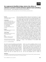
Fig. 2. Splicing of KcsA and RHCC. The left picture shows the
model of KcsA–RHCC. Only two subunits are shown for clarity.
The area enclosed by the square is where different splicing motifs
were tested in silico. It is displayed on the right in enlarged format
showing overlaps of different spliced structures.
RHCC domain imparts extreme thermostability to the KcsA channel Z. Yuchi et al.
6238 FEBS Journal 276 (2009) 6236–6246 ª 2009 The Authors Journal compilation ª 2009 FEBS
that of wtKcsA (Fig. 3A). The protein was purified to
homogeneity using a HisTrap
TM
HP column (Fig. 3B).
Biophysical characterization
The secondary and quaternary structures of KcsA–
RHCC were characterized by CD and gel filtration
chromatography (GFC), respectively. The CD data
showed that KcsA–RHCC is slightly more a-helical
than wtKcsA (64% versus 62%, respectively; Fig. 4A).
This is not surprising because RHCC existed predomi-
nantly as an a-helix in its crystallized form. The GFC
data showed that the majority of KcsA–RHCC is in a
tetrameric form, whereas a very small portion of it is
in a higher oligomeric form. This is very similar to that
of wtKcsA (Fig. 4B). Taken together, these two results
indicate that the gross biophysical nature of KcsA is
not altered by the addition of RHCC.
Thermostability test
The thermostability of wtKcsA, C-terminal deleted
KcsA (cdKcsA), KcsA–GCN4, KcsA–RHCC and
RHCC were compared by gel-shift assay (Fig. 5A).
The derived melting temperatures are shown in
Fig. 5D. Tetrameric KcsA is very stable and displays
properties of SDS resistance and heat resistance. Ther-
mostability in the presence of SDS is generally used to
indicate the stability of ion channels [20–22,29,30]. It is
usually reported as the temperature at which half the
tetrameric channels dissociate into monomers (T
m
). At
pH 8, the order of thermostability of the various con-
structs was KcsA–RHCC > wtKcsA > KcsA–
GCN4 > cdKcsA RHCC (Fig. 5B,D). Clearly, the
continuous coiled coil in KcsA–RHCC provided a
strong tetramerization force, as indicated by its ultra-
high T
m
value, which was much higher than 100 °C.
However, when two parts of KcsA–RHCC, namely,
A
kDa
WT GCN4125
120
RHCC
80
60
50
40
30
20
B
0 mM Imidazole gradient 500 mM
kDa
55
35
27
15
KcsA–RHCC
Fig. 3. (A) Western blot analysis of KcsA constructs. The same
number of E. coli cells (quantified by D
600
) expressing different
KcsA constructs were analysed using 15% SDS ⁄ PAGE. KcsA was
then identified by immunoblotting using an anti-his-tag IgG. WT:
KcsA 1–160; 125: KcsA 1–125; 120: KcsA 1–120; GCN4: KcsA–
GCN4; RHCC: KcsA–RHCC. (B) Purification of KcsA–RHCC by
HisTrap
TM
HP column. Proteins samples were run on a 4–12%
SDS ⁄ PAGE and stained with Commassie Blue. There was an
increasing amount of imidazole for the elution of protein samples
from the column present in the lanes from left to right. The arrow
indicates the position of purified KcsA–RHCC protein.
0
200
400
600
800
1000
1200
1400
1600
0.00 5.00 10.00 15.00 20.00 25.00
Absorbance at 280 nm (mAU)
Elution volume (mL)
KcsA–RHCC
wtKcsA
Tetramer
Higher
oligomer
–5000
–10 000
–15 000
–20 000
0
5000
10 000
15 000
20 000
25 000
A
B
198 218 238 258
Molar ellipticity (deg×cm
2
/decimole)
Wavelength (nm)
KcsA–RHCC
wtkcsA
Fig. 4. Biophysical characterization of KcsA–RHCC. (A) CD spectra
of tetrameric wtKcsA and KcsA–RHCC in LDAO. Estimated a-heli-
cal contents for wtKcsA and KcsA–RHCC are 62 and 64%, respec-
tively. (B) Elution profile of wtKcsA and KcsA–RHCC from the GFC
column. The estimated molecular mass of the tetrameric LDAO–
wtKcsA and LDAO–KcsA–RHCC micelles are 114 and 149 kDa,
respectively.
Z. Yuchi et al. RHCC domain imparts extreme thermostability to the KcsA channel
FEBS Journal 276 (2009) 6236–6246 ª 2009 The Authors Journal compilation ª 2009 FEBS 6239
cdKcsA and RHCC, were tested individually, both
of them displayed relatively low T
m
, suggesting that
the high stability of KcsA–RHCC is the result of
a co-operative effect. At pH 4, the order of
thermostability was KcsA–RHCC > cdKcsA >
KcsA–GCN4 > wtKcsA > RHCC (Fig. 5C,D). All
constructs except cdKcsA showed a decrease in T
m
upon pH change from 8 to 4, showing that all three
tetramerization domains are somewhat sensitive to pH
change. The pH effect on wtKcsA is well documented;
however, the acid labile nature of wild-type RHCC has
not been known until this investigation. The acid
labilities of GCN4 and RHCC may be due to the
weakening of intra- and⁄ or interhelical salt bridges
that stabilize their respective coiled coil structures
[23,24].
Electrophysiological test of KcsA–RHCC
When designing the KcsA–RHCC hybrid channel, we
expected the continuous coiled coil structure to keep the
inner helices and the channel permanently in the closed
form. However, the observed pH-sensitive nature of the
hybrid channel led us to speculate that KcsA–RHCC
may be conducting at acidic pH. This speculation was
confirmed by the measurement of its potassium con-
ducting activity with a planar bilayer system. KcsA–
RHCC can be opened at pH 4 and its apparent opening
probability (NPo) is 0.13, which is similar to that of
wtKcsA (NPo = 0.15) (Fig. 6A,B) [31]. However, its
zero-voltage conductance (44 pS) is lower than that of
wtKcsA (97 pS), and the outward rectifying property of
wtKcsA was not obvious in KcsA–RHCC (Fig. 6C)
[32]. When the buffer was changed to pH 8, channel
activity could barely be observed (Fig. 6A,B).
Discussion
Chimeric channel KcsA–RHCC was designed with the
aim of generating a more stable and robust channel
for structural and functional studies. Tetramerization
domains are present in many different families of ion
30 40 50 60 70 80 90 100 (°C) kDa
40
35
25
15
Tetramer
A
B
C
D
Dimer
Monomer
0
10
20
30
40
50
60
70
80
90
100
30 40 50 60 70 80 90 100
% of KcsA in tetrameric form
Temperature (°C)
pH8
wtKcsA
cdKcsA
KcsA–GCN4
KcsA–RHCC
RHCC
0
10
20
30
40
50
60
70
80
90
100
30 40 50 60 70 80 90 100
% of KcsA in tetrameric form
Temperature (°C)
pH4
wtKcsA
cdKcsA
KcsA–GCN4
KcsA–RHCC
RHCC
80.2
36.9
54.5
63.9
67.2
59.1
>100.0
78.9
52.9
0.0
0.0
10.0
20.0
30.0
40.0
50.0
60.0
70.0
80.0
90.0
100.0
pH8 pH4
Temperature (°C)
Tm at different pH
wtKcsA
cdKcsA
KcsA–GCN4
KcsA–RHCC
RHCC
Fig. 5. Thermostability determination of KcsA constructs. (A) Representative SDS ⁄ PAGE used in thermostability analyses (KcsA–RHCC at
pH 8). The tetramer, dimer and monomer bands of KcsA–RHCC are indicated on the left-hand side of the gel. The specific temperatures for
heat treatment are indicated above the gel. (B, C) Comparison of stability of wtKcsA, cdKcsA, KcsA–GCN4, KcsA–RHCC and RHCC at differ-
ent temperatures at (B) pH 8 and (C) pH 4. The fractional tetramer content in each sample was determined from the densitometry scans of
SDS ⁄ PAGE. The results shown in (B) and (C) are given as mean ± standard derivation (n = 3). (D) Comparison of T
m
values of three KcsA
constructs at pH 8 and pH 4. Each curve in (B) and (C) was fitted into the sigmoidal dose responsive (variable slope) model with R
2
> 0.97
using
GRAPHPAD PRISM software (La Jolla, CA, USA). The T
m
values were calculated from the corresponding equations of the models.
RHCC domain imparts extreme thermostability to the KcsA channel Z. Yuchi et al.
6240 FEBS Journal 276 (2009) 6236–6246 ª 2009 The Authors Journal compilation ª 2009 FEBS
channels. Our previous study established the impor-
tance of the native tetramerization domain in KcsA
and found that it can be replaced by an artificial tetra-
merization domain GCN4 [22].
In this study, the effects of different non-native tet-
ramerization domains and modes of linkage on the
stability of a KcsA hybrid channel were investigated.
It was found that whenever a flexible linker is present
the contributions of the different tetramerization
domains are very similar. However, a more stable con-
struct was obtained when two structurally compatible
domains were linked directly without a flexible linker.
Although GCN4 itself can form a stable tetramer, its
left-handed supercoil structure is not compatible with
the right-handed inner helix of KcsA. The linking of
these two structures could not be done without distort-
ing one or both of the contributing structures, which
would result in an unstable structure (Fig. 1B, middle).
On the other hand, the C-terminal domain of wtKcsA
was shown to adopt a right-handed four-helix bundle
structure linked to the inner helix via a less helical
structure [29,33] (Fig. 1B, left). The discontinuity in
the coiled coil structure may allow for flexibility and
suit its gating function; however, it will inevitably
compromise stability. In contrast, the continuous
RHCC design in KcsA–RHCC overcomes this prob-
lem and dramatically improves the stability of the
hybrid channel (Fig. 1B, right).
Previously we proposed a model of in vivo channel
assembly describing the correlation between channel
stability and protein expression level [22]. The results
reported in this study are consistent with this model.
A
B
C
Open
Close
10 pA
15 sec
pH 4
pH 8
pH 4
pH 4
pH 8
Current (pA)
0 2 4 6 8 10
Count (N)
0
100 000
200 000
Current (pA)
0 2 4 6 8 10
Count (N)
0
40 000
80 000
120 000
NPo = 0.014 ± 0.005
NPo = 0.129 ± 0.032
Close Open Close
–2
–4
–6
–8
0
2
4
6
8
10
–200 –50–100–150
0 50 100 150 200
V, mV
I, pA
Fig. 6. Measurement of currents conducted by KcsA–RHCC. (A) Representative current traces from a lipid bilayer containing KcsA–RHCC at
pH 4 (left panel) for a period of 1 min with an applied voltage of 200 mV followed by buffer exchange to pH 8 (middle panel) and back to pH
4 again (right panel). A high resolution detail of the measured current is shown at the top of the left panel, with the open and closed states
indicated at the side. (B) All-points amplitude histogram of single channel recordings for KcsA–RHCC at pH 4 and 8, respectively. The open
and closed states are indicated at the bottom of the chart. NPo values at pH 4 and 8 are indicated above the graphs. They are indicative of
the mean levels of activity from three recordings. (C) I–V curve at pH 4. Each data point represents mean current (± standard error, n = 3).
Z. Yuchi et al. RHCC domain imparts extreme thermostability to the KcsA channel
FEBS Journal 276 (2009) 6236–6246 ª 2009 The Authors Journal compilation ª 2009 FEBS 6241
The stability order deduced from a gel-shift assay was:
KcsA–RHCC > wtKcsA > KcsA–GCN4 > KcsA
1–125, which directly corresponds to the order of their
respective protein expression levels (Fig. 3A). How-
ever, it is puzzling that this correlation only holds well
for total proteins encompassing all oligomeric forms,
but not with the tetrameric form!
The coiled coil motif is a commonly found structure
in proteins. A statistical study from a genomic analysis
suggested that 5–10% of all protein sequences are in
coiled coils of various oligomeric states [34]. Typically
two to six a-helices wind around each other to form a
supercoil [35,36]. They are widely found in a diverse
array of proteins, such as transcription factors and
extracellular matrix proteins [37,38]. Because of its
simple and predictable folding properties, coiled coils
have been used as temperature regulators, antibody
stabilizers, anticancer drugs, purification tags, hydro-
gels and linker systems, etc [36].
In this study, we intended to fuse a right-handed
tetrameric coiled coil to KcsA to form a continuous
coiled coil. The multiplicity of coiled coil candidates
and the multiple possible splice junctions render
exhaustive experimental testing intractable. In silico
selection was therefore used to identify the optimal
splice variants. Both RHCC and left-handed coiled
coils were used as target candidates and our algorithm
easily identified the RHCC as better candidates. The
robustness of our computational algorithm was later
confirmed by the extreme thermostability of the
selected hybrid channel. This selection algorithm is
applicable to the design of other chimeric channels.
Regulation of ion channels by non-native domains
has been achieved in a large number of chimera experi-
ments. Most of these experiments involved intrafamily
sensor domain swapping, including the recent struc-
tural studies on Kv1.2–Kv2.1 [39], Kir3.1–prokaryotic
Kir [40], as well as many functional studies on a vari-
ety of ion channels [41–44]. Several chimera experi-
ments involved interfamily sensor swapping, including
using sensor domains from the Shaker channel, IRK1
channel and CNG channel to control the gating of
KcsA [45,46]. Meanwhile, utilization of a nonchannel
module to assist channel expression has also been
reported [25].
However, regulation of channel gating by a non-
channel module has not yet been reported. The fact
that KcsA–RHCC can conduct current opens up the
possibility of using RHCC as an alternative sensor
domain to the pore domain of other ion channels for
functional assays and drug screening. The pH depen-
dency of RHCC gating stems from its tetramerization
property. Its tetramer completely dissociates at acidic
pH (Fig. 5C,D), which is similar to the C-terminal
domain of KcsA [20].
Structural flexibility is a major obstacle in the pro-
duction of well-diffracting protein crystals due to its
effect on ordered crystal packing. The presence of a
flexible interdomain linker on wtKcsA reduced the dif-
fraction quality of its crystals (V. P. T. Pau, unpub-
lished results) [33]. Stiffening of KcsA by the addition
of RHCC should make it more prone to the yielding
of well-diffracting crystals. Currently, crystallization
conditions for KcsA–RHCC are being screened.
We propose that a similar engineering design may
also be applied to other ion channels as they all proba-
bly possess a RHCC structure at their respective
bundle crossing [1,47–51]. Functional minimal ion con-
ducting modules composed of S5–S6 helices from vari-
ous channels have been produced [52–55]. We envisage
that the expression of minimal channels may be facili-
tated by appropriate tetramerization domains and the
success of this effort will certainly open up the possi-
bilities in structural and functional characterization of
ion channels.
Materials and methods
Computational design of KcsA–RHCC
A comprehensive search of the Protein Data Bank [56] for
RHCC or parallel coiled coil structures that have four-fold
rotational symmetry retrieved four candidates. The pro-
gram fithelices (Doc. S1) was used to determine the
optimal splicing positions for joining the coiled coil fusion
candidate to KcsA. The indicator used by the program is
the root mean square deviation of the overlapping atoms
at the spliced site. The coordinates of the best spliced
structure for each fusion candidate were then Monte
Carlo minimized by program zmm-mvm (http://www.
zmmsoft.com/). The PDB file of the best minimized
structures can be found in Doc. S1.
Molecular cloning
The DNA sequence encoding residues Ala23-Val115 of
KcsA was amplified from pET28–KcsA [22], which con-
tains the wtKcsA gene of S. lividans, by PCR using Pfu
DNA polymerase (Fermentas, Burlington, Canada) with a
forward primer 5¢-GATTC
GGATCCGCGCTGCACTGG
AGGGC-3¢ and a reverse primer 5¢-TGATAACG GTGA
CGAACCAGGTGGCCAGCG-3¢. A gene encoding resi-
dues Thr16-Ile52 of RHCC (Table 2) was synthesized with
optimal codon usage for E. coli [57] and PCR amplified
with the following primers: forward: 5¢-CTGGTTCGT
CACCGTTA TCATCGACGAC-3¢ and reverse: 5¢-GAC
RHCC domain imparts extreme thermostability to the KcsA channel Z. Yuchi et al.
6242 FEBS Journal 276 (2009) 6236–6246 ª 2009 The Authors Journal compilation ª 2009 FEBS
TGA GAATTCTCATTAAATTGACGCCAGGATGGT-3¢
(the recognition sites of BamHI and EcoRI are underlined).
The two amplified fragments were joined by fusion PCR
[58], digested with the corresponding restriction enzymes
and cloned into pET28M, a modified pET28a expression
vector [20]. The sequence of this N-terminal his-tagged con-
struct, pET28M ⁄ kcsa-rhcc, was confirmed by dideoxynucle-
otide sequencing. The cloning of wtKcsA and KcsA–GCN4
was as previously described [22]. RHCC cloned in pET15b
was a gift from R. Kammerer (The University of
Manchester, UK).
Protein expression and purification
E. coli BL21(DE3) cells were transformed with
pET28M ⁄ kcsa-rhcc. A single colony was inoculated and
grown in 100 mL Luria–Bertani broth with 100 lgÆmL
)1
kanamycin (as the final concentration) at 37 °C overnight.
The culture was then diluted into 1 L Luria–Bertani broth
with 100 lgÆmL
)1
kanamycin and further grown for
100 min. Protein expression was induced by the addition of
isopropyl b-d-thiogalactopyranoside to a final concentra-
tion of 1 mm. Cells were pelleted after 3 h of incubation at
37 °C, resuspended in lysis buffer (20 mm Tris, pH 8,
150 mm KCl and 1 mm phenylmethanesulfonyl fluoride)
and subsequently lysed by French Press at 10 000 psi. The
cell lysate was centrifuged at 100 000 g for 1 h and the pel-
let was solubilized in 20 mL of 20 mm Tris, pH 8, 150 mm
KCl, 1 mm phenylmethanesulfonyl fluoride and 1% v ⁄ v
N,N-dimethyldodecylamine-N-oxide (LDAO) overnight at
4 °C. The resuspended mixture was centrifuged at
100 000 g for 1 h and the supernatant was loaded on to a
HisTrap
TM
HP column (GE Healthcare, Piscataway, NJ,
USA). Protein was purified using an FPLC system
(Pharmacia, Uppsala, Sweden) with a linear gradient of
0–500 mm imidazole. Purified proteins were analysed using
a NuPAGE Novex 4–12% Bistris midi gel (Invitrogen,
Carlsbad, CA, USA) with Coomassie Blue staining.
wtKcsA, KcsA–GCN4 and RHCC were expressed and
purified in a similar manner except the absence of detergent
during RHCC purification. cdKcsA was generated by
chymotrypsin digestion of wtKcsA [22].
Thermal stability determination
Protein of KcsA–RHCC was dialysed overnight against a
solution containing 150 mm KCl, 0.1% v ⁄ v LDAO, 20 mm
Tris, pH 8 (or 15 mm potassium citrate, pH 4) in dialysis
bags with a molecular mass cut-off of 3500 Da. The dialy-
sed sample was mixed with a loading solution containing
10% w ⁄ v SDS, 9.3% w ⁄ v dithiothreitol and 38% w ⁄ v glyc-
erol, heated for 30 min at various temperatures ranging
from 30 to 100 °C with a 10 °C increment, cooled to room
temperature and analysed by SDS ⁄ PAGE. Three indepen-
dent experiments were performed. Scanned images of the
gels were analysed using imagej ( />in Integrated-Intensity-Mode to determine the amounts of
tetramer and monomer in the samples. The fractional
tetramer content was calculated by dividing the integrated
density of tetramer by the combined integrated densities
of tetramer and monomer. Model fitting of the thermal
denaturation curves was carried out using prism 4.00
(GraphPad Software Inc., San Diego, CA, USA).
GFC
GFC was run on an FPLC system (Amersham Biosciences,
Piscataway, NJ, USA) using a Superdex 200 column
(Amersham Biosciences) equilibrated in 50 mm Tris buffer
(pH 8), 150 mm KCl and 0.1% v ⁄ v LDAO.
CD measurement
Protein samples at a 10 lm monomeric protein concentra-
tion were dissolved in 20 mm K
2
HPO
4
(pH 8), 150 mm
KCl and 0.1% v ⁄ v LDAO. CD spectra of the samples were
recorded in a 0.1 cm path length cell at 25 °C using a 410
CD spectrometer (AVIV Biomedical Inc., Lakewood, NJ,
USA). The secondary structure content of each sample was
quantified using the CD spectrum analysis program cdsstr
of the cdpro suite ( />CDPro ⁄ main.html).
Electrophysiology
Channel recordings were performed in a horizontal planar
lipid bilayer of 1-palmitoyl-2-oleoyl-phosphatidylethanol-
amine (POPE) and 1-palmitoyl-2-oleoyl-sn-phosphatidylgly-
cerol (POPG) (15 and 5 mgÆmL
)1
, respectively) at room
temperature. Both cis and trans chambers were filled with
solution at pH 4 (150 mm KCl, 20 mm potassium acetate)
at the start, which was changed to pH 8 (150 mm KCl,
20 mm Tris) when needed. Current records were acquired
at a sampling frequency >10 kHz and filtered to 1 kHz.
Acknowledgements
We thank Dr Richard Kammerer, University of Man-
chester, for providing us with the plasmid,
Table 2. DNA sequence of the synthesized rhcc gene.
ACCGTTATCATCGACGACCGTTACGAATCTCTGAAAAACCTGATCACCCTGCGTGCGGACCGTCTGGAAATGATTATCAACGACAACGTTTCTACCATCCTGGCGTCAATT
Z. Yuchi et al. RHCC domain imparts extreme thermostability to the KcsA channel
FEBS Journal 276 (2009) 6236–6246 ª 2009 The Authors Journal compilation ª 2009 FEBS 6243
pET15b ⁄ RHCC. We also thank Drs Richard M. Epand,
Raquel Epand and Vettai S. Ananthanarayanan,
McMaster University, for usage of CD spectrometers;
Dr Brad Rothberg, Temple University, for usage of
electrophysiological equipment and critical reading of
this manuscript. This work was supported by Microstar
Biotech Inc. and NSERC (DY).
References
1 Doyle DA, Morais Cabral J, Pfuetzner RA, Kuo A,
Gulbis JM, Cohen SL, Chait BT & MacKinnon R
(1998) The structure of the potassium channel:
molecular basis of K+ conduction and selectivity.
Science 280, 69–77.
2 Hille B (2001) Ion Channels of Excitable Membrane, 3rd
edn. Sinauer Associates, Sunderland, MA.
3 Shen NV, Chen X, Boyer MM & Pfaffinger PJ (1993)
Deletion analysis of K+ channel assembly. Neuron 11,
67–76.
4 Kreusch A, Pfaffinger PJ, Stevens CF & Choe S (1998)
Crystal structure of the tetramerization domain of the
Shaker potassium channel. Nature 392, 945–948.
5 Li M, Jan YN & Jan LY (1992) Specification of subunit
assembly by the hydrophilic amino-terminal domain
of the Shaker potassium channel. Science 257, 1225–
1230.
6 Jenke M, Sanchez A, Monje F, Stuhmer W, Weseloh
RM & Pardo LA (2003) C-terminal domains implicated
in the functional surface expression of potassium
channels. EMBO J 22, 395–403.
7 Tinker A, Jan YN & Jan LY (1996) Regions responsi-
ble for the assembly of inwardly rectifying potassium
channels. Cell 87, 857–868.
8 Jiang Y, Pico A, Cadene M, Chait BT & MacKinnon
R (2001) Structure of the RCK domain from the E. coli
K+ channel and demonstration of its presence in the
human BK channel. Neuron 29, 593–601.
9 Quirk JC & Reinhart PH (2001) Identification of a
novel tetramerization domain in large conductance
K(ca) channels. Neuron 32, 13–23.
10 Schmalhofer WA, Sanchez M, Dai G, Dewan A,
Secades L, Hanner M, Knaus HG, McManus OB,
Kohler M, Kaczorowski GJ et al. (2005) Role of the
C-terminus of the high-conductance calcium-activated
potassium channel in channel structure and function.
Biochemistry 44, 10135–10144.
11 Zhou L, Olivier NB, Yao H, Young EC & Siegelbaum
SA (2004) A conserved tripeptide in CNG and HCN
channels regulates ligand gating by controlling C-termi-
nal oligomerization. Neuron 44, 823–834.
12 Wei A, Jegla T & Salkoff L (1996) Eight potassium
channel families revealed by the C. elegans genome
project. Neuropharmacology 35, 805–829.
13 Covarrubias M, Wei AA & Salkoff L (1991) Shaker,
Shal, Shab, and Shaw express independent K+ current
systems. Neuron 7, 763–773.
14 VanDongen AM, Frech GC, Drewe JA, Joho RH &
Brown AM (1990) Alteration and restoration of K+
channel function by deletions at the N- and C-termini.
Neuron 5, 433–443.
15 Schulteis CT, Nagaya N & Papazian DM (1998)
Subunit folding and assembly steps are interspersed
during Shaker potassium channel biogenesis. J Biol
Chem 273 , 26210–26217.
16 Hopkins WF, Demas V & Tempel BL (1994) Both
N-and C-terminal regions contribute to the assembly
and functional expression of homo- and heteromulti-
meric voltage-gated K+ channels. J Neurosci 14,
1385–1393.
17 Deal KK, Lovinger DM & Tamkun MM (1994) The
brain Kv1.1 potassium channel: in vitro and in vivo
studies on subunit assembly and posttranslational
processing. J Neurosci 14, 1666–1676.
18 Xu J, Yu W, Jan YN, Jan LY & Li M (1995) Assembly
of voltage-gated potassium channels. Conserved hydro-
philic motifs determine subfamily-specific interactions
between the alpha-subunits. J Biol Chem 270, 24761–
24768.
19 Shen NV & Pfaffinger PJ (1995) Molecular recognition
and assembly sequences involved in the subfamily-
specific assembly of voltage-gated K+ channel subunit
proteins. Neuron 14, 625–633.
20 Pau VP, Zhu Y, Yuchi Z, Hoang QQ & Yang DS
(2007) Characterization of the C-terminal domain of a
potassium channel from Streptomyces lividans (KcsA).
J Biol Chem 282, 29163–29169.
21 Molina ML, Encinar JA, Barrera FN, Fernandez-
Ballester G, Riquelme G & Gonzalez-Ros JM (2004)
Influence of C-terminal protein domains and protein–
lipid interactions on tetramerization and stability of the
potassium channel KcsA. Biochemistry 43, 14924–
14931.
22 Yuchi Z, Pau VP & Yang DS (2008) GCN4 enhances
the stability of the pore domain of potassium channel
KcsA. FEBS J 275, 6228–6236.
23 Harbury PB, Zhang T, Kim PS & Alber T (1993) A
switch between two-, three-, and four-stranded coiled
coils in GCN4 leucine zipper mutants. Science 262,
1401–1407.
24 Stetefeld J, Jenny M, Schulthess T, Landwehr R, Engel
J & Kammerer RA (2000) Crystal structure of a
naturally occurring parallel right-handed coiled coil
tetramer. Nat Struct Biol 7, 772–776.
25 Zerangue N, Jan YN & Jan LY (2000) An artificial
tetramerization domain restores efficient assembly of
functional Shaker channels lacking T1. Proc Natl Acad
Sci USA 97, 3591–3595.
RHCC domain imparts extreme thermostability to the KcsA channel Z. Yuchi et al.
6244 FEBS Journal 276 (2009) 6236–6246 ª 2009 The Authors Journal compilation ª 2009 FEBS
26 Bowman GD, Nodelman IM, Levy O, Lin SL, Tian P,
Zamb TJ, Udem SA, Venkataraghavan B & Schutt CE
(2000) Crystal structure of the oligomerization domain
of NSP4 from rotavirus reveals a core metal-binding
site. J Mol Biol 304, 861–871.
27 Sales M, Plecs JJ, Holton JM & Alber T (2007) Struc-
ture of a designed, right-handed coiled-coil tetramer
containing all biological amino acids. Protein Sci 16,
2224–2232.
28 Kuhnel K, Jarchau T, Wolf E, Schlichting I, Walter U,
Wittinghofer A & Strelkov SV (2004) The VASP tetra-
merization domain is a right-handed coiled coil based
on a 15-residue repeat. Proc Natl Acad Sci USA 101,
17027–17032.
29 Cortes DM, Cuello LG & Perozo E (2001) Molecular
architecture of full-length KcsA: role of cytoplasmic
domains in ion permeation and activation gating.
J Gen Physiol 117, 165–180.
30 Perozo E, Cortes DM & Cuello LG (1999) Structural
rearrangements underlying K+-channel activation
gating. Science 285, 73–78.
31 Cordero-Morales JF, Cuello LG, Zhao Y, Jogini V,
Cortes DM, Roux B & Perozo E (2006) Molecular
determinants of gating at the potassium-channel
selectivity filter. Nat Struct Mol Biol 13, 311–318.
32 LeMasurier M, Heginbotham L & Miller C (2001)
KcsA: it’s a potassium channel. J Gen Physiol 118,
303–314.
33 Uysal S, Vasquez V, Tereshko V, Esaki K, Fellouse
FA, Sidhu SS, Koide S, Perozo E & Kossiakoff A
(2009) Crystal structure of full-length KcsA in its
closed conformation. Proc Natl Acad Sci USA 106,
6644–6649.
34 Mewes HW, Frishman D, Gruber C, Geier B, Haase D,
Kaps A, Lemcke K, Mannhaupt G, Pfeiffer F,
Schuller C et al. (2000) MIPS: a database for genomes
and protein sequences. Nucleic Acids Res 28, 37–
40.
35 Wolf E, Kim PS & Berger B (1997) MultiCoil: a pro-
gram for predicting two- and three-stranded coiled coils.
Protein Sci 6, 1179–1189.
36 Mason JM & Arndt KM (2004) Coiled coil domains:
stability, specificity, and biological implications.
ChemBioChem 5, 170–176.
37 Glover JN & Harrison SC (1995) Crystal structure of
the heterodimeric bZIP transcription factor c-Fos-c-Jun
bound to DNA. Nature 373, 257–261.
38 Frank S, Schulthess T, Landwehr R, Lustig A, Mini T,
Jeno P, Engel J & Kammerer RA (2002) Characteri-
zation of the matrilin coiled-coil domains reveals seven
novel isoforms. J Biol Chem 277, 19071–19079.
39 Long SB, Tao X, Campbell EB & MacKinnon R (2007)
Atomic structure of a voltage-dependent K+channel in
a lipid membrane-like environment. Nature 450,
376–382.
40 Nishida M, Cadene M, Chait BT & MacKinnon R
(2007) Crystal structure of a Kir3.1-prokaryotic Kir
channel chimera. EMBO J 26, 4005–4015.
41 Zhao X, Bucchi A, Oren RV, Kryukova Y, Dun W,
Clancy CE & Robinson RB (2009) In vitro character-
ization of HCN channel kinetics and frequency
dependence in myocytes predicts biological pacemaker
functionality. J Physiol 587, 1513–1525.
42 Urquhart W, Gunawardena AH, Moeder W, Ali R,
Berkowitz GA & Yoshioka K (2007) The chimeric cyc-
lic nucleotide-gated ion channel ATCNGC11 ⁄ 12 consti-
tutively induces programmed cell death in a Ca2+
dependent manner. Plant Mol Biol 65
, 747–761.
43 Varshney A & Mathew MK (2003) Inward and outward
potassium currents through the same chimeric human
Kv channel. Eur Biophys J 32, 113–121.
44 Cao Y, Crawford NM & Schroeder JI (1995) Amino
terminus and the first four membrane-spanning
segments of the Arabidopsis K+ channel KAT1 confer
inward-rectification property of plant–animal chimeric
channels. J Biol Chem 270, 17697–17701.
45 Lu Z, Klem AM & Ramu Y (2001) Ion conduction
pore is conserved among potassium channels. Nature
413, 809–813.
46 Ohndorf UM & MacKinnon R (2005) Construction of
a cyclic nucleotide-gated KcsA K+ channel. J Mol Biol
350, 857–865.
47 Jiang Y, Lee A, Chen J, Ruta V, Cadene M, Chait BT
& MacKinnon R (2003) X-ray structure of a voltage-
dependent K+ channel. Nature 423 , 33–41.
48 Long SB, Campbell EB & Mackinnon R (2005) Crystal
structure of a mammalian voltage-dependent Shaker
family K+ channel. Science 309, 897–903.
49 Jiang Y, Lee A, Chen J, Cadene M, Chait BT & MacK-
innon R (2002) Crystal structure and mechanism of a
calcium-gated potassium channel. Nature 417, 515–522.
50 Shi N, Ye S, Alam A, Chen L & Jiang Y (2006) Atomic
structure of a Na+- and K+-conducting channel.
Nature 440, 570–574.
51 Kuo A, Gulbis JM, Antcliff JF, Rahman T, Lowe ED,
Zimmer J, Cuthbertson J, Ashcroft FM, Ezaki T &
Doyle DA (2003) Crystal structure of the potassium
channel KirBac1.1 in the closed state. Science 300,
1922–1926.
52 Pincin C, Ferrera L & Moran O (2005) Minimal sodium
channel pore consisting of S5-P-S6 segments preserves
intracellular pharmacology. Biochem Biophys Res
Commun 334, 140–144.
53 Kang GB, Song HE, Song DW, Kim MK, Rho SH,
Kim do H & Eom SH (2007) Overexpression and purifi-
cation of the RyR1 pore-forming region. Protein Pept
Lett 14, 742–746.
54 Chen Z, Alcayaga C, Suarez-Isla BA, O’Rourke B,
Tomaselli G & Marban E (2002) A ‘‘minimal’’ sodium
channel construct consisting of ligated S5-P-S6 segments
Z. Yuchi et al. RHCC domain imparts extreme thermostability to the KcsA channel
FEBS Journal 276 (2009) 6236–6246 ª 2009 The Authors Journal compilation ª 2009 FEBS 6245
forms a toxin-activatable ionophore. J Biol Chem 277,
24653–24658.
55 Santos JS, Grigoriev SM & Montal M (2008) Molecular
template for a voltage sensor in a novel K+ channel.
III. Functional reconstitution of a sensorless pore
module from a prokaryotic Kv channel. J Gen Physiol
132, 651–666.
56 Berman HM, Westbrook J, Feng Z, Gilliland G, Bhat
TN, Weissig H, Shindyalov IN & Bourne PE (2000)
The Protein Data Bank. Nucleic Acids Res 28, 235–242.
57 Dong H, Nilsson L & Kurland CG (1996) Co-variation
of tRNA abundance and codon usage in Escherichia
coli at different growth rates. J Mol Biol 260, 649–663.
58 Shevchuk NA, Bryksin AV, Nusinovich YA, Cabello
FC, Sutherland M & Ladisch S (2004) Construction of
long DNA molecules using long PCR-based fusion of
several fragments simultaneously. Nucleic Acids Res 32,
e19.
59 Zhou Y, Morais-Cabral JH, Kaufman A & MacKinnon
R (2001) Chemistry of ion coordination and hydration
revealed by a K+ channel-Fab complex at 2.0 A
resolution. Nature 414, 43–48.
Supporting information
The following supplementary material is available:
Doc. S1. Program fithelices (in FORTRAN) and the
PDB coordinates of KcsA–RHCC.
This supplementary material can be found in the
online version of this article.
Please note: As a service to our authors and readers,
this journal provides supporting information supplied
by the authors. Such materials are peer-reviewed and
may be re-organized for online delivery, but are not
copy-edited or typeset. Technical support issues arising
from supporting information (other than missing files)
should be addressed to the authors.
RHCC domain imparts extreme thermostability to the KcsA channel Z. Yuchi et al.
6246 FEBS Journal 276 (2009) 6236–6246 ª 2009 The Authors Journal compilation ª 2009 FEBS