Báo cáo khoa học: The tetralogy of Fallot-associated G274D mutation impairs folding of the second epidermal growth factor repeat in Jagged-1 potx
Bạn đang xem bản rút gọn của tài liệu. Xem và tải ngay bản đầy đủ của tài liệu tại đây (561.23 KB, 11 trang )
The tetralogy of Fallot-associated G274D mutation
impairs folding of the second epidermal growth factor
repeat in Jagged-1
Corrado Guarnaccia, Somdutta Dhir, Alessandro Pintar and Sa
´
ndor Pongor
Protein Structure and Bioinformatics Group, International Centre for Genetic Engineering and Biotechnology (ICGEB), Trieste, Italy
Introduction
The Notch signaling pathway is a highly connected
and tightly regulated signal transduction framework
that, together with a restricted number of other signal-
ing networks, drives developmental processes in all
metazoans. Notch signaling controls cell lineage deci-
sions in tissues derived from all three primary germ
lines: endoderm, mesoderm and ectoderm, thus playing
an essential role in organogenesis [1–3]. Faults in the
Notch-mediated signaling network have been associ-
ated with very different pathologies, such as some
cancers (T-cell acute lymphoblastic leukemia,
mucoepidermoid carcinoma) [4–6]; several genetic
disorders [Alagille syndrome (AGS), tetralogy of
Fallot, spondylocostal dysostosis, cerebral autosomal
dominant arteriopathy with subcortical infarcts] [7];
and possibly autoimmune diseases, such as multiple
sclerosis [8].
Both receptors and ligands are membrane-bound
proteins, and this restricts signaling to adjacent cells.
Of the five Notch ligands identified in man, Jagged-1
Keywords
Alagille syndrome; disease mutation; limited
proteolysis; Notch signaling; oxidative
folding
Correspondence
A. Pintar or S. Pongor, AREA Science Park,
Padriciano 99, I-34149 Trieste, Italy
Fax: +39 040 226555
Tel: +39 040 3757354
E-mail: ;
(Received 30 June 2009, revised 25 August
2009, accepted 27 August 2009)
doi:10.1111/j.1742-4658.2009.07333.x
Notch signaling controls spatial patterning and cell-fate decisions in all
metazoans. Mutations in JAG1, one of the five Notch ligands in man, have
been associated with Alagille syndrome and with a familial form of tetral-
ogy of Fallot. A specific G274D mutation in the second epidermal growth
factor repeat of the Jagged-1 was found to correlate with tetralogy of
Fallot symptoms but not with usual Alagille syndrome phenotypes. To
investigate the effects of this mutation, we studied the in vitro oxidative
folding of the wild-type and mutant peptides encompassing the second epi-
dermal growth factor. We found that the G274D mutation strongly impairs
the correct folding of the epidermal growth factor module, and folding can-
not be rescued by compensative mutations. The 274 position displays very
low tolerance to substitution because neither the G274S nor the G274A
mutants could be refolded in vitro. A sequence comparison of epidermal
growth factor repeats found in human proteins revealed that the pattern
displayed by the second epidermal growth factor is exclusively found in
Notch ligands and that G274 is absolutely conserved within this group. We
carried out a systematic and comprehensive analysis of mutations found in
epidermal growth factor repeats and show that specific residue require-
ments for folding, structural integrity and correct post-translational
processing may provide a rationale for most of the disease-associated
mutations.
Abbreviations
AGS, Alagille syndrome; cbEGF, calcium-binding epidermal growth factor; DSL, Delta ⁄ Serrate ⁄ LAG2; EGF, epidermal growth factor; EGF1,
first epidermal growth factor; EGF2, second epidermal growth factor; Fmoc, 9-fluorenylmethyloxycarbonyl; GSH, reduced glutathione; GSSG,
oxidized glutathione; MIM, Mendelian Inheritance in Man; PDB, Protein Data Bank; TFA, trifluroacetic acid; TOF, tetralogy of Fallot.
FEBS Journal 276 (2009) 6247–6257 ª 2009 The Authors Journal compilation ª 2009 FEBS 6247
and -2 are orthologs of Drosophila Serrate, whereas
Delta-like-1, -3 and -4 are orthologs of Drosophila
Delta. Jagged-1 is a single pass type I membrane pro-
tein with a large extracellular region made of a poorly
characterized N-terminal region, a Delta ⁄ Serrate ⁄
LAG2 (DSL) domain, a series of 16 epidermal growth
factor (EGF) tandem repeats and a cysteine-rich juxt-
membrane region (Fig. 1). Mutations in the JAG1 gene
have been associated with AGS [Mendelian Inheritance
in Man (MIM) database: #118450], a rare genetic dis-
order that can affect several organs, such as the liver,
heart, eye, skeleton and kidneys [9]. More than 400
mutations in the JAG1 gene have been identified,
including missense, nonsense, deletion, insertion, splice
site mutations and even complete gene deletion.
AGS has an autosomal dominant inheritance and
haploinsufficiency has been indicated as the main
mechanism for the onset of this disorder. Recent stud-
ies, however, suggest that some mutations may have a
dominant-negative character, leading to truncated
soluble forms of JAG1 that can compete with the
membrane-bound ligand [10,11]. AGS is a complex
disorder with highly variable clinical symptoms and a
clear genotype–phenotype association cannot always
be established, with a few exceptions. A C234Y muta-
tion in the first epidermal growth factor (EGF1) of Jag-
ged-1 was found in a group of patients with deafness,
congenital heart defects and posterior embryotoxon
[12], and a G274D mutation in the second epidermal
growth factor (EGF2) repeat of Jagged-1 was found in
a familial form of tetralogy of Fallot (TOF) (MIM
database: #187500) [13], comprising a heart malforma-
tion involving a large ventricular septal defect, pulmo-
nary stenosis, right ventricular hypertrophy and an
overriding aorta. Although cardiac defects are fre-
quently found in AGS patients, none of the individuals
with the G274D mutation displayed any other relevant
clinical feature typical of AGS. The JAG1-G274D
mutant protein can actually be expressed in NIH-3T3
cells, although in two different forms [14]. A fraction
of JAG1-G274D is correctly processed, presented at
the cell surface, and is functional, whereas another
fraction is not fully glycosylated, is retained in the
intracellular compartment, and is therefore inactive.
This conclusion was supported by the sensitivity of
JAG1-G274D to endoglycosidase H, which removes
oligomannose and hybrid N-linked oligosaccharides,
but not complex carbohydrates, by the incomplete
digestion of the mutant protein in cells exposed to
trypsin, and by the partial activation of Notch signal-
ing detected through a reporter gene assay. The car-
diac-specific phenotype associated with this mutation
was explained in terms of a high sensitivity of the
developing heart to Jagged-1 levels [14].
Despite the fact that EGF repeats are widespread in
extracellular proteins and that hundreds of missense
mutations have been identified and associated with sev-
eral genetic diseases, the structural grounds of these
disorders have been investigated only in a few cases,
mostly related to calcium-binding EGFs (cbEGF).
Detailed biochemical studies were carried out on muta-
tions in CRB1 [15], the human orthologue of Drosoph-
ila Crumbs, fibrillin-1 [16–22], low density lipoprotein
receptor [23] and human factor IX [19], which are
associated with Leber congenital amaurosis, Marfan
syndrome, familial hypercholesterolemia and emophilia B,
respectively. The overall conclusion from these studies
is that, in multidomain proteins, mutations can have
different effects depending on the context, and a corre-
lation between the genotype and the phenotype is still
difficult.
To investigate the structural effects of the G274D
mutation, and to attempt to correlate them with the
available data obtained in vitro and in vivo , we initially
synthesized a peptide corresponding to EGF2 of
Jagged-1 (residues 263–295). This peptide, however,
MNNL
VWC
DSL
-27
-129
-146
-231
-251
-295
-335
-373
-411
-449
-465
-523
-573
-628
-666
-704
-742
-781
-790
-819
-857
-894
-972
-1016
-1066
-1218
1 2 3 4 5 6 7 8 9 10 11 12 13 14 15 16
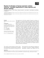
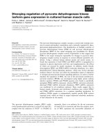
Fig. 1. Domain architecture of human Jagged-1. MNNL, N-terminal domain of Notch ligands; EGF domains are numbered progressively;
potential calcium-binding EGF domains are shaded in gray; VWC, von Willebrand factor type C domain; the transmembrane segment is
shown as a black bar; the receptor binding region is enclosed within a dashed rectangle. Amino acid residues corresponding to exon bound-
aries are shown above. The amino acid sequence of the J1ex6 peptide and the disulfide bond connectivities are also shown; the mutated
glycine (G274) is shown in bold and underlined.
Misfolding in Jagged-1 receptor binding region C. Guarnaccia et al.
6248 FEBS Journal 276 (2009) 6247–6257 ª 2009 The Authors Journal compilation ª 2009 FEBS
could not be refolded in vitro under the standard oxi-
dative folding conditions used for other EGFs. Stem-
ming from the observation that exon 6 of the JAG1
gene encodes not only EGF2, but also part of EGF1,
we speculated that exon 6 might encode an autono-
mously folding unit. We thus prepared a longer pep-
tide encompassing the C-terminal part of EGF1 and
the entire EGF2 (Fig. 1). This peptide, J1ex6 (residues
252–295), could be readily refolded in vitro and was
shown to yield a structured unit with a disulfide bond
topology typical of EGF repeats [24,25]. In the present
study, we show that the G274D mutation associated
with TOF strongly impairs the in vitro oxidative fold-
ing of this minimal folding and structural unit.
Results
The G274D mutation impairs folding of J1ex6
The solution structure of J1ex6 (residues 252–295)
determined by NMR [Protein Data Bank (PDB) code:
2KB9] showed that the N-terminal overhang corre-
sponding to the C-terminal part of EGF1 is not only
required for folding, but is also an integral part of a
structural unit that encompasses the EGF1 C-terminal
region and the entire EGF2 [25]. The solution struc-
ture of this unit, including the conformation of the
N-terminal overhang, is also very similar to the struc-
ture of the same region in a larger Jagged-1 construct
comprising the DSL and EGF1, 2 and 3 domains, for
which the crystal structure was determined recently
[26] (PDB code: 2VJ2). We used this minimal folding
and structural unit to address the structural grounds
for the misfolding of the G274D mutant.
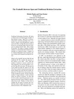
The results obtained with respect to the in vitro oxi-
dative folding for the wild-type J1ex6 and its variants
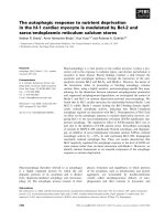
are summarized in Fig. 2. Folding of the wild-type
J1ex6 lead to a largely prevalent product (> 86% by
RP-HPLC area integration), with a very minor frac-
tion of products that could be identified by LC-MS as
mixed disulfides with glutathione (GSH). Because
GSH is hydrophilic, adducts with GSH usually display
shorter retention times in RP-HPLC compared to the
native folded species. The folded species has a reten-
tion time that is only slightly shorter ( 1 min) than
that of the reduced peptide, suggesting that J1ex6 lacks
a true hydrophobic core. Under the same experimental
conditions, oxidative folding of the G274D mutant
produced a very complex mixture. A clear separation
of the different products in the mixture could not be
achieved, but MS analysis revealed that most of the
RP-HPLC peaks arise from adducts with one or more
molecules of GSH. This suggests that, in the G274D
mutant, the correct folding and the complete forma-
tion of the four disulfide bonds cannot be accom-
plished, leaving one or more cysteines coupled to GSH
and leading to shorter retention time species. By con-
trast, products at longer retention times may contain
incorrect disulfide bonds that remain exposed to the
solvent. The same results were obtained using redox
buffers containing aromatic thiols, which were shown
to enhance both folding rates and yields [27]: wild-type
J1ex6 refolded in excellent yield, whereas the G274D
mutant remained trapped in mixed disulfides forms.
Figure 2 refers to a time point where equilibrium has
been reached, but the same trend was observed at
short refolding times. Although the RP-HPLC profile
for the wild-type J1ex6 already showed a major prod-
uct after 1 h, at the same time point, the profile of the
G274D mutant displayed a complex pattern.
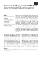
To confirm that the mixture of products obtained in
the refolding of the G274D mutant is actually com-
posed of misfolded species, we subjected it to proteoly-
sis using proline endopeptidase, and analyzed the
fragments by MALDI-TOF MS (Fig. 3). Proline endo-
peptidase was chosen because J1ex6 contains three
prolines (P267, P269 and P279), all in the central part
of the EGF2 sequence, and close to the mutated G274.
Although the wild-type J1ex6 was scarcely affected
after 20 h at 37 °C, the refolding mixture of the
G274D mutant digested under the same conditions dis-
played a completely different MS profile, with an
almost complete proteolysis of the G274D mutant into
Fig. 2. Oxidative folding. RP-HPLC profiles for the in vitro oxidative
folding of the wild-type J1ex6 and its variants in the presence of
the GSH ⁄ oxidized GSH (GSSG) redox couple. The RP-HPLC profile
of the purified, reduced J1ex6 peptide is also shown.
C. Guarnaccia et al. Misfolding in Jagged-1 receptor binding region
FEBS Journal 276 (2009) 6247–6257 ª 2009 The Authors Journal compilation ª 2009 FEBS 6249
small fragments. The much higher susceptibility of the
G274D mutant to proline endopeptidase suggests the
prevalence of misfolded species with a bead-like
arrangement of the core disulfide bonds.
From the 3D structure of J1ex6, we speculated that
misfolding of the G274D mutant could be a result of
electrostatic repulsion or a steric clash with E285. If
this was the case, the G274D mutant would be rescued
by a compensative mutation at position 285 aimed
either at neutralizing the negative charge or at reduc-
ing the steric hindrance of E285. Supported by the
observation that position 285 shows a high variability
(see below), we prepared the double mutants
G274D ⁄ E285Q and G274D ⁄ E285G, purified them,
and refolded under the same conditions used for the
wild-type J1ex6. RP-HPLC profiles (Fig. 2) reveal, as
for the G274D mutant, a complex pattern, suggesting
that the tested putative compensative mutations cannot
rescue the correct folding of the G274D mutant
in vitro. To test the tolerance of position 274 to amino
acid substitution, we prepared two additional mutants,
G274S and G274A, replacing glycine with two small
amino acids (i.e. serine or alanine, respectively). Also
in this case, however, RP-HPLC analysis of the oxida-
tive folding mixture (Fig. 2) showed a complex profile
and the lack of a major product. In conclusion, posi-
tion 274 is not tolerant to substitution, nor could puta-
tive compensative mutations rescue the folding of the
G274D mutant. From a closer inspection of the J1ex6
structure, it should be noted that, to maintain the cor-
rect stereochemistry, any side chain at position 274
would point towards the interior of the domain. In
other words, any amino acid other than glycine would
require a substantial rearrangement of the backbone to
reorient the side chain. The experiments performed in
the present study demonstrated that this region of
J1ex6 may be too rigid to allow for such a conforma-
tional change to occur.
The sequence pattern of EGF2 is unique
The very low tolerance of J1ex6 to amino acid substi-
tution at position 274 lead us to investigate whether
the sequence pattern associated with EGF2 is found in
other proteins. A pattern search in swiss-prot (http://
www.expasy.org/prosite/) produced 22 hits, which, sur-
prisingly, are all related to Notch ligands in different
organisms. In this dataset, G at position 274 is abso-
lutely conserved. Extending the pattern search to
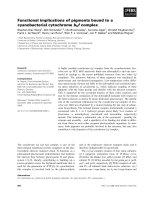
trEMBL, we obtained 115 hits. A plot of Shannon
entropy shows that, apart from cysteines, there are
only two additional positions that display no variabil-
ity at all, the first corresponds to G274 in the Jagged-1
sequence, and the second to G290 (Fig. 4). This sup-
ports the idea that, in this specially constrained type of
EGF, position 274 is not tolerant to substitution.
Analysis of disease-associated mutations
Because the EGF domain is one of the most common
structural building blocks in extracellular proteins
[28,29], we decided to undertake a global analysis of
disease-associated missense mutations found in EGF-
containing proteins (Tables S1–S4). By far the most
frequent disease-associated mutation found in EGF
domains involves cysteine (48%) followed by arginine
(11%) and glycine (10%). Although R fi X and
G fi X mutations are also involved in polymorphism,
C fi X mutations are almost exclusively disease-associ-
ated. To take into account the relative abundance of
certain amino acid types in EGF domains, which are
notably cysteine-rich, the number of observed muta-
tions was normalized for the amino acid content, and
this mutation frequency was compared with that calcu-
lated for the reference dataset. The ratio between these
two frequencies can be considered as a measure of the
relative impact of a certain AA
i
fi X mutation in a
Fig. 3. Probing folding by proteolysis. MALDI-TOF analysis of the
folding mixtures of (A) the wild-type (WT) and (B) the G274D
mutant peptides subjected to proteolysis with proline endopepti-
dase for 20 h at 37 °C. Cleavage sites are indicated by triangles
(.), the mutated glycine by an arrow; the m ⁄ z region of the intact
peptide is enclosed within a rectangle; for quantitative comparison,
the intensity ratio between the m ⁄ z values of the intact peptide
and the fragment of m ⁄ z = 2187 (labeled with an asterisk and cor-
responding to a C-terminal fragment of 16 residues) can be used.
Misfolding in Jagged-1 receptor binding region C. Guarnaccia et al.
6250 FEBS Journal 276 (2009) 6247–6257 ª 2009 The Authors Journal compilation ª 2009 FEBS
EGF domain (Fig. 5). Although normalization drasti-
cally reduces the weight of mutations involving cyste-
ine, it is apparent that mutations either removing
(C fi X) or introducing a cysteine (X fi C, similar to
Y fi C and R fi C) still have a great impact on EGF
domains. This effect can be easily explained by the
structural requirements of EGF domains, which, lack-
ing a true hydrophobic core, are mainly stabilized by
the three disulfide bridges. On the other hand, the
introduction of an additional cysteine is likely to
scramble the oxidative folding of EGF domains
in vivo. Oxidation of cysteines to yield disulfide bonds
is the most studied but not the only post-translational
modification found in EGF domains [30]. b-hydroxyl-
ation of aspartate and asparagine, as well as different
types of N- and O-glycosylation, has been reported.
Although the role of b-hydroxylation remains elusive,
correct O-glucosylation and O-fucosylation of ser-
ine ⁄ threonine residues has been shown to be required
for correct signaling mediated by Notch receptors
[31,32]. The impact of mutations involving these resi-
due types might be related to these post-translational
modifications, rather than to changes in the physico-
chemical properties of a specific amino acid.
To analyze this latter aspect, we compared the
disease-associated and neutral mutations in terms of
the chemical distance, as measured by the Grantham
score [33] (Fig. 6). Polymorphism-related mutations
follow an almost bell-like distribution centered on rela-
0.0
0.5
1.0
1.5
2.0
2.5
3.0
3.5
Shannon entropy
C i p
h p g C v
h G i C n e p w q C l C e t n w g G q l C
Amino acid sequence
Fig. 4. Sequence variability. Sequence variability in a set of 115
EGFs matching the pattern {C-X(5)-C-X(4)-C-X(5)-C-X-C-X(8)-C} plotted
as Shannon entropy versus position. Values for the Shannon entropy
can vary from zero (no variability) to a maximum of 4.3. The amino
acid sequence of Jagged-1 EGF2 (residues 265–293) is shown on
the x-axis; amino acids in capital letters are totally conserved.
0
1
2
3
4
5
6
7
8
9
10
WCF I YVLHMATRGQSNPDEK
Y(53)
R(35)
S(25)
G(15)
F(15)
W(11)
K(1)
C(11)
D(2)
K(9)
G(2)
D(1)
Q(1)
R(3)
H(1)
P(1)
C(22)
P(3)
W(3)
H(2)
Q(2)
G(1)
I(1)
K(1)
S(1)
S(6)
K(4)
H(2)
I(1)
Y(1)
E(1)
R(1)
T(6)
S(1)
Y(4)
N(3)
E(2)
G(2)
V(2)
A(1)
H(1)
C(1)
L(1)
V(1)
L(2)
I(2)
M(1)
P(1)
P(2)
N(1)
Y(1)
P(2)
T(1)
V(1)
I(2)
M(1)
R(9)
S(8)
D(5)
C(4)
E(4)
V(2)
C(2)
P(2)
R(1)
A(3)
R(2)
S(2)
L(1)
(0)
Mutation frequency ratio
Amino acid
Fig. 5. Disease-associated mutations in EGF domains. The ratio
between disease-associated mutation frequencies in EGF domains
and the reference data set is plotted for each amino acid type.
Amino acid types are shown in order of flexibility, as defined previ-
ously [41], from the least flexible (W) to the most flexible (K). The
resulting amino acid and the number of occurrences for each muta-
tion (in parenthesis) are shown above each bar. Mutations involving
cysteines are shown in bold.
0
10
20
30
40
25 50 75 100 125 150 175 200 225
Mutations (%)
Grantham score
G274D (94)
Fig. 6. Physico-chemical analysis of mutations. The percentage of
disease-associated mutations (black bars) and polymorphism-related
mutations (gray bars) are plotted versus their corresponding
Grantham score.
C. Guarnaccia et al. Misfolding in Jagged-1 receptor binding region
FEBS Journal 276 (2009) 6247–6257 ª 2009 The Authors Journal compilation ª 2009 FEBS 6251
tive small values of the Grantham score, whereas dis-
ease-associated mutations show an uneven distribution.
Overall, it can be concluded that mutations with a
high Grantham score are highly likely to be disease-
associated, but the contrasting case is not true, at least
for EGF domains, suggesting that the chemical dis-
tance is not the only determinant, as discussed above.
As a further step, we attempted to identify positions
in the EGF scaffold that are most sensitive to muta-
tions. This type of analysis, however, turned out to be
problematic because of the very high variability in the
amino acid sequence of EGF domains and in the
length of the loops, which together make both
sequence and structural alignments unreliable for this
purpose. We thus decided to carry out this type of
analysis on a coarser basis, dividing the sequence of
EGFs into seven windows, w1 to w7, and partitioning
mutations accordingly (Fig. 7). Polymorphism-related
mutations show a relatively homogeneous distribution
over the sequence, whereas disease-related mutations
are mainly found in w1, w3, w4 and, to a minor
extent, in w6. The relatively high frequency of disease-
associated mutations in the N-terminal region most
likely has no specific structural explanation, but is
rather related to the strict requirement of specific
amino acids (D ⁄ N) necessary for calcium coordination
in calcium-binding EGF domains. On the other hand,
mutations in w3 and w4 are more likely to disrupt the
two-strand antiparallel b-sheet that is the main (and
sometimes the only) secondary structure element in
EGF domains, or to involve residues that are required
for the correct formation of the interface between two
consecutive EGF repeats. A separate positional analy-
sis of cysteine mutations, which are all disease-associ-
ated, showed that they are equally distributed, with no
significant prevalence of any of the six positions.
Discussion
The G274D mutation in EGF2 of Jagged-1 is occur-
ring within the same window (w3 in Fig. 7) and at a
position that is structurally equivalent to G1127 in
fibrillin-1 and G106 in factor IX (Fig. 8). The G274D
mutation, however, appears to affect folding in a more
drastic way (Fig. 2) than the G1127S mutation in
fibrillin-1 and the G106S mutation in factor IX [19,20].
This is likely a result of the higher constraints in the
structure of this atypical EGF, as indicated by the
shorter B
N
-B
C
loop (ten residues, compared to 13 in
fibrillin-1 and 14 in factor IX) and spacing between
the first and last half-cystines (the A
N
–C
C
distance is
27 residues in Jagged-1 EGF2, compared to 35 in
fibrillin-1 and 30 in factor IX) and supported by the
observation that glycine at that position is totally con-
served in Notch ligands (Fig. 4). It is possible that the
G274D mutation, introducing a larger charged amino
acid, is more disrupting than a G fi S mutation (a dif-
ference of 94 in the Grantham score, compared to 56
for a G fi S mutation; Fig. 6). The misfolding of the
G274S and G274A mutants (Fig. 2), however, sup-
ports the hypothesis that no amino acid other than
glycine can be accommodated at that position, regard-
less of the substitution type. This low tolerance to
substitution is consistent with the positive / angle
measured for G274 (Tables S5). For steric reasons, in
protein structures, positive values of / are observed
0
2
4
6
8
10
Normalized mutation number
w1 w2 w3 w4 w5 w6 w7
A
N
B
N
A
C
B
C
C
N
C
C
w1
Sequence window
w2 w3 w4 w5 w6 w7
Fig. 7. Positional analysis of mutations. Disease-associated (black
bars) and polymorphism-related (gray bars) mutations in EGF
domains were partitioned according to their position in windows
w1 to w7 and normalized for the average window size. Mutations
involving cysteine were not considered. The six half-cystines are
named according to the A
N
B
N
A
C
B
C
C
N
C
C
annotation.
Fig. 8. Structural alignment. Multiple sequence alignment based on
the structural alignment of EGF2 from Jagged-1 (JAG1; PDB code:
2VJ2), cbEGF1 from factor IX (FA9, PDB code: 1EDM) and
cbEGF13 from fibrillin-1 (FBN1; PDB code: 1LMJ). Despite some
discrepancy in the N-terminal region, half-cystines (boxed) and the
mutated glycines (in bold) are aligned. Structure comparison was
made using
STAMP [42].
Misfolding in Jagged-1 receptor binding region C. Guarnaccia et al.
6252 FEBS Journal 276 (2009) 6247–6257 ª 2009 The Authors Journal compilation ª 2009 FEBS
almost exclusively for glycine residues, and glycines
that are both buried and have a positive / angle tend
to be highly conserved [34]. Misfolding of the G274S
and G274A mutants suggests that the positive / angle
cannot be maintained upon introduction of any side
chain, and also indicates that the backbone in this
region of J1ex6 may be too rigid to allow extensive
rearrangements to occur.
Additional missense mutations reported for exon 6
of JAG1 and expected to induce an amino acid
replacement include G256S in EGF1 [35], P269L [36],
C271R [35], C284F [10,11,37], and W288C [10,37] in
EGF2. All these six missense mutations share a com-
mon feature; they occur at residues that are either
completely (positions 256, 271, 274 and 284) or very
highly (positions 269 and 288) conserved in the amino
acid sequence (Fig. 4). When considering all the 17
missense mutations occurring in the 16 EGF repeats of
Jagged-1, ten involve either the replacement or the
introduction of a cysteine, and are thus likely to be
structurally disrupting (Fig. 5). Previously reported
mappings of mutations over the Jagged-1 sequence
[35–37] did not indicate the presence of any hot spot
of critical region. Such mapping, however, was per-
formed considering all types of possible mutations,
including premature termination, and partitioning
them over the 26 exons of the JAG1 gene. Taking into
account only missense mutations, which are likely to
be more informative with respect to structural changes,
and partitioning them over domains, rather than
exons, it appears that the segment comprising the
N-terminal domain, the DSL and the first two EGFs
is most sensitive to missense mutations (Figure S1 and
Tables S4). This is consistent with the DSL ⁄ EGF1-2
region being involved in receptor binding [26,38] and
points to a key role of the N-terminal domain. From
this map, it can be speculated that two additional
regions, one extending over EGF12–14 and the other
including the von Willebrand factor type C domain,
might also play a yet unidentified structural or func-
tional role.
The JAG1-G274D mutant cloned into a retroviral
expression vector and expressed in NIH-3T3 cells was
shown to be partially retained in the intracellular com-
partment and partially presented at the cell surface in
a functional form. The cardiac-specific phenotype asso-
ciated with this mutation was explained in terms of a
high sensitivity of the developing heart to Jagged-1
levels [14], in accordance with a haploinsufficiency
mechanism of the disease. The severe impairment of
EGF2 folding observed in vitro and caused by the
G274D mutation may actually reflect the in vivo
misfolding and retention in the endoplasmic reticulum
of Jagged-1, and is in agreement with the prevalent
intracellular localization of the mutated Jagged-1 in
NIH-3T3 cells. The question arises as to whether the
fraction of the mutated Jagged-1 that is presented at
the cell surface is correctly folded. The results obtained
in the present study suggest the opposite. There are
several lines of evidence in support of this hypothesis:
the dramatic impairment of the oxidative folding in vi-
tro induced by the G274D mutation, the misfolding of
the G274S and G274A mutants, the impossibility of
rescuing the G274D mutation with compensatory
mutations, the sensitivity of the G274D mutant folding
mixture to proteolytic cleavage, the steric requirements
at position 274, the relatively highly constrained nature
of this atypical EGF, and the strict conservation of
G274 emerging from sequence analysis. Thus, it is
unlikely that the EGF2 containing the G274D muta-
tion can be correctly refolded, even minimally. It is
possible, however, that the structural changes induced
by the G274D mutation remain confined to EGF2,
and that in vivo, the mutated Jagged-1 can be still be
correctly processed and transported to the cell surface,
as observed in NIH-3T3 cells. Correct trafficking has
been reported for the G1127S mutant of fibrillin-1,
which is normally secreted [21], and for a C284F
mutant of Jagged-1 [11]. The C284F mutant was found
to be correctly processed, glycosylated and targeted to
the plasma membrane, despite the fact that this muta-
tion is expected to disrupt the C-terminal disulfide of
EGF2. Of the additional missense mutations reported
for exon 6 of JAG1, no detailed biochemical studies
are available for the G256S, P269L and C271R
mutants. A normal level of mRNA transcript was
detected for the W288C mutant, suggesting also in this
case that the protein is likely to be expressed [10].
These results suggest that large multidomain proteins
such as Jagged-1 can escape degradation and undergo
normal trafficking if misfolding is restricted to a small
region. Depending on the type and position of the
mutation, folding kinetics and post-translational modi-
fications also may play an important role. We could
not identify any straightforward correlation between
missense mutations within this region of Jagged-1 and
a particular phenotype. Although the G274D mutation
has been reported to affect heart development almost
exclusively, the other mutations are associated with
more classical symptoms of Alagille syndrome (e.g.
liver, heart, face, eye and skeleton defects), although
with slightly different patterns.
JAG1-G274D expressed in NIH-3T3 cells was
shown to activate a response in NIH-3T3 cells trans-
fected with a reporter plasmid containing a luciferase
gene downstream of a Notch-activated promoter [14].
C. Guarnaccia et al. Misfolding in Jagged-1 receptor binding region
FEBS Journal 276 (2009) 6247–6257 ª 2009 The Authors Journal compilation ª 2009 FEBS 6253
The response varied by approximately 20–60%
compared to that of the wild-type, depending on the
temperature. This experiment confirmed that the
JAG1-G274D expressed at the cell surface is func-
tional, although it was not conclusive with respect to
binding efficiency because the activity was normalized
for the total protein content, and not for the Jagged-1
actually expressed at the surface. How can this finding
be reconciled with the ‘local misfolding’ model
proposed above? Deletion studies on mouse Jagged-1
constructs demonstrated that the DSL domain is nec-
essary and sufficient for binding to Notch receptors,
with EGF1 and 2 substantially increasing the binding
[38]. Although the structural determinants of the Not-
ch ⁄ ligands interactions are not yet known in detail, the
X-ray structure of a receptor binding module compris-
ing the DSL and EGF1-3 domains revealed the pres-
ence of a patch of highly conserved residues on the
DSL domain, which were shown to be functionally
important [26]. It is therefore possible that the G274D
mutation, although disrupting the correct fold of
EGF2, leaves the DSL and EGF1 unaffected, thereby
reducing, but not abolishing, binding to the receptor.
Altered flexibility in the rod-like structure of the
DSL ⁄ EGF1-3 structure [26] might also affect the dock-
ing of the ligand to the receptor.
The oxidative folding in vitro of larger constructs
comprising modules adjacent to EGF2 may provide
additional clues regarding the effects of mutations on
the folding, structure and flexibility of this region.
Experimental procedures
Peptide synthesis
Peptides (44 amino acid long) corresponding to residues
252–295 of human Jagged-1 and its variants were synthe-
sized on solid phase using 9-fluorenylmethyloxycarbonyl
(Fmoc) ⁄ O-benzotriazolyl-1,1,3,3-tetramethyluronium hexa-
fluorophosphate chemistry on a 0.05 mmol scale. The
syntheses were automatically performed on a home-built
automatic synthesizer based on a Gilson Aspec XL SPE
(Gilson Inc., Middleton, WI, USA). All amino acids except
cysteines were introduced as double couplings using a
four-fold excess of amino acid (Fmoc-AA ⁄ [(1H-6-chlo-
robenzotriazol-1 -yl)(dimethylamino)meth ylene]-N-methylm-
ethanaminium hexafluorophosphate N-oxide ⁄ diisopro-
pylethylamine; 1 : 1 : 2). Cysteine residues were instead
introduced by double coupling as N-a-Fmoc-S-trityl-l-cyste-
ine pentafluorophenyl ester to avoid cysteine racemization.
Peptide cleavage ⁄ deprotection was performed by treatment
with trifluroacetic acid (TFA) ⁄ ethandithiol ⁄ triisopropylsi-
lane ⁄ H
2
O (90 : 5 : 2.5 : 2.5) for 3 h at room temperature.
The peptides were then precipitated with diethylether,
washed and freeze-dried. The crude peptides were reduced
by Tris(2-carboxyethyl) phosphine hydrochloride and puri-
fied by RP-HPLC on a Zorbax 300SB-C18 9.4 · 250 mm
semipreparative column (Agilent Technologies Inc., Santa
Clara, CA, USA) using H
2
O ⁄ 0.1% TFA and MeCN ⁄ 0.1%
TFA as the A and B eluents, respectively. The purified pep-
tide fractions were analyzed by LC-MS to verify purity and
molecular mass. The purified reduced peptide fractions were
quantified by measuring UV A
280
using a calculated extinc-
tion coefficient of 19630 m
)1
Æcm
)1
and immediately used for
oxidative folding experiments.
Oxidative folding
Fractions from RP-HPLC were diluted to a final peptide
concentration of 0.1 mgÆmL
)1
in the degassed refolding buf-
fer (0.25 m Tris-HCl, 2 m m EDTA, 3.7 mm GSH, 3.7 mm
GSSG, pH 8) and refolded for 18 h at 4 °C. The folding
reactions were stopped by acid quenching (TFA addition)
and analyzed by RP-HPLC using a Zorbax SB300-C18
5 lm 4.6 · 150 mm column (Agilent Technologies Inc.)
connected to a Gilson analytical HPLC using UV detection
at 214 nm and MS detection (Applied Biosystems API
150EX; Applied Biosystems Inc., Foster City, CA, USA).
The gradient for separation was 18–38% B in 40 min with
H
2
O ⁄ 0.1% TFA and MeCN ⁄ 0.1% TFA as the A and B el-
uents respectively.
Resilience to proteolysis was evaluated as follows. Equal
amounts ( 3 mg) of purified J1ex6 and of the G274D
mutant were refolded as described above. Equal aliquots
( 100 lg in 1 mL) of each folding mixture was quenched
by addition of 20 lL of TFA and quickly desalted by RP-
HPLC on a C18 analytical column (Zorbax SB300-C18,
4.6 · 150 mm; Agilent Technologies Inc.). The full range of
peptides (including folding intermediates and mixed disul-
fides) was collected for both peptides and, in the case of
J1ex6, the purified peak alone corresponding to the native
folded form was also collected for comparison. Amounts of
approximately 20 lg of each peptide were subjected to pro-
teolysis with proline endopeptidase (peptide ⁄ protease;
20 : 1) for 20 h at 37 °Cin20lL of ammonium acetate
buffer (0.1 m, pH 5.8) containing 2.5 mm CaCl
2
. Aliquots
(1 lL) of the digestion mixtures were mixed with 9 lLof
HCCA matrix (10 mgÆmL
)1
) and analyzed by MALDI-
TOF (Applied Biosystems 4800 Proteomics Analyzer) in
reflector positive ion mode.
Sequence analysis
Sequence retrieval, filtering and analysis were carried out
using in-house written perl scripts. Multiple sequence
alignment was performed using clustalw (http://www.
ebi.ac.uk/clustalw/).
Misfolding in Jagged-1 receptor binding region C. Guarnaccia et al.
6254 FEBS Journal 276 (2009) 6247–6257 ª 2009 The Authors Journal compilation ª 2009 FEBS
Pattern searches ( in
swiss-prot (release 55.1) or trembl (release 38.1) databases
were carried out using either the {C-X(8)-C-X(1,2)-C-X(5)-
C-X(4)-C-X(5)-C-X-C-X(8)-C} eight-cysteine motif that
includes the EGF2 signature and the preceding disulfide
bond loop or the {C-X(5)-C-X(4)-C-X(5)-C-X-C-X(8)-C}
six-cysteine motif that characterizes EGF2. Sequence vari-
ability was estimated from the Shannon entropy calculated
using the Sequence Variability Server (http://immunax.
dfci.harvard.edu/Tools/svs.html).
Sequences of EGF domains containing annotated dis-
ease-associated mutations were retrieved from swiss-prot
(release 55.6). Only EGF domains with three-disulfide
bonds were considered for the present study, thus discard-
ing the laminin and integrin-like EGF domains, which have
one additional disulfide bond. Domain boundaries were
considered as annotated in swiss-prot. Disease-associated
mutations and neutral mutations (polymorphism) were col-
lected separately. A total of 325 disease-associated muta-
tions from 105 EGF domains in 21 different proteins were
obtained (Tables S1 and S2). The neutral mutation dataset
consisted of a total of 67 mutations from 64 EGF domains
in 38 proteins (Tables S3). As a reference dataset, we used
a collection of all disease-associated mutations described in
the MIM database [39] and annotated in swiss-prot. This
dataset comprises a total of 4236 mutations from 436 genes,
regardless of protein function, cellular localization and
domain type [40]. To compare the frequency of each dis-
ease-associated mutation type observed in EGF domains
with that in the reference dataset, all disease mutations of
the type AA
i
fi X, where X is any amino acid, were col-
lected, summed up for each amino acid type AA
i
, and
divided by the number of occurrences of AA
i
, to obtain a
normalized mutation frequency F
i
for the EGF domain
dataset and f
i
for the reference dataset. The ratio F
i
⁄ f
i
between these two frequencies was plotted for each amino
acid type. To account for the very different size of the two
datasets, the number of observed mutations in the reference
dataset was first downscaled to the size of the EGF dataset.
Disease-associated and neutral mutations in EGF
domains were also analyzed in terms of the Grantham score
[33] associated with every mutation type. The Grantham
score is a composite measure of the chemical distance
between two amino acid types, and takes into account the
molecular volume, polarity and side-chain composition of
amino acid pairs. Grantham scores are in the range 5–215,
with a higher number reflecting less conservative changes.
Mapping of disease-associated and neutral mutations
onto the sequence of EGF domains was achieved by divid-
ing the EGF sequence into seven windows, w1 to w7 (with
w1 comprising the N-terminal residues, w2 to w6 compris-
ing the residues delimited by disulfide bonds half-cystines,
and w7 the C-terminal linker residues), counting the muta-
tions occurring in each window, and dividing these values
by the average number of residues in the window.
Acknowledgements
We thank Franco Pagani (ICGEB) for critically read-
ing the manuscript.
References
1 Hurlbut GD, Kankel MW, Lake RJ & Artavanis-
Tsakonas S (2007) Crossing paths with Notch in the
hyper-network. Curr Opin Cell Biol 19, 166–175.
2 Ehebauer M, Hayward P & Arias AM (2006) Notch,
a universal arbiter of cell fate decisions. Science 314,
1414–1415.
3 Bray SJ (2006) Notch signalling: a simple pathway
becomes complex. Nat Rev Mol Cell Biol 7, 678–689.
4 Roy M, Pear WS & Aster JC (2007) The multifaceted role
of Notch in cancer. Curr Opin Genet Dev 17, 52–59.
5 Koch U & Radtke F (2007) Notch and cancer: a double-
edged sword. Cell Mol Life Sci 64, 2746–2762.
6 Miele L, Golde T & Osborne B (2006) Notch signaling
in cancer. Curr Mol Med 6, 905–918.
7 Gridley T (2003) Notch signaling and inherited disease
syndromes. Hum Mol Genet 12, R9–R13.
8 John GR, Shankar SL, Shafit-Zagardo B, Massimi A,
Lee SC, Raine CS & Brosnan CF (2002) Multiple
sclerosis: re-expression of a developmental pathway that
restricts oligodendrocyte maturation. Nat Med 8, 1115–
1121.
9 Spinner NB, Colliton RP, Crosnier C, Krantz ID,
Hadchouel M & Meunier-Rotival M (2001) Jagged1
mutations in alagille syndrome. Hum Mutat 17, 18–33.
10 Boyer J, Crosnier C, Driancourt C, Raynaud N,
Gonzales M, Hadchouel M & Meunier-Rotival M (2005)
Expression of mutant JAGGED1 alleles in patients with
Alagille syndrome. Hum Genet 116, 445–453.
11 Boyer-Di Ponio J, Wright-Crosnier C, Groyer-Picard
MT, Driancourt C, Beau I, Hadchouel M & Meunier-
Rotival M (2007) Biological function of mutant forms
of JAGGED1 proteins in Alagille syndrome: inhibitory
effect on Notch signaling. Hum Mol Genet 16, 2683–
2692.
12 Le Caignec C, Lefevre M, Schott JJ, Chaventre A,
Gayet M, Calais C & Moisan JP (2002) Familial
deafness, congenital heart defects, and posterior
embryotoxon caused by cysteine substitution in the first
epidermal-growth-factor-like domain of jagged 1.
Am J Hum Genet 71, 180–186.
13 Eldadah ZA, Hamosh A, Biery NJ, Montgomery RA,
Duke M, Elkins R & Dietz HC (2001) Familial Tetral-
ogy of Fallot caused by mutation in the jagged1 gene.
Hum Mol Genet 10, 163–169.
14 Lu F, Morrissette JJ & Spinner NB (2003) Conditional
JAG1 mutation shows the developing heart is more sen-
sitive than developing liver to JAG1 dosage. Am J Hum
Genet 72, 1065–1070.
C. Guarnaccia et al. Misfolding in Jagged-1 receptor binding region
FEBS Journal 276 (2009) 6247–6257 ª 2009 The Authors Journal compilation ª 2009 FEBS 6255
15 Davis JA, Handford PA & Redfield C (2007) The
N1317H substitution associated with Leber congenital
amaurosis results in impaired interdomain packing in
human CRB1 epidermal growth factor-like (EGF)
domains. J Biol Chem 282, 28807–28814.
16 Downing AK, Knott V, Werner JM, Cardy CM,
Campbell ID & Handford PA (1996) Solution structure
of a pair of calcium-binding epidermal growth factor-
like domains: implications for the Marfan syndrome
and other genetic disorders. Cell 85 , 597–605.
17 Suk JY, Jensen S, McGettrick A, Willis AC, Whiteman
P, Redfield C & Handford PA (2004) Structural conse-
quences of cysteine substitutions C1977Y and C1977R
in calcium-binding epidermal growth factor-like domain
30 of human fibrillin-1. J Biol Chem 279, 51258–51265.
18 Yuan X, Werner JM, Lack J, Knott V, Handford PA,
Campbell ID & Downing AK (2002) Effects of the
N2144S mutation on backbone dynamics of a
TB-cbEGF domain pair from human fibrillin-1. J Mol
Biol 316, 113–125.
19 Whiteman P, Downing AK, Smallridge R, Winship PR
& Handford PA (1998) A Gly –> Ser change causes
defective folding in vitro of calcium-binding epidermal
growth factor-like domains from factor IX and
fibrillin-1. J Biol Chem 273, 7807–7813.
20 Whiteman P, Smallridge RS, Knott V, Cordle JJ,
Downing AK & Handford PA (2001) A G1127S change
in calcium-binding epidermal growth factor-like domain
13 of human fibrillin-1 causes short range conforma-
tional effects. J Biol Chem 276, 17156–17162.
21 Whiteman P & Handford PA (2003) Defective secretion
of recombinant fragments of fibrillin-1: implications of
protein misfolding for the pathogenesis of Marfan syn-
drome and related disorders. Hum Mol Genet 12, 727–737.
22 Whiteman P, Willis AC, Warner A, Brown J, Redfield
C & Handford PA (2007) Cellular and molecular stud-
ies of Marfan syndrome mutations identify co-operative
protein folding in the cbEGF12-13 region of fibrillin-1.
Hum Mol Genet 16, 907–918.
23 Saha S, Boyd J, Werner JM, Knott V, Handford PA,
Campbell ID & Downing AK (2001) Solution structure
of the LDL receptor EGF-AB pair: a paradigm for the
assembly of tandem calcium binding EGF domains.
Structure 9, 451–456.
24 Guarnaccia C, Pintar A & Pongor S (2004) Exon 6 of
human Jagged-1 encodes an autonomously folding unit.
FEBS Lett 574, 156–160.
25 Pintar A, Guarnaccia C, Dhir S & Pongor S (2009)
Exon 6 of human JAG1 encodes a conserved structural
unit. BMC Struct Biol 9, 43.
26 Cordle J, Johnson S, Tay JZ, Roversi P, Wilkin MB,
de Madrid BH, Shimizu H, Jensen S, Whiteman P, Jin
B et al. (2008) A conserved face of the Jagged ⁄ Serrate
DSL domain is involved in Notch trans-activation and
cis-inhibition. Nat Struct Mol Biol 15, 849–857.
27 Gurbhele-Tupkar MC, Perez LR, Silva Y & Lees WJ
(2008) Rate enhancement of the oxidative folding of
lysozyme by the use of aromatic thiol containing redox
buffers. Bioorg Med Chem 16, 2579–2590.
28 Campbell ID & Bork P (1993) Epidermal growth fac-
tor-like modules. Curr Opin Struct Biol 3, 385–392.
29 Wouters MA, Rigoutsos I, Chu CK, Feng LL, Sparrow
DB & Dunwoodie SL (2005) Evolution of distinct EGF
domains with specific functions. Protein Sci 14, 1091–
1103.
30 Harris RJ & Spellman MW (1993) O-linked fucose and
other post-translational modifications unique to EGF
modules. Glycobiology 3, 219–224.
31 Haines N & Irvine KD (2003) Glycosylation regulates
Notch signalling. Nat Rev Mol Cell Biol 4, 786–797.
32 Stanley P (2007) Regulation of Notch signaling by
glycosylation. Curr Opin Struct Biol 17, 530–535.
33 Grantham R (1974) Amino acid difference formula to
help explain protein evolution. Science 185
, 862–864.
34 Overington J, Donnelly D, Johnson MS, Sali A &
Blundell TL (1992) Environment-specific amino acid
substitution tables: tertiary templates and prediction of
protein folds. Protein Sci 1, 216–226.
35 Warthen DM, Moore EC, Kamath BM, Morrissette JJ,
Sanchez P, Piccoli DA, Krantz ID & Spinner NB
(2006) Jagged1 (JAG1) mutations in Alagille syndrome:
increasing the mutation detection rate. Hum Mutat 27,
436–443.
36 Ropke A, Kujat A, Graber M, Giannakudis J &
Hansmann I (2003) Identification of 36 novel Jagged1
(JAG1) mutations in patients with Alagille syndrome.
Hum Mutat 21, 100.
37 Crosnier C, Driancourt C, Raynaud N, Dhorne-Pollet
S, Pollet N, Bernard O, Hadchouel M & Meunier-
Rotival M (1999) Mutations in JAGGED1 gene are
predominantly sporadic in Alagille syndrome.
Gastroenterology 116, 1141–1148.
38 Shimizu K, Chiba S, Kumano K, Hosoya N, Takahashi
T, Kanda Y, Hamada Y, Yazaki Y & Hirai H (1999)
Mouse jagged1 physically interacts with notch2 and
other notch receptors. Assessment by quantitative
methods. J Biol Chem 274, 32961–32969.
39 Hamosh A, Scott AF, Amberger JS, Bocchini CA &
McKusick VA (2005) Online Mendelian Inheritance in
Man (OMIM), a knowledgebase of human genes and
genetic disorders. Nucleic Acids Res 33, D514–D517.
40 Vitkup D, Sander C & Church GM (2003) The amino-
acid mutational spectrum of human genetic disease.
Genome Biol 4, R72.
41 Vihinen M, Torkkila E & Riikonen P (1994) Accuracy
of protein flexibility predictions. Proteins 19, 141–149.
42 Russell RB & Barton GJ (1992) Multiple protein
sequence alignment from tertiary structure comparison:
assignment of global and residue confidence levels.
Proteins 14, 309–323.
Misfolding in Jagged-1 receptor binding region C. Guarnaccia et al.
6256 FEBS Journal 276 (2009) 6247–6257 ª 2009 The Authors Journal compilation ª 2009 FEBS
Supporting information
The following supplementary material is available:
Fig. S1. Plot of missense mutations found in JAG1
and associated with Alagille syndrome.
Table S1. List of EGF-containing proteins and the
names of the mutation-associated diseases.
Table S2. List of disease-associated missense mutations
found in EGF repeats.
Table S3. List of neutral mutations found in EGF
repeats.
Table S4. List of missense mutations found in JAG1
and associated with Alagille syndrome.
Table S5. Solvent accessible surface, / angles and con-
servation of glycines in J1ex6.
This supplementary material can be found in the
online version of this article.
Please note: As a service to our authors and readers,
this journal provides supporting information supplied
by the authors. Such materials are peer-reviewed and
may be re-organized for online delivery, but are not
copy-edited or typeset. Technical support issues arising
from supporting information (other than missing files)
should be addressed to the authors.
C. Guarnaccia et al. Misfolding in Jagged-1 receptor binding region
FEBS Journal 276 (2009) 6247–6257 ª 2009 The Authors Journal compilation ª 2009 FEBS 6257