Báo cáo khoa học: Mutagenesis at the a–b interface impairs the cleavage of the dystroglycan precursor doc
Bạn đang xem bản rút gọn của tài liệu. Xem và tải ngay bản đầy đủ của tài liệu tại đây (874.41 KB, 13 trang )
Mutagenesis at the a–b interface impairs the cleavage of
the dystroglycan precursor
Francesca Sciandra
1,
*, Manuela Bozzi
2,
*, Simona Morlacchi
1,3
, Antonio Galtieri
4
, Bruno Giardina
1,2
and Andrea Brancaccio
1
1 Istituto di Chimica del Riconoscimento Molecolare (CNR), c ⁄ o Istituto di Biochimica e Biochimica Clinica, Universita
`
Cattolica del
Sacro Cuore, Rome, Italy
2 Istituto di Biochimica e Biochimica Clinica, Universita
`
Cattolica del Sacro Cuore, Rome, Italy
3 Dipartimento di Biologia Animale ed Ecologia Marina, Universita
`
degli Studi di Messina, Italy
4 Dipartimento di Chimica Organica e Biologica, Universita
`
di Messina, Italy
Introduction
Dystroglycan (DG) is a ubiquitous membrane-span-
ning protein complex that was originally identified and
characterized in rabbit skeletal muscle [1–3]. DG is
expressed in skeletal and cardiac muscle, in the central
Keywords
alanine scanning; dystroglycan; dystroglycan
precursor; laminin binding; post-translational
processing
Correspondence
A. Brancaccio, Istituto di Chimica del
Riconoscimento Molecolare (CNR), c ⁄ o
Istituto di Biochimica e Biochimica Clinica,
Universita
`
Cattolica del Sacro Cuore,
L.go F. Vito 1, 00168 Rome, Italy
Fax: +39 6 3053598
Tel: +39 6 3057612
E-mail:
*These two authors contributed equally to
this work
(Received 29 April 2009, revised 10 June
2009, accepted 3 July 2009)
doi:10.1111/j.1742-4658.2009.07196.x
The interaction between a-dystroglycan (a-DG) and b-dystroglycan (b-DG),
the two constituent subunits of the adhesion complex dystroglycan, is crucial
in maintaining the integrity of the dystrophin–glycoprotein complex. The
importance of the a–b interface can be seen in the skeletal muscle of humans
affected by severe conditions, such as Duchenne muscular dystrophy, where
the a–b interaction can be secondarily weakened or completely lost, causing
sarcolemmal instability and muscular necrosis. The reciprocal binding epi-
topes of the two subunits reside within the C-terminus of a-DG and the
ectodomain of b-DG. As no ultimate structural data are yet available on the
a–b interface, site-directed mutagenesis was used to identify which specific
amino acids are involved in the interaction. A previous alanine-scanning
analysis of the recombinant b-DG ectodomain allowed the identification of
two phenylalanines important for a-DG binding, namely F692 and F718. In
this article, similar experiments performed on the a-DG C-terminal domain
pinpointed two residues, G563 and P565, as possible binding counterparts
of the two b-DG phenylalanines. In 293-Ebna cells, the introduction of ala-
nine residues instead of F692, F718, G563 and P565 prevented the cleavage
of the DG precursor that liberates a- and b-DG, generating a pre-DG of
about 160 kDa. This uncleaved pre-DG tetramutant is properly targeted at
the cell membrane, is partially glycosylated and still binds laminin in pull-
down assays. These data reinforce the notion that DG processing and its
membrane targeting are two independent processes, and shed new light on
the molecular mechanism that drives the maturation of the DG precursor.
Structured digital abstract
l
MINT-7214494: alpha DG (uniprotkb:Q62165) binds (MI:0407)tobeta DG (uni-
protkb:
Q62165)bysolid phase assay (MI:0892)
l
MINT-7214516: laminin (uniprotkb:P19137) binds (MI:0407)tobeta DG (uniprotkb:Q62165)
by pull down (
MI:0096)
Abbreviations
DG, dystroglycan; DGC, dystrophin–glycoprotein complex; EGFP, enhanced green fluorescent protein; WGL, wheat germ lectin.
FEBS Journal 276 (2009) 4933–4945 ª 2009 The Authors Journal compilation ª 2009 FEBS 4933
and peripheral nervous system and in several epithelial
tissues [3,4]. Homozygous null mice for the DG gene
dag-1 die early during embryogenesis, at day E6.5, as a
result of defects in Reichert’s membrane, the first
extra-embryonic basement membrane deposited during
murine development [5].
Indeed, DG plays a crucial role in the assembly of
several basement membranes, promoting the recruit-
ment of laminins and other extracellular matrix mole-
cules during morphogenesis, tissue remodelling, cell
polarization and wound healing [6–10]. DG is also
implicated in the myelinization of nerves and in the sta-
bilization of the neuromuscular junction [11,12]. More-
over, in skeletal muscle, together with sarcoglycans,
dystrobrevins, syntrophins and sarcospan, DG forms
the dystrophin–glycoprotein complex (DGC), which
connects the extracellular matrix to the actin cytoskele-
ton, and is thought to offer stabilization to the muscle
fibres during the contraction–relaxation cycle [13].
Although no primary genetic alterations of DG have
been linked to human diseases to date, mutations in
other components of the DGC are associated with dis-
tinct forms of muscular dystrophy. Primary mutations
in dystrophin, laminin-2 and any of the sarcoglycans
cause Duchenne muscular dystrophy, congenital mus-
cular dystrophy and limb-girdle muscular dystrophy,
respectively [2]. In these forms of muscular dystrophy,
DG membrane targeting and stability can be strongly
perturbed.
DG is composed of two interacting subunits, a and
b, which are translated from a single mRNA molecule,
generating a precursor protein of 895 residues that is
post-translationally cleaved into the two noncovalently
associated subunits [1]. The cleavage site is highly con-
served among vertebrates and lies between residues
G653 and S654 [14,15]. The detailed mechanism and
functional significance of the post-translational pro-
cessing of the DG precursor are still largely unknown,
but experimental evidence has demonstrated its impor-
tance for the correct function of DG. Indeed, a trans-
genic mouse overexpressing the uncleaved precursor
developed muscular dystrophy, and the expression of
the noncleavable DG protein in neuroepithelial cells
reduced their proliferation and differentiation in
neurons [16,17].
b-DG is a transmembrane protein whose cytoplas-
mic domain binds actin via the interaction with dystro-
phin, and may act as a scaffold platform for signalling
proteins interacting with the adaptor protein Grb2,
but also with ezrin and extracellular signal-regulated
kinase [18]. a-DG, in turn, is a peripheral protein char-
acterized by a dumbbell-like structure with two globu-
lar domains at the N- and C-termini, separated by an
elongated central and highly glycosylated mucin-like
domain [19]. a-DG binds with high affinity a variety
of extracellular matrix molecules, such as laminin,
agrin and perlecan. The reduction of the glycosylated
shell of DG is thought to perturb its binding affinity
towards extracellular matrix molecules [20]. Indeed,
several forms of congenital muscular dystrophy are
caused by mutations in a number of known or putative
glycosyltransferases, leading to hypoglycosylation of
a-DG in both skeletal muscle and brain [21].
However, a-DG retains contact with the plasma
membrane through binding with b-DG, and the inter-
action is independent of glycosylation [22,23]. The
interaction between the two subunits involves the
C-terminal domain of a-DG and the extracellular
domain of b-DG, which belongs to the increasingly
populated family of natively unfolded proteins, charac-
terized by high conformational plasticity [22,24]. The
reciprocal binding epitopes have been mapped between
amino acids 550 and 565 of the C-terminal domain of
a-DG and in the region located between the amino
acid positions 691 and 719 of b-DG [24,25]. Recently,
detailed mutagenesis analysis of the interaction
between the two DG subunits identified two phenylala-
nine residues (F692 and F718), belonging to the b-DG
ectodomain, that are essential for the binding to a-DG
in vitro [26]. In this study, extending the molecular
analysis to the C-terminal portion of the b-DG binding
epitope of a-DG [25], we identified some new residues
that are important for the stability of the a–b inter-
face.
Results
Alanine scanning of the b-DG binding epitope
within the C-terminal domain of a-DG
We have previously demonstrated that a linear amino
acid sequence of 15 residues between positions 550 and
565 of a-DG is sufficient to interact with b-DG in
experiments carried out with recombinant proteins
[25]. Following these preliminary data, alanine scan-
ning was performed on three amino acid positions
belonging to the N-terminal portion of this linear
sequence, namely W551, F554 and N555, in order to
evaluate the contribution of each amino acid side-
chain to the stability of the a–b interface [26]. As none
of these three mutations seem to significantly affect the
interaction with b-DG, we extended our alanine-scan-
ning approach to the C-terminus of the 550–565 linear
sequence.
We expressed and purified a series of recombinant
proteins spanning the C-terminal domain of a-DG,
Mutagenesis induces an uncleaved dystroglycan F. Sciandra et al.
4934 FEBS Journal 276 (2009) 4933–4945 ª 2009 The Authors Journal compilation ª 2009 FEBS
a-DG(485–630), carrying the following point muta-
tions: S556A, Q559A, M561A, Y562A, G563A, L564A
and P565A (Fig. 1). The affinity of each mutant
towards the soluble recombinant biotinylated b-DG
ectodomain, b-DG(654–750), was measured by solid-
phase binding assays. Although solid-phase binding
assays were carried out in nonequilibrium conditions,
they provide apparent dissociation constants that are
fully comparable with those measured with more accu-
rate techniques, such as surface plasmon resonance
[26]. a-DG(485–630) and its mutants a-DG(485–630)
S556A, a-DG(485–630)Q559A, a-DG(485–630)M561A,
a-DG(485–630)Y562A, a-DG(485–630)G563A, a-DG
(485–630)L564A and a–DG(485–630)P565A were coated
onto a microtitre plate, whereas biotinylated b-DG(654–
750) was used as a soluble ligand at increasing concentra-
tions (up to 20 lm).
The mutants a-DG(485–630)S556A, a-DG(485–630)
Y562A and a-DG(485–630)L564A bind b-DG(654–
750) with the same affinity as the wild-type (see
Fig. 2A), whereas a-DG(485–630)Q559A, a-DG(485–
630)M561A, a-DG(485–630)G563A and a-DG(485–
630)P565A show a slightly reduced affinity for
b-DG(654–750), suggesting that these latter mutations
might destabilize the a–b interface (see Fig. 2B). In
Table 1, it can be seen that the lowest affinities (corre-
sponding to the highest apparent dissociation
constants) refer to the mutants a-DG(485–630)G563A
and a-DG(485–630)P565A. In order to further validate
these results, we have produced the double mutant
a-DG(485–630)G563A-P565A and measured its affin-
ity towards b-DG(654–750). The double substitution
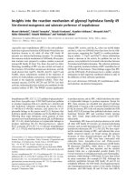
Fig. 1. Panel of mutants of murine DG fused to GFP. The a- and
b-subunits of mammalian DG contain several well-conserved
domains: (a) the N-terminal domain, the mucin-like region and the
C-terminal region of a-DG, the latter containing the b-DG binding epi-
tope (amino acids 550–565); (b) the ectodomain, the transmembrane
region (TM) and the cytosolic domain of b-DG. The b-DG binding epi-
tope was mutated by alanine scanning to produce the following
mutants: S556A, Q559A, M561A, Y562A, G563A, L564A and
P565A. A mutant deleted of the whole b-DG binding epitope
(DGD550–565) was also generated. All the mutations were intro-
duced into the wild-type murine DG cDNA sequence and cloned into
a pEGFP vector for cell transfection experiments, or introduced into
a plasmid, allowing quantitative expression of recombinant C-termi-
nal a-DG peptides in E. coli cells (see Experimental procedures).
A
B
C
Fig. 2. Solid-phase binding assays. a-DG(485–630) (black) and its
mutants, a-DG(485–630)S556A (red), a-DG(485–630)Y562A (green),
a-DG(485–630)L564A (blue) (A), a-DG(485–630)Q559A (red), a-DG
(485–630)M561A (green), a-DG(485–630)G563A (blue), a-DG(485–
630)P565A (magenta) (B) and a-DG(485–630)G563A-P565A (red)
(C), were coated onto a microtitre plate, whereas biotinylated
b-DG(654–750) was used as a soluble ligand at increasing concen-
trations. Each continuous line corresponds to a representative
experiment (from a set of at least three experiments with similar
results), and was obtained by fitting experimental data to a single
class of equivalent binding sites equation (see Experimental
procedures).
F. Sciandra et al. Mutagenesis induces an uncleaved dystroglycan
FEBS Journal 276 (2009) 4933–4945 ª 2009 The Authors Journal compilation ª 2009 FEBS 4935
of both G563 and P565 with alanine completely inhib-
ited the interaction between a-DG(485–630) and
b-DG(654–750), at least in the ligand concentration
range explored (Fig. 2C). Interestingly, G563 and P565
are also fully conserved in DGs from phylogenetically
distant species (Fig. S1, see Supporting information).
These results indicate that, together, G563 and P565
might significantly contribute to the a–b interface and
to the stability of the whole DG complex.
Transfection of 293-Ebna cells with mutated DGs:
western blot and fluorescence microscopy
In order to analyse in eukaryotic cells the effects of the
point mutations that impair the interaction between a-
and b-DG, the same mutations tested in solid-phase
binding assays were introduced within the entire mur-
ine DG cDNA, which was cloned into the pEGFP vec-
tor and used to transiently transfect 293-Ebna cells.
Enhanced green fluorescent protein (EGFP) was fused
at the C-terminal region of b-DG to increase its molec-
ular mass by 25 kDa; the presence of GFP allows
endogenous b-DG to be distinguished unambiguously
from exogenous b-DG-EGFP in western blot analysis.
Western blot of total protein extracts of cells overex-
pressing DG-EGFP constructs carrying the single
point mutations S556A, Q559A, M561A, G563A,
L564A and P565A confirmed the presence of the
expected 68 kDa band corresponding to exogenous
b-DG-EGFP when the samples were probed with both
anti-b-DG and anti-EGFP IgG (Fig. 3A,B).
However, G563A displayed an additional faint band
of about 100 ⁄ 200 kDa (Fig. 3A,B). Interestingly, the
same band was also detectable in the two double
mutants, G563A ⁄ P565A and F692A ⁄ F718A (Fig. 3C–
E). The latter mutant hits the two phenylalanines
belonging to the b-DG ectodomain, F692A and F718A,
that have been shown previously to be key residues for
binding with a-DG in vitro [26]. This higher band is
likely to correspond to the unprocessed DG precursor
(hereafter pre-DG), as the mutation S654A, located at
the physiological a ⁄ b maturation cleavage site G653–
S654, produces a single band with a molecular weight
estimated at 160 kDa that has the same electrophoretic
mobility as displayed by the double mutants G563A ⁄
P565A and F692A ⁄ F718A (Fig. 3C,D) [16,27,28].
On the basis of these results, we hypothesized that
perturbation of the network of interactions that is
likely to stabilize the a–b interface within the DG com-
plex may interfere with the cleavage of the DG precur-
sor. To further validate this hypothesis, we generated
two additional constructs, one carrying the four
mutations G563A, P565A, F692A and F718A,
DGG563A_P565A_F692A_F718A, and the second
with deletion of the whole b-DG binding epitope
between amino acids 550 and 565 within the C-termi-
nal domain of a-DG, DGD550–565 [25]. As expected,
the products of both constructs appeared on SDS-
PAGE as a single 160 kDa band, albeit less intense
than that observed for the mutant S654A, indicating
an instability and a major susceptibility to degradation
of the former mutant pre-DGs (Fig. 3C–E). A
possible scale in the amounts of pre-DG is as fol-
lows: DGS654A > DGG563A_P565A_F692A_F718A
> DG(D550–565) > DGF692AF718A ‡ DGG563A
P565A > DGG563A (Fig. 3E).
Fluorescence microscopy analysis showed that the
DG precursors are likely to be properly targeted at
the plasma membrane, as cells expressing the un-
cleavable DG mutants are indistinguishable from
those expressing wild-type DG (Fig. 4). In addition,
the quadruple mutation G563A ⁄ P565A
⁄ -
F692A ⁄ F718A and the deletion of the 550–565
region did not significantly affect the trafficking or
membrane targeting of pre-DG (Fig. 4). The diffused
and punctuated label throughout the cytoplasm and
around the plasma membrane, featured by cells
transfected with both wild-type and mutated DG,
was probably a result of overexpression of exogenous
EGFP-tagged proteins.
Wheat germ lectin (WGL)-driven enrichment of
mutant pre-DGs
The DG gene encodes a unique polypeptide precur-
sor consisting of 895 amino acids with a calculated
Table 1. Apparent equilibrium dissociation constants (K
D
) calcu-
lated by solid-phase binding assays. Mean apparent K
D
values and
relative standard deviations, calculated for the interaction between
a-DG(485–630) and its mutants and b-DG(654–750) in solid-phase
binding assays. The values were averaged over a number of inde-
pendent experiments indicated in parentheses. For the a-DG(485–
630) double mutant, showing a strongly reduced affinity towards
b-DG(654–750), the K
D
value could not be calculated (n.d.; see
Experimental procedures).
Immobilized protein ⁄ biotinylated protein K
D,app
(lM)
a-DG
wt
⁄ b-DG
wt
3.3 ± 1.0 (6)
a-DG(S556A) ⁄ b-DG
wt
3.2 ± 1.2 (4)
a-DG(Q559A) ⁄ b-DG
wt
4.3 ± 0.3 (3)
a-DG(M561A) ⁄ b-DG
wt
4.5 ± 1.4 (3)
a-DG(Y562A) ⁄ b-DG
wt
3.1 ± 0.8 (5)
a-DG(G563A) ⁄ b-DG
wt
4.7 ± 0.6 (3)
a-DG(L564A) ⁄ b-DG
wt
3.5 ± 1.4 (3)
a-DG(P565A) ⁄ b-DG
wt
5.5 ± 1.2 (4)
a-DG(G563A–P565A) ⁄ b-DG
wt
n.d. (3)
Mutagenesis induces an uncleaved dystroglycan F. Sciandra et al.
4936 FEBS Journal 276 (2009) 4933–4945 ª 2009 The Authors Journal compilation ª 2009 FEBS
molecular mass of about 98 kDa. Based on their
apparent mobility on SDS-PAGE, pre-DGG
563A_P565A, pre-DGG563A_P565A_F692A_F718A,
pre-DG(D550–565) and pre-DGS654A should be
highly, or at least partially, glycosylated. In order to
further clarify this aspect, total protein extracts
obtained from 293-Ebna cells transfected with the
uncleavable DG mutants were incubated with aga-
rose-immobilized WGL that specifically binds N-acet-
ylglucosamine residues (Fig. 5A). All the pre-DG
mutants were pulled down and enriched by this pro-
cedure, suggesting the presence of N-acetylglucos-
amine moieties within the uncleaved precursors
(Fig. 5B). Densitometric analysis confirmed the minor
stability of preDGG563A_P565A, preDGG563A_
P565A_F692A_F718A and preDG(D550–565) when
compared with pre-DGS654A (Fig. 5C).
Laminin binding properties of mutant pre-DGs
DG serves as a receptor for a variety of extracellular
ligands, such as laminin, agrin and perlecan. Full chemi-
cal deglycosylation of a-DG in vitro is known to disrupt
its ability to bind other extracellular matrix proteins
[29,30]. Therefore, laminin conjugated to Sepharose
beads was used to test the capacity of the mutant
pre-DGs (pre-DGG563A_P565A_F692A_F718A, pre-
DG
D550–565
and pre-DGS654A) to interact with com-
mercial mouse laminin-1 (Fig. 6A). Mutant pre-DGs
remained bound to laminin even after several washing
steps (Fig. 6B). This interaction was inhibited using
EDTA, suggesting that the binding between laminin and
the mutant pre-DGs is reversible and dependent on
divalent calcium cations, as expected for the laminin–
DG interaction (data not shown) [29,31].
A
B
C D
E
Fig. 3. Western blot of total protein
extracts. 293-Ebna cells were transfected
with DG mutants and their protein extracts
were probed with anti-b-DG (anti-43-DAG)
(A and C) or anti-GFP (B and D). The DG
mutants carrying the point mutations
S556A, Q559A, M561A, G563A, L564,
P565A display a single band corresponding
to the cleaved b-DG-GFP (A and B). The
double DG mutants, G563A ⁄ P565A and
F692A ⁄ F718A, show the presence of an
additional higher band at 160 kDa that is
likely to correspond to the unprocessed
pre-DG (C and D). Pre-DG is also expressed
in the presence of the mutation S654A,
the quadruple mutation G563A ⁄ P565A ⁄
F692A ⁄ F718A and the deletion of the entire
b-DG binding epitope between amino acids
550–565 (C and D). A lower band, at about
50 kDa, probably originates from further pro-
teolysis of b-DG-GFP. The black boxes indi-
cate pre-DG-GFP and b-DG-GFP. The
amounts of cleaved DG-GFP (open bars) and
unprocessed pre-DG (filled bars) were quan-
tified by densitometry, averaging the values
of the band intensities obtained from five
independent experiments (E). Such a quanti-
tative analysis shows how the differences in
band intensities between some of the
mutants are not significant.
F. Sciandra et al. Mutagenesis induces an uncleaved dystroglycan
FEBS Journal 276 (2009) 4933–4945 ª 2009 The Authors Journal compilation ª 2009 FEBS 4937
Discussion
Towards the identification of single amino acids
within the C-terminal region of a-DG that are
crucial for the interaction with b-DG
In this study, we focused alanine scanning on the
a-DG amino acid linear sequence 550–565 in an
attempt to identify which residues were responsible for
binding with b-DG. A series of point mutations,
S556A, Q559A, M561A, Y562A, G563A, L564A and
P565A, was introduced into the recombinant protein
a-DG(485–630), and their affinities towards recombi-
nant biotinylated b-DG(654–750) were measured. The
K
D
values reported in Table 1 show that only the point
mutations hitting odd positions (i.e. G563A and
P565A) elicit some slight effect on the interaction with
b-DG, whereas those at even positions (i.e. S556A,
Y562A and L564A) do not produce any effect (see
Fig. 2A,B and Table 1). Moreover, the double muta-
tion G563A ⁄ P565A completely inhibits the interaction
between a- and b-DG (Fig. 2C).
The interaction between a- and b-DG seems to
induce some local secondary structures. Indeed, our
results may suggest that the a-DG linear sequence
QLMYGLP assumes a b-strand conformation, with
the amino acids Q559, M561, G563 and P565 pointing
towards the b-DG ectodomain and interacting with it.
Retrospectively, our previous NMR experiments, car-
ried out by exploiting the synthetic peptide a-DG(550–
585) in free and b-DG-bound fashion, suggested a
greater involvement of Q559, M561 and G563 than of
S558, L560, Y562 and L564 in binding the recombi-
nant b-DG ectodomain; the alternate fashion of these
side-chain contributions could indeed be reminiscent of
a b-strand conformation (see Fig. 4 of [25]). Further-
more, the hypothesis that the QLMYGLP amino acid
stretch would assume a b-strand conformation is
corroborated by a model of the a-DG C-terminal
domain based on sequence homology with a member
of the cadherin family [32]. However, further experi-
ments are needed to validate this hypothesis.
Interestingly, the crucial importance of G563 and
P565 could be deduced from the analysis of a multiple
alignment of DG sequences from species phylogeneti-
cally distant from humans or mouse, including lower
vertebrate and several invertebrate species (Fig. S1, see
Supporting information), where these amino acids are
always conserved despite a very low overall sequence
homology. Our new data on G563 and P565, together
with the results of our previous study, in which two
phenylalanines belonging to the b-DG ectodomain,
F692 and F718, were recognized as key residues for
the interaction with a-DG, point towards the identifi-
cation of the major molecular cornerstones of the a–b
interface.
Fig. 4. Immunofluorescence of 293-Ebna cells transfected with the pEGFP vector, empty or carrying wild-type or mutated DGs. All the
uncleavable mutants are expressed and targeted to the plasma membrane (open arrowheads), showing a fluorescence pattern similar to that
of wild-type DG (WT). GFP was expressed throughout the cytoplasm.
Mutagenesis induces an uncleaved dystroglycan F. Sciandra et al.
4938 FEBS Journal 276 (2009) 4933–4945 ª 2009 The Authors Journal compilation ª 2009 FEBS
The a–b interface is essential for the correct
cleavage of the DG precursor
A heterologous cell expression system was used to
verify whether the mutations analysed in vitro might
also influence the expression and stability of DG in
cells. 293-Ebna cells were transfected with the entire
DG gene carrying the single mutations, S556A,
Q559A, M561A, G563A, L564A and P565A, and
cloned into a pEGFP vector. As demonstrated by
western blot of total cell extracts, the single point
mutations do not drastically alter the stability of DG,
which is correctly processed into the two subunits
(Fig. 3A,B). Only the mutant G563A showed an addi-
tional faint band at about 160 kDa, probably caused
by a small amount of the uncleaved DG precursor,
pre-DG, which spans both the a- and b-subunits
of DG (Fig. 3A,B,E). Interestingly, the two DG
A
B
C
Fig. 5. WGL enrichment of total protein extracts of untransfected
cells (NT) and cells transfected with wild-type or mutated DGs. (A)
Assay rationale: WGL specifically binds to the N-acetylglucosamine
moieties covalently linked to the core protein of a-DG. Therefore,
b-DG-GFP, which is noncovalently associated with a-DG (or directly
pre-DG), can be retained from the immobilized WGL molecules.
(B) Western blot carried out with the anti-b-DG IgG clearly shows
that both wild-type (b-DG-GFP) and mutant (b-DG-GFP and mainly
pre-DG) DG proteins can be specifically eluted by WGL beads.
Only the eluted fractions, collected upon extensive washing, were
loaded onto the gel; the wash fractions did not contain any rele-
vant signal (data not shown). (C) The amounts of cleaved DG-GFP
and unprocessed pre-DG were quantified by densitometry, averag-
ing the values of the band intensities obtained from three indepen-
dent experiments.
A
B
Fig. 6. Laminin-Sepharose pull-down of mutant pre-DGs. (A) Assay
rationale: laminin, covalently bound to CNBr-Sepharose, interacts
with a-DG. b-DG-GFP is retained by laminin-Sepharose beads
through the interaction with the a-subunit. (B) Pull-down of wild-
type (WT) DG or DG carrying the mutations indicated on the spe-
cific panels. Western blot carried out with the anti-b-DG IgG clearly
shows that both wild-type and mutant DG proteins specifically bind
laminin (lane E: elution fraction) after extensive washing (lanes W1
and W5); FT (flow-through). The black boxes indicate b-DG-GFP and
pre-DG-GFP.
F. Sciandra et al. Mutagenesis induces an uncleaved dystroglycan
FEBS Journal 276 (2009) 4933–4945 ª 2009 The Authors Journal compilation ª 2009 FEBS 4939
constructs carrying the double mutations,
G563A ⁄ P565A and F692A ⁄ F718A, which, in solid-
phase assays completely inhibit the binding between
a- and b-DG (Fig. 2C) [26], display significant
amounts of pre-DG, with respect to the correctly
cleaved b-DG-EGFP (Fig. 3C–E).
The correct cleavage is completely inhibited in the
DG construct carrying the four mutations G563A,
P565A, F692A and F718A, (Fig. 3C–E), suggesting
that interaction between the reciprocal binding epi-
topes of the two subunits forming the mature DG
complex is necessary for correct processing of the DG
precursor. Consistent with this hypothesis, the entire
deletion (knockin) of the b-DG binding epitope within
the a-DG subunit (positions 550–565) abolishes the
cleavage, producing the 160 kDa pre-DG (Fig. 3C–E).
The mutants pre-DGG563A_P565A_F692A_F718A,
pre-DG
D550–565
and pre-DGS654A specifically bind
WGL, which indicates that they are at least partially
glycosylated (Fig. 5B); furthermore, laminin pull-down
experiments show that mutated DG precursors har-
bour some laminin binding epitopes (Fig. 6B), clearly
indicating a residual functionality of hypoglycosylated
and unprocessed pre-DGs.
Depicting a possible model for DG precursor
processing
The mechanism and functional significance of DG pre-
cursor processing still remain largely elusive. In several
human and murine cell lines and tissues, DG was
always detected as a heteromeric complex, suggesting
that precursor cleavage is a very early post-transla-
tional event along the route of DG maturation. Muta-
tions in the amino acid sites crucial for the interaction
between a- and b-DG, namely G563 and P565 within
the C-terminal domain of a-DG and their counterparts
F692 and F718 within the b-DG ectodomain, ‘freeze’
the DG precursor as a relatively stable and partially
glycosylated monomeric intermediate. Our results
strongly suggest that the network of interactions
important for the build up of the a–b interface on pre-
cursor cleavage is already established within the unc-
leaved DG precursor and is strictly necessary for
processing into the two subunits. The impairment of
the correct formation of the a–b interface may destabi-
lize pre-DGs; indeed, both pre-DGG563A_P565A_
F692A_F718A and pre-DG(D550–565) display lower
expression levels compared with pre-DGS654A, in
which most of the interactions underlying the a–b
interface are still likely to take place (Fig. 3E). Such a
network of interactions may also influence the glyco-
sylation pattern of the DG precursor. This could be
inferred from the different electrophoretic behaviour
displayed by the uncleavable pre-DGs. Indeed, pre-
DG
S654A
displays in western blot as a broader band
(which could imply the presence of more carbohydrate
groups) with respect to pre-DGG563A_P565A_
F692A_F718A and pre-DG(D550–565), where most of
the a–b interactions cannot be established (Fig. 3C,D).
The correct folding of the DG precursor may there-
fore be important for the recognition by glyco-
syltransferases, which should primarily take place at
the level of the N-terminal portion of a-DG [33,34]. In
particular, O-glycosyltransferases are thought to be
crucial, especially for the extensive sugar decoration of
the DG central mucin-like domain [19]. Apart from
the correct folding of what could be defined as the
‘pre-a–b interface’, a few other factors have been
proposed to play an important role in DG precursor
processing: for example, the disulfide bridge between
C669 and C713, within the b-DG ectodomain [35], and
N-glycosylation [27]. The formation of this disulfide
bridge may also contribute to the stabilization of the
correct folding of the DG precursor necessary for spe-
cific cleavage. As far as N-glycosylation is concerned,
it has been shown by others that alanine substitution
of N662, a putative N-glycosylation site in the b-DG
ectodomain, prevents the cleavage of the precursor and
strongly reduces its expression [27]. However, whether
N-glycosylation really influences DG precursor cleav-
age is still a matter of debate: other studies have
shown that blocking N-glycosylation does not prevent
cleavage [36].
On the basis of our data and other evidence from
the literature, we propose the following scenario for
DG maturation (shown in Fig. 7): immediately after
translation, the DG core protein is translocated
into the endoplasmic reticulum, where it is likely to
adopt a stable three-dimensional conformation prior
to any post-translational modifications. At this stage,
an essential contribution for achieving a confor-
mation that will allow subsequent cleavage is pro-
vided by a network of interactions (in which G563,
P565, F692 and F718 play a crucial role) that are
likely to stabilize the mature a–b interface also on
cleavage.
It is still unclear whether cleavage is carried out by
an unidentified protease or whether it occurs via an
autocatalytic mechanism [28]. However, our data
clearly show that precursor cleavage is dispensable for
correct trafficking and membrane targeting of DG, as
all our novel uncleavable mutants can be detected at
the plasma membrane, and their localization is indis-
tinguishable from that characterizing wild-type DG
(Fig. 4); furthermore, they are still capable of binding
Mutagenesis induces an uncleaved dystroglycan F. Sciandra et al.
4940 FEBS Journal 276 (2009) 4933–4945 ª 2009 The Authors Journal compilation ª 2009 FEBS
laminin, fulfilling one of the most important functions
of DG (Fig. 6).
Conclusions
Based on the available evidence, during evolution,
there was a ‘free choice’ for the liberation, or not, of
the two DG subunits. For example, in Caenorhabditis
elegans, in which not only DG but a whole DGC
orthologue has been identified and functionally charac-
terized [37], the maturation of the DG complex into
two subunits has not been observed and, accordingly,
the motif Gly-Ser (653–654) at the cleavage site has
not been conserved [38]. Clearly, further work is
needed in order to fully understand the biological sig-
nificance of why the two DG subunits are liberated
[39].
How an abnormal a–b interface would affect human
DG function is not yet known, as no primary muta-
tions of the dag1 gene have been identified so far.
However, in principle, it should be possible to find spe-
cific mutations, or more likely polymorphisms, which,
in mammals, would interfere with DG processing with-
out grossly impairing DG function and displaying very
mild phenotypic signs in virtually asymptomatic carri-
ers. This is suggested by recent papers from other
laboratories showing that DG does not take part in
the later stages of embryonic development, or that hy-
poglycosylated DG can be partially functional [40–42].
However, it will also be important to rule out the pos-
sibility that the presence of an uncleaved DG precur-
sor may, instead, be linked to severe neuromuscular
pathologies.
A concerted effort of biochemical, genetic and clini-
cal studies is needed in order to finally address these
points. At the present stage, our identification of mul-
tiple point mutations that inhibit or affect the DG
maturation pathway may provide a useful tool to
investigate and shed light on the molecular details of
such an important and mysterious process.
Experimental procedures
DNA manipulation
The full-length cDNA encoding for murine DG was used
as a template to generate, by PCR, two DNA constructs,
one corresponding to the N-terminal region of b-DG,
b-DG(654–750), and the other to the C-terminal region of
a-DG, a-DG(485–630) [22]. Appropriate primers were used
to amplify the DNA sequences of interest. For b-DG(654–
750): forward, 5¢-CCCGGATCCTCTATCGTGGTGG
AATGGACCAACA-3¢; reverse, 5¢-CCCGAATTCTTAG
TAAACATCGTCCTCACTGCTCTCTTC-3¢ (BamHI and
EcoRI restriction sites are given in italic type). For
a-DG(485–630): forward, 5¢-CCCGTCGACAGTGGAGTG
CCCCGTGGGGGAGAAC-3¢; reverse, 5¢-CCCGAATTC
TTATACCAAAGCAATTTTTCTTGTGAATG-3¢ (SalI
and EcoRI restriction sites are given in italic type). Single
point mutations were introduced into the murine DG gene,
cloned into the pEGFP vector, using the QuikChange site-
PreDG
AB C
PreDG
Fig. 7. Schematic model showing the influ-
ence of the a–b interface on pre-DG cleav-
age. (A) In the wild-type pre-DG, the correct
interaction between the a- and b-domains
stabilizes pre-DG in a conformation that can
be proteolytically processed at its G ⁄ S
cleavage site, liberating the a- and b-subun-
its. The black double-headed arrow indicates
the pre-a–b interface. (B) When S654, part
of the cleavage site, is mutated, pre-DG is
not proteolytically processed. It is possible
that the interactions within the a–b interface
are formed even in the uncleaved precursor,
ensuring a certain stability of pre-DG. The
black double-headed arrow indicates the
pre-a–b interface. (C) When the a–b inter-
face is impaired by specific mutations hitting
the amino acids G563, P565 (within a-DG),
F692 and F718 (within b-DG), pre-DG does
not reach a conformation suitable for proteo-
lytic cleavage.
F. Sciandra et al. Mutagenesis induces an uncleaved dystroglycan
FEBS Journal 276 (2009) 4933–4945 ª 2009 The Authors Journal compilation ª 2009 FEBS 4941
directed mutagenesis kit (StratageneÒ, Cedar Creek, TX,
USA); all constructs were verified by automated sequenc-
ing. The primers used for mutagenesis are reported below
with the mutated codons in italic:
S556A forward: 5¢-TGGGTTCAGTTTAACGCCAACA
GCCAGCTCATG-3¢
S556A reverse: 5¢-CATGAGCTGGCTGTTGGCGTTA
AACTGAACCCA-3¢
Q559A forward: 5¢-TTTAACAGCAACAGCGCGCTC
ATGTATGGCCTG-3¢
Q559A reverse: 5¢-CAGGCCATACATGAGCGCGCT
GTTGCTGTTAAA-3¢
M561A forward: 5¢-AGCAACAGCCAGCTCGCGTAT
GGCCTGCCTGAC-3¢
M561A reverse: 5¢-GTCAGGCAGGCCATACGCGAG
CTGGCTGTTGCT-3¢
Y562A forward: 5¢-AACAGCCAGCTCATGGCT
GGCCTGCCTGACAGC-3¢
Y562A reverse: 5¢-GCTGTCAGGCAGGCCAGCCAT
GAGCTGGCTGTT-3¢
G563A forward: 5¢-AGCCAGCTCATGTATGCCCTG
CCTGACAGCAGC-3¢
G653A reverse: 5¢-GCTGCTGTCAGGCAGGGCATA
CATGAGCTGGCT-3¢
L564A forward: 5¢-CAGCTCATGTATGGCGCGCCTG
ACAGCAGCCAT-3¢
L564A reverse: 5¢-ATGGCTGCTGTCAGGCGCGCC
ATACATGAGCTG-3¢
P565A forward: 5
¢-CTCATGTATGGCCTGGCTGAC
AGCAGCCATGTG-3¢
P565A reverse: 5¢-CACATGGCTGCTGTCAGCCAG
GCCATACATGAG-3¢
S654A forward: 5¢-CAGAACATCACTCGGGGCGC
TATCGTGGTGGAATGGACC-3¢
S654A reverse: 5¢-GGTCCATTCCACCACGATAGCGC
CCCGAGTGATGTTCTG-3¢
G563AP565A forward:5¢-AGCCAGCTCATGTATG
CCCTGGCTGACAGCAGC-3¢
G563AP565A reverse: 5¢-GCTGCTGTCAGCCAGGG
CATACATGAGCTGGCT-3¢
The full-length DNA constructs carrying the point muta-
tions were also used as templates to generate, by PCR, the
DNA constructs for the expression of the a-DG(485–630)
mutants in the Escherichia coli recombinant system (see
below), employing the same primers as used to amplify the
wild-type a-DG(485–630) sequence.
For the production of the DG(D550–565) deletion
mutant, the knocked-in DNA construct was generated by
the overlap extension method [43] using 5¢-CCCGAAT
TCATGTCTGTGGACAACTGGCTACTG-3¢ and 5¢-
TTTCTCACCTACTAACTGCTGCTCT-3¢ as forward and
reverse primers, respectively, for the first PCR, and 5¢-
CAGTTAGTAGGTGAGAAAGACAGCAGCCATGTG-3¢
and 5¢-CCCGAATTCGGCTAGGGGGAACATACGGAG
GGGG-3¢ for the second PCR.
Protein expression, purification and biotinylation
The DNA constructs were cloned into a bacterial vector
that was appropriate for the expression of the protein as a
thioredoxin fusion product, also containing an N-terminal
6His tag and a thrombin cleavage site [44]. The recombi-
nant fusion proteins were expressed in E. coli BL21(DE3)
Codon Plus RIL strain and purified using nickel affinity
chromatography. The fragments of interest were obtained
on thrombin cleavage. Tricine ⁄ SDS-PAGE was used to
check the purity of the recombinant proteins under analy-
sis. For solid-phase binding assays, recombinant
b-DG(654–750) was biotinylated in 5 mm sodium phos-
phate buffer at pH 7.4, with 0.5 mgÆmL
)1
sulfo-N-hydroxyl-
succinimido-biotin (S-NHS-biotin, PierceÒ, Rockford, IL,
USA). The reaction was carried out for 30 min on ice and
in the dark, and dialysed overnight against 10 mm
Tris ⁄ HCl, 150 mm NaCl, pH 7.4. The optimal dilution for
signal detection was determined by dot blot analysis and
revealed by enhanced chemiluminescence (PierceÒ).
Solid-phase binding assays
To assess the binding properties of recombinant a-DG(485–
630) and its mutants with respect to biotinylated recombi-
nant b-DG(654–750), solid-phase assays were performed as
follows: approximately 0.5 lgofa-DG(485–630), its
mutants and BSA were immobilized on microtitre plates in
coating buffer (50 mm NaHCO
3
, pH 9.6) overnight at 4 °C.
After washing with NaCl ⁄ P
i
buffer (2.5 mm KCl, 2 mm
KH
2
PO
4
,2mm Na
2
HPO
4
, 140 mm NaCl, pH 7.4) contain-
ing 0.05% (v ⁄ v) Tween-20, 1.25 mm CaCl
2
and 1 mm
MgCl
2
, wells were incubated with decreasing concentrations
of recombinant biotinylated b-DG(654–750) in NaCl ⁄ P
i
containing 0.05% (v ⁄ v) Tween-20, 3% (w ⁄ v) BSA, 1.25 mm
CaCl
2
and 1 mm MgCl
2
for 3 h at room temperature. After
washing, the biotinylated b-DG(654–750) bound fraction
was detected with alkaline phosphatase Vectastain AB
Complex (Vector LaboratoriesÒ, Burlingame, CA, USA).
Five milligrams of p-nitrophenyl phosphate dissolved in
10 mL of 10 mm diethanolaminine and 0.5 m MgCl
2
were
added to every well containing 100 lL of this solution, and
used as a substrate for the reaction with alkaline phospha-
tase; the absorbance values were recorded at 405 nm. For
each b-DG(654–750) concentration, the absorbance value
(A
i
) originating from coated BSA was subtracted from the
values obtained with the coated wild-type or mutated a-DG
samples under analysis. The data were fitted using a single
class of equivalent binding sites equation, A
i
= A-
sat
[c ⁄ (K
D
+ c)+A
0
], where A
i
represents the absorbance
measured at increasing concentrations of ligand, K
D
is the
dissociation constant, c is the concentration of ligand, bioti-
nylated b-DG(654–750), and A
sat
and A
0
are the absor-
bances at saturation and in the absence of ligand,
respectively. Data were normalized and reported as the
Mutagenesis induces an uncleaved dystroglycan F. Sciandra et al.
4942 FEBS Journal 276 (2009) 4933–4945 ª 2009 The Authors Journal compilation ª 2009 FEBS
fractional saturation (%): 100· [(A
i
) A
0
) ⁄ (A
sat
) A
0
)]. For
the double mutant a-DG(485–630)G563A-P565A, which
displayed a strong reduction in binding affinity, the data
could not be fitted according to the equation above, and
were simply normalized, setting the maximal binding of
wild-type a-DG(485–630), extrapolated by the fitting, as
100%. The K
D
values reported in Table 1 were averaged
over three or more independent experiments and their stan-
dard deviations were calculated and reported.
Cell culture, transfection and fluorescence
microscopy
293-Ebna cells were grown in DMEM supplemented with
antibiotics and 10% (v ⁄ v) fetal bovine serum. About 1 lg
of wild-type or mutated pEGFP
DG
was transiently trans-
fected into 293-Ebna cells using Fugene-6 (RocheÒ, Basle,
Switzerland), according to the manufacturer’s instructions.
On transfection (24 h), the cells were fixed with 4% (v ⁄ v)
paraformaldehyde at room temperature for 30 min and
observed under a fluorescence microscope (Nikon
Ò
, Tokyo,
Japan).
About 20 lg of wild-type or mutated pEGFP
DG
were
also transfected using the calcium phosphate method:
briefly, DNA was mixed with 125 mm CaCl
2
and 50 mm
Bes (Bes-buffered saline). The DNA–calcium phosphate
complex was added to the cells. After 24 h, the cells were
collected for western blot analysis.
Total protein extraction and western blot
Cells transfected with empty pEGFP, wild-type or mutated
pEGFP
DG
were lysed with NaCl ⁄ P
i
containing 1% Triton
X-100 and protease inhibitors and centrifuged at 15 000 g
for 10 min at 4 °C; 20 lg of each lysate were resolved by
10% SDS-PAGE. For western blot analysis, proteins were
transferred to nitrocellulose and probed with the following
IgG: anti-b-DG (Novocastra, Newcastle Upon Tyne, UK),
diluted 1 : 50, and anti-EGFP (Clontech, Mountain View,
CA, USA), diluted 1 : 250. The nitrocellulose was incu-
bated with horseradish peroxidase-conjugated secondary
antibody (Sigma–Aldrich, St Louis, MO, USA), diluted
1 : 5000; the reactive products were revealed using the lumi-
nol-based ECL system (PierceÒ).
WGL enrichment assay
Total protein extracts of cells transfected with pEGFP
DG
,
wild-type, and carrying the mutations S654A,
G563A ⁄ P565A, G563A ⁄ P565A ⁄ F692A ⁄ F718A and the
deletion D550–565 were incubated with WGL Sepharose
6MB (Vector Laboratories) and equilibrated in NaCl ⁄ P
i
and 1% Triton X-100 overnight at 4 °C. After extensive
washing with 50 mm Tris ⁄ HCl, pH 7.8, 500 mm NaCl,
0.1% Triton X-100, bound glycoproteins were eluted in the
same buffer with 250 mm N-acetylglucosamine and
analysed by western blot.
Western blot quantification
Densitometric analyses of different films were performed
using a GS-800 imaging densitometer (Bio-Rad, Hercules,
CA, USA) and analysed using Bio-Rad Quantity-One soft-
ware. The signal intensities of cleaved b-DG and unpro-
cessed pre-DG bands were determined using volume
analysis with object average background correction applied.
The volume is defined as the ‘sum of the intensities of the
pixels within the volume boundary · pixel area’ and vol-
ume units are calculated as ‘intensity units · mm
2
’. The
volumes of at least three independent experiments were nor-
malized with respect to the endogenous DG volume and
used to calculate the mean values. Data were exported to
MicrosoftÔ Excel to generate the plot.
Preparation of laminin-Sepharose
Preactivated CNBr Sepharose (Vector Laboratories) was sus-
pended in 1 mm HCl for 15 min and washed with 1 mm HCl
on a sintered glass filter. EHS tumor laminin (Sigma–
Aldrich) (300 lg) in coupling buffer (0.1 m NaHCO
3
, 0.5 m
NaCl, pH 9) was mixed with 3 mL of CNBr Sepharose for
2 h at room temperature. The laminin-Sepharose was incu-
bated for 2 h with blocking buffer (0.1 m Tris-base, pH 8.0),
and then washed with three cycles of alternating pH. Each
cycle consists of a wash with 0.1 m acetic acid, followed by a
wash with 0.1 m Tris-base pH 8.0 containing 0.5 m NaCl.
Laminin-Sepharose was stored in NaCl ⁄ P
i
at 4 °C.
Laminin-Sepharose pull-down assay
Total protein extracts of cells transfected with pEGFP
DG
,
wild-type, and carrying the mutations S654A,
G563A ⁄ P565A, G563A ⁄ P565A ⁄ F692A ⁄ F718A and the
deletion D550–565 were diluted with NaCl ⁄ P
i
containing
1.25 mm CaCl
2
, 1.25 mm MgCl
2
, and incubated overnight
at 4 °C with 200 lL of laminin-Sepharose in the presence
of 1.25 mm CaCl
2
, 1.25 mm MgCl
2
. Laminin-Sepharose
was extensively washed with NaCl ⁄ P
i
containing 1.25 mm
CaCl
2
, 1.25 mm MgCl
2
until the absorbance at 280 nm of
the supernatant was zero. Laminin-coupled proteins were
eluted with 100 lLof1m glycine, pH 3. The pH was equil-
ibrated to 7.4 with 1 m Tris-base, pH 9, and the samples
were analysed by western blot.
Acknowledgements
This work was supported by Telethon grant
GGP06225 and ISS ⁄ NIH collaborative project (7DR1)
F. Sciandra et al. Mutagenesis induces an uncleaved dystroglycan
FEBS Journal 276 (2009) 4933–4945 ª 2009 The Authors Journal compilation ª 2009 FEBS 4943
to A.B. The authors wish to thank Ernesto Pavoni,
Maria Giovanna Desimio and Salvatore Meo for tech-
nical assistance, Maria Giulia Bigotti (Rome) and
Tamara C. Petrucci (Rome) for critical reading of the
manuscript, and Kevin P. Campbell (Iowa University)
for his advice on the laminin pull-down experiments.
References
1 Ibraghimov-Beskrovnaya O, Ervasti JM, Leveille CJ,
Slaughter CA, Sernett SW & Campbell KP (1992) Pri-
mary structure of dystrophin-associated glycoproteins
linking dystrophin to the extracellular matrix. Nature
355, 696–702.
2 Barresi R & Campbell KP (2006) Dystroglycan: from
biosynthesis to pathogenesis of human disease. J Cell
Sci 119, 199–207.
3 Cohn RD (2005) Dystroglycan: important player in
skeletal muscle and beyond. Neuromuscul Disord 15,
207–217.
4 Durbeej M & Campbell KP (1999) Biochemical charac-
terization of the epithelial dystroglycan complex. J Biol
Chem 274, 26609–26616.
5 Williamson RA, Henry MD, Daniels KJ, Hrstka RF,
Lee JC, Sunada Y, Ibraghimov-Beskrovnaya O &
Campbell KP (1997) Dystroglycan is essential for
early embryonic development: disruption of Reichert’s
membrane in Dag1-null mice. Hum Mol Genet 6,
831–841.
6 Henry MD & Campbell KP (1998) A role for dystrogly-
can in basement membrane assembly. Cell 95, 859–870.
7 Colognato H, Winkelmann DA & Yurchenco PD
(1999) Laminin polymerization induces a receptor-cyto-
skeleton network. J Cell Biol 145, 619–631.
8 Deng WM, Schneider M, Frock R, Castillejo-Lopez C,
Gaman EA, Baumgartner S & Ruohola-Baker H (2003)
Dystroglycan is required for polarizing the epithelial cells
and the oocyte in Drosophila. Development 130, 173–184.
9 Weir ML, Oppizzi ML, Henry MD, Onishi A, Camp-
bell KP, Bissell MJ & Muschler JL (2006) Dystroglycan
loss disrupts polarity and b-casein induction in mam-
mary epithelial cells by perturbing laminin anchoring.
J Cell Sci 119, 4047–4058.
10 Peng H, Shah W, Holland P & Carbonetto S (2008)
Integrins and dystroglycan regulate astrocyte wound
healing: the integrin-b1 subunit is necessary for process
extension and orienting the microtubular network. Dev
Neurobiol 68, 559–574.
11 Peng HB, Xie H, Rossi SG & Rotundo RL (1999)
Acetylcholinesterase clustering at the neuromuscular
junction involves perlecan and dystroglycan. J Cell Biol
145, 911–921.
12 Saito F, Moore SA, Barresi R, Henry MD, Messing A,
Ross-Barta SE, Cohn RD, Williamson RA, Sluka KA,
Sherman DL et al. (2003) Unique role of dystroglycan
in peripheral nerve myelination, nodal structure, and
sodium channel stabilization. Neuron 38, 747–758.
13 Ervasti JM & Sonnemann KJ (2008) Biology of the
striated muscle dystrophin–glycoprotein complex. Int
Rev Cytol 265, 191–225.
14 Smalheiser NR & Kim E (1995) Purification of cranin,
a laminin binding membrane protein. Identity with dys-
troglycan and reassessment of its carbohydrate moieties.
J Biol Chem 270, 15425–15433.
15 Deyst KA, Bowe MA, Leszyk JD & Fallon JR (1995)
The a-dystroglycan–b-dystroglycan complex. Membrane
organization and relationship to an agrin receptor.
J Biol Chem 270, 25956–25959.
16 Jayasinha V, Nguyen HH, Xia B, Kammesheidt A,
Hoyte K & Martin PT (2003) Inhibition of dystroglycan
cleavage causes muscular dystrophy in transgenic mice.
Neuromuscul Disord 13, 365–375.
17 Schro
¨
der JE, Tegeler MR, Grobhans U, Porten E,
Blank M, Lee J, Esapa C, Blake DJ & Kro
¨
ger S (2007)
Dystroglycan regulates structure, proliferation and dif-
ferentiation of neuroepithelial cells in the developing
vertebrate CNS. Dev Biol 307, 62–78.
18 Batchelor CL, Higginson JR, Chen YJ, Vanni C, Eva A
& Winder SJ (2007) Recruitment of Dbl by ezrin and
dystroglycan drives membrane proximal Cdc42 activa-
tion and filopodia formation. Cell Cycle 6, 353–363.
19 Brancaccio A, Schulthess T, Gesemann M & Engel J
(1995) Electron microscopic evidence for a mucin-like
region in chick muscle a-dystroglycan. FEBS Lett 368,
139–142.
20 Michele DE, Barresi R, Kanagawa M, Saito F, Cohn
RD, Satz JS, Dollar J, Nishino I, Kelley RI, Somerk H
et al. (2002) Post-translational disruption of dystrogly-
can–ligand interactions in congenital muscular dystro-
phies. Nature 418, 417–421.
21 Muntoni F, Brockington M, Blake DJ, Torelli S &
Brown SC (2002) Defective glycosylation in muscular
dystrophy. Lancet 360, 1419–1421.
22 Sciandra F, Schneider M, Giardina B, Baumgartner S,
Petrucci TC & Brancaccio A (2001) Identification of the
b-dystroglycan binding epitope within the C-terminal
region of a-dystroglycan. Eur J Biochem 268, 4590–
4597.
23 Bonuccelli G, Sotgia F, Schubert W, Park DS, Frank
PG, Woodman SE, Insabato L, Cammer M, Minetti C
& Lisanti P (2003) Proteasome inhibitor (MG-132)
treatment of mdx mice rescues the expression and mem-
brane localization of dystrophin and dystrophin-associ-
ated proteins. Am J Pathol 163, 1663–1675.
24 Bozzi M, Bianchi M, Sciandra F, Paci M, Giardina B,
Brancaccio A & Cicero DO (2003) Structural character-
ization by NMR of the natively unfolded extracellular
domain of b-dystroglycan: toward the identification of
Mutagenesis induces an uncleaved dystroglycan F. Sciandra et al.
4944 FEBS Journal 276 (2009) 4933–4945 ª 2009 The Authors Journal compilation ª 2009 FEBS
the binding epitope for a-dystroglycan. Biochemistry 42 ,
13717–13724.
25 Bozzi M, Veglia G, Paci M, Sciandra F, Giardina B &
Brancaccio A (2001) A synthetic peptide corresponding
to the 550–585 region of a-dystroglycan binds b-dystro-
glycan as revealed by NMR spectroscopy. FEBS Lett
499, 210–214.
26 Bozzi M, Sciandra F, Ferri L, Torreri P, Pavoni E, Pet-
rucci TC, Giardina B & Brancaccio A (2006) Concerted
mutation of Phe residues belonging to the b-dystrogly-
can ectodomain strongly inhibits the interaction with
a-dystroglycan in vitro. FEBS J 273, 4929–4943.
27 Esapa CT, Bentham GR, Schroder JE, Kroger S &
Blake DJ (2003) The effects of post-translational pro-
cessing on dystroglycan synthesis and trafficking. FEBS
Lett 555, 209–216.
28 Akhavan A, Crivelli SN, Singh M, Lingappa VR &
Muschler JL (2008) SEA domain proteolysis determines
the functional composition of dystroglycan. FASEB J
22, 612–621.
29 Ervasti JM & Campbell KP (1993) A role for the
dystrophin–glycoprotein complex as a transmembrane
linker between laminin and actin. J Cell Biol 122 ,
809–823.
30 Sugita S, Saito F, Tang J, Satz J, Campbell KP &
Su
¨
dhof TC (2001) A stoichiometric complex of
neurexins and dystroglycan in brain. J Cell Biol 154,
435–445.
31 Hohenester E, Tisi D, Talts JF & Timpl R (1999) The
crystal structure of a laminin G-like module reveals the
molecular basis of a-dystroglycan binding to laminins,
perlecan, and agrin. Mol Cell 4, 783–792.
32 Dickens NJ, Beatson S & Ponting CP (2002) Cadherin-
like domains in a-dystroglycan, a ⁄ e-sarcoglycan and
yeast and bacterial proteins. Curr Biol 2, 197–199.
33 Kanagawa M, Saito F, Kunz S, Yoshida-Moriguchi T,
Barresi R, Kobayashi YM, Muschler J, Dumanski JP,
Michele DE, Oldstone MB et al. (2004) Molecular
recognition by LARGE is essential for expression of
functional dystroglycan. Cell 117, 953–964.
34 Bozic D, Sciandra F, Lamba D & Brancaccio A (2004)
The structure of the N-terminal region of murine skele-
tal muscle a-dystroglycan discloses a modular architec-
ture. J Biol Chem 279, 44812–44816.
35 Watanabe N, Sasaoka T, Noguchi S, Nishino I &
Tanaka T (2007) Cys669–Cys713 disulfide bridge for-
mation is a key to dystroglycan cleavage and subunit
association. Genes Cells 12, 75–88.
36 Holt KH, Crosbie RH, Venzke DP & Campbell KP
(2000) Biosynthesis of dystroglycan: processing of a
precursor propeptide. FEBS Lett 468, 79–83.
37 Grisoni K, Martin E, Gieseler K, Mariol MC & Segalat
L (2002) Genetic evidence for a dystrophin–glycoprotein
complex (DGC) in Caenorhabditis elegans . Genes 294,
77–86.
38 Johnson RP, Kang SH & Kramer JM (2006) C. elegans
dystroglycan DGN-1 functions in epithelia and neurons,
but not muscle, and independently of dystrophin. Devel-
opment 133, 1911–1921.
39 Bozzi M, Morlacchi S, Bigotti MG, Sciandra F & Bran-
caccio A (2009) Functional diversity of dystroglycan.
Matrix Biol 28, 179–187.
40 Satz JS, Barresi R, Durbeej M, Willer T, Turner A,
Moore SA & Campbell KP (2008) Brain and eye mal-
formations resembling Walker–Warburg syndrome are
recapitulated in mice by dystroglycan deletion in the
epiblast. J Neurosci 28, 10567–10575.
41 Jimenez-Mallebrera C, Torelli S, Feng L, Kim J,
Godfrey C, Clement E, Mein R, Abbs S, Brown SC,
Campbell KP et al. (2008) Comparative study of
a-dystroglycan glycosylation in dystroglycanopathies
suggests that the hypoglycosylation of a-dystroglycan
does not consistently correlate with clinical severity.
Brain Pathol. doi:10.1111/j.1750-3639.2008.00198.
42 Puckett R, Moore SA, Winder TL, Willer T, Romansky
SG, Covault KK, Campbell KP & Abdenur JE (2009)
Further evidence of Fukutin mutations as a cause of
childhood onset limb-girdle muscular dystrophy
without mental retardation. Neuromuscul Disord 19,
352–356.
43 Horton RM (1993) PCR protocols: current methods
and applications. In Methods in Molecular Biology:
PCR protocols – Current Methods and Application,
vol. 15 (White BA, ed.) pp. 251–261. Humana Press,
Totowa, NJ.
44 Kammerer RA, Schulthess T, Landwehr R, Lustig A,
Fischer D & Engel J (1998) Tenascin-C hexabrachion
assembly is a sequential two-step process initiated by
coiled-coil alpha-helices. J Biol Chem 273, 10602–10608.
45 Thompson JD, Higgins DG & Gibson TJ (1994)
CLUSTAL W: improving the sensitivity of progressive
multiple sequence alignment through sequence weight-
ing, position-specific gap penalties and weight matrix
choice. Nucleic Acids Res 22, 4673–4680.
Supporting information
The following supplementary material is available:
Fig. S1. Multiple alignments of Mus musculus and
other available DG sequences.
This supplementary material can be found in the
online article.
Please note: As a service to our authors and readers,
this journal provides supporting information supplied
by the authors. Such materials are peer-reviewed and
may be re-organized for online delivery, but are not
copy-edited or typeset. Technical support issues arising
from supporting information (other than missing files)
should be addressed to the authors.
F. Sciandra et al. Mutagenesis induces an uncleaved dystroglycan
FEBS Journal 276 (2009) 4933–4945 ª 2009 The Authors Journal compilation ª 2009 FEBS 4945