Báo cáo khoa học: Light-induced gene expression of fructose 1,6-bisphosphate aldolase during heterotrophic growth in a cyanobacterium, Synechocystis sp. PCC 6803 ppt
Bạn đang xem bản rút gọn của tài liệu. Xem và tải ngay bản đầy đủ của tài liệu tại đây (483.3 KB, 12 trang )
Light-induced gene expression of fructose
1,6-bisphosphate aldolase during heterotrophic growth
in a cyanobacterium, Synechocystis sp. PCC 6803
Yosuke Tabei, Katsuhiko Okada, Nobuaki Makita and Mikio Tsuzuki
School of Life Sciences, Tokyo University of Pharmacy and Life Sciences, Japan
In photosynthetic organisms, light activates various
signal transduction pathways regulating the growth
rate, the expression of genes involved in various meta-
bolic processes and the activation of enzymes related
to energy processes. In higher plants, various pheno-
mena concerning photogermination, phototropism and
photoperiodism, including flower initiation, are regu-
lated by light as a signal via phytochromes and ⁄ or
other photoreceptors [1,2]. In cyanobacteria, resetting
of the circadian rhythm [3] and phototaxis [4] have
been analyzed at the gene expression level. In the light,
cyanobacteria assimilate CO
2
via the reductive pentose
phosphate pathway using ATP and NAD(P)H gener-
ated through photosynthesis to produce glycogen and
other organic compounds. In the dark, glucose resi-
dues derived from glycogen are catabolized via glycoly-
sis, the oxidative pentose phosphate pathway (OPPP)
and the incomplete tricarboxylic acid cycle, leading to
the production of NAD(P)H and biosynthetic inter-
mediates for maintenance and growth [5,6]. Both
Keywords
cyanobacteria; glucose degradation; light;
signal transduction; Synechocystis
Correspondence
K. Okada, School of Life Sciences, Tokyo
University of Pharmacy and Life Sciences,
Horinouchi, Hachioji, Tokyo 192-0392, Japan
Fax: +81 42 676 6721
Tel: +81 42 676 6716
E-mail:
(Received 8 August 2008, revised 18
October 2008, accepted 30 October 2008)
doi:10.1111/j.1742-4658.2008.06772.x
Synechocystis sp. PCC 6803 exhibits light-activated heterotrophic growth
(LAHG) under dark conditions with glucose as a carbon source. The light
activation is remarkable at a late period of photoautotrophic preculture,
such as the late-linear and stationary growth phases. To understand the
physiological effects of light irradiation and glucose under LAHG condi-
tions, their effects on the expression of soluble proteins were analyzed by
means of 2D-PAGE. Various soluble proteins, which were minimal under
photoautotrophic preculture conditions, were observed clearly under
LAHG conditions, suggesting that proteins were synthesized actively under
these conditions. Fructose 1,6-bisphosphate aldolase, one of the glycolytic
enzymes, was found to be induced under LAHG conditions on 2D-PAGE.
The activity of fructose 1,6-bisphosphate aldolase, which had decreased
during photoautotrophic preculture, also increased under LAHG condi-
tions, similar to the mRNA level of the encoding gene, fbaA. In addition,
we found that a deletion mutant of sll1330, a putative gene containing a
helix-turn-helix DNA-binding motif, could not grow under LAHG condi-
tions, whereas it could grow photoautotrophically. The increases in the
protein level of FbaA and fbaA gene expression observed in wild-type cells
under LAHG conditions were greatly inhibited in the deletion mutant.
These results suggest that the regulation of fbaA gene expression by way of
sll1330 is one of the important processes in Synechocystis sp. PCC 6803
under light pulse LAHG conditions.
Abbreviations
EMSA, electrophoretic mobility shift assay; FBA, fructose 1,6-bisphosphate aldolase; G6PD, glucose 6-phosphate dehydrogenase; GAPDH,
glyceraldehyde 3-phosphate dehydrogenase; GK, glucokinase; HTH, helix-turn-helix; LAHG, light-activated heterotrophic growth; OPPP,
oxidative pentose phosphate pathway; PGI, glucose 6-phosphate isomerase; PK, pyruvate kinase.
FEBS Journal 276 (2009) 187–198 ª 2008 The Authors Journal compilation ª 2008 FEBS 187
metabolic processes occur in the same compartment in
prokaryotic cells [7].
Synechocystis sp. PCC 6803 (hereafter referred to as
Synechocystis), a unicellular cyanobacterium, is a use-
ful model species for research on biological responses
to the environment because its whole genome sequence
has been determined [8] and molecular techniques are
available, including transformation and homologous
recombination. Exhaustive analyses of gene expression
(such as of the transcriptome and proteome) are now
possible for this organism. For example, new insights
into the role of thioredoxin in the regulation of cellular
processes and the function of the cell membrane in
cyanobacteria have been obtained through proteomic
analysis [9]. Moreover, Synechocystis can grow hetero-
trophically on glucose. Light irradiation, however, is
required for heterotrophic growth, which occurs even
with a low light intensity at which photosynthesis can
scarcely proceed. The cells can grow even with occa-
sional light pulses (e.g. for 5 min every 24 h) [10].
Therefore, so far, this phenomenon has been desig-
nated as light-activated heterotrophic growth (LAHG).
Although hik8 and sigE have been reported to be
necessary for LAHG [11,12], the exact mechanism
underlying LAHG has not yet been determined.
Genome sequencing for several cyanobacteria has
revealed the presence of all the genes required for glu-
cose metabolism, and biochemical evidence of the
functional roles of the gene products has been
obtained [8,13]. The genes and ⁄ or enzymes involved in
glycolysis and the OPPP, as studied to date in some
detail in cyanobacteria, include glucose 6-phosphate
dehydrogenase (G6PD) [14], 6-phosphogluconate dehy-
drogenase [15], glyceraldehyde 3-phosphate dehydroge-
nase (GAPDH; EC 1.2.1.12) [16–18], pyruvate kinase
(PK; EC 2.7.1.40) [19], phosphoenolpyruvate carboxyl-
ase [20], fructose 1,6-bisphosphate aldolase (FBA; EC
4.1.2.13) [21] and phosphofructokinase [6,22]. How-
ever, the regulation of glucose metabolism under dark
conditions and the coordination of its control in
cyanobacteria remain poorly understood. It is impor-
tant to note that the reductive pentose phosphate path-
way as well as the glycolysis and gluconeogenesis
pathways function in the same compartment of the
cyanobacterial cells. In addition, cyanobacterial thyla-
koid membranes harbor both photosynthetic and
respiratory electron transport chains, and the same
components are shared by the two processes [23].
Therefore, it is important to strictly regulate the direc-
tion of reactions in the anabolic and catabolic path-
ways in response to environmental conditions.
For characterization of LAHG, the analysis of gene
expression involved in carbon metabolism that is
induced by light, especially of glycolytic enzymes, is
important. In the present study, we provide clear
evidence of LAHG with cells at the late-linear and
stationary growth phases of photoautotrophic precul-
ture. In addition, we show that, under LAHG condi-
tions FBA, a glycolytic enzyme, is regulated by light
and glucose via sll1330, a putative gene containing a
helix-turn-helix (HTH) DNA-binding motif.
Results
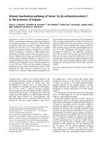
Growth of Synechocystis and its glucose
utilization under heterotrophic growth conditions
Photoautotrophically grown cells at the late-linear
growth phase were cultured heterotrophically with or
without short periods (5 min) of light every 3 h, which
is considered as LAHG in the present study. The cells
grew with approximately 30 and 66 h of doubling time
on culture with or without light pulses, respectively.
They grew to a two-fold higher level with 10 mm glu-
cose (6.0 · 10
8
cellsÆmL
)1
) than with 5 mm glucose in
approximately 1 week (2.8 · 10
8
cellsÆmL
)1
) (Fig. 1).
However, in continuous darkness (dark heterotrophic
conditions), the cells did not grow so much, even in
the presence of either 5 or 10 mm glucose. Without
glucose, the cells could not grow either with or without
the light pulses. Whereas glucose in the medium was
almost completely consumed under LAHG conditions
in 8 days, glucose remained in the medium even after
cell growth had ceased under dark heterotrophic con-
ditions (Fig. 1B). These results suggest that the supply
of glucose in the medium was the sole carbon and
energy source for the cells to grow and that the cells
could not fully utilize glucose until they were exposed
to light pulses.
During culture, the amount of chlorophyll a in the
cells grown photoautotrophically decreased under both
LAHG and dark heterotrophic conditions from 3.0 to
0.9 · 10
)14
gÆcell
)1
in 8 days. The amount of
soluble protein was maintained at approximately
2.0 · 10
)14
gÆcell
)1
under LAHG conditions, but
decreased to 0.8 · 10
)14
gÆcell
)1
in 8 days under dark
heterotrophic conditions. The results obtained suggest
that soluble protein synthesis continued at a normal
level under LAHG conditions, but not in the dark.
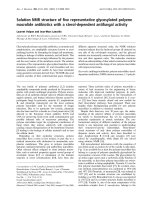
Dependence of LAHG on the cell conditions for
photoautotrophic preculture
The cell conditions for photoautotrophic preculture
greatly affected the subsequent LAHG and heterotro-
phic growth profiles (Fig. 2). The cells harvested at the
Activation of expression of fbaA in Synechocystis Y. Tabei et al.
188 FEBS Journal 276 (2009) 187–198 ª 2008 The Authors Journal compilation ª 2008 FEBS
logarithmic growth phase could grow fast heterotro-
phically either with or without light pulses (Fig. 2B).
By contrast, those harvested at the stationary growth
phase of the preculture scarcely grew, even in the pres-
ence of glucose in the dark (Fig. 2F). Cells harvested
at any phase of the preculture could grow under
LAHG conditions, although the induction period was
longer with a longer preculture period (Fig. 2C–E).
Therefore, the difference in cell growth between
LAHG and dark heterotrophic conditions was obvious
at a late period of culture, such as the late-linear
growth phase, where reactivation of some processes by
light pulses may occur under LAHG conditions. Cells
harvested on day 8 of photoautotrophic preculture
(Fig. 2E) were used for subsequent experiments.
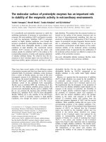
Proteome analysis of cells grown under
photoautotrophic preculture, LAHG and
dark heterotrophic conditions
The compositions of soluble proteins on culture under
LAHG, dark heterotrophic and the respective precul-
ture conditions were analyzed by 2D-PAGE. Under
photoautotrophic preculture and LAHG conditions
(Fig. 3A,B), over 90 protein spots were observed,
whereas less than half this number was observed under
heterotrophic conditions. These results imply that some
of the proteins appearing during photoautotrophic pre-
culture disappeared under dark heterotrophic condi-
tions. We also found that 32 of the spots observed were
thicker for LAHG than the corresponding spots for
photoautotrophic preculture, suggesting a greater syn-
thesis of the respective proteins with the addition of
glucose. Moreover, 20 of these 32 spots were thicker
than the corresponding spots under dark heterotrophic
conditions, suggesting a greater synthesis of the respec-
tive proteins with light in the presence of glucose.
The putative genes and their products for individual
spots that were increased by the glucose supply with and
without the short periods of light were determined from
the molecular weights of fragments by means of
MALDI-TOF MS (Table 1). Protein syntheses with
various categories of genes in relation to metabolism,
transport and the machinery of gene expression were
enhanced under LAHG conditions. The proteins
involved in cellular processes, GroEL1 (spot 6), Tig (8)
and GreP (32), were induced under LAHG conditions.
HtpG (spot 3), DnaK2 (4) and GroEL2 (5, 7) appeared
under both LAHG and dark heterotrophic conditions.
Rps1a (spot 20) and Tsf (26) involved in the translation
machinery were induced under LAHG conditions,
whereas Fus (spot 2), Tuf A (14) and Rpl12 (31) were
induced under dark heterotrophic conditions. These
results suggest that cellular processes and translation
machinery function differently with or without light in
the presence of glucose. AtpB (spot 9), NatB (10), UrtA
(11), RbcL (17) and CpcB (29), formerly regarded as
representing peripheral membrane proteins [24], were
abundant in the soluble fraction of the cells grown under
LAHG conditions. They might have been extracted dur-
ing our sample preparation due to good solubilization,
or the association between the proteins and the
membrane might change with growth conditions.
0
0.5
1
1.5
A
B
02468
Time (days)
Cell concentration (A
730
)
0
2
4
6
8
10
02468
Time (days)
Glucose concentration (mM)
Fig. 1. Growth of Synechocystis under heterotrophic conditions
with (open circles and open squares) or without (filled circles and
filled squares) light pulses for 5 min every 3 h (A) and the glucose
concentration in the medium during culture (B). The cultures grown
photoautotrophically at the late-linear growth phase were incubated
in BG-11 in the presence of 5 m
M (open circles and filled circles) or
10 m
M (open squares and filled squares) glucose, and in the
absence of glucose (open triangles and filled triangles) at 30 °C.
The decrease in the glucose concentration was determined at the
indicated times thereafter. Error bars are for three separately grown
cultures.
Y. Tabei et al. Activation of expression of fbaA in Synechocystis
FEBS Journal 276 (2009) 187–198 ª 2008 The Authors Journal compilation ª 2008 FEBS 189
2D-PAGE also revealed that some proteins, including
RbcL (spot 17), Gap2 (24) and Prk (19) in photosyn-
thetic carbon metabolism, and FbaA (18) in glucose
metabolism, were synthesized to a greater extent under
LAHG conditions than under any of the other condi-
tions. The reason why photosynthetic genes were
induced by glucose was probably a result of the higher
protein-synthetic activity. Enzymes involved in glycoly-
sis and the OPPP other than FbaA were not included in
Table 1, probably due to their low contents or to them
being less changeable in their amounts. The increase in
the protein level of FbaA under LAHG conditions com-
pared to under dark heterotrophic conditions suggested
that glucose degradation was induced by light and glu-
cose in Synechocystis.
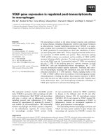
The activity of FBA, which decreased from 88 to
46 unitsÆmg
)1
protein during autotrophic preculture,
was determined for both LAHG and dark heterotro-
phic cultures (Fig. 4). The FBA activity increased
under LAHG conditions, whereas, under dark hetero-
trophic conditions, it remained constant for 4 days
and then decreased gradually. This finding is consistent
with the protein level of FBA, which was increased by
light pulses in the presence of glucose.
Possible enhancement of glucose metabolism
during LAHG
When 5 mmd-[
14
C(U)]glucose was supplied to cells
that had been incubated in continuous darkness with
5mm glucose for 6 days, most of the radioactivity
incorporated was found in phosphate esters and a
slight amount in glutamate at 1 h after the supply of
d-[
14
C(U)]glucose (data not shown). Thus, we pre-
sumed that even the cells cultured in continuous dark-
ness metabolized it to phosphorylated sugars, but to a
lesser extent to other organic compounds.
2D TLC of a cell extract with detection with ninhy-
drin revealed that glutamate was a major amino acid
in cells incubated under both LAHG and dark hetero-
trophic conditions, but that the amount under the
former was much higher than that under the latter (see
Fig. S1). The l-glutamate contents in the cells at 0 and
8 days under LAHG, and at 8 days in continuous
darkness, were 0.13, 0.20 and 0.05 · 10
)11
gÆcell
)1
,
respectively. Considering that glutamate is synthesized
from 2-oxoglutarate via the tricarboxylic acid cycle by
way of an aminotransferase reaction, we could deduce
that the glucose metabolism is more active under
Cell concentration (A
730
)
Cell concentration (A
730
)
Time (days)
Time (days)
0
0.2
0.4
0.6
0.8
1.0
0281012
14
12d
02468
8d
0
0.2
0.4
0.6
0.8
1.0
7d
02468
0
0.2
0.4
0.6
0.8
1.0
5d
02468
0
0.2
0.4
0.6
0.8
1.0
02468
4d
0
0.2
0.4
0.6
0.8
1.0
0
1
2
3
4
AB C
D
EF
4d
8d
7d
5d
0246812
12d
Fig. 2. Effects of pre-culture conditions on the heterotrophic growth of Synechocystis. Growth of Synechocystis on photoautotrophic precul-
ture (A). Growth of Synechocystis under the heterotrophic conditions (B–F) with (open circles and open squares) or without (filled circles and
filled squares) light pulses for 5 min every 3 h. The cultures were carried out in BG-11 in the presence of 5 m
M glucose (open circles and
filled circles) and in the absence of glucose (open squares and filled squares). (B–F) Growth when the cells started to be cultured in the pres-
ence of glucose at 4, 5, 7, 8 and 12 days, respectively, in (A). The data are representative of three separate cultures.
Activation of expression of fbaA in Synechocystis Y. Tabei et al.
190 FEBS Journal 276 (2009) 187–198 ª 2008 The Authors Journal compilation ª 2008 FEBS
LAHG conditions than under dark heterotrophic
conditions.
Changes in some enzymatic activities on glucose
metabolism
The maximum enzymatic activites of glycolytic
enzymes under dark heterotrophic and LAHG condi-
tions for 8 days were determined in the presence of
dithiothreitol (Table 2). The activities of glucokinase
(GK; EC 2.7.1.2) and PK were constantly maintained
under the three sets of conditions. The glucose 6-phos-
phate isomerase (PGI; EC 5.3.1.9) and FBA activities
were relatively higher than those of the other enzymes.
The total activity of FBA under LAHG conditions
was higher than the initial level, whereas that under
dark heterotrophic conditions was lower. The activity
of GAPDH was increased by the addition of glucose.
Therefore, we concluded that FBA was enhanced at
both the protein level and with respect to its activity
8
LAHG conditions
kDa
AB
C
E
D
250
75
50
25
p
I
4
Autotrophic conditions
1
3
4
5
6
2
18
9
7
8
10
11
23
24
26
25
27
28
29
31
30
32
22
21
19
20
17
12
13
14
15
16
Dark heterotrophic conditions
kDa
Δsll1330 LAHG conditions
Fig. 3. Proteome analyses of the soluble protein fraction of Synechocystis under various conditions. The soluble proteins were extracted
from Synechocystis cultured under photoautotrophic preculture (A), LAHG (B) and dark heterotrophic (C) conditions for 8 days. Equivalent
amounts of proteins were separated by 2D-PAGE and stained with Coomassie brilliant blue. The profile shown in (E) is identical to (B) with
circles and numbers for annotation purposes. The soluble proteins of Dsll1330 cultured under LAHG conditions for 8 days (D) were also
analyzed. The positions of molecular size markers are indicated in kDa on the left. FBA is indicated by an arrow.
Y. Tabei et al. Activation of expression of fbaA in Synechocystis
FEBS Journal 276 (2009) 187–198 ª 2008 The Authors Journal compilation ª 2008 FEBS 191
under LAHG conditions compared to in continuous
darkness. This result is consistent with the enhance-
ment of fbaA gene expression by light pulses in the
presence of glucose [25].
Changes in the transcript level of fbaA during
heterotrophic culture
Figure 5 shows the changes in the mRNA level of
fbaA under LAHG and dark heterotrophic conditions.
Considering that the cells at 0 days in Fig. 5 had been
grown under photoautotrophic conditions, and that
the mRNA level of fbaA increased under LAHG con-
ditions, we deduced that the increase in the mRNA
level of fbaA was induced by both light pulses and
glucose. On the other hand, the mRNA level of fbaA
under dark heterotrophic conditions increased slightly
until 6 days, and then decreased gradually. The level
under the dark heterotrophic conditions was approxi-
mately one-tenth of that under LAHG conditions after
8 days. These results suggested that light irradiation
was required for the expression of fbaA.
Table 1. Proteins were observed under LAHG conditions to a 1.5-fold greater extent than under autotrophic conditions in Synechocystis.
Category ORF Putative gene product
Spot no. on
2D-PAGE
Light
inducible
a
Amino acid biosynthesis sll1931 Serine hydroxymethyltrasferase (GlyA) 15 s
sll1363 Ketol-acid reductoisomerase (IlvC) 21
Cellular processes sll0430 Heat shock protein 90 molecular
chaperone (HtpG)
3
sll0170 70 kDa heat shock protein (DnaK2) 4
sll0416 60 kDa chaperonin (GroEL2) 5, 7
slr2076 60 kDa chaperonin (GroEL1) 6 s
sll0533 Trigger factor (Tig) 8 s
sll0057 Heat shock protein GrpE (GreP) 32 s
Energy metabolism sll1234 Adenosylhomocysteinase 16 s
Photosynthesis and respiration,
energy metabolism
slr1329 ATP synthase b subunit (AtpB) 9 s
sll1577 Phycocyanine b subunit (CpcB) 29 s
slr1561 Superoxide dismutase (SodB) 29 s
slr1963 Water-soluble carotenoid protein 22 s
slr0009 Ribulose bisphosphate carboxylase
large subunit (RbcL)
17 s
sll1525 Phosphoribulokinase (Prk) 19 s
sll1342 Glyceraldehyde 3-phosphate
dehydrogenase (Gap2)
24 s
sll0018 Fructose 1,6-bisphosphate
aldolase (FbaA)
18 s
Transport and binding proteins slr0559 Periplasmic binding protein of ABC
transporter for natural amino acids (NatB)
10 s
sll1447 Periplasmic protein, ABC-type urea
transporter system substrate
binding protein (UrtA)
11
sll1450 Nitrate ⁄ nitrite transporter system
substrate binding protein (NrtA)
13
DNA replication, restriction, modification,
recombination, and repair
sll8006 Type I restriction-modification
system S subunit
12 s
sll1868 DNA primase (DnaG) 1
Translation slr1356 30s ribosomal protein S1 (Rps1a) 20 s
sll1261 Elongation factor Ts (Tsf) 26 s
sll1749 50s ribosomal protein L12 (Rpl12) 31
slr1463 Elongation factor (Fus) 2
sll1099 Elongation factor Tu (TufA) 14
Others slr1198 Antioxidant protein 27 s
sll1621 AhpC ⁄ TSA family protein, 28 s
Unidentified 25 s
Unidentified 23
a
Genes enhanced under LAHG conditions to a 1.5-fold greater extent than under dark conditions are indicated (s).
Activation of expression of fbaA in Synechocystis Y. Tabei et al.
192 FEBS Journal 276 (2009) 187–198 ª 2008 The Authors Journal compilation ª 2008 FEBS
LAHG and the protein composition of
Dsll1330, encoding a two-component system
response regulator
Because many transcriptional factors contain the HTH
DNA-binding motif, there is a possibility that some
HTH DNA-binding proteins may be involved in the
fbaA gene expression induced by light and glucose. We
have searched the Synechocystis genome sequence
databank and Cyanobase, and identified 57 ORFs
encoding putative HTH motif-containing transcrip-
tional factors. Among these candidates, we successfully
mutated the sll1330 gene encoding a putative two-
component response regulator by means of targeted
mutagenesis with a spectinomycin resistance cassette.
A resultant sll1330 deletion mutant, Dsll1330, could
grow under photoautotrophic and mixotrophic (i.e.
under continuous light illumination with glucose) con-
ditions as fast as the wild-type (Fig. 6A,B). However,
Dsll1330 did not grow well either under LAHG or
dark heterotrophic conditions compared to the wild-
type (Fig. 6C,D). This can be explained by a deficiency
in the ability to use glucose as a carbon source for
growth.
Proteome analysis of Dsll1330 showed relatively
lower contents of various proteins [e.g. Gap2 (spot 24)
and Tsf (spot 26)]. The FbaA protein (spot 18; Fig. 3,
arrow) was also less in Dsll1330 (Fig. 3B,D) and was
as high as that in wild-type cells incubated under dark
conditions. The FbaA protein appeared to be more
stable than its mRNA. In any case, these results, when
taken together with those obtained previously [25],
suggest that one of the reasons for the inability of
LAHG by Dsll1330 is a lack of enhancement of the
protein level of FBA with light pulses in the presence
of glucose.
FBA activity (mU·mg
–1
protein)
100
0 8 6 4 2
80
60
40
20
0
Time (da
y
s)
Fig. 4. The FBA activities of Synechocystis under LAHG and het-
erotrophic conditions in BG-11 containing 5 m
M glucose. Cells were
incubated under LAHG (white bars) and dark heterotrophic (black
bars) conditions for 2, 4, 6 and 8 days (0 days indicates the activity
of FBA on photoautotrophic preculture on day 8). The error bars
represent three separately grown cultures.
Table 2. Glycolytic enzyme activities of cells grown under dark heterotrophic and LAHG conditions for 8 days.
Enzyme ORF
Activity (mUÆmg
)1
protein ± SE)
Initial
(0 days)
Dark heterotroph
(8 days)
LAHG
(8 days)
GK sll0593 12.0 ± 2.9 8.8 ± 2.6 9.9 ± 1.8
PGI slr1349 370 ± 26 363 ± 17 204 ± 11
FBA (total) sll0018, slr0943 45.9 ± 4.5 31.7 ± 5.6 61.6 ± 7.2
FBA (class-I)
a
slr0943 7.4 ± 0.8 3.2 ± 1.2 5.7 ± 0.8
GAPDH slr0884 2.8 ± 0.8 12.1 ± 3.4 12.2 ± 5.0
Enolase sll0752 2.9 ± 1.3 1.2 ± 0.9 3.1 ± 1.2
PK sll0587, sll1275 13.3 ± 2.4 12.2 ± 1.4 10.2 ± 1.2
a
Class-II was inhibited by the addition of 1 mM EDTA.
fbaA
rRNA
0864208642
LAHG Dark heterotrophic
(days)
Fig. 5. Northern analysis of fbaA gene expression in Synechocystis
under LAHG and dark heterotrophic conditions in BG-11 containing
5m
M glucose. RNA was isolated from cells grown photoautotrophi-
cally on day 8 (0 days) and cells incubated under LAHG and dark
heterotrophic conditions for 2, 4, 6 and 8 days. Total RNA (10 lg)
was then subjected to northern hybridization with a probe specific
for the fbaA gene. The lower panel shows rRNA stained with ethi-
dium bromide as a control. The data are representative of three
independent experiments.
Y. Tabei et al. Activation of expression of fbaA in Synechocystis
FEBS Journal 276 (2009) 187–198 ª 2008 The Authors Journal compilation ª 2008 FEBS 193
Interaction of the Sll1330 protein with the fbaA pro-
moter region was additionally examined by means of
the electrophoretic mobility shift assay (EMSA) using
DNA fragments of the upstream region of fbaA (see
Fig. S2). The DNA fragment comprising )331 to
)186 bp from the putative transcription start point of
the fbaA gene was found to bind with Sll1330, which
was overexpressed in Escherichia coli (see Fig. S2).
These results suggest that Sll1330 binds to this putative
promoter region of the fbaA gene in PCC 6803 cells.
Discussion
In the present study, the effects of light on heterotro-
phic growth of Synechocystis were precisely investi-
gated. First, we confirmed that Synechocystis could
not grow in complete darkness, as reported previously
[10]. However, the cells at the logarithmic growth
phase could grow heterotrophically even without illu-
mination. This would be because glucose-degrading
machineries were active in the photoautotrophic pre-
culture at relatively early stages. The total FBA activ-
ity in the late-linear growth phase was lower than that
in the exponential growth phase (Table 2). This is
consistent with the fact that the mRNA levels of some
glycolytic genes were suppressed at the stationary
growth phase compared to at the exponential growth
phase [26]. Physiological changes occur at different
growth phases with respect to the accumulation of
inhibitory metabolites [27,28] and gene expression [29].
The expression of 10% of the genes was different in
the linear growth phase compared to in the logarithmic
growth phase [30]. Considering the close correlation
between nitrogen and carbon metabolism [13], we
assume the decrease in gene expression during growth
is due to the lower level of nitrogen in the medium.
Alternatively, because of the higher concentration of
cells during photoautotrophic preculture, the amount
of light absorbed by each cell decreased in these condi-
tions. In any case, when using cells precultured at a
late period of the preculture (i.e. after 8 days of cul-
ture), an effect of light on the heterotrophic growth
was clearly observed. Therefore, LAHG might be due
to reactivation of some biochemical activities.
We have attempted proteome analysis using the com-
bination of 2D-PAGE and MALDI-TOF MS of tryptic
digests. Considerable amounts of soluble proteins were
found in the cells under LAHG conditions compared to
those after 8 days of photoautotrophic preculture. Con-
sidering that various kinds of proteins were present at
higher levels under LAHG conditions than under dark
heterotrophic conditions, we deduced that the light
pulses triggered the transcription and translation of a
whole set of proteins essential for growth with glucose.
In this respect, the transcriptional levels of molecular
chaperones groESL and cpn60 are known to be signifi-
cantly increased by light irradiation in Synechocystis
[30]. Moreover, the DnaK2 protein is one of the abun-
dant soluble proteins, with dnaK gene expression having
0.6
A
B
D
C
0.4
0.2
0
02468
1.5
1.0
0.5
0
02468
1.0
2.0
3.0
0
02
46
8
0.4
0.2
0
02468
Cell concentration (A
730
)
Time (days)
0.5
0.3
0.1
Fig. 6. Growth of the wild-type (circles) and Dsll1330 (triangles)
under photoautotrophic (continuous light; A), mixotrophic (con-
tinuous light + 10 m
M glucose; B), LAHG (light pulses + 10 mM
glucose; C) and dark heterotrophic (dark + 10 mM glucose; D)
conditions. Error bars indicate the SD for three independent
experiments.
Activation of expression of fbaA in Synechocystis Y. Tabei et al.
194 FEBS Journal 276 (2009) 187–198 ª 2008 The Authors Journal compilation ª 2008 FEBS
been reported to obey up-regulation with the lights on
and down-regulation with the lights off (i.e. in the man-
ner of a circadian rhythm) [31], which is consistent with
the results shown in Fig. 3. DnaK2 and rehydrin may
protect the cells from continuous light irradiation and
facilitate the functional assembly of photosynthetic
machinery proteins [32]. The SodB protein (Spot 29) cat-
alyzes the dismutation of O
2
)
to H
2
O
2
and O
2
, in which
sodB gene and SodB protein expression are induced by
continuous light irradiation [33]. Light induction of
putative 30S ribosomal protein S1 (sll1356) and elonga-
tion factor Ts (sll1261), which are involved in transla-
tion processes, suggests that protein synthesis is
enhanced during cell growth under LAHG conditions.
This is supported by the appearance of more spots on
2D-PAGE of cells under LAHG conditions than under
heterotrophic conditions.
In the present study, the expression of GroEL1, Tig
and GreP was also up-regulated under LAHG condi-
tions compared to under both photoautotrophic and
dark heterotrophic conditions. These results suggest
the higher activities of cellular processes (e.g. protein
synthesis and metabolic processes) under LAHG con-
ditions. FbaA, one of the components of the glucose-
metabolism machinery, was also up-regulated by light
under glucose-utilizing conditions. The protein level of
FbaA was high compared to that of other glycolytic
enzymes. The increase in the maximum Fba activity in
8 days (Fig. 4) under LAHG conditions, but not under
dark heterotrophic conditions, suggested the light acti-
vation of Fba synthesis. This is also supported by the
higher content of glutamate in the cells. Therefore,
cells under LAHG conditions may be more active than
without light.
We found also that, in the sll1330 deletion mutant
(Dsll1330), which could not grow heterotrophically
(Fig. 6), the protein level of FbaA was repressed
(Fig. 3D), even under LAHG conditions, and the
increases in the mRNA levels of five glycolytic genes,
including fbaA, were repressed by light and glucose [25].
We also found binding of Sll1330 with the promoter
region of the fbaA gene. Because the Dsll1330 mutant
could grow photoautotrophically with the expression of
fbaA, the sll1330 gene is not required for the expression
of fbaA under photoautotrophic conditions. This means
that fbaA gene expression is controlled through two
signal pathways: presumably photosynthetic processes
and light pulses in the presence of glucose. Gene expres-
sion of fbaA may be important for survival in various
environments, including both under photoautotrophic
and heterotrophic conditions. In the latter case, glucose
induces the expression of several genes, such as those
for GK, phosphofructokinase, phosphoglycerate mutase
and PK, including sll1330 [34]. In this respect, glucose
degradation as a whole may be regulated by the expres-
sion of glycolytic genes via sll1330.
Experimental procedures
Algal cells and culture conditions
A glucose tolerant strain of Synechocystis [35] was cultivated
photoautotrophically in BG-11 containing 20 mm Tes-KOH
(pH 8.2) with continuous illumination by a bank of fluores-
cent lamps at 10 WÆm
)2
and bubbled with 2% (v ⁄ v) CO
2
in
glass tubes or flasks [36]. The LAHG cultures were incu-
bated in the dark with 5 min of light irradiation (5 WÆ m
)2
),
eight times a day in the presence of 5 and 10 mm glucose,
whereas the heterotrophic cultures were incubated without
light irradiation in the presence of glucose. Growth and cell
density were followed by measuring the absorption at
730 nm with a spectrophotometer (DU640; Beckman
Coulter, Fullerton, CA, USA). Cell concentrations were
determined with a hemacytometer (Bu
¨
rker-Turk; Erma Opti-
cal Works, Tokyo, Japan). Glucose uptake was assayed by
measurement of the concentration of glucose in the medium
with a glucose CII kit (Wako Pure Chemical Industries,
Osaka, Japan). In the mutant, the wild-type gene for sll1330
was disrupted by the insertion of a spectinomycin resistance
cassette [25].
Determination of intracellular chlorophyll a,
soluble protein and
L-glutamate
Chlorophyll a was measured by the procedure described by
Mackinney [37], with some modifications. Soluble protein
was assayed by measurement of the concentration of a par-
ticular protein with a BCA protein assay reagent kit (Pierce
Biotechnology, Rockford, IL, USA). Cells were harvested
at a certain time, centrifuged, and then disrupted with a
French press at 28 000 p.s.i. The lysate was centrifuged and
the resulting supernatant was used for the measurement
of soluble protein. Intracellular l-glutamate was assayed
by measurement of the concentration of l-glutamate with
a l-glutamate determination kit (Yamasa Corp., Chiba,
Japan).
2D-PAGE and in-gel digestion
The materials precipitated from 300 lg of soluble protein
were resuspended in 8 m urea, 1% (v ⁄ v) Triton X-100,
ampholine (pH 3.5–10.0) and 2-mercaptoethanol. The mix-
ture was centrifuged at 10 000 g for 10 min at 4 °C. The
supernatant was applied to a linear immobilized pH gradi-
ent strip (pH 4.0–8.0). Isoelectric focusing and subsequent
SDS ⁄ PAGE were then performed. Proteins were stained
with Coomassie brilliant blue and then scanned using an
Y. Tabei et al. Activation of expression of fbaA in Synechocystis
FEBS Journal 276 (2009) 187–198 ª 2008 The Authors Journal compilation ª 2008 FEBS 195
image scanner. In-gel digestion was performed as described
for an in-gel digest kit (Millipore, Billerica, MA, USA).
The gel pieces were re-swollen by adding a trypsin solution.
Samples were incubated overnight at 30 °C and the protein
fragments were extracted.
MALDI-TOF analysis
Peptide extracts were loaded onto the target plate by the
dried droplet method using a-cyano-4-hydroxycinnamic
acid as a matrix. MALDI-TOF analysis was performed in
the reflector mode with a Voyager-DE PRO mass spec-
trometer (Applied Biosystems, Foster City, CA, USA).
Database search
Proteins were identified by searching the Synechocystis
database using ms-fit (Protein Prospector; UCSF, San
Francisco, CA, USA). The following criteria were used for
a positive identification in the ms-fit database search: (a)
at least four matching peptide masses; (b) at least 50% of
the measured masses must match the theoretical masses;
and (c) 0.01% or better mass accuracy.
Assaying enzyme activity
The cells cultivated under LAHG and heterotrophic condi-
tions were harvested by centrifugation. The pellets were
resuspended and then lysed by three passages through a
French press at 28 000 p.s.i., followed by clarification by cen-
trifugation at 35 000 g for 10 min. The enzyme activities
were measured using previously described procedures
[7,21,38] with some modifications. The assay mixtures com-
prised: GK [50 mm Hepes-KOH (pH 8.0), 1 mm dithiothrei-
tol, 10 mm MgCl
2
, 2.5 mm glucose, 5 mm ATP, 0.5 mm
NAD
+
and 5 unitsÆ mL
)1
G6PD]; PGI [50 mm Hepes-KOH
(pH 8.0), 1 mm dithiothreitol, 10 mm MgCl
2
, 1.7 mm
NAD
+
,2mm fructose 6-phosphate and 5 unitsÆmL
)1
G6PD]; PFK [50 mm Hepes-KOH (pH 8.0), 1 mm dith-
iothreitol, 10 m m MgCl
2
, 0.15 mm NADH, 2 mm fruc-
tose 6-phosphate, 2.5 mm ATP, 1 unitÆmL
)1
aldolase,
10 unitsÆmL
)1
triosephosphate isomerase and 1 unitÆmL
)1
glycerophosphate dehydrogenase]; FBA [50 m m Tris–HCl
(pH 7.5), 0.2 mm CoCl
2
, 0.2 mm NADH, 1 unitÆmL
)1
glyc-
erol 3-phosphate dehydrogenase, 1 unitÆmL
)1
triosephos-
phate isomerase and 1 m m fructose 1,6-bisphosphate];
GAPDH [50 mm Tris–HCl (pH 7.5), 5 m m MgCl
2
,3mm
3-phosphoglycerate, 1 mm EDTA, 5 unitsÆmL
)1
3-phospho-
glycerate kinase, 2 mm ATP and 0.2 mm NADH]; enolase
[50 mm Hepes-KOH (pH 8.0), 1 mm dithiothreitol, 10 mm
MgCl
2
, 0.15 mm NADH, 3 mm ADP, 3 mm 2-phospho-
glycerate, 5 unitsÆmL
)1
PK and 6 unitsÆmL
)1
lactate
dehydrogenase]; and PK [50 mm Hepes-NaOH (pH 7.0),
30 mm MgCl
2
, 0.15 mm NADH, 2.5 mm phosphoenolpyr-
uvate, 1 mm ADP and 2 unitsÆmL
)1
lactate dehydrogenase].
RNA isolation and northern hybridization
Total RNA was extracted and purified by phenol–chloro-
form extraction [39]. For northern hybridization, the DNA
fragments of fbaA were amplified by PCR, using primers
5¢-ATTTCGATCATGCAGGCCG-3¢ and 5¢-GGAAGAAC
CGTGCATTACC-3¢, and labeled with [a-
32
P]dCTP using a
Megaprime labeling kit (Amersham Pharmacia, Piscataway,
NJ, USA). Hybridization signals were detected with a BAS-
2000 bio-imaging analyzer (Fuji Film, Tokyo, Japan).
Expression of the Sll1330 protein in E. coli
Overexpression of Sll1330 was carried out as described pre-
viously [40] with some modification. The sll1330 gene was
cloned from PCC 6803 genomic DNA after PCR amplifica-
tion using specific oligonucleotides. The sll1330 gene was
amplified using primers 1330OX-F (5¢-GTCTAGA
CAT
ATGAATCCAG-3¢), comprising nucleotides )10 to +10
of the sll1330 coding region, and 1330OX-R (5¢-CG
GGA
TCCAGGGTAACAGGTTTCTCCG-3¢), comprising nucle-
otides +704 to +730 of the sll1330 coding region. NdeI
and BamHI sites were introduced into the 1330OX-F and
1330OX-R primers, respectively (underlined).
EMSA
A 135-bp DNA fragment, prom1, corresponding to posi-
tions )86 to +49 upstream of the translation initiation site
of the fbaA gene was amplified, using forward primer
5¢-GCAGAAACTAGCCTAAGATG-3¢ and reverse primer
5¢-CCATTTTCCGCCGCATGGTC-3¢. A 121-bp DNA
fragment, prom2, corresponding to positions )190 to )70
upstream of the translation initiation site of the fbaA gene
was amplified with forward primer 5¢-CTCAGCCATAGA
CTAGGGTG-3¢ and reverse primer 5¢-ATCTTAGGCTA
GTTTCTGC-3¢. A 126-bp fragment, prom3, corresponding
to positions )311 to )186 upstream of the translation initi-
ation site of the fbaA gene was amplified with forward
primer 5¢-GCTAAAGGAAGTATTTGCTAC-3¢ and
reverse primer 5¢-CTGAGTTAACCAAGTCCAGG-3¢.A
134 bp DNA fragment, prom4, corresponding to positions
)438 to )305 upstream of the translation initiation site of
the fbaA gene was amplified with forward primer 5¢-CA
ATAAATTTGCCCGTTTCC-3¢ and reverse primer
5¢-CCTTAGCGACGGCAATGGTC-3¢. EMSA was carried
out as described previously [41].
Acknowledgements
The authors are indebted to Dr S. Fujiwara for helpful
discussion and Mr N. J. Halewood for his kind help in
the correction of the English. They are also indebted
to Mr Masato Sasahara, Ms Yuka Katsuki and
Activation of expression of fbaA in Synechocystis Y. Tabei et al.
196 FEBS Journal 276 (2009) 187–198 ª 2008 The Authors Journal compilation ª 2008 FEBS
Ms Miwako Itoh for their experimental support. This
work was supported by Grants-in-Aid from the Minis-
try of Education, Science, Sports and Culture, Japan;
the Promotion and Mutual Aid Corporation for Pri-
vate Schools to M. T.; and a Sasakawa Scientific
Research Grant from the Japan Science Society to
Y. T.
References
1 Briggs WR & Christie JM (2002) Phototropins 1 and 2:
versatile plant blue-light receptors. Trends Plant Sci 7,
204–210.
2 Gyula P, Scha
¨
fer E & Nagy F (2003) Light perception
and signaling in higher plants. Curr Opin Plant Biol 6,
446–452.
3 Mackey SR & Golden SS (2007) Winding up the cyano-
bacterial circadian clock. Trends Microbiol 15, 381–388.
4 Yoshihara S & Ikeuchi M (2004) Phototactic motility in
the unicellular cyanobacterium Synechocystis sp. PCC
6803. Photochem Photobiol Sci 3, 512–518.
5 Stal LJ & Moezelaar R (1997) Fermentation in cyano-
bacteria. FEMS Microbiol Rev 21, 179–211.
6 Knowles VL & Plaxton WC (2003) From genome to
enzyme: analysis of key glycolytic and oxidative pen-
tose-phosphate pathway enzymes in the cyanobacterium
Synechocystis sp. PCC 6803. Plant Cell Physiol 44, 758–
763.
7 Ball SG & Morell MK (2003) From bacterial glycogen
to starch: understanding the biogenesis of the plant
starch granule. Annu Rev Plant Biol 54, 207–233.
8 Kaneko T, Sato S, Kotani H, Tanaka A, Asamizu E,
Nakamura Y, Miyajima N, Hirosawa M, Sugiura M,
Sasamoto S et al. (1996) Sequence analysis of the gen-
ome of the unicellular cyanobacterium Synechocystis sp.
strain PCC 6803. II. Sequence determination of the
entire genome and assignment of potential protein-
coding regions. DNA Res 3, 109–136.
9 Florencio FJ, Pe
´
rez-Pe
´
rez ME, Lo
´
pez-Maury L,
Mata-Cabana A & Lindahl M (2006) The diversity and
complexity of the cyanobacterial thioredoxin systems.
Photosynth Res 89, 157–171.
10 Anderson SL & McIntosh L (1991) Light-activated het-
erotrophic growth of the cyanobacterium Synechocystis
sp. strain PCC 6803: a blue-light-requiring process.
J Bacteriol 173, 2761–2767.
11 Singh AK & Sherman LA (2005) Pleiotropic effect of a
histidine kinase on carbohydrate metabolism in Syn-
echocystis sp. strain PCC 6803 and its requirement for
heterotrophic growth. J Bacteriol 187, 2368–2376.
12 Osanai T, Kanesaki Y, Nakano T, Takahashi H, Asay-
ama M, Shirai M, Kanehisa M, Suzuki I, Murata N &
Tanaka K (2005) Positive regulation of sugar catabolic
pathways in the cyanobacterium Synechocystis sp. PCC
6803 by the group 2 sigma factor SigE. J Biol Chem
280, 30653–30659.
13 Yang C, Hua Q & Shimizu K (2002) Integration of the
information from gene expression and metabolic fluxes
for the analysis of the regulatory mechanisms in Syn-
echocystis. Appl Microbiol Biotechnol 58, 813–822.
14 Scanlan DJ, Newman J, Sebaihia M, Mann NH & Carr
NG (1992) Cloning and sequence analysis of the glu-
cose-6-phosphate dehydrogenase gene from the cyano-
bacterium Synechococcus PCC 7942. Plant Mol Biol 19 ,
877–880.
15 Broedel SE Jr & Wolf RE Jr (1990) Genetic tagging,
cloning, and DNA sequence of the Synechococcus sp.
strain PCC 7942 gene (gnd) encoding 6-phosphogluco-
nate dehydrogenase. J Bacteriol 172, 4023–4031.
16 Valverde F, Losada M & Serrano A (1997) Functional
complementation of an Escherichia coli gap mutant sup-
ports an amphibolic role for NAD(P)-dependent glycer-
aldehyde 3-phosphate dehydrogenase of Synechocystis
sp. PCC 6803. J Bacteriol 179, 4513–4522.
17 Koksharova O, Schubert M, Shestakov S & Cerff R
(1998) Genetic and biochemical evidence for distinct
key functions of two highly divergent GAPDH genes in
catabolic and anabolic carbon flow of the cyanobacte-
rium Synechocystis sp. PCC 6803. Plant Mol Biol 36,
183–194.
18 Figge RM, Cassier-Chauvat C, Chauvat F & Cerff R
(2000) The carbon metabolism-controlled Synechocystis
gap2 gene harbours a conserved enhancer element and a
Gram-positive-like –16 promoter box retained in some
chloroplast genes. Mol Microbiol 36, 44–54.
19 Knowles VL, Smith CS, Smith CR & Plaxton WC
(2001) Structural and regulatory properties of pyruvate
kinase from the cyanobacterium Synechococcus PCC
6301. J Biol Chem 276, 20966–20972.
20 Luinenburg I & Coleman JR (1990) A requirement for
phosphoenolpyruvate carboxylase in the cyanobacte-
rium Synechococcus PCC 7942. Arch Microbiol 154,
471–474.
21 Nakahara K, Yamamoto H, Miyake C & Yokota A
(2003) Purification and characterization of class-I and
class-II fructose-1,6-bisphosphate aldolases from the
cyanobacterium Synechocystis sp. PCC 6803. Plant Cell
Physiol 44, 326–333.
22 Pelory RA, Levine GA & Bassham JA (1976) Kinetics
of light-dark CO
2
fixation and glucose assimilation by
Aphanocapsa 6714. J Bacteriol 128, 633–643.
23 Scherer S (1990) Do photosynthetic and respiratory
electron transport chains share redox proteins? Trends
Biochem Sci 15, 458–462.
24 Wang Y, Sun J & Chitnis PR (2000) Proteomic study
of the peripheral proteins from thylakoid membranes of
the cyanobacterium Synechocystis sp. PCC 6803. Elec-
trophoresis 21, 1746–1754.
Y. Tabei et al. Activation of expression of fbaA in Synechocystis
FEBS Journal 276 (2009) 187–198 ª 2008 The Authors Journal compilation ª 2008 FEBS 197
25 Tabei Y, Okada K & Tsuzuki M (2007) Sll1330 con-
trols the expression of glycolytic genes in Synechocystis
sp. PCC 6803. Biochem Biophys Res Commun 355,
1045–1050.
26 Foster JS, Singh AK, Rothschild LJ & Sherman LA
(2007) Growth-phase dependent differential gene expres-
sion in Synechocystis sp. strain PCC 6803 and regula-
tion by a group 2 sigma factor. Arch Microbiol 187,
265–279.
27 Siegele DA & Kolter R (1992) Life after log. J Bacteriol
174, 345–348.
28 Nystro
¨
m T (2004) Stationary-phase physiology. Annu
Rev Microbiol 58, 161–181.
29 Tani TH, Khodursky A, Blumenthal RM, Brown PO &
Matthews RG (2002) Adaptation to famine: a family of
stationary-phase genes revealed by microarray analysis.
Proc Natl Acad Sci USA 99, 13471–13476.
30 Glatz A, Horva
´
th I, Varvasovszki V, Kova
´
cs E, To
¨
ro
¨
k
Z & Vigh L (1997) Chaperonin genes of the Synechocys-
tis PCC 6803 are differentially regulated under light-
dark transition during heat stress. Biochem Biophys Res
Commun 239, 291–297.
31 Aoki S, Kondo T & Ishiura M (1995) Circadian
expression of the dnaK gene in the cyanobacterium
Synechocystis sp. strain PCC 6803. J Bacteriol 177,
5606–5611.
32 Choi JS, Kim DS, Lee J, Kim SJ, Kim SI, Kim YH,
Hong J, Yoo JS, Suh KH & Park YM (2000) Proteome
analysis of light-induced proteins in Synechocystis sp.
PCC 6803: identification of proteins separated by
2D-PAGE using N-terminal sequencing and MALDI-
TOF MS. Mol Cells 10, 705–711.
33 Kim JH & Suh KH (2005) Light-dependent expression of
superoxide dismutase from cyanobacterium Synecho-
cystis sp. strain PCC 6803. Arch Microbiol 183, 218–223.
34 Lee S, Ryu JY, Kim SY, Jeon JH, Song JY, Cho HT,
Choi SB, Choi D, de Marsac NT & Park YI (2007)
Transcriptional regulation of the respiratory genes in
the cyanobacterium Synechocystis sp. PCC 6803 during
the early response to glucose feeding. Plant Physiol 145,
1018–1030.
35 Williams JGK (1988) Construction of specific mutants
in photosystem II photosynthetic reaction center by
genetic engineering methods in Synechocystis 6803.
Methods Enzymol 167, 766–778.
36 Rippka R, Deruelles J, Waterbury JB, Herdman M &
Stanier RY (1979) Generic assignments, strain histories
and properties of pure cultures of cyanobacteria. J Gen
Microbiol 111, 1–61.
37 Mackinney G (1941) Absorption of light by chlorophyll
solutions. J Biol Chem 140, 315–322.
38 Sparla F, Pupillo P & Trost P (2002) The C-terminal
extension of glyceraldehyde-3-phosphate dehydrogenase
subunit B acts as an autoinhibitory domain regulated
by thioredoxins and nicotinamide adenine dinucleotide.
J Biol Chem 277, 44946–44952.
39 Los DA, Ray MK & Murata N (1997) Differences in
the control of the temperature-dependent expression of
four genes for desaturases in Synechocystis sp. PCC
6803. Mol Microbiol 25, 1167–1175.
40 Gutekunst K, Phunpruch S, Schwarz C, Schuchardt S,
Schulz-Friedrich R & Appel J (2005) LexA regulates
the bidirectional hydrogenase in the cyanobacterium
Synechocystis sp. PCC 6803 as a transcription activator.
Mol Microbiol 58, 810–823.
41 Oliveira P & Lindblad P (2005) LexA, a transcription
regulator binding in the promoter region of the
bidirectional hydrogenase in the cyanobacterium
Synechocystis sp. PCC 6803. FEMS Microbiol Lett 251,
59–66.
Supporting information
The following supplementary material is available:
Fig. S1. Amino acid compositions of soluble fractions
of Synechocystis.
Fig. S2. EMSA analysis of Sll1330 and the promoter
region of the fbaA gene.
This supplementary material can be found in the
online version of this article.
Please note: Wiley-Blackwell is not responsible for
the content or functionality of any supplementary
materials supplied by the authors. Any queries (other
than missing material) should be directed to the corre-
sponding author for the article.
Activation of expression of fbaA in Synechocystis Y. Tabei et al.
198 FEBS Journal 276 (2009) 187–198 ª 2008 The Authors Journal compilation ª 2008 FEBS