Báo cáo khoa học: ADPase activity of recombinantly expressed thermotolerant ATPases may be caused by ppt
Bạn đang xem bản rút gọn của tài liệu. Xem và tải ngay bản đầy đủ của tài liệu tại đây (378.83 KB, 9 trang )
ADPase activity of recombinantly expressed
thermotolerant ATPases may be caused by copurification
of adenylate kinase of Escherichia coli
Baoyu Chen
1,
*, Tatyana A. Sysoeva
2
, Saikat Chowdhury
2
, Liang Guo
3
and B. Tracy Nixon
2
1 Integrative Biosciences Graduate Degree Program – Chemical Biology, The Pennsylvania State University, University Park, PA, USA
2 Department of Biochemistry and Molecular Biology, The Pennsylvania State University, University Park, PA, USA
3 BioCAT, Advanced Photon Source, Argonne National Lab and Illinois Institute of Technology, Chicago, IL, USA
ATPases associated with various cellular activities
(AAA+ ATPases) form a large family of chaperone-like
proteins that use ATP hydrolysis to remodel numerous
macromolecular complexes [1]. The NtrC1 protein of
Aquifex aeolicus is one such ATPase, belonging to the
subfamily whose members are called bacterial enhancer
binding proteins (EBPs). EBPs use ATP hydrolysis to
activate transcription by the r54-dependent form of
RNA polymerase [2]. Although some AAA+ ATPases
can operate by hydrolyzing other NTPs or even dNTP
and ddNTPs [3,4], they specifically target the phospho-
diester bond between b-phosphates and c -phosphates of
the nucleotides. They do not hydrolyze ADP, even
though such hydrolysis releases free energy similar to
that released by cleavage of the bond to the c-phos-
phate. To our knowledge, such high specificity for the
Keywords
AAA+ ATPase; adenylate kinase; ADPase;
r54; thermophilic proteins
Correspondence
B. Tracy Nixon, 406 S, Frear Lab,
Biochemistry and Molecular Biology, The
Pennsylvania State University, University
Park, PA 16802, USA
Fax: +1 814 863 7024
Tel: +1 814 863 4904
E-mail:
*Present address
UT Southwestern Medical Center at Dallas,
TX, USA
(Received 18 September 2008, revised 24
November 2008, accepted 2 December
2008)
doi:10.1111/j.1742-4658.2008.06825.x
Except for apyrases, ATPases generally target only the c-phosphate of a
nucleotide. Some non-apyrase ATPases from thermophilic microorganisms
are reported to hydrolyze ADP as well as ATP, which has been described
as a novel property of the ATPases from extreme thermophiles. Here, we
describe an apparent ADP hydrolysis by highly purified preparations of the
AAA+ ATPase NtrC1 from an extremely thermophilic bacterium, Aqui-
fex aeolicus. This activity is actually a combination of the activities of the
ATPase and contaminating adenylate kinase (AK) from Escherichia coli,
which is present at 1 ⁄ 10 000 of the level of the ATPase. AK catalyzes
conversion of two molecules of ADP into AMP and ATP, the latter being
a substrate for the ATPase. We raise concern that the observed thermo-
tolerance of E. coli AK and its copurification with thermostable proteins
by commonly used methods may confound studies of enzymes that specifi-
cally catalyze hydrolysis of nucleoside diphosphates or triphosphates. For
example, contamination with E. coli AK may be responsible for reported
ADPase activities of the ATPase chaperonins from Pyrococcus furiosus,
Pyrococcus horikoshii, Methanococcus jannaschii and Thermoplasma acido-
philum; the ATP ⁄ ADP-dependent DNA ligases from Aeropyrum pernix K1
and Staphylothermus marinus; or the reported ATP-dependent activities of
ADP-dependent phosphofructokinase of P. furiosus. Purification methods
developed to separate NtrC1 ATPase from AK also revealed two distinct
forms of the ATPase. One is tightly bound to ADP or GDP and able to
bind to Q but not S ion exchange matrixes. The other is nucleotide-free
and binds to both Q and S ion exchange matrixes.
Abbreviations
AAA+ ATPases, ATPases associated with various cellular activities; AK, adenylate kinase; Ap5A, diadenosine pentaphosphate; EBP,
enhancer binding protein; Mg-ADP-BeF
x
, ATP ground state analog composed of a complex of ADP and magnesium and beryllofluoride ions
(x denotes uncertain stoichiometry of fluorine atoms); SAXS, small-angle solution X-ray scattering.
FEBS Journal 276 (2009) 807–815 ª 2009 The Authors Journal compilation ª 2009 FEBS 807
c phosphate bond is also true for all members of the
P-loop NTPase superfamily and most other nucleotide-
binding proteins.
One well-known exception is apyrase (or NTPDase)
of eukaryotic cells [5], which breaks both phosphodi-
ester bonds of a nucleotide, hydrolyzing ATP and
ADP to AMP and orthophosphate(s). Also, a novel
ADPase activity of ATPases from thermophilic organ-
isms, including four different chaperonins [6] and two
DNA ligases [7,8], has been reported. It was hypothe-
sized that using ADP as an energy source instead of
ATP in thermophilic organisms may be beneficial
because ATP is less stable at high temperatures. Fur-
thermore, there are controversial observations that
some ADP-dependent glucokinases and phospho-
fructokinases in thermophilic archeaons can also use
ATP as a phosphoryl transfer donor [9–12].
Here we report an apparent ADPase activity in
preparations of the recombinant ATPase domain of
the AAA+ ATPase NtrC1 (NtrC1
C
) from the extre-
mely thermophilic bacterium A. aeolicus purified from
Escherichia coli. Although conversion of ADP to AMP
and P
i
depends upon intact catalytic activity of the
NtrC1 ATPase, we show that it also depends upon the
action of 0.01% contamination by E. coli adenylate
kinase (AK). Apparent catalysis of ADP hydrolysis by
NtrC1
C
was in fact conversion of two ADP molecules
to ATP and AMP by AK followed by hydrolysis of
ATP to ADP and P
i
by NtrC1
C
.
Proteins that tolerate high temperatures, such as
NtrC1, are popular subjects of structural studies. They
are often purified by a similar strategy, which takes
advantage of their thermostability. Our observation
that AK of E. coli survives, and is indeed copurified,
by such a method raises a concern about possible con-
tamination of other protein preparations with AK.
The presence of tiny amounts of this contaminant
could confound studies of any nucleotide-hydrolyzing
enzymes from thermophilic organisms. Chromato-
graphic methods developed to remove the AK contam-
ination revealed a heterogeneity in the ATPase
preparation, yielding two subfractions. The resulting,
more homogeneous preparation of an NtrC1
C
variant
bearing a single amino acid substitution has led to
diffracting crystals (to be described elsewhere).
Results
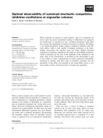
Highly pure NtrC1
C
preparation catalyzes
hydrolysis of ADP
NtrC1
C
purified by heat denaturation and anion
exchange chromatography was highly pure (> 99%)
as judged from SDS ⁄ PAGE (Fig. 1A) and gel
filtration (not shown). However, addition of 5 mm
ATP to the protein produced 8–10 mm free P
i
(data
not shown), suggesting further hydrolysis of ADP.
This was confirmed by ion exchange chromatography
permitting quantification of fluxes in the concen-
trations of ATP, ADP and AMP, beginning
with an initial concentration of 10 mm ATP
(Fig. 1B).
The apparent ADPase activity displays high
thermal stability, requires an ATPase-competent
NtrC1
C
protein, and is associated with structural
changes in NtrC1
C
To determine how the apparent ADPase activity is
associated with the NtrC1 ATPase, we first examined
the rate of ADP turnover by NtrC1
C
preparations
that had been pre-equilibrated to different tempera-
tures. The optimal temperature for ADP hydrolysis
was 60 °C, which is somewhat lower than the 82 °C
optimum seen for ATP hydrolysis by NtrC1
C
A
B
Fig. 1. Apparent ADP hydrolysis by highly pure NtrC1
C
. (A) SDS ⁄
PAGE of purified NtrC1
C
(initial preparation) and subsequent
Q-fraction and S-fraction. Ten micrograms of each protein was
loaded. (B) Products of ATP hydrolysis by 2 mgÆmL
)1
NtrC1
C
Q-frac-
tion at 60 °C, as quantified by anion exchange chromatography.
Copurification of NtrC1 ATPase and adenylate kinase B. Chen et al.
808 FEBS Journal 276 (2009) 807–815 ª 2009 The Authors Journal compilation ª 2009 FEBS
(Fig. 2). The ratio of ADP turnover to ATP
turnover remained constant and close to 20% over a
wide range of temperatures, from 0 °C to about
60 °C. At higher temperatures, ADP turnover started
to decrease, and it ceased above 70 °C. After ther-
mal inactivation by incubation at 80 °C for 30 min,
the ADPase activity was completely recovered as
soon as it could be measured upon cooling to 60 °C
(not shown). Studies of several NtrC1
C
single amino
acid substitution variants showed that both ADPase
and ATPase activities require the same active site
residues (Table 1).
Using small-angle solution X-ray scattering (SAXS)
and size exclusion chromatography, we previously
established that a large conformational change in
NtrC1
C
is stabilized upon binding of ADP-BeF
x
,a
ground state analog of ATP. This conformational
change allows NtrC1
C
to interact with r54 [13]. Here,
we used the same methods to determine whether the
apparent hydrolysis of ADP is associated with struc-
tural changes in NtrC1 ATPase. Substitution of the
conserved glutamate of the Walker B motif (Glu239)
by alanine abolished ATP hydrolysis, but the altered
protein still underwent a conformational change simi-
lar to the wild-type when ATP was added, and it could
then bind to r54. Likewise, this substitution abolished
hydrolysis of ADP, but addition of ADP caused a
conformational change similar to that seen upon the
addition of ATP and it promoted binding to r54
(Fig. 3A,B).
Table 1. ATPase and ADPase activities of NtrC1
C
variants with
single amino acid substitutions. The location of each substitution
shows the structural role of the residue in the function of the
ATPase [2,21]. ‘+’ and ‘)’ represent the presence or absence of
specified activities, respectively. For a given ATP-hydrolyzing
mutant protein, the rate of ADP turnover was typically 10–20% of
ATP hydrolysis.
Substitution Location ATPase ADPase
Wild-type + +
T217A GAFTGA loop + +
N280A Sensor 1 + +
K173A Walker A + +
E239A Walker B ))
R299A Arg-finger ))
R357A Sensor 2 ))
A
B
Fig. 3. Structural and functional effects of turning over ADP. (A)
Small-angle solution scattering from 10 mgÆmL
)1
NtrC1
C
wild-type
(WT) and the E239A variant in the presence of 5 m
M specified
nucleotides or analogs (Q-fraction and S-fractions are specifically
noted for E239A; otherwise, similar results were seen for the initial
preparation, Q-fraction and S-fraction). The shaded area contains
signatures of relevant conformational changes, with the ‘bending-
up’ and ‘bending-down’ trajectories (arrows) suggesting either a
flattened, non-r54-binding, or a pore-region extruded, r54-binding
conformation, respectively (shapes illustrated as space-filled mod-
els) [13]. (B) Gel filtration chromatography profiles of NtrC1
C
E239A
in
the presence of 2 m
M ADP, monitoring complexation of the
ATPase with r54.
Fig. 2. Thermostability of ATP and apparent ADP hydrolysis. The
initial rate of P
i
release was measured upon addition of 5 mM ATP
(open circles) or ADP (open triangles) to the NtrC1
C
Q-fraction
(2 mgÆmL
)1
) incubated with 5 mM MgCl
2
at the desired tempera-
tures. The ratio of ADPase activity to ATPase activity is shown as
filled rectangles. Data for AK of E. coli (filled triangles) were taken
from [14] and plotted using arbitrary units to show its optimal
temperature for activity.
B. Chen et al. Copurification of NtrC1 ATPase and adenylate kinase
FEBS Journal 276 (2009) 807–815 ª 2009 The Authors Journal compilation ª 2009 FEBS 809
Cation exchange chromatography separates the
NtrC1
C
preparation into two fractions, one of
which lost the apparent ADPase activity and the
other of which was enriched for ADPase activity
Despite the fact that the above results were consistent
with NtrC1 ATPase being able to hydrolyze ADP, the
ADPase activity could not be visualized on native gels
by enzymatic staining (Fig. 4A). This suggested the
presence of another factor in the apparent ADP hydro-
lysis reaction. As the protein was purified by anion
exchange chromatography, we tried cation exchange
for further purification. The protein fractionated into
two parts (Fig. 4B). Both the S-fraction (bound to the
SP HP column) and Q-fraction (in the flow-though)
had similar ATPase activities, and MS showed similar
molecular masses for the respective proteins (S-frac-
tion, 30 537.5 ± 6 Da; Q-fraction, 30 537.0 ± 6 Da).
However, the S-fraction lost apparent ADPase activity
and the Q-fraction had an elevated apparent ADPase
activity. [Note that this cation exchange chromatogra-
phy was performed at room temperature (22 °C); when
it was performed at 4 °C, the resulting S-fraction did
not lose the apparent ADPase activity (not shown).]
Chromatography of the E239A variant also yielded a
Q-fraction and an S-fraction. Only the Q-fraction
showed conformational change and binding to r54
when presented with ADP. These results suggest that
at room temperature, a separate factor needed for
apparent ADP hydrolysis activity does not bind to the
S-column, but that the column does nonetheless bind
to a subfraction of NtrC1 ATPase.
The Q-fraction has tightly bound nucleotides, but
this does not cause the apparent ADPase activity
We searched for differences between the Q-fraction
and S-fraction that could shed light on the source of
the apparent ADPase activity. No differences were
observed by staining SDS/PAGE (Fig. 1A) or 2D elec-
trophoresis gels with Coomassie Blue, or by gel filtra-
tion chromatography and in vitro transcription assay
(not shown). A major difference was that the Q-frac-
tion but not the S-fraction of NtrC1
C
retained nucleo-
tides (ADP > GDP >>; AMP > GMP, data not
shown) that were released when heated in the presence
of 8 m urea at 70 °C (Fig. 5). Under native conditions,
dialysis of the Q-fraction against four changes of buf-
fer containing 5 mm EDTA for 4 days at 22 °C failed
to release these ‘tightly bound’ nucleotides (not
shown). However, they could be released by repeated
dilution and spin-concentration of the Q-fraction. As
the ATPase functions as a ring-shaped heptamer that
is unstable below a concentration of a few mgÆmL
)1
[13], this manipulation presumably cycled the protein
through disassembled and assembled states, releasing
the nucleotides. Release of the bound nucleotides from
the Q-fraction did not affect the ATPase or apparent
ADPase activity, or enable the Q-fraction to bind to
the HP SP column (not shown). The S-fraction of pro-
tein remained free of ‘tightly bound’ nucleotide after it
A
B
Fig. 4. Apparent ADPase activity is separable from NtrC1
C
ATPase
activity. (A) Native gels showing in situ enzymatic staining for ATP
or ADP hydrolysis activity of the Q-fraction of NtrC1
C
. Arrows indi-
cate positions of the NtrC1
C
and apyrase proteins located by Coo-
massie Blue staining (not shown). Similar enzymatic staining for an
apyrase (Sigma) is shown in parallel as a positive control for this
method in detecting P
i
released from ATP or ADP hydrolysis. Both
regular cathode native PAGE for acidic proteins and anode native
PAGE for basic proteins were performed to ensure that the uniden-
tified ADPase-stimulating factor migrated into the gel. Electrode
directions are shown by vertical arrows, with È representing the
anode and É the cathode. (B) Further purification of NtrC1
C
with a
5 mL SP HP cation exchange column at 22 °C. The flow-through is
the Q-fraction and the elution is the S-fraction. The relative rate of
ATP or ADP turnover is shown as bars aligned to corresponding
fractions of the chromatography profile.
Copurification of NtrC1 ATPase and adenylate kinase B. Chen et al.
810 FEBS Journal 276 (2009) 807–815 ª 2009 The Authors Journal compilation ª 2009 FEBS
was incubated at various incubation temperatures and
for various times with numerous combinations of
nucleotides in the presence or absence of Mg
2+
.
Contamination of NtrC1
C
ATPase with AK causes
the apparent ADP hydrolysis
A different form of the NtrC1 ATPase domain that
has a C-terminal His6 tag, NtrC1
Cshort-his6
, behaved
similarly to NtrC1
C
in purification and functional
assays (not shown) – however, it could be further puri-
fied by nickel affinity chromatography, due to the His6
tag. The eluate from the nickel resin retained 98% of
the applied NtrC1
Cshort-his6
, and it was free of apparent
ADP hydrolysis activity. Fractionation of the flow-
through by gel filtration showed that the apparent
ADPase activity coeluted with the remaining
NtrC1
Cshort-his6
(not shown). Our previous work estab-
lished that NtrC1 ATPase oligomerizes from a mixture
of monomers and dimers into a heptamer ring in the
presence of ADP-BeF
x
, resulting in a dramatic shift of
its elution peak in gel filtration. To test whether
the ADPase-causing factor still coeluted with the
NtrC1
Cshort-his6
when the ATPase oligomerized, we
fractionated the flow-through by gel filtration in
the presence of ADP-BeF
x
(Fig. 6A). The fraction of
oligomerized ATPase lost the apparent ADP hydro-
lysis activity. Examination of all the gel filtration frac-
tions identified an ‘ADPase-stimulating’ peak that
itself could not hydrolyze ATP or ADP, but when
added to several ADPase-free ATPase preparations
caused the latter to appear to hydrolyze ADP (not
shown). The tested ATPases included the S-fraction of
NtrC1
C
ATPase, two other EBPs (PspF and NtrC),
the more distantly related ClpX ATPase, and the
transcription terminator Rho. Hence, the apparent
ADP hydrolysis was clearly stimulated by a factor that
was copurified in the NtrC1
Cshort-his6
ATPase Q-frac-
tion. The Q-fractions of purified ATPase-deficient
NtrC1
C
variants listed in Table 1 also contained such
a factor. When tested separately, these Q-fractions did
not stimulate ADP turnover; however, apparent hydro-
lysis was observed when these Q-fractions were mixed
with the S-fraction of the wild-type NtrC1
C
(itself
competent to hydrolyze ATP but devoid of ADP
hydrolysis activity).
Further fractionation of the ‘ADPase-stimulating’
fraction from the above by MonoQ chromatography
and analysis by MS (MALDI and, separately, LC ⁄ MS)
identified AK as a contaminant that could cause the
apparent ADP hydrolysis activity when coupled with
the ATPase activity of NtrC1 (Fig. 6B; MALDI and
LC ⁄ MS identified masses matching tryptic fragments of
AK of E. coli that covered 72 or 25.4% of the entire
polypeptide, respectively). Other identified contami-
nants include YjgF and the x-subunit of RNA polymer-
ase. The presence of AK was confirmed by the ability of
the fraction to catalyze the reaction 2ADP , ATP +
AMP (Fig. 6C). This reaction and the apparent hydro-
lysis of ADP by the Q-fraction of NtrC1
C
were both
strongly inhibited by the specific AK inhibitor diadeno-
sine pentaphosphate (Ap5A) (Fig. 6D). Addition of
purified recombinant AK to ADPase-free NtrC1 AT-
Pase caused similar apparent ADPase activity (not
shown). The total yield of AK from 30 g of E. coli cell
paste was 50 lg, 10 000 times less than the yield of
NtrC1 ATPase. The presence of trace quantities of AK
thus caused the apparent ADP hydrolysis, by generating
ATP to be used by the ATPase.
Discussion
It is widely known that proteins cannot be purified
from biological samples to 100% purity, even though
many published studies describe their samples as ‘puri-
fied to homogeneity’. For at least three reasons, poten-
tial contamination can easily be overlooked in the
purification of recombinant proteins of thermophilic
organisms that are expressed in E. coli. First, the puri-
fication involves heating at 60–80 °C. Most E. coli pro-
teins irreversibly denature and aggregate at such
temperatures. The identities of the E. coli proteins that
do survive the heat treatment are not known, and they
are thus largely overlooked. Second, the activity assays
for thermophilic proteins are usually performed at
relatively high temperatures, again presumed to inacti-
vate most E. coli proteins. Third, these thermophilic
proteins are usually expressed at high levels, so
preparations of them contain such low levels of impu-
Fig. 5. The Q-fraction of NtrC1
C
contained tightly bound nucleo-
tides. After denaturation in 8
M urea at 70 °C, 50 mg of the NtrC1
C
Q-fraction or S-fraction were applied to a 24 mL Superdex 200
column with 8
M urea included in the elution buffer.
B. Chen et al. Copurification of NtrC1 ATPase and adenylate kinase
FEBS Journal 276 (2009) 807–815 ª 2009 The Authors Journal compilation ª 2009 FEBS 811
rities that the latter go unnoticed. Finally, even within
a ‘pure’ population of protein molecules, differences in
ligand occupancies or conformational states can gener-
ate diversity. Here we report an example where these
issues turn out to have important, confounding
impacts on studies of an AAA+ ATPase.
We see that a common method for purifying ther-
mophilic proteins (by ion exchange and size exclusion
chromatography of cleared, heated extracts) yields a
few hundredths of a per cent of residual E. coli
proteins, one of which is AK. This enzyme is a strong
catalyst, stimulating the reaction 2ADP , ATP +
AMP with a maximum k
cat
of 1400 s
)1
at 50 °C
(Fig. 2 and [14]). Given its high catalytic activity and
K
m
for ADP of 90 lm [15], nanomolar concentrations
of AK are sufficient to generate ATP from ADP to
fuel the NtrC1
C
ATPase and cause the effect of appar-
ent hydrolysis of ADP by NtrC1
C
. Similar contamina-
tion by AK may be relevant to other studies of
thermophilic proteins. ADPase activities were reported
for thermophilic chaperonins ( Pyrococcus furiosus,
Pyrococcus horikoshii, Methanococcus jannaschii, and
Thermoplasma acidophilum) and a DNA ligase (Aero-
pyrum pernix K1 and Staphylothermus marinus). These
proteins were purified in ways similar to that reported
here [6–8]. Although the chaperonins exhibited an
ADPase activity at 80 °C, at which the E. coli AK is
inactive, it is possible that the chaperonin protected
A
B
C
D
Fig. 6. Identification of AK contamination. (A) Gel filtration profile (solid line) of the flow-through from a nickel column of NtrC1
Cshort-his6
in
the presence of 1 m
M ADP-BeF
x
. Each 200 lL fraction was diluted 100-fold before being mixed 1 : 1 with 1.5 mgÆmL
)1
ADPase-free
NtrC1
Cshort-his6
to measure apparent ADP hydrolysis. The metal fluoride ATP analog stabilized assembly of the residual NtrC1
C
ATPase into
its ring form (eluting at 12.8 mL; arrow) and clearly separated it from material that stimulated apparent ADP hydrolysis (dashed line, peak at
$ 16.8 mL). (B) Further fractionation of the pooled ‘ADPase-stimulating’ fractions in (A) (16–17.5 mL) by MonoQ chromatography. Stimula-
tion of apparent ADPase activity was measured as in (A), with the peak fraction denoted as F*. SDS ⁄ PAGE analysis of the first six fractions
shows that the stimulating activity coincides with enrichment of E. coli AK (arrow; purified recombinant AK is shown as a reference). The
flow-through from the nickel column shows overlap between residual NtrC1
Cshort-his6
and AK, plus all other impurities. (C) Interconversion of
ADP and ATP ⁄ AMP by fraction F*. Solutions containing MgCl
2
and ADPase-free NtrC1
Cshort-his6
or fraction F* were equilibrated at the given
temperatures and mixed with the indicated nucleotides (5 m
M ATP or AMP, 10 mM ADP). After 5 min of incubation, 100 lL of each reaction
was applied to a 5 mL Q HP column. Bound nucleotides were eluted with a 120 mL gradient of 0–1
M KCl, but only the 83–333 mM range
is shown. Labels and dotted lines indicate elution condition for standards of AMP, ADP and ATP. (D) Ap5A blocks conversion of ADP to ATP
and AMP. The above reaction with 10 m
M ATP was repeated in the absence or presence of 10 mM Ap5A. Data for a single time point show
that the inhibitor does not block ATP hydrolysis, but does prevent production of AMP.
Copurification of NtrC1 ATPase and adenylate kinase B. Chen et al.
812 FEBS Journal 276 (2009) 807–815 ª 2009 The Authors Journal compilation ª 2009 FEBS
AK from thermal inactivation just as it protected
malate dehydrogenase from thermal unfolding for
60 min at 80 °C [6]. The observations of these ADPase
activities were novel and unexpected, and were dis-
cussed in the context of possible metabolic differences
between mesophilic and thermophilic organisms. It is
important to establish that the reported ADPase activi-
ties are indeed intrinsic for the enzymes and were not
caused by the interconversion of adenine nucleotides
catalyzed by AK. AK is also able to produce ADP
from ATP and AMP, the latter of which is often pres-
ent (or slowly generated) at low levels in most ATP
preparations. The reported ATP dependence of the
ADP-dependent phosphofructokinase from P. furiosus
may thus also be caused by contamination with AK
[9]. Once ADP hydrolysis begins, fresh AMP would be
produced to feed the coupled catalysis.
It is also clear from this study that prior prepara-
tions of AAA+ NtrC1
C
ATPase domain were not
homogeneous. An uncharacterized conformational
difference must exist that causes a 2 : 1 partitioning of
Q-column binding material into forms that bind or fail
to bind to an S-column. Also, mixed purine nucleotides
are tightly bound to the non-S-binding fraction, but
this does not explain the partitioning among the ion
exchange resins, because the nucleotides can be
removed by cycles of dilution and reconcentration
without affecting the charge-based partitioning. It
remains to be determined whether the heterogeneity
revealed here has significance for how the NtrC1
AAA+ ATPase functions. We have noted no distinc-
tion between the SAXS signals for the Q-fraction and
S-fraction of NtrC1
C
in the apo state or when provided
with different nucleotides or nucleotide analogs [13]
(B. Chen and B. T. Nixon, unpublished observations).
This suggests that the tightly bound nucleotide diphos-
phates participate in (or at least do not interfere with)
intersubunit communication that occurs in response
to subsequently bound nucleotides or metal fluorides.
We have been able to generate diffracting crystals of
the S-fraction of the E239A substitution variant bound
to Mg
2+
-ATP (to be described elsewhere). Examples
of nucleotides being tightly bound to AAA+ ATPases
have been reported [16], as have sites of differential
affinity for nucleotides [17–19], but how these are inte-
grated into ATPase function is not yet clear [18–20].
Experimental procedures
Protein preparation
Two NtrC1 ATPase constructs from A. aeolicus (GI
#2983588) were used: NtrC1
C
(residues 121–387) [21] and
NtrC1
Cshort-his6
(residues 137–387 plus a C-terminal His6
tag). Both proteins were overexpressed from pET21 vectors
in Rosetta E. coli cells (Novagen). Typically, 15–20 g of
frozen cell paste was resuspended in chilled buffer A
[20 mm Tris, 5% (w ⁄ v) glycerol, pH 8.0] plus 500 mm KCl,
5mm EDTA and EDTA-free complete protease inhibitor
(Roche Diagnostics Corporation, Indianapolis, IN, USA),
and disrupted by sonication as previously described [21].
Lysate was cleared by centrifugation at 100 000 g for
45 min at 4 °C, incubated at 70 °C for 30 min, and recle-
ared by centrifugation as before. Supernatant was applied
to a Sephacryl S-200 HR 26 ⁄ 60 column (GE Healthcare
Bio-Sciences Corp., Piscataway, NJ, USA) equilibrated with
buffer B (20 mm Tris, 5 mm EDTA, pH 8), giving fractions
containing NtrC1 ATPase that were applied to a 70 mL
Q Sepharose Fast Flow column or 5 mL HiTrap Q HP
column (GE Healthcare) and eluted with a salt gradient
(0.05–1 m KCl added to buffer A, 5 °C). Additional purifi-
cation of protein diluted to 50 mm final KCl concentration
was achieved at 22 °C, using a 5 mL cation exchange
HiTrap SP HP column (GE HealthCare), which split the
protein into two portions: two-thirds bound to and eluted
from the S-column with a similar salt gradient (named the
S-fraction), and one-third failed to bind (named the Q-frac-
tion). Also at 22 °C, the Q-fraction of NtrC1
Cshort-his6
was
bound to and eluted from a 5 mL nickel affinity column
(Sigma) using imidazole (500 mm), and the flow-through
was concentrated by filter-centrifugation at 3000 g for three
minute intervals (Amicon Ultra-15 10K; Millipore). The
concentrated flow-through was supplemented with 1 m m
Mg-ADP-BeF
x
), and fractionated on a Superdex 200 10 ⁄ 30
size exclusion column (GE Healthcare) equilibrated with
buffer A containing Mg-ADP-BeF
x
(1 mm) to promote
oligomerization of NtrC1. This caused it to elute at
12.5 mL, well ahead of fractions peaking at 16.7 mL, which
enabled the S-fraction of NtrC1
Cshort-his6
(ADPase-free) to
‘hydrolyze’ ADP. The pooled active fractions were desalted
into low-salt buffer (20 mm Tris, 5% glycerol, pH 8.0) and
further fractionated on a MonoQ HR 5 ⁄ 5 column using a
gradient of KCl.
r54 with His6 tag from Klebsiella pneumoniae was puri-
fied as previously described [22]. SDS ⁄ PAGE, native PAGE
[13,23], IEF and analytical gel filtration chromatography
were used to determine the protein composition of various
fractions.
Functional and structural assays
Nucleotide hydrolysis was measured by determining the
concentration of released P
i
using a heteropolyacid system
with slight modifications [24]. NtrC1 ATPase was pre-equil-
ibrated with 5 mm MgCl
2
in buffer A at the desired tem-
perature (typically 60 °C) for 3 min before 5 mm ADP or
ATP was added to start the reaction. At each time point,
5 lL of the reaction mixture was aliquoted into 270 lLof
B. Chen et al. Copurification of NtrC1 ATPase and adenylate kinase
FEBS Journal 276 (2009) 807–815 ª 2009 The Authors Journal compilation ª 2009 FEBS 813
0.88 m HNO
3
to quench the reaction. Finally, 225 lLof
color-developing solution (44.4 mm bismuth nitrate, 0.6 m
HNO
3
, 31.1 mm ammonium molybdate, 0.11% ascorbic
acid, freshly mixed from stock solutions) was added, and
A
700 nm
was measured after 3 min. Alternatively, free nucle-
otides were separated from protein by centrifugation at
10 000 g for 20 s through Nanosep 3K membranes (Pall
Life Sciences Corp., New York, NY, USA). Recovered
nucleotides were identified and quantified by anion exchange
chromatography and UV spectroscopy, using known nucle-
otides as standards (Sigma-Aldrich Corp., St Louis, MO,
USA) [13]. Nucleotides tightly bound to protein in the
Q-fraction were released by either repeated dilution and
concentration (Amicon Ultra-15 10K; Millipore) or incuba-
tion in buffer A supplemented with 8 m urea at 70 °C for
30 min followed by gel filtration on a Superdex 200 10 ⁄ 30
column equilibrated with the urea buffer. Enzymatic stain-
ing on native gels was performed by trapping the P
i
released
from ADP or ATP hydrolysis at 60 °C as previously
described [25]. To track the activity of AK during its enrich-
ment (and prior to its identification), the fractions were
diluted and mixed with the S-fraction of NtrC1
Cshort-his6
(ADPase-free) to measure apparent ADP hydrolysis. The
single-round in vitro transcription assay, SAXS and gel fil-
tration experiment to measure the complexation of NtrC1
C
with r54 were performed as previously described [13,26].
Acknowledgements
This work was funded by NIH grant GM069937 to
B. T. Nixon. Use of the Advanced Photon Source
was supported by the DOE, and the BioCAT is an
NIH-supported Research Center. EIF and MS were
performed by Hassan Koc and Emine Koc (Penn
State) and by the Proteomics and Mass Spectrometry
Facility of the Huck Institutes of the Life Sciences
at Penn State. AK, ClpX ATPase and Rho were
generous gifts from H. Yang (Chemistry, University of
California, Berkeley, CA, USA), R. T. Sauer (Biology,
Massachusetts Institute of Technology, MA, USA),
P. Babitzke (Biochemistry and Molecular Biology, The
Pennsylvania State University, PA, USA), respectively.
References
1 Erzberger JP & Berger JM (2006) Evolutionary relation-
ships and structural mechanisms of AAA+ proteins.
Ann Rev Biophys Biomol Struct 35, 93–114.
2 Rappas M, Bose D & Zhang X (2007) Bacterial enhan-
cer-binding proteins: unlocking sigma54-dependent gene
transcription. Curr Opin Struct Biol 17, 110–116.
3 Berger B, Wilson DB, Wolf E, Tonchev T, Milla M &
Kim PS (1995) Predicting coiled coils by use of pairwise resi-
due correlations. Proc Natl Acad Sci USA 92, 8 259–8263.
4 Lee JH, Scholl D, Nixon BT & Hoover TR (1994) Con-
stitutive ATP hydrolysis and transcription activation by
a stable, truncated form of Rhizobium meliloti DCTD, a
sigma 54-dependent transcriptional activator. J Biol
Chem 269, 20401–20409.
5 Komoszynski M & Wojtczak A (1996) Apyrases (ATP
diphosphohydrolases, EC 3.6.1.5): function and
relationship to ATPases. Biochim Biophys Acta 1310,
233–241.
6 Hongo K, Hirai H, Uemura C, Ono S, Tsunemi J,
Higurashi T, Mizobata T & Kawata Y (2006) A novel
ATP ⁄ ADP hydrolysis activity of hyperthermostable
group II chaperonin in the presence of cobalt or man-
ganese ion. FEBS Lett 580, 34–40.
7 Jeon SI & Ishikawa K (2003) A novel ADP-dependent
DNA ligase from Aeropyrum pernix K1. FEBS Lett 55,
69–73.
8 Seo MS, Kim YJ, Choi JJ, Lee MS, Kim JH, Lee JH &
Kwon ST (2007) Cloning and expression of a DNA
ligase from the hyperthermophilic archaeon Staphyloth-
ermus marinus and properties of the enzyme. J Biotech-
nol 128, 519–530.
9 Kengen SW, Tuininga JE, de Bok FA, Stams AJ & de
Vos WM (1995) Purification and characterization of a
novel ADP-dependent glucokinase from the hyper-
thermophilic archaeon Pyrococcus furiosus. J Biol Chem
270, 30453–30457.
10 Ronimus RS, Koning J & Morgan HW (1999) Purifica-
tion and characterization of an ADP-dependent phos-
phofructokinase from Thermococcus zilligii.
Extremophiles 3, 121–129.
11 Verhees CH, Tuininga JE, Kengen SW, Stams AJ, van
der Oost J & de Vos WM (2001) ADP-dependent
phosphofructokinases in mesophilic and thermophilic
methanogenic archaea. J Bacteriol 183, 7145–7153.
12 Verhees CH, Koot DG, Ettema TJ, Dijkema C, de Vos
WM & van der Oost J (2002) Biochemical adaptations
of two sugar kinases from the hyperthermophilic archa-
eon Pyrococcus furiosus. Biochem J 366, 121–127.
13 Chen B, Doucleff M, Wemmer DE, De Carlo S, Huang
HC, Nogales E, Hoover TR, Kondrashkina E, Guo L
& Nixon BT (2007) ATP ground- and transition states
of bacterial enhancer binding AAA+ ATPases support
complex formation with their target protein, r54. Struc-
ture 15, 429–440.
14 Wolf-Watz M, Thai V, Henzler-Wildman K, Hadjipav-
lou G, Eisenmesser EZ & Kern D (2004) Linkage
between dynamics and catalysis in a thermophilic–meso-
philic enzyme pair. Nat Struct Mol Biol 11, 945–949.
15 Monnot M, Gilles A-M, Girons IS, Michelson S, Barzu
O & Fermandjian S (1987) Circular dichroism investiga-
tion of Escherichia coli adenylate kinase. J Biol Chem
262
, 2502–2506.
16 Zhang X, Shaw A, Bates PA, Newman RH, Gowen B,
Orlova E, Gorman MA, Kondo H, Dokurno P, Lally J
Copurification of NtrC1 ATPase and adenylate kinase B. Chen et al.
814 FEBS Journal 276 (2009) 807–815 ª 2009 The Authors Journal compilation ª 2009 FEBS
et al. (2000) Structure of the AAA ATPase p97. Mol
Cell 6, 1473–1484.
17 DeLaBarre B & Brunger AT (2003) Complete structure
of p97 ⁄ valosin-containing protein reveals communica-
tion between nucleotide domains. Nat Struct Biol 10,
856–863.
18 Hersch GL, Burton RE, Bolon DN, Baker TA & Sauer
RT (2005) Asymmetric interactions of ATP with the
AAA+ ClpX6 unfoldase: allosteric control of a protein
machine. Cell 121, 1017–1027.
19 Schumacher J, Joly N, Claeys-Bouuaert IL, Azia SA,
Rappas M, Zhang X & Buck M (2008) Mechanisms of
homotropic control to coordinate hydrolysis in a hexa-
meric AAA+ ring ATPase. J Mol Biol 381, 1–12.
20 Martin A, Baker TA & Sauer RT (2007) Distinct static
and dynamic interactions control ATPase–peptidase com-
munication in a AAA+ protease. Mol Cell 27, 41–52.
21 Lee SY, DeLaTorre A, Yan D, Kustu S, Nixon BT &
Wemmer DE (2003) Regulation of the transcriptional
activator NtrC1: structural studies of the regulatory and
AAA+ ATPase domains. Genes Dev 17, 2552–2563.
22 Rappas M, Schumacher J, Beuron F, Niwa H, Bordes
P, Wigneshweraraj SR, Keetch CA, Robinson CV,
Buck M & Zhang X (2005) Structural insights into the
activity of enhancer-binding proteins. Science 307,
1972–1975.
23 Reisfeld RA, Lewis UJ & Williams DE (1962) Disk
electrophoresis of basic proteins and peptides on poly-
acrylamide gels. Nature 195, 281–283.
24 Chen B, Guo Q, Guo Z & Wang X (2003) An improved
activity assay method for arginine kinase based on a
ternary heteropolyacid system. Tsinghua Sci Technol 8,
422–427.
25 Zlotnick GW & Gottlieb M (1986) A sensitive staining
technique for the detection of phosphohydrolase activi-
ties after polyacrylamide gel electrophoresis. Anal
Biochem 153, 121–125.
26 Xu H, Gu B, Nixon BT & Hoover TR (2004)
Purification and characterization of the AAA+
domain of Sinorhizobium meliloti DctD, a sigma54-
dependent transcriptional activator. J Bacteriol 186,
3499–3507.
B. Chen et al. Copurification of NtrC1 ATPase and adenylate kinase
FEBS Journal 276 (2009) 807–815 ª 2009 The Authors Journal compilation ª 2009 FEBS 815