Báo cáo khoa học: Ligand binding promotes prion protein aggregation – role of the octapeptide repeats potx
Bạn đang xem bản rút gọn của tài liệu. Xem và tải ngay bản đầy đủ của tài liệu tại đây (982.05 KB, 12 trang )
Ligand binding promotes prion protein aggregation – role
of the octapeptide repeats
Shuiliang Yu
1
, Shaoman Yin
1
, Nancy Pham
2
, Poki Wong
1
, Shin-Chung Kang
1
, Robert B. Petersen
1
,
Chaoyang Li
1
and Man-Sun Sy
1
1 Department of Pathology, Case Western Reserve University, Cleveland, OH, USA
2 Cleveland Clinic Lerner College of Medicine of Case Western Reserve University, Cleveland, OH, USA
Prion diseases are a group of fatal neurodegenerative
disease in humans and animals. It is believed that all
prion diseases are caused by the conversion of a nor-
mal cellular prion protein (PrP
C
) to a pathogenic and
infectious isoform, commonly referred to as scrapie
prion (PrP
Sc
) or proteinase-resistant prion (PrP
RES
) [1].
The majority of human prion diseases are sporadic,
and the cause of the disease is not known. A small
number of prion diseases, such as Kuru, iatrogenic
Creutzfeldt–Jacob disease and variant Creutzfeldt–
Jacob disease are contracted through an infectious
mechanism. By contrast, familial or inherited human
prion disease, which accounts for 10–15% of human
prion diseases, is the result of mutations in the germ-
line prion protein gene, PRNP. More than 30 different
pathogenic mutations in human PRNP have been iden-
tified [2,3]. These mutations are either insertional or
point mutations. The insertion mutations occur solely
in the octapeptide-repeat region; wild-type human PrP
has five octapeptide repeats. The number of pathogenic
insertions ranges from two to nine. However, point
mutations occur along the entire PrP molecule, but
tend to cluster in the C-terminal globular domain. It is
thought that the mutant prion protein is inherently
Keywords
aggregation; copper; glycosaminoglycan;
octapeptide repeat; prion
Correspondence
M S. Sy, Room 5131 Wolstein Research
Bldg, School of Medicine, Case Western
Reserve University, 2103 Cornell Road,
Cleveland, OH 44106-7288, USA
Fax: +1 216 368 1357
Tel: +1 216 368 1268
E-mail:
(Received 7 July 2008, revised 18 August
2008, accepted 10 Sepember 2008)
doi:10.1111/j.1742-4658.2008.06680.x
Aggregation of the normal cellular prion protein, PrP, is important in the
pathogenesis of prion disease. PrP binds glycosaminoglycan (GAG) and
divalent cations, such as Cu
2+
and Zn
2+
. Here, we report our findings that
GAG and Cu
2+
promote the aggregation of recombinant human PrP
(rPrP). The normal cellular prion protein has five octapeptide repeats. In
the presence of either GAG or Cu
2+
, mutant rPrPs with eight or ten octa-
peptide repeats are more aggregation prone, exhibit faster kinetics and
form larger aggregates than wild-type PrP. When the GAG-binding motif,
KKRPK, is deleted the effect of GAG but not that of Cu
2+
is abolished.
By contrast, when the Cu
2+
-binding motif, the octapeptide-repeat region,
is deleted, neither GAG nor Cu
2+
is able to promote aggregation. There-
fore, the octapeptide-repeat region is critical in the aggregation of rPrP,
irrespective of the promoting ligand. Furthermore, aggregation of rPrP in
the presence of GAG is blocked with anti-PrP mAbs, whereas none of the
tested anti-PrP mAbs block Cu
2+
-promoted aggregation. However, a mAb
that is specific for an epitope at the N-terminus enhances aggregation in
the presence of either GAG or Cu
2+
. Therefore, although binding of either
GAG or Cu
2+
promotes the aggregation of rPrP, their aggregation pro-
cesses are different, suggesting multiple pathways of rPrP aggregation.
Abbreviations
GAG, glycosaminoglycan; PBST, NaCl ⁄ P
i
⁄ 0.05% Tween; PrP, prion protein; PrP
C
, normal cellular form of PrP; PrP
Sc
, the infectious and
pathogenic scrapie PrP; rPrP, recombinant wild-type PrP; rPrP
D51-90
, recombinant PrP with deletion of octapeptide-repeat region; rPrP
DKKRPK
,
recombinant PrP with deletion of GAG binding motif, KKRPK at the beginning of the N-terminal; rPrP
10OR
, recombinant PrP with 10
octapeptide-repeats; rPrP
8OR
, recombinant PrP with 8 octapeptide-repeats.
5564 FEBS Journal 275 (2008) 5564–5575 ª 2008 The Authors Journal compilation ª 2008 FEBS
unstable, prone to misfold and aggregate, forming a
structure which acts as a ‘seed’ to recruit additional
mutant proteins, eventually leading to the formation
of pathogenic and infectious PrP
Sc
[4].
Recombinant bacterial-produced wild-type PrP, rPrP
and rPrP with pathogenic mutations have been used
extensively as model systems for studying the conver-
sion processes [5]. Some mutant rPrPs have been
shown to acquire certain physical characteristics simi-
lar to PrP
Sc
, such as the content of b-sheet structure,
partial resistance to proteinase K and a propensity to
aggregate [6–8]. However, the mechanisms leading to
these changes are not completely understood. Biophy-
sical studies suggest that thermo-instability is not the
major contributing factor in the conversion process [9].
Accumulated in vivo and in vitro evidence suggest that
the conversion process may require the participation of
other proteins, such as ‘protein X’ or non-protein mac-
romolecules, such as nucleic acids, glycosaminoglycans,
lipids or divalent cations [1,10].
Recently, we found that rPrP with a pathogenic
mutation of three additional insertions, rPrP
8OR
, has a
more exposed N-terminus, binds better to glycosami-
noglycans (GAGs) and is more susceptible to oxidative
attack than wild-type rPrP. The aberrant properties
associated with rPrP
8OR
are also observed in another
insertion mutant prion protein with five extra repeats,
rPrP
10OR
; the aberrations are even more profound in
rPrP
10OR
[11]. In addition, we also found that under
denaturing conditions and low pH, the insertion
mutant proteins are more prone to aggregate, and the
degree and kinetics of aggregation are proportional to
the number of inserts [12].
Here we report further studies on the consequences
of binding of GAG and Cu
2+
to rPrPs. We found that
both GAG and Cu
2+
promote the aggregation of rPrP
in proportion to the number of inserts. Furthermore,
we found that the octapeptide-repeat region is critical
for rPrP aggregation irrespective of whether aggrega-
tion is promoted by GAG or Cu
2+
. Blocking with
anti-PrP mAb revealed that GAG and Cu
2+
promote
the aggregation of rPrP differently. Because aggrega-
tion is an essential step in PrP
C
to PrP
Sc
conversion,
the significance of these findings with respect to the
pathogenesis of inherited human prion disease is dis-
cussed.
Results
Enhancement of rPrP aggregation with GAG
We previously reported that insertion mutant rPrPs
such as rPrP
8OR
and rPrP
10OR
bind much better to
GAG than rPrP [11]. Furthermore, the level of GAG
binding is proportional to the number of inserts [11].
We also showed that at low pH, for example pH 4.0,
rPrPs aggregate spontaneously, again proportional to
the number of inserts [12]. We therefore investigated
whether heparin, a GAG, promotes the aggregation of
rPrPs, and whether the degree of enhancement is pro-
portional to the number of inserts. These experiments
were carried out in NaCl ⁄ P
i
at pH 7.4, with low con-
centrations of rPrPs and GAG; conditions that are
more physiological. At pH 7.4, heparin enhances the
aggregation of all three rPrPs, and the enhancement is
greatest for rPrP
10OR
followed by rPrP
8OR
and then
rPrP (Fig. 1). Heparin does not promote the aggre-
gation of rPrP
DKKRPK
, which lacks the GAG-binding
motif, KKRPK, the first five amino acids at the
Fig. 1. Aggregation of rPrP is enhanced by heparin. (A) Comparison
of the heparin-enhanced aggregations of rPrP, rPrP
8OR
, rPrP
10OR
and rPrP
DKKRPK
. rPrPs (1 lM) were mixed with various concentra-
tions of heparin in NaCl ⁄ P
i
(pH 7.4) at 25 °C, and A
405
was
recorded 300 s after mixing. The results are means ± SEM for
three experiments. (B) Kinetics of the heparin-enhanced aggrega-
tions of rPrP, rPrP
8OR
, rPrP
10OR
and rPrP
DKKRPK
. rPrPs (1 lM) were
mixed with 1 lgÆmL
)1
heparin in NaCl ⁄ P
i
at 25 °C, and A
405
was
monitored as described in the Experimental Procedures. The
enhanced aggregation is given here as an increased percentage of
starting turbidities [P =(T ⁄ T
0
)1) · 100; P, percentage increase; T,
turbidity; T
0
, starting turbidity]. All experiments were carried out at
least three times with different batches of rPrPs.
S. Yu et al. Aberrant features of insertion mutant prion proteins
FEBS Journal 275 (2008) 5564–5575 ª 2008 The Authors Journal compilation ª 2008 FEBS 5565
N-terminus. These findings provide the first evidence
that enhanced GAG binding has biological conse-
quences on insertion mutant proteins, allowing them
to bind GAG better, which then facilitates aggregate
formation.
Because commercially purchased heparin is heteroge-
neous in its molecular mass, we next investigated
whether heparin with a defined molecular mass of
3 kDa, which contains nine sugar residues, also pro-
motes rPrPs aggregation. We obtained similar results
with this low molecular mass heparin. However, hepa-
rin with only two sugar residues did not promote
aggregation, indicating that a minimal size is required
for aggregate promotion (Fig. 2A,B).
We next used an ELISA to determine whether the
aggregates contain GAG. A biotinylated GAG was
used to promote the aggregation of rPrP or
rPrP
DKKRPK
. After aggregation, the rPrP aggregates
were collected by repeated centrifugation and washing.
Aggregates were then resuspended, diluted in various
amounts of NaCl ⁄ P
i
and added to individual ELISA
wells, which had been pre-coated with an anti-PrP
mAb, 11G5, to capture the rPrP. An avidin-conju-
gated enzyme was then added to the wells to detect
bound biotinylated GAG. Much stronger immunore-
activity is detected in samples containing rPrP than
rPrP
DKKRPK
, which cannot bind GAG (Fig. 2C).
These results suggest that rPrP aggregates indeed
contain GAG.
Sucrose-gradient centrifugation of rPrP–GAG
aggregates
We used sucrose-gradient centrifugation to compare
the relative sizes of rPrP–GAG and PrP
10OR
–GAG
aggregates. rPrP
DKKRPK
was used as a control. rPrP–
GAG aggregates and controls (without GAG) were
centrifuged on 5–50% sucrose gradients. Ten fractions
from each gradient were collected, run on 12%
SDS ⁄ PAGE and immunoblotted with mAb 8H4.
Without GAG, rPrP, rPrP
DKKRPK
and rPrP
10OR
were
detected in the upper fractions (Fig. 3). By contrast,
when mixed with 3 kDa GAG, rPrP immunoreactivity
is detected in all fractions, with the bottom fractions
containing most immunoreactivity. These results sug-
gest that rPrP–GAG aggregates exist in different sizes.
By contrast, when rPrP
10OR
is mixed with 3 kDa
GAG, all the immunoreactivity is detected in the bot-
tom fraction. Therefore, rPrP
10OR
forms much larger
aggregates than wild-type rPrP. In rPrP
DKKRPK
, which
does not bind GAG, when mixed with GAG and
centrifuged under identical conditions, all the immuno-
reactivity remained on the top of the gradient.
Fig. 2. Characterization of the heparin enhanced aggregation of
rPrP. (A) Comparison of the aggregation of rPrP enhanced by hepa-
rin, low molecular mass heparin (LMW heparin, 3 kDa) and heparin
disaccharide. rPrP (5 l
M) was mixed with various concentrations of
heparin, LMW heparin or heparin disaccharide, respectively in
NaCl ⁄ P
i
at 25 °C, and the A
405
was recorded 300 s after mixing.
The results are means ± SEM for three experiments. (B) Kinetics
of the aggregation of rPrP enhanced by heparin, LMW heparin and
heparin disaccharide. rPrP (5 l
M) was mixed with 10 lgÆmL
)1
of
heparin, LMW heparin or heparin disaccharide respectively in
NaCl ⁄ P
i
at 25 °C, and the A
405
was monitored as described in the
text. (C) Detection of biotinylated heparin in the aggregates of rPrP.
rPrP (5 l
M) was mixed with 10 lgÆmL
)1
biotinylated heparin in
NaCl ⁄ P
i
and the aggregates were harvested by centrifugation at
13 000 g for 10 min. The pellet was washed with NaCl ⁄ P
i
three
times and dissolved in NaCl ⁄ P
i
containing 0.1% Triton X-100 as
described in the text. Various dilutions of the resolved aggregate
solution were incubated with mAb 11G5 pre-coated plates and the
biotinylated heparin, which bound in the aggregates was detected
using horseradish peroxidase–streptavidin.
Aberrant features of insertion mutant prion proteins S. Yu et al.
5566 FEBS Journal 275 (2008) 5564–5575 ª 2008 The Authors Journal compilation ª 2008 FEBS
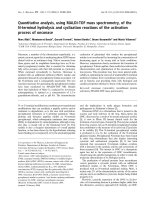
Enhancement of rPrP aggregation by Cu
2+
or
Zn
2+
but not Mg
2+
or Mn
2+
rPrP binds divalent cations such as Cu
2+
and Zn
2+
[13,14]. We next determined whether Cu
2+
or Zn
2+
influences the aggregation of rPrP, rPrP
8OR
and
rPrP
10OR
. At low pH, neither Cu
2+
nor Zn
2+
has any
effect on the aggregation of rPrP (not shown). The
failure of these cations to modulate rPrP aggregation
is most likely due to the effects of pH on the octapep-
tide repeat, rendering it unable to bind divalent cations
[15]. However, when the aggregation assay was carried
out at pH 7.4, Cu
2+
and Zn
2+
, but not Mg
2+
or
Mn
2+
, promote rPrP aggregation in a concentration-
dependent manner (Fig. 4A–D). Again, the levels of
enhancement are proportional to the number of
inserts. These results are in good accord with earlier
findings that PrP binds Cu
2+
and Zn
2+
but not Mg
2+
or Mn
2+
[13,16].
Furthermore, although the KKRPK deletion mutant
was totally unable to form aggregates in the presence
of heparin, in the presence of Cu
2+
, the KKRPK dele-
tion mutant behaved identically to wild-type rPrP
(Fig. 5A,B). In addition to the octapeptide-repeat
region, two additional Cu
2+
-binding sites have been
identified in the rPrP C-terminal globular domain
[16,17]. To investigate whether the octapeptide-repeat
region is important in Cu
2+
-induced aggregation, we
deleted the octapeptide-repeat region and created
rPrP
D51-90
. In contrast to wild-type rPrP, Cu
2+
does
not promote the aggregation of rPrP
D51-90
(Fig. 5C,D).
Therefore, the octapeptide-repeat region is the critical
motif that mediates Cu
2+
-induced rPrP aggregation.
Unexpectedly, GAG also failed to promote the
aggregation of rPrP
D51-90
(Fig. 6A). This deficit is not
because rPrP
D51-90
does not bind GAG. rPrP
D51-90
does bind GAG albeit with lower avidity (Fig. 6B).
Fig. 3. Sucrose-gradient centrifugation of rPrP–GAG aggregates.
rPrP (1 l
M) was mixed with 5 lgÆmL
)1
low molecular mass heparin
(3 kDa) in NaCl ⁄ P
i
and incubated at 25 °C for 30 min. The mixture
was loaded on to a 5–50% sucrose gradient and centrifuged at
4 °C, 100 000 g for 2 h. Ten fractions were drawn from top to bot-
tom. An equal volume of each fraction was loaded onto a 12%
SDS ⁄ PAGE and PrPs were detected by immunoblotting with mAb
8H4.
Fig. 4. Aggregation of rPrPs is enhanced
by metal ions. One micromole rPrP, rPrP
8OR
or rPrP
10OR
was mixed with various concen-
trations of CuCl
2
(A), ZnCl
2
(B), MnCl
2
(C)
and MgCl
2
(D) respectively in NaCl ⁄ P
i
, and
A
405
was recorded 300 s after mixing. The
results are means ± SEM of at least three
experiments. All the enhanced aggregation
are given here as an increased percentage
of starting turbidities [P =(T ⁄ T
0
)1) · 100;
P, percentage increase; T, turbidity; T
0
,
starting turbidity].
S. Yu et al. Aberrant features of insertion mutant prion proteins
FEBS Journal 275 (2008) 5564–5575 ª 2008 The Authors Journal compilation ª 2008 FEBS 5567
At higher protein concentrations, rPrP
D51-90
and rPrP
have comparable GAG-binding activity (Fig. 6B).
These results suggest that the octapeptide-repeat
region is the nucleation center of rPrP aggregation,
irrespective of whether aggregation is initiated with
GAG or Cu
2+
. Furthermore, these results also pro-
vide strong evidence that although the KKRPK motif
is the GAG-binding site, the octapeptide-repeat region
also contributes to the total affinity between PrP and
GAG. Although Cu
2+
and GAG bind to different
sites on PrP, we did not observe a synergistic effect
when both Cu
2+
and GAG were added to the rPrPs
(results not shown).
Sucrose-gradient centrifugation of rPrP–Cu
2+
aggregates
We also used sucrose-gradient centrifugation to com-
pare the relative sizes of rPrP–Cu
2+
and PrP
10OR
–
Cu
2+
aggregates. rPrP
D51-90
was used as a control. As
expected, without Cu
2+
, rPrP, rPrP
D51-90
and
rPrP
10OR
were detected in the top fractions (Fig. 7).
By contrast, when rPrP is mixed with Cu
2+
, most of
the PrP immunoreactivity is detected in the bottom
fractions. However, upon longer exposure, PrP immu-
noreactivity is also present in the intermediate frac-
tions (not shown). By contrast, when rPrP
10OR
is
mixed with Cu
2+
, all the immunoreactivity is detected
in the bottom fraction. Therefore, rPrP
10OR
also
forms much larger aggregates than wild-type rPrP.
rPrP
D51-90
, which does not bind Cu
2+
, remained on
the top of the gradient.
Modulation of GAG- or Cu
2+
-promoted
aggregation of rPrP
10OR
with anti-PrP mAbs
We next investigated whether GAG- or Cu
2+
-pro-
moted aggregation of rPrP
10OR
can be amended with
anti-PrP mAbs. The epitopes of these mAbs are dia-
grammatically presented in Fig. 8A. Of all the anti-PrP
mAbs tested, one, 8B4, consistently enhanced the
aggregation of rPrP
10OR
in the presence of either GAG
or Cu
2+
(Fig. 8B,C). mAb 8B4 alone does not induce
the aggregation of rPrP
10OR
without the PrP ligands.
Four mAbs, SAF32, 11G5, 7A12 and 8H4 consis-
tently blocked the aggregation of rPrP
10OR
in the pres-
ence of GAG (Fig. 8B). However, none of the tested
mAb was able to block the effects of Cu
2+
(Fig. 8C).
The inability of these mAbs to block Cu
2+
induced
aggregation is not because Cu
2+
prevents the binding
of these mAbs as shown by ELISA; Cu
2+
does not
inhibit the binding of these mAbs to rPrP (results not
shown).
Discussion
Aggregation of PrP is an essential step in the conver-
sion of PrP to PrP
Sc
[1]. Here we describe four new
findings on the aggregation of rPrPs: (a) in the pres-
ence of PrP ligands, such as GAG or the divalent
cation Cu
2+
, rPrPs aggregate in proportion to the
number of octapeptide inserts, thus rPrPs with inser-
tional mutations, such as rPrP
8OR
and rPrP
10OR
form
more and larger aggregates with faster kinetics than
wild-type rPrP; (b) whereas GAG-induced aggregation
Fig. 5. Copper enhances aggregation of
rPrP and rPrP
DKKRPK
, but not rPrP
D51-90
.
Various dilutions of CuCl
2
were mixed with
rPrP (A), rPrP
DKKRPK
(B) and rPrP
D51-90
(C),
respectively in NaCl ⁄ P
i
and A
405
was
recorded 300 s after mixing. (D) A compari-
son of the aggregation of rPrP, rPrP
DKKRPK
and rPrP
D51-90
enhanced by 50 lM CuCl
2
in
NaCl ⁄ P
i
. All the enhanced aggregation are
given here as an increased percentage of
starting turbidities [P =(T ⁄ T
0
)1) · 100; P,
percentage increase; T, turbidity; T
0
, starting
turbidity]. And the results are means ± SEM
for at least three experiments.
Aberrant features of insertion mutant prion proteins S. Yu et al.
5568 FEBS Journal 275 (2008) 5564–5575 ª 2008 The Authors Journal compilation ª 2008 FEBS
requires the GAG-binding motif, Cu
2+
-induced aggre-
gation requires the octapeptide repeat; (c) the octapep-
tide-repeat region is essential for both GAG- and
Cu
2+
-promoted rPrP aggregation; (d) aggregation
induced by GAG and Cu
2+
share common features,
yet each one has its own unique features, suggesting
multiple pathways leading to rPrP aggregation.
Bacterial produced rPrP has been used extensively as
a model system for studying the aggregation process
[5]. In previous studies, aggregation of rPrP required
denaturation, low pH and relatively high concentra-
tions of rPrP [18–22]. In this study, aggregation of
rPrP was carried out at pH 7.4 and with relatively low
concentrations of full-length rPrP; these conditions are
physiologically more relevant. Accumulated evidence
suggests that binding of GAG may be important in
the pathogenesis of prion diseases [23–27]. PrP
Sc
parti-
cles formed in vivo contain GAG [28]. In vitro, GAG
facilitates the conversion of PrP to PrP
Sc
[24], and
greatly increases the infectivity of non-aggregated
PrP
res
[25]. Reduction of cellular GAG significantly
decreases the biogenesis of PrP
Sc
in scrapie-infected
cells [29]. Cell-surface GAG has also been reported to
be the receptor for PrP
Sc
[23,27]. However, exogenous
GAG and GAG analogs, such as low molecular mass
heparin, suramin, pentosan polysulfate and dextran
sulfate can inhibit PrP
Sc
formation in cells, and pro-
long the incubation time of experimental prion diseases
[10]. It has been postulated that exogenous GAG and
GAG analogs block PrP
Sc
formation by competing
with the endogenous GAG which is critical for PrP
Sc
generation [10].
GAG may function as a scaffold for concentrating
PrP, creating a reservoir of PrP for conversion. We
reported earlier that rPrP
8OR
and rPrP
10OR
bind GAG
better than rPrP, and the level of binding is propor-
tional to the number of inserts [11]. Our current find-
ings that GAG also promotes the aggregation of
rPrP
8OR
and rPrP
10OR
proportional to the number of
inserts are in good accord with our earlier results.
Enhancement of rPrP aggregation is most apparent
when the concentration of rPrP is low, such as 1 lm.
At this concentration, rPrP by itself does not aggre-
gate. A small GAG, with nine sugar residues is as
Fig. 6. Heparin enhances aggregation of rPrP, but not rPrP
DKKRPK
and rPrP
D51-90
. (A) rPrPs (1, 3, 5 lM) were mixed with 5 lgÆmL
)1
heparin in NaCl ⁄ P
i
and A
405
was measured 300 s after mixing. All
the enhanced aggregation are given here as an increased percent-
age of starting turbidities [P =(T ⁄ T
0
)1) · 100; P, percentage
increase; T, turbidity; T
0
, starting turbidity]. The results herein are
means ± SEM for three experiments. (B) Detection of rPrP
D51-90
binding to heparin by ELISA. Heparin (10 lgÆmL
)1
) was coated onto
plates at 4 °C overnight and blocked with 3% BSA. BSA was
coated as a control. Different concentrations of rPrP, rPrP
DKKRPK
or
rPrP
D51-90
were incubated with the plates for 2 h at 25 °C. After
three washes with PBST, appropriate dilution of mAb 8H4 was
used to detect the bound rPrP. The results are means ± SEM for
three wells and this experiment was repeated at least three times.
Fig. 7. Sucrose-gradient centrifugation of rPrP–Cu
2+
aggregates.
rPrPs (1 l
M) was mixed with 20 lM CuCl
2
in NaCl ⁄ P
i
and incubated
at 25 °C for 30 min. The mixture was loaded on top of a 5–50%
sucrose gradient and centrifuged at 4 °C, 100 000 g for 2 h. Ten
fractions were drawn from top to bottom. An equal volume of each
fraction was loaded onto 12% SDS ⁄ PAGE and the PrPs were
detected by immunoblotting with mAb 8H4.
S. Yu et al. Aberrant features of insertion mutant prion proteins
FEBS Journal 275 (2008) 5564–5575 ª 2008 The Authors Journal compilation ª 2008 FEBS 5569
effective as larger GAG in promoting rPrP aggrega-
tion. However, a disaccharide of GAG is unable to
cause aggregation, suggesting that the minimum unit
of GAG required for rPrP aggregation is between
three and nine sugar residues. The promotion of aggre-
gation by GAG is not only limited to rPrP with inser-
tion mutations. GAG also promotes the aggregation
of rPrPs with pathogenic point mutations, albeit at
lower levels [30]. We hypothesize that enhanced bind-
ing to GAG, leading to aggregation is a common
feature in inherited human prion disease.
The precise mechanism by which GAG promotes
rPrP aggregation is not known. GAG may promote
aggregation by serving as a scaffold. If this is the case,
the rPrP aggregates should contain GAG. Alterna-
tively, GAG may simply serve as a platform for rPrPs
to be physically close to each other, resulting in aggre-
gation between rPrPs, without including GAG. Our
ELISA results suggest that some rPrP–GAG aggre-
gates contain GAG. However, our sucrose-gradient
centrifugation experiments revealed that rPrP–GAG
aggregates exist in many different sizes. Because the
GAG used in these experiments has a molecular mass
of 3 kDa, it is probable that some of the larger rPrP–
GAG aggregates are composed mainly of rPrP. Thus,
GAG serves as a scaffold as well as a platform in facil-
itating rPrP aggregation. In contrast to rPrP, when
mixed with 3 kDa GAG, all the rPrP
10OR
is detected
in the bottom fraction of the sucrose gradient. This is
in good accordance with our earlier finding that under
denaturing and low pH condition; rPrP
10OR
has the
propensity to spontaneously aggregate, in a protein
concentration-dependent manner. When incubated
with GAG, rPrP
10OR
is concentrated, thus able to
form much larger aggregates. It is interesting to note
that in PrP
Sc
infected mouse brain homogenate centri-
fuged under identical conditions, most of the PrP
immunoreactivity is present in the bottom fractions of
the sucrose gradient [31]. However, in contrast to
in vivo-derived PrP
Sc
aggregates, the rPrP aggregates
formed in the presence of GAG are PK sensitive
(results not shown).
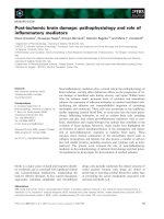
Fig. 8. Blocking of rPrP
10OR
aggregation
enhanced by heparin or copper using anti-
PrP mAbs. (A) The location of mAb-binding
epitopes along the length of PrP. LS, leader
sequence; GPI, glycosylphosphatidylinositol
anchor. (B) Blocking of the aggregation of
rPrP
10OR
enhanced by heparin. rPrP
10OR
(1 lM) was mixed with 1 lgÆmL
)1
heparin
and 0.125 l
M mAbs in NaCl ⁄ P
i
and A
405
was monitored as described in text. NS
mAb, non-specific mAb. (C) Blocking of the
aggregation of rPrP
10OR
enhanced by cop-
per. rPrP
10OR
(1 lM) was mixed with 20 lM
CuCl
2
and 0.125 lM mAbs in NaCl ⁄ P
i
and
A
405
was recorded. The aggregation is given
as an increased percentage of starting tur-
bidities [P =(T ⁄ T
0
)1) · 100; P, percentage
increase; T, turbidity; T
0
, starting turbidity].
The two experiments were repeated at least
three times.
Aberrant features of insertion mutant prion proteins S. Yu et al.
5570 FEBS Journal 275 (2008) 5564–5575 ª 2008 The Authors Journal compilation ª 2008 FEBS
rPrP binds divalent cations, such as Cu
2+
and Zn
2+
but not Mg
2+
or Mn
2+
[32]. A metal imbalance in the
central nervous system has been speculated to play a
role in neurodegenerative diseases, including prion dis-
ease [32]. However, the physiological significance of
the interaction between PrP and Cu
2+
remains poorly
understood. Some studies found that Cu
2+
causes
aggregation of rPrP [33–35]. Others reported that
Cu
2+
inhibits rPrP conversion to amyloid [36,37].
Copper chelators also inhibit PrP
Sc
replication in vitro
[38]. Some studies suggest that treatment with Cu
2+
causes PrP to acquire PK resistance [34–36,39,40].
However, this interpretation is complicated by the
recent finding that Cu
2+
inhibits proteinase K activity
[41].
We found that at neutral pH and low concentrations
of rPrPs, Cu
2+
and Zn
2+
but not Mg
2+
and Mn
2+
promote aggregation of rPrP, rPrP
8OR
and rPrP
10OR
in
a concentration-dependent manner. For rPrP
8OR
and
rPrP
10OR
, the enhancement can be observed in as low
as 1 lm of Cu
2+
or Zn
2+
, a concentration that is
physiologically relevant [42]. Again the degree of
enhancement is proportional to the number of octa-
peptide repeats, and Cu
2+
is consistently more efficient
in promoting aggregation than Zn
2+
.Cu
2+
does not
promote the aggregation of rPrP
D51-90
, which lacks the
octapeptide-repeat region. Therefore, the octapeptide-
repeat region is important in rPrP aggregation. This
finding is consistent with an earlier report suggesting
that the octapeptide-repeat region constitutes a pH-
dependent folding and aggregation site of PrP [22].
Our result is also consistent with another study show-
ing that when Cu
2+
binds to the octapeptide-repeat
region, it serves as a ‘copper switch’, which is impor-
tant in PrP aggregation [43]. However, we were sur-
prised to find that GAG was also unable to promote
the aggregation of rPrP
D51-90
, because the octapeptide-
repeat region is not required for the binding of GAG.
Furthermore, under high rPrP
D51-90
concentration, low
pH and denaturing conditions, rPrP
D51-90
also failed to
aggregate spontaneously (results not shown). There-
fore, the octapeptide-repeat region is critical for rPrP
aggregation irrespective of whether aggregation is
ligand initiated or spontaneous. It should be noted
that others have identified additional Cu
2+
-binding
sites at the C-terminus of PrP [16]. It is possible that
these binding sites may not be essential for PrP aggre-
gation.
The precise mechanisms by which the divalent
cations promote aggregation are not known. Cu
2+
and
Zn
2+
can bind PrP intramolecularly as well as inter-
molecularly [44]. We speculate that it is the inter-
molecular binding of Cu
2+
that enhances aggregation.
Presumably, by having more octapeptide repeats,
rPrP
10OR
is more readily to interact with Cu
2+
and
Zn
2+
. The failure of either Mg
2+
or Mn
2+
to enhance
aggregation provides the most appropriate control for
the specificity of the interactions. This interpretation is
also supported by results from the sucrose gradient
centrifugation experiments.
It has been reported that GAG promotes the aggre-
gation of rPrP and that the aggregate is stabilized by
the binding of Cu
2+
[26]. However, we did not observe
a synergistic effect between GAG and Cu
2+
in our
aggregation assay (results not shown). It should be
noted that in our assay the concentrations of rPrPs (1
versus 4 lm), GAG (0.1 versus 2 lm) as well as Cu
2+
(1–20 versus 500 lm) were much lower than is typically
used in this type of experiments. Furthermore, our
assay only detects the amount of aggregate that is gen-
erated rather than the stability of the aggregate.
Hence, it is possible that the aggregate formed with
GAG alone is different from the aggregate formed in
the presence of high concentrations of GAG and
Cu
2+
.
We reported earlier that under low pH and denatur-
ing conditions, only mAbs which react with an epitope
in the octapeptide-repeat region and the helix 1 region
respectively, block the spontaneous aggregation of
rPrPs [12]. In the current study, we found that mAb
8B4, which reacts with an epitope at the N-terminus
further promotes GAG- and Cu
2+
-induced rPrP
aggregation. We suggest that mAb 8B4 is able to align
the rPrP in the same orientation, in parallel, pairing
the N-terminus of two PrPs, which then facilitates the
binding of either GAG or Cu
2+
. It should be noted
that mAb 8B4 does not cause the aggregation of rPrP
without the participation of either GAG or Cu
2+
.
In addition to mAbs that are specific for the octa-
peptide-repeat region, such as SAF32, other mAbs,
such as 7A12, 11G5 and 8H4 also blocked GAG-
induced aggregation. These results suggest that the
entire C-globular domain including the helix 1, b2 and
helix 2 regions are all important in the aggregation
process. We could not evaluate whether mAb 8H4
inhibits spontaneous aggregation of rPrP because mAb
8H4 does not bind PrP at pH 4.0. This observation is
in good accordance with a recent study suggesting that
the opening of the helix 1 region, followed by confor-
mational changes in helix 2 of rPrP, is critical in rPrP
aggregation [45]. Finally, we showed that mAb 8F9,
which reacts with an epitope at the C-terminal end,
does not block GAG-induced aggregation. These
results suggest that in the presence of GAG, aggrega-
tion of rPrP starts at the end of N-terminus, proceed-
ing into the octapeptide-repeat region, the b1-sheet
S. Yu et al. Aberrant features of insertion mutant prion proteins
FEBS Journal 275 (2008) 5564–5575 ª 2008 The Authors Journal compilation ª 2008 FEBS 5571
region, helix 1 region and then the helix 2, in a ‘zip-
per’-like manner. This interpretation is also in good
agreement with another recent finding showing that
PrP fibril formation proceeds by aligning PrP mole-
cules in parallel, face to back, like a ‘zipper’ [46].
The underlying reason that none of the anti-PrP
mAbs is able to block Cu
2+
-induced rPrP aggregation
is not known. Accumulated evidence suggests that
there are multiple pathways in the PrP aggregation
process [10,47]. Our results suggest that GAG-induced
and Cu
2+
-induced aggregation proceed via different
pathways.
All the studies described here were based on findings
using rPrPs. Normal PrP has two highly conserved
N-linked glycosylation sites and is present on the cell
membrane with a glycosylphosphatidylinositol anchor.
Therefore, it is possible that the presence of N-linked
glycans as well as the placement of the cell membrane
can further modulate the interactions between PrP and
its ligands. Based on our findings, we hypothesize that
an increase in the number of octapeptide repeats
causes conformational changes at the N-terminus,
resulting in an enhancement in the binding of PrP
ligands, such as GAG, eventually leading to PrP aggre-
gation. Because all these aberrant features are propor-
tional to the number of insertions, our earlier and
current findings provide a biochemical explanation for
the observation that patients with more octapeptide-
repeat insertions have earlier disease onset and shorter
disease duration [3,48].
Experimental procedures
Plasmid construction and recombinant protein
preparation
Cloning, generation and purification of human rPrP,
rPrP
8OR
, rPrP
10OR
and rPrP
DKKRPK
were performed as
described previously with slight modification [11,30]. After
refolding and purification, these rPrPs were dialyzed against
20 mm NaAc, pH 5.5 and filtered through a 0.2 lm mem-
brane. For human rPrP
D51-90
, codons 51–90 were removed
from the prion protein coding sequence by annealing the
primer 5¢-GGCAACCGCTACCCA ⁄ CAAGGAGGTGG
CACC-3¢ ( ⁄ marks the site between codon 50 and codon 91)
to a phagemid containing the PrP-coding sequence. Muta-
genesis was performed using the BioRad Muta-Gene phage-
mid in vitro mutagenesis kit. The PrP mature fragment
(codons 23–231 with deletion of residues 51–90) was cloned
to the vector of pET42a(+) (Novagen, Gibbstown, NJ,
USA) [11], termed pET–rPrP
D51-90
. The insertion sequence
was verified by using the Applied Biosystems 3730 sequen-
cer (Foster City, CA, USA).
Freshly transformed BL21 (DE3) star Escherichia coli
(Invitrogen, Carlsbad, CA, USA) containing plasmid pET–
rPrP
D51-90
was transferred to 1 L Luria–Bertani media with
50 lgÆmL
)1
kanamycin at 37 °C until A
600
reached 0.6 and
induced for 4 h with 1 mm isopropyl thio-b-d-galactoside.
Bacteria were harvested by centrifugation at 4000 g for
15 min at 4 °C, resuspended in 20 mm Tris ⁄ HCl, pH 7.4,
150 mm NaCl, 1 mm phenylmethanesulfonyl fluoride,
0.1 mgÆmL
)1
lysozyme, 1 mm EDTA, 0.1% Triton X-100
and incubated at 25 ° C for 30 min before further lysis by
sonication. Samples were centrifuged at 13 000 g for
15 min, and the protein pellets were extensively washed
using 20 mm Tris ⁄ HCl, pH 7.4 with 0.5% Triton X-100
twice, then washed with the same buffer containing 2 m
NaCl and 2 m urea respectively. The pellets were then
resuspended in 20 mm Tris ⁄ HCl, pH 8.0, 8 m urea, 10 mm
b-mercaptoethanol. The protein was refolded by dialysis
against 20 mm Tris ⁄ HCl, pH 8.0 buffer with decreasing
urea and b-mercaptoethanol gradient concentrations. All
refolded rPrPs were further dialyzed against 20 mm NaAc,
pH 5.5, filtered through 0.2 lm membrane, stored at
)80 °C and used for experiments within one week after
refolding. SDS ⁄ PAGE and Coomassie Brilliant Blue stain-
ing showed that the purity of the recombinant protein is
consistently > 95% (not shown). Protein concentration
was determined with a Bio-Rad Protein Assay Kit. All of
the recombinant prion proteins were freshly purified before
use.
Antibodies
The generation, purification and characterization of all the
anti-PrP murine mAbs have been described in detail previ-
ously [49,50]. mAb 8B4 recognizes an epitope at residues
35–45; SAF32 reacts with residues 63–94 covering the octa-
peptide-repeat sequences [51]; 7A12 interacts with helix 1
between residues 143 and 155; 11G5 reacts with residues
115–130 covering b-sheet 1; 8H4 recognizes residues 175–
185 of helix 2; 8F9 reacts with residues 220–231. mAbs
8B4, SAF32, 7A12, 8H4 and 8F9 are IgG
1
, whereas mAb
11G5 is IgG
2b
. All mAbs were affinity purified using
Protein G chromatography. The concentration of mAbs
was determined with a BCA protein assay Kit (Pierce,
Rockford, IL, USA).
Turbidity measurement
The assays were performed at 25 °C in flat-bottomed
96-well plates. Heparin (from porcine intestinal mucosa;
Sigma, St Louis, MO, USA) or CuCl
2
was added into the
wells before addition of 200 lL NaCl ⁄ P
i
(pH 7.4) contain-
ing 1 lm rPrPs. After mixing as quickly as possible, turbidi-
ties were monitored within 15 s by reading the absorbance
at 405 nm in a Beckman Coulter AD340 micro-ELISA
Aberrant features of insertion mutant prion proteins S. Yu et al.
5572 FEBS Journal 275 (2008) 5564–5575 ª 2008 The Authors Journal compilation ª 2008 FEBS
plate reader, using a kinetic photometric model (interval
time 30 s, 30 cycles with 1 s shaking before every cycle).
Similar processes were performed with ZnCl
2
, MgCl
2
and
MnCl
2
.
To investigate whether anti-PrP mAbs can block the hep-
arin enhanced aggregation of rPrP, 10 lL heparin (final con-
centration 1 lgÆmL
)1
) was mixed with 2.5 lL mAbs (final
concentration 0.125 lm). Then 200 lL NaCl ⁄ P
i
containing
1 lm rPrP
10OR
was added and mixed quickly. Turbidities
were recorded as described in above. A similar procedure
was carried out to investigate the effect of anti-PrP mAbs on
copper enhanced aggregation of rPrP. An irrelevant mAb
9C1, anti-(brain-derived neurotrophic factor), was used as a
negative control. All experiments were carried out at least
three times with different batches of rPrPs.
Detection of rPrP binding to heparin
Flat-bottomed, 96-well Costar plates (Corning, Corning,
NY, USA) were coated with 10 lgÆmL
)1
heparin at 4 °C
overnight and blocked with 3% BSA in NaCl ⁄ P
i
at 25 °C
for 3 h. BSA was coated onto the plates as a control.
Appropriate dilutions of rPrP
D51-90
or rPrP were added into
the plates in triplicate and incubated at 25 °C for 2 h. After
three washes with phosphate-buffered saline ⁄ 0.05% Tween
(PBST), bound rPrP was detected with mAb 8H4. Horse-
radish peroxidase-conjugated goat anti-mouse IgG (Chem-
icon, Billerica, MA, USA) was used as the secondary
antibody and A
405
was measured for 2,2¢-azinobis-(3-ethyl-
benzthiazoline-6-sulfonic acid) (Roche Diagnostics, India-
napolis, IN, USA). All experiments were carried out at
least three times with different batches of rPrPs.
Detection of biotinylated heparin in the
aggregates of rPrPs
mAb 11G5 was previously shown to be able to react with
PrP aggregates [52]. mAb 11G5 was coated onto the flat-
bottomed, 96-well Costar plates at 5 lgÆmL
)1
at 4 °C over-
night and blocked with 3% BSA in NaCl ⁄ P
i
at 25 °C for
3 h. BSA was coated onto the plates as a control. Five
micromoles of either rPrP or rPrP
DKKRPK
was mixed with
10 lgÆmL
)1
biotinylated heparin (from porcine intestinal
mucosa, Sigma) in 400 lL NaCl ⁄ P
i
respectively and incu-
bated at 25 °C for 30 min. The aggregates were collected by
centrifugation at 16 000 g for 10 min at 25 °C. Supernatants
were removed and the pellets were washed three times with
NaCl ⁄ P
i
by vortexing followed by centrifugation at 16 000 g
for 5 min. The aggregates were dissolved with 50 lL
NaCl ⁄ P
i
containing 0.1% Triton X-100 by incubation at
42 °C for 10 min. NaCl ⁄ P
i
(450 lL) was then added into
the Eppendorf tubes to a final volume of 500 lL. Various
dilutions of this original aggregate solution in NaCl ⁄ P
i
were
then incubated with mAb 11G5-coated plates at 4 °C over-
night. After three washes with PBST, the bound biotinylated
heparin was detected by adding horseradish peroxidase-con-
jugated streptavidin (Chemicon) at 1 : 10 000 dilutions. 2,2¢-
Azinobis-(3-ethylbenzthiazoline-6-sulfonic acid) was added
and A
405
was recorded. All experiments were carried out at
least three times with different batches of rPrPs.
Sucrose-gradient fractionation
To form a 5–50% step sucrose gradient, 5, 10, 15, 20, 30,
40, 50% sucrose solution prepared in NaCl ⁄ P
i
were loaded
into ultraclear centrifuge tubes (13 · 51 mm). rPrPs (1 lm)
were mixed with 5 lgÆmL
)1
low molecular mass heparin
(3 kDa) or 20 lm CuCl
2
in NaCl ⁄ P
i
. After incubation for
30 min at 25 °C, 0.5 mL of the mixture was loaded on top
of the sucrose gradient. Ultracentrifugation was carried out
in SW55 rotor (Beckman, Fullerton, CA, USA) at
100 000 g,4°C for 2 h. Fractions of 0.5 mL were collected
from the top of the tubes. rPrP present in different sucrose-
gradient fractions was detected by immunoblotting. 10 lL
of each fraction was mixed with 2· SDS loading buffer and
heated at 95 °C for 10 min before separation on 12%
SDS ⁄ PAGE. The gel was transferred to a nitrocellulose
membrane and probed with mAb 8H4. Blue dextran
(Sigma) with a molecular mass of 2000 kDa was used as a
marker in the gradient.
Statistical analysis
A two-way ANOVA program was used to determine the
P-value between various groups. P > 0.05 is considered to
be not significant (ns).
Acknowledgements
We would like to thank Dr Jacques Grassi (Atomic
Energy Commission, Saclay, France) for his gift of
mAb SAF32. This work was supported in part by
NIH (National Institutes of Health) grant NS-045981-
01 and an award ⁄ contract from the US Department of
the Army, DAMD17-03-1- 286 (to MSS).
References
1 Prusiner SB (1998) Prions. Proc Natl Acad Sci USA 95,
13363–13383.
2 Mead S (2006) Prion disease genetics. Eur J Hum Genet
14, 273–281.
3 Kovacs GG, Trabattoni G, Hainfellner JA, Ironside
JW, Knight RS & Budka H (2002) Mutations of the
prion protein gene phenotypic spectrum. J Neurol 249,
1567–1582.
4 Cohen FE, Pan KM, Huang Z, Baldwin M, Fletterick
RJ & Prusiner SB (1994) Structural clues to prion repli-
cation. Science 264, 530–531.
S. Yu et al. Aberrant features of insertion mutant prion proteins
FEBS Journal 275 (2008) 5564–5575 ª 2008 The Authors Journal compilation ª 2008 FEBS 5573
5 Riesner D (2003) Biochemistry and structure of PrP(C)
and PrP(Sc). Br Med Bull 66, 21–33.
6 Cappai R, Stewart L, Jobling MF, Thyer JM, White
AR, Beyreuther K, Collins SJ, Masters CL & Barrow
CJ (1999) Familial prion disease mutation alters the sec-
ondary structure of recombinant mouse prion protein:
implications for the mechanism of prion formation.
Biochemistry 38, 3280–3284.
7 Swietnicki W, Petersen RB, Gambetti P & Surewicz
WK (1998) Familial mutations and the thermodynamic
stability of the recombinant human prion protein. J Biol
Chem 273, 31048–31052.
8 Vanik DL & Surewicz WK (2002) Disease-associated
F198S mutation increases the propensity of the recom-
binant prion protein for conformational conversion to
scrapie-like form. J Biol Chem 277, 49065–49070.
9 Liemann S & Glockshuber R (1999) Influence of amino
acid substitutions related to inherited human prion dis-
eases on the thermodynamic stability of the cellular
prion protein. Biochemistry 38 , 3258–3267.
10 Caughey B (2003) Prion protein conversions: insight
into mechanisms, TSE transmission barriers and strains.
Br Med Bull 66, 109–120.
11 Yin S, Yu S, Li C, Wong P, Chang B, Xiao F, Kang
SC, Yan H, Xiao G, Grassi J et al. (2006) Prion pro-
teins with insertion mutations have altered N-terminal
conformation and increased ligand binding activity and
are more susceptible to oxidative attack. J Biol Chem
281, 10698–10705.
12 Yu S, Yin S, Li C, Wong P, Chang B, Xiao F, Kang
SC, Yan H, Xiao G, Tien P et al. (2007) Aggregation
of prion protein with insertion mutations is propor-
tional to the number of inserts. Biochem J 403, 343–
351.
13 Brown LR & Harris DA (2003) Copper and zinc cause
delivery of the prion protein from the plasma mem-
brane to a subset of early endosomes and the Golgi.
J Neurochem 87, 353–363.
14 Thompsett AR, Abdelraheim SR, Daniels M & Brown
DR (2005) High affinity binding between copper and
full-length prion protein identified by two different tech-
niques. J Biol Chem 280, 42750–42758.
15 Miura T, Sasaki S, Toyama A & Takeuchi H (2005)
Copper reduction by the octapeptide-repeat region of
prion protein: pH dependence and implications in cellu-
lar copper uptake. Biochemistry 44, 8712–8720.
16 Jackson GS, Murray I, Hosszu LL, Gibbs N, Waltho
JP, Clarke AR & Collinge J (2001) Location and prop-
erties of metal-binding sites on the human prion pro-
tein. Proc Natl Acad Sci USA 98, 8531–8535.
17 Jones CE, Abdelraheim SR, Brown DR & Viles JH
(2004) Preferential Cu
2+
coordination by His96 and
His111 induces beta-sheet formation in the unstructured
amyloidogenic region of the prion protein. J Biol Chem
279, 32018–32027.
18 Bocharova OV, Breydo L, Parfenov AS, Salnikov VV
& Baskakov IV (2005) In vitro conversion of full-length
mammalian prion protein produces amyloid form with
physical properties of PrP(Sc). J Mol Biol 346, 645–659.
19 Jackson GS, Hosszu LL, Power A, Hill AF, Kenney J,
Saibil H, Craven CJ, Waltho JP, Clarke AR & Collinge
J (1999) Reversible conversion of monomeric human
prion protein between native and fibrilogenic conforma-
tions. Science 283, 1935–1937.
20 Sokolowski F, Modler AJ, Masuch R, Zirwer D, Baier
M, Lutsch G, Moss DA, Gast K & Naumann D (2003)
Formation of critical oligomers is a key event during
conformational transition of recombinant Syrian ham-
ster prion protein. J Biol Chem 278, 40481–40492.
21 Swietnicki W, Morillas M, Chen SG, Gambetti P &
Surewicz WK (2000) Aggregation and fibrillization of
the recombinant human prion protein huPrP90-231.
Biochemistry 39, 424–431.
22 Zahn R (2003) The octapeptide repeats in mammalian
prion protein constitute a pH-dependent folding and
aggregation site. J Mol Biol 334, 477–488.
23 Horonchik L, Tzaban S, Ben-Zaken O, Yedidia Y,
Rouvinski A, Papy-Garcia D, Barritault D, Vlodavsky
I & Taraboulos A (2005) Heparan sulfate is a cellular
receptor for purified infectious prions. J Biol Chem 280,
17062–17067.
24 Wong C, Xiong LW, Horiuchi M, Raymond L, Wehrly
K, Chesebro B & Caughey B (2001) Sulfated glycans
and elevated temperature stimulate PrP(Sc)-dependent
cell-free formation of protease-resistant prion protein.
EMBO J 20, 377–386.
25 Shaked GM, Meiner Z, Avraham I, Taraboulos A &
Gabizon R (2001) Reconstitution of prion infectivity
from solubilized protease-resistant PrP and nonprotein
components of prion rods. J Biol Chem 276, 14324–
14328.
26 Gonzalez-Iglesias R, Pajares MA, Ocal C, Espinosa JC,
Oesch B & Gasset M (2002) Prion protein interaction
with glycosaminoglycan occurs with the formation of
oligomeric complexes stabilized by Cu(II) bridges.
J Mol Biol 319, 527–540.
27 Hijazi N, Kariv-Inbal Z, Gasset M & Gabizon R (2005)
PrPSc incorporation to cells requires endogenous gly-
cosaminoglycan expression. J Biol Chem 280, 17057–
17061.
28 Snow AD, Kisilevsky R, Willmer J, Prusiner SB &
DeArmond SJ (1989) Sulfated glycosaminoglycans in
amyloid plaques of prion diseases. Acta Neuropathol
(Berl) 77, 337–342.
29 Ben-Zaken O, Tzaban S, Tal Y, Horonchik L, Esko
JD, Vlodavsky I & Taraboulos A (2003) Cellular hepa-
ran sulfate participates in the metabolism of prions.
J Biol Chem 278, 40041–40049.
30 Yin S, Pham N, Yu S, Li C, Wong P, Chang B, Kang
SC, Biasini E, Tien P, Harris DA et al. (2007) Human
Aberrant features of insertion mutant prion proteins S. Yu et al.
5574 FEBS Journal 275 (2008) 5564–5575 ª 2008 The Authors Journal compilation ª 2008 FEBS
prion proteins with pathogenic mutations share com-
mon conformational changes resulting in enhanced
binding to glycosaminoglycans. Proc Natl Acad Sci
USA 104, 7546–7551.
31 Tzaban S, Friedlander G, Schonberger O, Horonchik
L, Yedidia Y, Shaked G, Gabizon R & Taraboulos A
(2002) Protease-sensitive scrapie prion protein in aggre-
gates of heterogeneous sizes. Biochemistry 41, 12868–
12875.
32 Lehmann S (2002) Metal ions and prion diseases. Curr
Opin Chem Biol 6, 187–192.
33 Treiber C, Simons A & Multhaup G (2006) Effect of
copper and manganese on the de novo generation of
protease-resistant prion protein in yeast cells. Biochemis-
try 45, 6674–6680.
34 Tsiroulnikov K, Rezaei H, Dalgalarrondo M, Chobert
JM, Grosclaude J & Haertle T (2006) Cu(II) induces
small-size aggregates with amyloid characteristics in two
alleles of recombinant ovine prion proteins. Biochim
Biophys Acta 1764, 1218–1226.
35 Kuczius T, Buschmann A, Zhang W, Karch H, Becker
K, Peters G & Groschup MH (2004) Cellular prion pro-
tein acquires resistance to proteolytic degradation fol-
lowing copper ion binding. Biol Chem 385, 739–747.
36 Bocharova OV, Breydo L, Salnikov VV & Baskakov IV
(2005) Copper(II) inhibits in vitro conversion of prion
protein into amyloid fibrils. Biochemistry 44, 6776–6787.
37 Hijazi N, Shaked Y, Rosenmann H, Ben-Hur T & Gab-
izon R (2003) Copper binding to PrPC may inhibit
prion disease propagation. Brain Res 993, 192–200.
38 Sigurdsson EM, Brown DR, Alim MA, Scholtzova H,
Carp R, Meeker HC, Prelli F, Frangione B & Wisniew-
ski T (2003) Copper chelation delays the onset of prion
disease. J Biol Chem 278, 46199–46202.
39 Qin K, Yang DS, Yang Y, Chishti MA, Meng LJ,
Kretzschmar HA, Yip CM, Fraser PE & Westaway D
(2000) Copper(II)-induced conformational changes and
protease resistance in recombinant and cellular PrP.
Effect of protein age and deamidation. J Biol Chem
275, 19121–19131.
40 Quaglio E, Chiesa R & Harris DA (2001) Copper con-
verts the cellular prion protein into a protease-resistant
species that is distinct from the scrapie isoform. J Biol
Chem 276, 11432–11438.
41 Stone LA, Jackson GS, Collinge J, Wadsworth JD &
Clarke AR (2007) Inhibition of proteinase K activity by
copper(II) ions. Biochemistry 46, 245–252.
42 Kramer ML, Kratzin HD, Schmidt B, Romer A, Windl
O, Liemann S, Hornemann S & Kretzschmar H (2001)
Prion protein binds copper within the physiological con-
centration range. J Biol Chem 276, 16711–16719.
43 Leliveld SR, Dame RT, Wuite GJ, Stitz L & Korth C
(2006) The expanded octarepeat domain selectively
binds prions and disrupts homomeric prion protein
interactions. J Biol Chem 281, 3268–3275.
44 Morante S, Gonzalez-Iglesias R, Potrich C, Meneghini
C, Meyer-Klaucke W, Menestrina G & Gasset M
(2004) Inter- and intra-octarepeat Cu(II) site geometries
in the prion protein: implications in Cu(II) binding coo-
perativity and Cu(II)-mediated assemblies. J Biol Chem
279, 11753–11759.
45 Eghiaian F, Daubenfeld T, Quenet Y, van Audenhaege
M, Bouin AP, van der Rest G, Grosclaude J & Rezaei
H (2007) Diversity in prion protein oligomerization
pathways results from domain expansion as revealed by
hydrogen ⁄ deuterium exchange and disulfide linkage.
Proc Natl Acad Sci USA 104, 7414–7419.
46 Sawaya MR, Sambashivan S, Nelson R, Ivanova MI,
Sievers SA, Apostol MI, Thompson MJ, Balbirnie M,
Wiltzius JJ, McFarlane HT et al.
(2007) Atomic struc-
tures of amyloid cross-beta spines reveal varied steric
zippers. Nature 447, 453–457.
47 Baskakov IV, Legname G, Baldwin MA, Prusiner SB &
Cohen FE (2002) Pathway complexity of prion protein
assembly into amyloid. J Biol Chem 277, 21140–21148.
48 Croes EA, Theuns J, Houwing-Duistermaat JJ, Der-
maut B, Sleegers K, Roks G, Van den Broeck M, van
Harten B, van Swieten JC, Cruts M et al. (2004) Octa-
peptide repeat insertions in the prion protein gene and
early onset dementia. J Neurol Neurosurg Psychiatry 75,
1166–1170.
49 Li R, Liu T, Wong BS, Pan T, Morillas M, Swietnicki W,
O’Rourke K, Gambetti P, Surewicz WK & Sy MS (2000)
Identification of an epitope in the C-terminus of normal
prion protein whose expression is modulated by binding
events in the N-terminus. J Mol Biol 301, 567–573.
50 Zanusso G, Liu D, Ferrari S, Hegyi I, Yin X, Aguzzi
A, Hornemann S, Liemann S, Glockshuber R, Manson
JC et al. (1998) Prion protein expression in different
species: analysis with a panel of new mAbs. Proc Natl
Acad Sci USA 95, 8812–8816.
51 Feraudet C, Morel N, Simon S, Volland H, Frobert Y,
Creminon C, Vilette D, Lehmann S & Grassi J (2005)
Screening of 145 anti-PrP monoclonal antibodies for
their capacity to inhibit PrPSc replication in infected
cells. J Biol Chem 280, 11247–11258.
52 Pan T, Chang B, Wong P, Li C, Li R, Kang SC, Rob-
inson JD, Thompsett AR, Tein P, Yin S et al. (2005)
An aggregation-specific enzyme-linked immunosorbent
assay: detection of conformational differences between
recombinant PrP protein dimers and PrP(Sc) aggre-
gates. J Virol 79, 12355–12364.
S. Yu et al. Aberrant features of insertion mutant prion proteins
FEBS Journal 275 (2008) 5564–5575 ª 2008 The Authors Journal compilation ª 2008 FEBS 5575