Báo cáo khoa học: A role of monocyte chemoattractant protein-4 (MCP-4)/CCL13 from chondrocytes in rheumatoid arthritis doc
Bạn đang xem bản rút gọn của tài liệu. Xem và tải ngay bản đầy đủ của tài liệu tại đây (649.6 KB, 9 trang )
A role of monocyte chemoattractant protein-4
(MCP-4)/CCL13 from chondrocytes in rheumatoid arthritis
Takuji Iwamoto
1,2
, Hiroshi Okamoto
1
, Shu Kobayashi
1,2
, Katsunori Ikari
1
, Yoshiaki Toyama
2
,
Taisuke Tomatsu
1
, Naoyuki Kamatani
1
and Shigeki Momohara
1
1 Institute of Rheumatology, Tokyo Women’s Medical University, Japan
2 Department of Orthopedic Surgery, School of Medicine, Keio University, Tokyo, Japan
Rheumatoid arthritis (RA) is a chronic, symmetric poly-
articular joint disease that primarily affects the small
joints of the hands and feet [1]. It is characterized by
infiltration of inflammatory cells such as monocytes
and T-lymphocytes into the joints, leading to synovial
proliferation and progressive destruction of cartilage
and bone [2]. Although the basic mechanisms of RA
are widely accepted, the pathogenesis of the disease is
not fully understood.
Chemokines in humans comprise more than 50 small
(8–10 kDa) heparin-binding proteins that were origi-
nally identified by their chemotactic activity on bone
marrow-derived cells [3,4]. They are classified into
four families on the basis of the location of cysteine
residues. The four chemokine groups are CC, C, CXC,
and CX3C, and their receptors are consequently classi-
fied as CCR, CR, CXCR, and CX3CR. Chemokines
and chemokine receptors have been shown to be
involved in a variety of inflammatory diseases by
recruiting leukocytes to the inflammatory site [5]. It is
well known that synovial tissue and synovial fluid from
RA patients contain increased concentrations of sev-
eral chemokines, such as interleukin (IL)-8) ⁄ CXCL8,
interferon-c (IFN-c)-inducible protein-10 ⁄ CXCL10,
monokine induced by interferon-c ⁄ CXCL9, stromal
cell-derived factor-1 ⁄ CXCL12, monocyte chemotactic
protein (MCP)-1 ⁄ CCL2, macrophage inflammatory
protein-1a ⁄ CCL3, and fractalkine ⁄ CXC3CL1 [6].
Keywords
chondrocytes; extracellular signal-regulated
kinase (ERK); monocyte chemoattractant
protein-4 (MCP-4) ⁄ CCL13; rheumatoid
arthritis
Correspondence
H. Okamoto, Institute of Rheumatology,
Tokyo Women’s Medical University, 10-22
Kawada-cho, Shinjuku, Tokyo 162-0054,
Japan
Fax: +81 3 5269 1726
Tel: +81 3 5269 1725
E-mail:
(Received 14 April 2007, revised 6 July
2007, accepted 26 July 2007)
doi:10.1111/j.1742-4658.2007.06013.x
We studied the role of monocyte chemoattractant (MCP)-4 ⁄ CCL13 in the
pathogenesis of rheumatoid arthritis (RA). MCP-4 was highly expressed in
cartilage from RA patients. Interferon-c significantly stimulated MCP-4 ⁄
CCL13 production in human chondrocytes, and this effect was enhanced in
combination with interleukin-1b or tumor necrosis factor-a. MCP-4 ⁄
CCL13 induces the phosphorylation of extracellular signal-regulated kinase
in fibroblast-like synoviocytes and activates cell proliferation, and PD98059
completely inhibits these effects. These data suggest that interferon-c in
combination with interleukin-1b ⁄ tumor necrosis factor-a activates the pro-
duction of MCP-4 ⁄ CCL13 from chondrocytes in RA joints, and that
secreted MCP-4 ⁄ CCL13 enhances fibroblast-like synoviocyte proliferation
by activating the extracellular signal-regulated kinase mitogen-activated
protein kinase cascade.
Abbreviations
DAB, 3¢3-diaminobenzidine tetrahydrochloride; ERK, extracellular signal-regulated kinase; FLS, fibroblast-like synoviocyte; IFN-c, interferon-c;
IL, interleukin; MCP, monocyte chemoattractant protein; OA, osteoarthritis; RA, rheumatoid arthritis; SNP, single-nucleotide polymorphism;
TNF-a, tumor necrosis factor-a; XTT, sodium 3¢-[1-(phenylaminocarbonyl)-3,4-tetrazolium]-bis (4-methoxy-6-nitro) benzene sulfonic acid
hydrate.
4904 FEBS Journal 274 (2007) 4904–4912 ª 2007 The Authors Journal compilation ª 2007 FEBS
These chemokines are implicated in RA pathogenesis
via the recruitment and retention of leukocytes into
the joints. In addition to functioning in cell traffic,
several chemokines are reported to enhance the prol-
iferation of fibroblast-like synoviocytes (FLSs) and
upregulate gelatinase and collagenase production by
FLSs [7]. Thus, chemokines are key molecules in RA
pathogenesis and are potential therapeutic targets for
RA [8].
Although macrophages and FLSs are considered to
be the most potent producers of chemokines in the
synovial compartment, chondrocytes also have the
ability to produce chemokines [8–10]. In our previous
report, we found that mRNA expression of MCP-
4 ⁄ CCL13 was significantly higher in cartilage from RA
patients than from osteoarthritis (OA) patients or nor-
mal controls, and the concentration of MCP-4 ⁄ CCL13
protein in synovial fluid was also significantly higher in
RA patients than in OA patients [11].
MCP-4 ⁄ CCL13 is a recently identified CC chemo-
kine from a human cDNA library that directs the
migration of eosinophils, monocytes and T-lympho-
cytes through several chemokine receptors, including
CCR-2 and CCR-3 [12,13]. The role of MCP-4 ⁄
CCL13 in disease is less well defined, but recent studies
suggest that it is involved in inflammatory cell recruit-
ment in allergic disorders such as asthma and atopic
dermatitis [14–17].
In the present study, we further determined the role
of MCP-4 ⁄ CCL13 in RA pathogenesis. We investi-
gated the role of several stimuli on the expression of
MCP-4 ⁄ CCL13 by human chondrocytes, and the sig-
nal transduction pathways controlling FLS prolifera-
tion by MCP-4 ⁄ CCL13. In addition, we conducted a
case-control study using single-nucleotide polymor-
phisms (SNPs) to determine whether MCP-4 ⁄ CCL13
could be a genetic risk factor for RA.
Results
Production of MCP-4
⁄
CCL13 by human
chondrocytes
To identify the stimulatory signals that activate the
production of MCP-4 ⁄ CCL13 from human chondro-
cytes, we investigated the effect of several cytokines
reported to have roles in RA pathogenesis. Human
chondrocytes from RA or OA patients were cultured
in the presence of IL-1b, tumor necrosis factor-a
(TNF-a) or IFN-c, and various combinations of these
three cytokines. Chondrocytes from both RA and
OA patients gave similar results, and we present the
data obtained with RA-derived chondrocytes. MCP-
4 ⁄ CCL13 protein concentrations in culture superna-
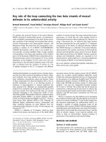
tants were evaluated by ELISA. IFN-c significantly
stimulated MCP-4 ⁄ CCL13 production in a dose-depen-
dent manner, whereas IL-1b and TNF-a had no signif-
icant effect (Fig. 1A). Interestingly, stimulation of
MCP-4 ⁄ CCL13 production by IFN-c was significantly
and remarkably enhanced when IFN-c was combined
with IL-1b or TNF-a (Fig. 1B).
To determine whether these observed effects occur
at the transcriptional level, quantitative real-time PCR
analysis was performed on IL-1b, TNF-a, and IFN-c.
Consistent with the ELISA data, IFN-c significantly
stimulated mRNA expression of MCP-4 ⁄ CCL13 in
MCP-4 concentration (pg/mL)
MCP-4 concentration (pg/mL)
0
100
200
300
IFN-γ
γ
0 1 10 1000100
400
A
B
0 10010 0 10010
IL-1
β
TNF-
α
(ng/mL)
*
*
0
100
200
300
IFN-
γ
(ng/mL)
400
500
600
IL-1
β
(ng/mL)
TNF-
α
(ng/mL)
10 10
10
10
10
100 100
10
100
10
#
#
1000
Fig. 1. MCP-4 ⁄ CCL13 protein production by human chondrocytes
from RA and OA patients. Human chondrocytes were cultured in
the presence of (A) IL-1b (0–100 ngÆmL
)1
; open bars), TNF-a
(0–100 ngÆmL
)1
; solid bars) and IFN-c (0–1000 ngÆmL
)1
; shaded
bars) for 48 h, or (B) IFN-c (shaded bars), IFN-c + IL-1b (open bars),
and IFN-c + TNF-a (solid bars) for 48 h. MCP-4 ⁄ CCL13 protein
concentrations in these cell culture supernatants were evaluated by
ELISA. Bars show the mean and SD of four separate experiments.
Statistical evaluation was performed using one-way ANOVA
followed by Tukey’s method for multiple comparisons. *P<0.01
compared with vehicle-treated control.
#
P<0.01 compared with
the sample cultured with IFN-c alone.
T. Iwamoto et al. Role of MCP-4 ⁄ CCL13 in rheumatoid arthritis
FEBS Journal 274 (2007) 4904–4912 ª 2007 The Authors Journal compilation ª 2007 FEBS 4905
a dose-dependent manner. IL-1b or TNF-a also
enhanced the mRNA expression of MCP-4 ⁄ CCL13
induced by IFN-c. Chondrocytes from both RA and
OA patients gave similar results, and we present the
data obtained with RA-derived chondrocytes. These
results suggest that these stimulatory effects occur at
the transcriptional level (Fig. 2A,B).
Effect of MCP-4
⁄
CCL13 on extracellular
signal-regulated kinase (ERK) phosphorylation
Western blot analysis was performed to investigate
whether MCP-4⁄ CCL13 could activate ERK, which is
widely known to play a major role in cell prolifera-
tion [20]. The FLSs from both RA and OA patients
were similar, and we present the data from the
RA-derived FLSs. As expected, phosphorylation of
ERK was induced by stimulation with MCP-4 ⁄
CCL13 in RA FLSs. This activation peaked at about
10–20 min, and returned to basal levels within 60 min
(Fig. 3A). Incubation with 100 lm PD98059, a spe-
cific ERK activation inhibitor, was sufficient to abol-
ish ERK activation by MCP-4 ⁄ CCL13 in RA FLSs
(Fig. 3B).
To confirm the involvement of ERK phosphorylation
in the proliferative effect on RA FLSs of MCP-4 ⁄
CCL13, an XTT {sodium 3¢-[1-(phenylaminocarbonyl)-
3,4-tetrazolium]-bis-(4-methoxy-6-nitro) benzene sulfo-
nic acid hydrate} cell proliferation assay was performed,
using PD98059 to antagonize the phosphorylation of
ERK. As representative results of western blot ana-
lyses were obtained at a concentration of 100 ngÆmL
)1
,
an XTT cell proliferation assay was performed with
the same concentrations. MCP-4 ⁄ CCL13 enhanced the
proliferation of FLSs in a dose-dependent manner,
and PD98059 completely inhibited the stimulatory
effect of MCP-4 ⁄ CCL13 on FLS proliferation.
PD98059 did not inhibit basal proliferation of RA
FLSs (Fig. 4A). The FLSs from both RA and OA
patients were similar, and we present the data from the
RA-derived FLSs.
Association study using SNPs of MCP-4
⁄
CCL13
As we found that MCP-4 ⁄ CCL13 was one of the key
molecules in the pathogenesis of RA, we studied the
association of the MCP-4 ⁄ CCL13 gene with RA sus-
ceptibility. We selected two SNPs in MCP-4 ⁄ CCL13
(T887C and rs159313), for the following reasons. Two
single-nucleotide T–to–C polymorphisms (T896C and
T887C) were reported in the MCP-4 ⁄ CCL13 core pro-
moter region [22]. They are located 896 and 887 bp
before the transcription initiation site, and were
reported to have direct effects on the transcript level of
the gene [22]. After preliminary analysis, we selected
T887C for a larger-scale study, because T896C and
T887C were in complete linkage disequilibrium (D¢ ¼
1.0, r
2
¼ 1.0). On the basis of information from the
National Center of Biotechnology Information data-
base, three SNPs (rs3136677, rs159313 and rs2072069)
were found in MCP-4 ⁄ CCL13. Among them, we
selected SNP rs159313 for the study, because one SNP
(rs3136677) was nonpolymorphic in Japanese popula-
tions, and other two SNPs (rs159313 and rs2072069)
were in complete linkage disequilibrium (D¢ ¼ 1.0,
r
2
¼ 1.0) according to the International HapMap pro-
ject (public release 19) [23].
08412
IFN-γ
IFN-γ
1000 ng/mL
IFN-γ
100 ng/ml
IFN-γ
10 ng/ml
1 ng/ml
IFN-γ
0.1 ng/ml
IFN-γ
1000
IFN-γ
100 ng/mL
IFN-γ
10 ng/mL
1 ng/mL
IFN-γ
0.1 ng/mL
-delta delta Ct-delta delta Ct
Time (hours)
*
*
*
*
2
4
6
8
A
B
IFN-γ 10 ng/mL
+ Il -1β 10 ng/mL
IFN-γ
+ IL-1β
IFN-γ 10 ng/mL
Il -1β 10 ng/mL
+ TNF-α 10 ng/mL
IL-1β
+ TNF-α
TNF-α 10 ng/mLTNF-α
Il -1β 10 ng/mLIL-1β
IFN-γ 10 ng/mL
+ TNF-α 10 ng/mL
IFN-γ
+ TNF-α
2
4
0
6
8
08412
Time (hours)
#
#
Fig. 2. MCP-4 ⁄ CCL13 mRNA expression by human chondrocytes
from RA and OA patients. Total RNA from chondrocytes stimulated
with different cytokines for 4–12 h was harvested and transcribed
to cDNA by reverse transcription. cDNA was used for TaqMan
quantitative real-time PCR: (A) with IFN-c (0–1000 ngÆmL
)1
), and (B)
with IL-1b, TNF-a, IFN-c, IL-1b + TNF-a, IFN-c + IL-1b, and
IFN-c + TNF-a. The figure shows expression of MCP-4 ⁄ CCL13
mRNA relative to time-matched vehicle-treated controls using the
comparative threshold cycle (Ct) method. Data are mean ± SD of
four separate experiments. Statistical evaluation was performed
using one-way ANOVA followed by Tukey’s method for multiple
comparisons. *P<0.01 compared with vehicle-treated control.
#
P<0.01 compared with the sample cultured with IFN-c alone.
Role of MCP-4 ⁄ CCL13 in rheumatoid arthritis T. Iwamoto et al.
4906 FEBS Journal 274 (2007) 4904–4912 ª 2007 The Authors Journal compilation ª 2007 FEBS
According to the genotyping results, these two SNPs
were present in Hardy–Weinberg equilibrium in both
cases and controls. No statistically significant differ-
ences in genotype or allele frequencies were observed
between cases and controls. We failed to find signifi-
cant differences even when RA patients were stratified
according to the rheumatoid factor status (Table 1).
These data indicate that the MCP-4 ⁄ CCL13 gene may
not be responsible for the onset of RA.
Discussion
MCP-1 ⁄ CCL2, MCP-2 ⁄ CCL8, MCP-3 ⁄ CCL7 and
MCP-4 ⁄ CCL13 constitute a subfamily of CC chemo-
kines that share structural and functional features.
MCP-1 ⁄ CCL2 was the first to be identified [24], and
MCP-4 ⁄ CCL13 is the most recently identified chemo-
kine, and is a potent chemoattractant for eosinophils,
monocytes and T-lymphocytes [12,13]. MCP-4⁄ CCL13
expression is upregulated at sites of inflammation in a
number of different diseases, including asthma [14–16],
atherosclerosis [25], acute renal inflammation [26], and
atopic dermatitis [17]. MCP-4 ⁄ CCL13 was also highly
expressed in articular cartilage from patients with RA
[11].
In the present study we demonstrated MCP-
4 ⁄ CCL13 protein production by human chondrocytes,
and showed for the first time that MCP-4 ⁄ CCL13
from chondrocytes is actively involved in RA patho-
genesis. We also demonstrated that IFN-c was the
main stimulus for MCP-4 ⁄ CCL13 production, and that
TNF-a and IL-1b enhanced the stimulatory effect of
IFN-c. According to the analysis of the human MCP-
4 ⁄ CCL13 gene in dermal fibroblasts, the core promoter
region contained IFN-c-response elements as well as
nuclear factor-jB-like consensus sequences [27]. Fur-
thermore, MCP-4 ⁄ CCL13 mRNA expression was
reported to be upregulated by stimulation with TNF-a
and IFN-c in dermal fibroblasts [27]. Similar results
were obtained in human airway epithelial cell lines
after stimulation with the cytokine TNF-a alone or in
combination with IFN-c [28]. In contrast, our experi-
ments indicated that IFN-c was the main stimulus for
MCP-4 ⁄ CCL13 production by human chondrocytes.
We observed no significant stimulation of induction of
MCP-4 ⁄ CCL13 mRNA expression by TNF-a alone in
human chondrocytes, whereas TNF-a greatly enhanced
the expression in combination with IFN-c.
IFN-c is produced by T-cells and by natural killer
cells infiltrating the inflamed synovium, and is secreted
into the joint space, although its role in the progres-
sion of articular injury remains controversial [29]. To
date, divergent in vitro effects of IFN- c have been
reported in the literature. IFN-c induces the produc-
tion of nitric oxide, IL-6 and prostaglandin E
2
by
human chondrocytes [30]. In contrast, IFN-c inhibits
TNF-a- and IL-1b-induced collagenase and stromely-
sin production by chondrocytes, as well as TNF-a-
and IL-1b-stimulated proteoglycan degradation [31,32].
Furthermore, the effects of IFN-c in the treatment of
pERK
A
B
1/2
ERK 1/2
MCP-4 (min)
025 2010 30 60 120
Relative Intensity 1.00 1.03 16.55 47.73 46.16 23.29 1.04 1.05
Relative Intensity 1.00 0.96 0.94 0.95 0.98 0.97 0.99 1.01
PD98059
IL-1β
pERK 1/2
ERK 1/2
MCP-4
+++
10μ
Μ
100μ
Μ
Relative Intensity 1.00 40.01 9.20 0.96 27.88
Relative Intensity 1.00 0.96 0.98 1.01 1.03
Fig. 3. Induction of ERK phosphorylation by
MCP-4 ⁄ CCL13 in RA FLSs. FLSs were cul-
tured overnight in serum-free DMEM. (A)
FLSs were incubated in the presence of
MCP-4 ⁄ CCL13 (100 ngÆmL
)1
) for an addi-
tional 0–120 min. (B) FLSs were incubated
in the presence of MCP-4 ⁄ CCL13
(100 ngÆmL
)1
) for 20 min with or without
PD98059 (10–100 lgÆmL
)1
). As a positive
control, FLSs were incubated with IL-1b
(5 ngÆmL
)1
) for 20 min. Cell lysates were
examined for ERK activation by western
blotting with phospho-p44 ⁄ 42 MAP kinase
mouse monoclonal antibody (pERK1 ⁄ 2).
Total p44 ⁄ 42 MAP kinase antibody was
used to verify equal protein loading. The
result is one representative example from
three independent experiments.
T. Iwamoto et al. Role of MCP-4 ⁄ CCL13 in rheumatoid arthritis
FEBS Journal 274 (2007) 4904–4912 ª 2007 The Authors Journal compilation ª 2007 FEBS 4907
RA are unclear. A statistically significant improvement
was observed among the RA patients treated with
recombinant IFN-c in one double-blind study of 91
patients [33], whereas current evidence shows that anti-
IFN-c therapy is significantly superior to placebo in 30
patients with RA [34]. It is widely accepted that
TNF-a is the key molecule in RA pathogenesis, as
demonstrated by the clinical benefit of TNF-a-neutra-
lizing therapy [35]. Although approximately 40% of
patients show dramatic responses, the remainder show
some evidence of persistent synovitis or minimal clini-
cal benefit [1]. The results of the present study suggest
that IFN-c may contribute to the progression of joint
inflammation, in part by modulating MCP-4 ⁄ CCL13
production by human chondrocytes.
We have also demonstrated the ability of MCP-
4 ⁄ CCL13 to phosphorylate ERK mitogen-activated
protein (MAP) kinase, and have shown that induction
of FLS proliferation by MCP-4 ⁄ CCL13 is dependent
on the phosphorylation of ERK MAP kinase. It is
widely accepted that the progressive destruction of
articular cartilage is reliant on the evolution of hyper-
plastic synovial tissue, and that hyperplasia of FLSs is
dependent on dysregulated proliferation and apoptosis
[1,36]. Key regulators of this proliferation include the
recently recognized macrophage migration inhibitory
factor and proinflammatory cytokines such as TNF-a
and IL-1b through the nuclear factor- jB and ⁄ or MAP
kinase signal transduction pathways [37–40]. To date,
several chemokines, including MCP-1, stromal cell-
derived factorF-1a, IFN-c-inducible protein, monokine
induced by IFN-c and MCP-4 ⁄ CCL13 are also known
to enhance FLS proliferation, although the signal
transduction pathways underlying the proliferation
remain unclear [7,11]. We hypothesized that ERK acti-
vation might be involved in the proliferative effect
of MCP-4 ⁄ CCL13, as the ERK cascade has been
reported to be a central pathway that transmits signals
from many extracellular agents to regulate cellular pro-
cesses such as proliferation, differentiation and cell
cycle progression in various cells [22,41]. As expected,
the MCP-4 ⁄ CCL13–ERK cascade was indeed involved
in the proliferation of synovial cells, as shown here. In
addition, ERK is reported to have a role in the expres-
sion of matrix metalloproteinases (MMPs), such as
MMP-1, MMP-2, and MMP-9, and contributes to the
degradation of extracellular matrix for the invasion of
melanoma cells [42]. Several lines of evidence have
shown that MMPs are involved in the joint degrada-
tion process in RA. Thus, MCP-4 ⁄ CCL13 might have
roles not only in the proliferation of synovial cells but
also in the invasion of synovial cells, resulting in pan-
nus formation and destruction of joints in RA. Taken
together with these results, MCP-4⁄ CCL13 secreted
from chondrocytes in the joints plays an important
role in the development of aggressive synovial tissues
in RA, as illustrated in Fig. 4B. There are numerous
reports showing the importance of synovial cells in RA
pathogenesis. Our data support the notion that chon-
drocytes are also actively involved in RA pathogenesis.
In conclusion, we have shown that MCP-4 ⁄ CCL13
is produced by human chondrocytes from RA patients
Absorbance
0.8
0.9
1.0
1.1
MCP-4 (ng/mL)
PD98059
*
**
*
100
+
0 100
+
10010
+
**
1.2
A
B
Chondrocytes
MCP-4
Proliferation
IL-1β, TNF-α
IFN-γ
Th1 cells
Synovial Cells
IL-18
Fig. 4. (A) Effects of inhibition of ERK phosphorylation on RA
FLS proliferation. FLSs were treated with MCP-4 ⁄ CCL13 (0–
100 ngÆmL
)1
) with and without the addition of MAP kinase kinase
inhibitor PD98059 (100 lgÆmL
)1
) for 48 h. MCP-4 ⁄ CCL13 signifi-
cantly increased RA FLS proliferation, and inhibition of ERK phos-
phorylation by PD98059 significantly inhibited FLS proliferation.
PD98059 did not inhibit basal proliferation of RA FLSs. Bars show
the mean and SD of three indepemdent experiments. Statistical
evaluation was performed using one-way ANOVA followed by
Tukey’s method for multiple comparisons.*P<0.05; **P<0.01.
(B) Schematic representation of the role of MCP-4 ⁄ CCL13 in RA. A
vicious circle is formed between chondrocytes and synovium in the
affected joint. IFN-c is produced by Th1 (T helper 1) cells infiltrating
the synovium and activates the expression of MCP-4 ⁄ CCL13. Then,
MCP-4 ⁄ CCL13 stimulates the proliferation of synovial cells, which
produce inflammatory cytokines (IL-b, TNF-a). Together, these cyto-
kines, with IFN-c, further enhance the production of MCP-4 ⁄ CCL13
by chondrocytes.
Role of MCP-4 ⁄ CCL13 in rheumatoid arthritis T. Iwamoto et al.
4908 FEBS Journal 274 (2007) 4904–4912 ª 2007 The Authors Journal compilation ª 2007 FEBS
stimulated by IFN-c and TNF-a ⁄ IL-1b. In addition,
MCP-4 ⁄ CCL13 has significant effects on FLS prolifer-
ation that are dependent on the activation of ERK
MAP kinase. These data suggest that MCP-4 ⁄ CCL13
is a significant contributor to synovial hyperplasia in
RA, and that MCP-4 ⁄ CCL13 may serve as a new tar-
get for anti-RA therapy.
Experimental procedures
Preparation of articular cartilage and synovial
tissue
Human articular cartilage and synovial tissue were obtained
from OA and RA patients (n ¼ 5 in each group) who were
undergoing total knee replacement at Tokyo Women’s
Medical University, Tokyo, Japan. OA was diagnosed by
physical examination along with radiographic findings, and
RA patients met the 1987 disease criteria of the American
College of Rheumatology [18]. All samples were obtained
with informed consent. All experiments were approved
by the Ethical Committee of Tokyo Women’s Medical
University.
Isolation and culture of chondrocytes and FLSs
Tissue was obtained under aseptic conditions and was finely
minced. Chondrocytes were isolated by sequential enzy-
matic digestion at 37 °C: 5 mgÆmL
)1
pronase (Kaken Phar-
maceutical Co., Ltd, Tokyo, Japan) for 1 h, followed by
2mgÆmL
)1
collagenase (Sigma Chemical Co., St Louis,
MO, USA) for 6 h at 37 °C in DMEM (Nikken Bio Medi-
cal Laboratory, Kyoto, Japan) with antibiotics (100 unitsÆ
mL
)1
penicillin, 100 lgÆmL
)1
streptomycin; Gibco BRL,
Grand Island, NY, USA). FLSs were also isolated by diges-
tion with 1 mgÆmL
)1
collagenase for 3 h at 37 °Cin
DMEM. The digested tissue was briefly subjected to centri-
fugation at 1500 g at 37 °C for 15 min using an MX-100
centrifuge (TOMY Seiko, Tokyo, Japan) with TMP-11
angle-type rotor, and the resulting pellet was washed three
times in NaCl ⁄ P
i
. The isolated cells were seeded at high
density in tissue culture flasks and cultured in DMEM sup-
plemented with 10% heat-inactivated fetal bovine serum
(Tissue Culture Biologicals, Tulare, CA, USA) at 37 °Cin
a humidified atmosphere of 5% CO
2
⁄ 95% air. The culture
medium was changed every 3–5 days, and nonadherent
lymphoid cells were removed. At confluence, chondrocytes
and FLSs were detached and passaged once, and then
seeded at high density and allowed to grow in DMEM
supplemented as above. Chondrocytes were used between
passages 1 and 3, and FLSs were used between passages
5 and 8 for the following experiments. In some cases, carti-
lage tissue slices were obtained for immunohistochemical
analysis.
Effect of cytokines on MCP-4 production by
human chondrocytes
Human chondrocytes from four RA patients and three OA
patient were cultured in DMEM supplemented with 10%
fetal bovine serum in 12-well culture plates. At confluence,
the culture medium was replaced with serum-free DMEM.
After 24 h, chondrocytes were incubated for an additional
48 h in the absence or presence of recombinant human
IL-1b (0–100 ngÆmL
)1
; R&D Systems), recombinant human
TNF-a (0–100 ngÆmL
)1
; R&D Systems), recombinant
human IFN-c (0–1000 ngÆmL
)1
; R&D Systems) and combi-
nations of these cytokines. The culture supernatant was
collected and stored at ) 80 °C. MCP-4 concentrations in
these supernatants were evaluated as described above.
Experiments were performed three times with each of the
four independent cultures.
Table 1. Summary of the association of MCP-4 in rheumatoid arthritis cases and controls. The major allele was always referred to as allele 1
and the minor allele as allele 2. SNP, single-nucleotide polymorphism; RF, rheumatoid factor; MAF, minor allele frequency; OR, odds ratio;
95% CI, confidence interval.
SNP
Genotype
Cases Controls
Allele 1 versus allele 2
a
1 ⁄ 11⁄ 22⁄ 2 Total MAF 1 ⁄ 11⁄ 22⁄ 2 Total MAF v
2
OR (95% CI) P
rs159313
total 400 533 189 1122 0.41 152 227 75 454 0.42 0.23 0.96 (0.82–1.13) 0.63
RF + 350 467 167 984 0.41 0.17 0.97 (0.82–1.14) 0.68
RF - 50 66 22 138 0.4 0.24 0.93 (0.70–1.24) 0.62
T-887C
total 924 193 9 1126 0.094 368 75 4 447 0.093 0.006 1.01 (0.77–1.34) 0.94
RF + 810 168 9 987 0.094 0.014 1.02 (0.77–1.35) 0.91
RF – 114 25 0 139 0.09 0.022 0.96 (0.58–1.56) 0.88
a
Distribution of the frequency of allele 1 versus allele 2 in the cases compared with the controls.
T. Iwamoto et al. Role of MCP-4 ⁄ CCL13 in rheumatoid arthritis
FEBS Journal 274 (2007) 4904–4912 ª 2007 The Authors Journal compilation ª 2007 FEBS 4909
Quantitative real-time PCR
Total RNA was harvested from chondrocytes stimulated
with cytokines for 4–12 h using the RNeasy Mini Kit
according to the manufacturer’s instructions (Qiagen, Chats-
worth, CA, USA). cDNA was synthesized from 0.3 lgof
total RNA in a 20 lL reaction using TaqMan Reverse Tran-
scription Reagents (Applied Biosystems, Tokyo, Japan).
TaqMan quantitative real-time PCR was performed using
the ABI Prism 7900HT sequence detection system and Taq-
Man PCR Master Mix according to the manufacturer’s pro-
tocol (Applied Biosystems). Primers and probes for human
MCP-4 ⁄ CCL13 and human glyceraldehyde-3-phosphate
dehydrogenase were purchased from Applied Biosystems.
RNA samples lacking reverse transcriptase were used with
each real-time PCR experiment to verify the absence of
genomic DNA. The incubation was initiated at 50 °C for
2 min, and this was followed by 95 °C for 10 min, and 40
cycles at 95 °C for 15 s and 65 °C for 1 min. Samples were
compared using the comparative threshold cycle (Ct)
method to determine MCP-4 mRNA expression relative to
the time-matched vehicle-treated control. The parameter Ct
is the PCR cycle number at which the fluorescence generated
by cleavage of the probe reaches a fixed threshold above
baseline. For each sample, the MCP-4 ⁄ CCL13 Ct value was
normalized using DCt ¼ MCP-4 ⁄ CCL13 Ct ) glyceralde-
hyde-3-phosphate dehydrogenase Ct. To determine relative
expression levels, the following formula was used: DDCt ¼
sample DCt ) time-matched control DCt, and the value used
to plot relative MCP-4 ⁄ CCL13 expression of each sample
was calculated using the expression 2
–DDCt
.
Western blot analysis
The phosphorylation of p44 ⁄ 42 MAP kinase, or ERK, was
assessed by western blotting. In brief, FLSs were cultured in
DMEM supplemented with 10% fetal bovine serum on a
10 cm culture dish. At 80% confluence, the culture medium
was replaced with serum-free DMEM. After 24 h, FLSs
were incubated in the presence of recombinant human
MCP-4 (rHuMCP-4, 100 ngÆmL
)1
; R&D Systems) for an
additional 0–120 min. In addition, FLSs were incubated in
the presence of recombinant human MCP-4 (100 ngÆmL
)1
)
for 20 min with or without a specific inhibitor of MAP
kinase kinase, PD98059 (Calbiochem, San Diego, CA, USA;
10–100 lgÆmL
)1
). As a positive control, FLSs were incu-
bated with recombinant human IL-1b (5 ngÆmL
)1
) for
20 min. Cells were lysed with Cell Lysis Buffer (Cell Signal-
ing Technology, Beverly, MA, USA). After incubation on
ice for 10 min, the protein concentration was determined,
and the lysates were stored at ) 80 °C. Equal amounts of
cellular proteins were separated by SDS ⁄ PAGE and trans-
ferred to Immune-Blot poly(vinylidene difluoride) mem-
brane (Bio-Rad, Hercules, CA, USA). Immunoblotting was
performed using phospho-p44 ⁄ 42 MAP kinase mouse
monoclonal antibody (Cell Signaling Technology; diluted
1 : 5000) and p44 ⁄ 42 MAP kinase antibody (Cell Signaling
Technology; diluted 1 : 1000) to verify equal protein
loading.
Cell proliferation assay
FLSs were seeded at a density of 1 · 10
3
cells per well in
96-well microtiter plates in 100 lL of serum-free DMEM
per well, and were treated with recombinant human MCP-
4 (0–100 ngÆmL
)1
) for 48 h. The activation of ERK was
antagonized with PD98059 (100 lgÆmL
)1
). Cell prolifera-
tion was evaluated by measuring the number of viable
cells using the XTT assay by using the XTT Cell Proli-
feration Kit II (Roche Applied Science, Mannheim,
Germany) [19]. Formazan product in the supernatant was
measured in terms of absorbance values at 490 nm by
using an ELISA plate reader. The absorbance values
obtained from culture medium without cells were sub-
tracted from the values obtained with cells. Experiments
were performed six times with each of the three indepen-
dent cultures.
Genetic association study using SNPs
The study was part of an RA cohort project (IORRA:
Institute of Rheumatology RA cohort), and was approved
by Tokyo Women’s Medical University Genome Ethics
Committee [20]. Out of the registered RA patients, DNA
samples were obtained from 1284. Informed written consent
was obtained from every subject. Of these, 1128 samples
were randomly selected for this study. Eighty-eight per cent
of them were rheumatoid factor positive. They were mostly
females (82.6%), and the mean age of the patients was
57.6 years (range: 19–85 years). Four hundred and fifty-five
population-based control DNA samples were obtained
from the Pharma SNP consortium ( />psc/index.html). All control subjects were matched for sex,
ethnic origin, and geographical area.
SNP genotyping was performed using the TaqMan fluoro-
genic 5¢-nuclease assay (Applied Biosystems) according to
the manufacturer’s instructions, as described previously [21].
Statistical methods
Data are presented as the mean ± standard deviation
(SD). Statistical comparisons were performed using either
the Mann–Whitney U-test or one-way ANOVA followed
by Tukey’s method for multiple comparisons, as appropri-
ate. Hardy–Weinberg equilibrium and associations between
RA and each of the SNPs were estimated by the chi-square
test. Statistical significance was established at the P<0.05
level. All analyses were carried out using the r software
package, version 2.0.1 ( />Role of MCP-4 ⁄ CCL13 in rheumatoid arthritis T. Iwamoto et al.
4910 FEBS Journal 274 (2007) 4904–4912 ª 2007 The Authors Journal compilation ª 2007 FEBS
Acknowledgements
This work was supported, in part, by grants-in-aid
from the Ministry of Education, Culture, Sports, Sci-
ence and Technology of Japan. The expert technical
help of Yukiko Katagiri is gratefully acknowledged.
References
1 Firestein GS (2003) Evolving concepts of rheumatoid
arthritis. Nature 423, 356–361.
2 Feldmann M, Brennan FM & Maini RN (1996)
Rheumatoid arthritis. Cell 85, 307–310.
3 Charo IF & Ransohoff RM (2006) The many roles of
chemokines and chemokine receptors in inflammation.
N Engl J Med 354, 610–621.
4 Sallusto F, Mackay CR & Lanzavecchia A (2001) The
role of chemokine receptors in primary, effector, and
memory immune responses. Annu Rev Immunol 18,
593–620.
5 Luster AD (1998) Chemokines ) chemotactic
cytokines that mediate inflammation. N Engl J Med
338, 436–445.
6 Koch AE (2005) Chemokines and their receptors in
rheumatoid arthritis: future targets? Arthritis Rheum 52,
710–721.
7 Garcia-Vicuna R, Gomez-Gaviro MV, Dominguez-Luis
MJ, Pec MK, Gonzalez-Alvaro I, Alvaro-Gracia JM &
Diaz-Gonzalez F (2004) CC and CXC chemokine
receptors mediate migration, proliferation, and matrix
metalloproteinase production by fibroblast-like
synoviocytes from rheumatoid arthritis patients.
Arthritis Rheum 50, 3866–3877.
8 Haringman JJ, Ludikhuize J & Tak PP (2004)
Chemokines in joint disease: the key to inflammation?
Ann Rheum Dis 63 , 1186–1194.
9 Pulsatelli L, Dolzani P, Piacentini A, Silvestri T,
Ruggeri R, Gualtieri G, Meliconi R & Facchini A
(1999) Chemokine production by human chondrocytes.
J Rheumatol 26, 1992–2001.
10 Villiger PM, Terkeltaub R & Lotz M (1992) Monocyte
chemoattractant protein-1 (MCP-1) expression in
human articular cartilage. Induction by peptide regula-
tory factors and differential effects of dexamethasone
and retinoic acid. J Clin Invest 90, 488–496.
11 Iwamoto T, Okamoto H, Iikuni N, Takeuchi M,
Toyama Y, Tomatsu T, Kamatani N & Momohara S
(2006) Monocyte chemoattractant protein-4 (MCP-
4) ⁄ CCL13 is highly expressed in cartilage from patients
with rheumatoid arthritis. Rheumatology (Oxford) 45,
421–424.
12 Garcia-Zepeda EA, Combadiere C, Rothenberg ME,
Sarafi MN, Lavigne F, Hamid Q, Murphy PM & Luster
AD (1996) Human monocyte chemoattractant protein
(MCP)-4 is a novel CC chemokine with activities on
monocytes, eosinophils, and basophils induced in aller-
gic and nonallergic inflammation that signals through
the CC chemokine receptors (CCR)-2 and -3. J Immunol
157, 5613–5626.
13 Uguccioni M, Loetscher P, Forssmann U, Dewald B, Li
H, Lima SH, Li Y, Kreider B, Garotta G, Thelen M
et al. (1996) Monocyte chemotactic protein 4 (MCP-4),
a novel structural and functional analogue of MCP-3
and eotaxin. J Exp Med 183, 2379–2384.
14 Lamkhioued B, Garcia-Zepeda EA, Abi-Younes S,
Nakamura H, Jedrzkiewicz S, Wagner L, Renzi PM,
Allakhverdi Z, Lilly C, Hamid Q et al. (2000) Monocyte
chemoattractant protein (MCP)-4 expression in the air-
ways of patients with asthma. Induction in epithelial
cells and mononuclear cells by proinflammatory cyto-
kines. Am J Respir Crit Care Med 162 (2 Part 1), 723–
732.
15 Kalayci O, Sonna LA, Woodruff PG, Camargo CA Jr,
Luster AD & Lilly CM (2004) Monocyte chemotactic
protein-4 (MCP-4; CCL-13): a biomarker of asthma.
J Asthma 41, 27–33.
16 Taha RA, Minshall EM, Miotto D, Shimbara A, Luster
A, Hogg JC & Hamid QA (1999) Eotaxin and mono-
cyte chemotactic protein-4 mRNA expression in small
airways of asthmatic and nonasthmatic individuals.
J Allergy Clin Immunol 103 (3 Part 1), 476–483.
17 Taha RA, Minshall EM, Leung DY, Boguniewicz M,
Luster A, Muro S, Toda M & Hamid QA (2000)
Evidence for increased expression of eotaxin and
monocyte chemotactic protein-4 in atopic dermatitis.
J Allergy Clin Immunol 105 , 1002–1007.
18 Arnett FC, Edworthy SM, Bloch DA, McShane DJ,
Fries JF, Cooper NS, Healey LA, Kaplan SR, Liang
MH, Luthra HS et al. (1998) The American Rheuma-
tism Association 1987 revised criteria for the classifica-
tion of rheumatoid arthritis. Arthritis Rheum 31,
315–324.
19 Okamoto H, Cujec TP, Okamoto M, Peterlin BM, Baba
M & Okamoto T (2000) Inhibition of the RNA-depen-
dent transactivation and replication of human immuno-
deficiency virus type 1 by a fluoroquinoline derivative
K-37. Virology 272, 402–408.
20 Matsuda Y, Singh G, Yamanaka H, Tanaka E, Urano
W, Taniguchi A, Saito T, Hara M, Tomatsu T &
Kamatani N (2003) Validation of a Japanese version of
the Stanford Health Assessment Questionnaire in 3,763
patients with rheumatoid arthritis. Arthritis Rheum 49,
784–788.
21 Ikari K, Kuwahara M, Nakamura T, Momohara S, Hara
M, Yamanaka H, Tomatsu T & Kamatani N (2005)
Association between PADI4 and rheumatoid arthritis: a
replication study. Arthritis Rheum 52, 3054–3057.
22 Kalayci O, Birben E, Wu L, Oguma T, Storm Van’s
Gravesande K, Subramaniam V, Sheldon HK,
T. Iwamoto et al. Role of MCP-4 ⁄ CCL13 in rheumatoid arthritis
FEBS Journal 274 (2007) 4904–4912 ª 2007 The Authors Journal compilation ª 2007 FEBS 4911
Silverman ES & Lilly CM (2003) Monocyte
chemoattractant protein-4 core promoter genetic
variants: influence on YY-1 affinity and plasma levels.
Am J Respir Cell Mol Biol 29, 750–756.
23 Altshuler D, Brooks LD, Chakravarti A, Collins FS,
Daly MJ & Donnelly PA (2005) Haplotype map of the
human genome. Nature 437, 1299–1320.
24 Furutani Y, Nomura H, Notake M, Oyamada Y, Fukui
T, Yamada M, Larsen CG, Oppenheim JJ &
Matsushima K (1989) Cloning and sequencing of the
cDNA for human monocyte chemotactic and activating
factor (MCAF). Biochem Biophys Res Commun 159,
249–255.
25 Berkhout TA, Sarau HM, Moores K, White JR,
Elshourbagy N, Appelbaum E, Reape RJ, Brawner M,
Makwana J, Foley JJ et al. (1997) Cloning, in vitro
expression, and functional characterization of a novel
human CC chemokine of the monocyte chemotactic
protein (MCP) family (MCP-4) that binds and signals
through the CC chemokine receptor 2B. J Biol Chem
272, 16404–16413.
26 Chakravorty SJ, Howie AJ, Girdlestone J, Gentle D &
Savage CO (2001) Potential role for monocyte chemo-
tactic protein-4 (MCP-4) in monocyte ⁄ macrophage
recruitment in acute renal inflammation. J Pathol 194,
239–246.
27 Hein H, Schluter C, Kulke R, Christophers E, Schroder
JM & Bartels J (1999) Genomic organization, sequence
analysis and transcriptional regulation of the human
MCP-4 chemokine gene (SCYA13) in dermal fibro-
blasts: a comparison to other eosinophilic beta-chemo-
kines. Biochem Biophys Res Commun 255, 470–476.
28 Stellato C, Collins P, Ponath PD, Soler D, Newman W,
La Rosa G, Li H, White J, Schwiebert LM, Bickel C
et al. (1997) Production of the novel C-C chemokine
MCP-4 by airway cells and comparison of its biological
activity to other C-C chemokines. J Clin Invest 99, 926–
936.
29 Feldmann M, Brennan FM & Maini RN (1996) Role of
cytokines in rheumatoid arthritis. Annu Rev Immunol
14, 397–440.
30 Henrotin YE, Zheng SX, Labasse AH, Deby GP, Criel-
aard JM & Reginster JY (2000) Modulation of human
chondrocyte metabolism by recombinant human inter-
feron. Osteoarthritis Cartilage 8, 474–482.
31 Bunning RA & Russell RG (1989) The effect of tumor
necrosis factor alpha and gamma-interferon on the
resorption of human articular cartilage and on the pro-
duction of prostaglandin E and of caseinase activity by
human articular chondrocytes. Arthritis Rheum 32,
780–784.
32 Andrews HJ, Bunning RA, Plumpton TA, Clark IM,
Russell RG & Cawston TE (1990) Inhibition of interleu-
kin-1-induced collagenase production in human articular
chondrocytes in vitro by recombinant human interferon-
gamma. Arthritis Rheum 33, 1733–1738.
33 Lemmel EM, Brackertz D, Franke M, Gaus W, Hartl
PW, Machalke K, Mielke H, Obert HJ, Peter HH,
Sieper J et al. (1988) Results of a multicenter placebo-
controlled double-blind randomized phase III clinical
study of treatment of rheumatoid arthritis with recom-
binant interferon-gamma. Rheumatol Int 8, 87–93.
34 Sigidin YA, Loukina GV, Skurkovich B & Skurkovich
S (2001) Randomized, double-blind trial of anti-inter-
feron-gamma antibodies in rheumatoid arthritis. Scand
J Rheumatol 30, 203–207.
35 Lipsky PE, van der Heijde DM, St Clair EW, Furst
DE, Breedveld FC, Kalden JR, Smolen JS, Weisman
M, Emery P, Feldmann M et al. (2000) Infliximab and
methotrexate in the treatment of rheumatoid arthritis.
Anti-Tumor Necrosis Factor Trial in Rheumatoid
Arthritis with Concomitant Therapy Study Group.
N Engl J Med 343, 1594–1602.
36 Qu Z, Garcia CH, O’Rourke LM, Planck SR, Kohli M
& Rosenbaum JT (1994) Local proliferation of fibro-
blast-like synoviocytes contributes to synovial hyperpla-
sia. Results of proliferating cell nuclear antigen ⁄ cyclin,
c-myc, and nucleolar organizer region staining.
Arthritis
Rheum 37, 212–220.
37 Lacey D, Sampey A, Mitchell R, Bucala R, Santos L,
Leech M & Morand E (2003) Control of fibroblast-like
synoviocyte proliferation by macrophage migration
inhibitory factor. Arthritis Rheum 48, 103–109.
38 Inoue H, Takamori M, Nagata N, Nishikawa T, Oda
H, Yamamoto S & Koshihara Y (2001) An investiga-
tion of cell proliferation and soluble mediators induced
by interleukin 1beta in human synovial fibroblasts: com-
parative response in osteoarthritis and rheumatoid
arthritis. Inflamm Res 50, 65–72.
39 Schett G, Tohidast-Akrad M, Smolen JS, Schmid BJ,
Steiner CW, Bitzan P, Zenz P, Redlich K, Xu Q &
Steiner G (2000) Activation, differential localization,
and regulation of the stress-activated protein kinases,
extracellular signal-regulated kinase, c-JUN N-terminal
kinase, and p38 mitogen-activated protein kinase, in
synovial tissue and cells in rheumatoid arthritis.
Arthritis Rheum 43, 2501–2512.
40 Youn J, Kim HY, Park JH, Hwang SH, Lee SY, Cho
CS & Lee SK (2002) Regulation of TNF-alpha-medi-
ated hyperplasia through TNF receptors, TRAFs, and
NF-kappaB in synoviocytes obtained from patients with
rheumatoid arthritis. Immunol Lett 83, 85–93.
41 Rubinfeld H & Seger R (2005) The ERK cascade: a
prototype of MAPK signaling. Mol Biotechnol 31,
151–174.
42 Smalley KS (2003) A pivotal role for ERK in the onco-
genic behaviour of malignant melanoma? Int J Cancer
104, 527–532.
Role of MCP-4 ⁄ CCL13 in rheumatoid arthritis T. Iwamoto et al.
4912 FEBS Journal 274 (2007) 4904–4912 ª 2007 The Authors Journal compilation ª 2007 FEBS