Báo cáo khoa học: hhLIM is a novel F-actin binding protein involved in actin cytoskeleton remodeling ppt
Bạn đang xem bản rút gọn của tài liệu. Xem và tải ngay bản đầy đủ của tài liệu tại đây (405.26 KB, 11 trang )
hhLIM is a novel F-actin binding protein involved in actin
cytoskeleton remodeling
Bin Zheng, Jin-kun Wen and Mei Han
Department of Biochemistry and Molecular Biology, Hebei Medical University, Shijiazhuang, China
The actin cytoskeleton is a highly organized and
dynamic structure present in all eukaryotic cells, where
it plays a central role in many processes including
intracellular transport and cell growth, signaling, and
division. Many of the actin-binding proteins affect the
cytoskeletal structure and architecture by mediating
the association of actin filaments into cables and bun-
dles and cross-linking these structures into complex
networks [1]. The data presented here demonstrate that
human heart LIM protein (hhLIM) is an actin-binding
protein that participates in remodeling of the actin
cytoskeleton, possibly by promoting actin bundling.
The LIM domain [CX2CX16–23HX2CX2CX2-
CX16–21CX2(C ⁄ H ⁄ D), where X denotes any amino
acid] is a cysteine-rich zinc-finger motif found in a
large family of proteins and now recognized as a key
component of the regulatory machinery of the cell
[2–4]. Recent studies have indicated that proteins
containing LIM domains have diverse cellular roles as
regulators of gene expression, cytoarchitecture, cell
adhesion, cell motility and signal transduction [3,5].
hhLIM, also named hLIM3 (GenBank AF121260),
was cloned by three-element PCR-select cDNA subtrac-
tion from the embryo heart cDNA library [6]. Using
insulin-like growth factor-1 and endothelin-1 as con-
trols, our previous studies have shown that: (a) expres-
sion of the hhLIM gene is tightly linked to cardiac
and skeletal specification, (b) hhLIM plays an impor-
tant role in cardiac hypertrophy, (c) hhLIM can shuttle
between the nucleus and the cytoplasm and initiate
Keywords
actin-binding protein; cytoskeleton; F-actin;
hhLIM; LIM domain
Correspondence
M. Han, Department of Biochemistry and
Molecular Biology, Hebei Medical
University, No. 361, Zhongshan East Road,
Shijiazhuang 050017, China
Fax: +86 311 8669 6826
Tel: +86 311 8626 5563
E-mail:
(Received 21 November 2007, revised 15
January 2008, accepted 30 January 2008)
doi:10.1111/j.1742-4658.2008.06315.x
Human heart LIM protein (hhLIM) is a newly cloned protein. In vitro
analyses showed that green fluorescent protein (GFP)-tagged hhLIM pro-
tein accumulated in the cytoplasm of C2C12 cells and colocalized with
F-actin, indicating that hhLIM is an actin-binding protein in C2C12 cells.
Overexpression of hhLIM–GFP in C2C12 cells significantly stabilized actin
filaments and delayed depolymerization of the actin cytoskeleton induced
by cytochalasin B treatment. Expression of hhLIM–GFP in C2C12 cells
also induced significant changes in the organization of the actin cytoskele-
ton, specifically, fewer and thicker actin bundles than in control cells, sug-
gesting that hhLIM functions as an actin-bundling protein. This hypothesis
was confirmed using low-speed co-sedimentation assays and direct observa-
tion of F-actin bundles that formed in vitro in the presence of hhLIM.
hhLIM has two LIM domains. To identify the essential regions and sites
for association, a series of truncated mutants was constructed which
showed that LIM domain 2 has the same activity as full-length hhLIM. To
further characterize the binding sites, the LIM domain was functionally
destructed by replacing cysteine with serine in domain 2, and results
showed that the second LIM domain plays a central role in bundling of
F-actin. Taken together, these data identify hhLIM as an actin-binding
protein that increases actin cytoskeleton stability by promoting bundling of
actin filaments.
Abbreviations
CRP, cysteine-rich protein; GFP, green fluorescent protein; GST, glutathione S-transferase; hhLIM, human heart LIM protein; MLP, muscle
LIM protein.
1568 FEBS Journal 275 (2008) 1568–1578 ª 2008 The Authors Journal compilation ª 2008 FEBS
cardiac hypertrophy, and (d) hhLIM is a member of
the group of cytosolic LIM proteins and interacts with
skeletal a-actin in the cytoplasm. However, little is
known about the mechanism whereby hhLIM interacts
with skeletal a-actin and regulates the organization
and rearrangement of the actin cytoskeleton [7].
hhLIM contains two LIM domains and is most
homologus to the cysteine-rich protein (CRP) family,
which comprises three members (CRP1, CRP2 and
CRP3). hhLIM displayed nuclear, actin-associated,
and nuclear plus actin-associated distributions similar
to those of CRPs. But the one-LIM motif Drosophila
protein (DMLP1) displayed a diffuse cytosolic pattern
in subset of cells [8]. The LIM homeo-domain protein
Apterous and Isl-1 almost exclusively accumulated in
the nucleus [9]. The nuclear functions of CRPs have
been studied over the past two decades and it is now
well established that this subset of LIM proteins are
important regulators of cell differentiation and tran-
scription. By contrast, their actin cytoskeleton-related
roles have remained obscure. CRPs were first believed
to interact with actin filaments in an indirect manner
through the intermediation of actin-binding protein
partners such as a-actinin or zyxin [10]. However, in
agreement with our data on hhLIM, it has been dem-
onstrated that CRP1 and CRP2 have the ability to
interact with actin filaments in a direct manner. Impor-
tantly, CRP1 has been shown to induce actin filament
bundling in vitro, as well as in transformed rat embry-
onic fibroblasts [11,12]. Taken together, these data
strongly suggest that CRPs and CRP-related LIM pro-
teins participate in regulation of the actin cytoskeleton
architecture [13]. Understanding the mechanism of
actin filament stabilization and bundling triggered by
hhLIM and CRPs requires, in the first instance, identi-
fication of their actin-binding domains. To date, none
of the actin-binding domain sequences registered in
databases is present in hhLIM or CRPs. The goals of
this study were to define the actin-binding properties
of hhLIM and determine the precise actin-binding sites
of hhLIM. Our results show that hhLIM binds to fila-
mentous (F) actin and the second LIM domain of
hhLIM plays a central role in this interaction.
Results
hhLIM interacts and colocalizes with F-actin
in the cytoplasm of C2C12 cells
Using confocal microscopy we have identified that
hhLIM is colocalized with actin filaments [7]. To fur-
ther confirm this interaction, coimmunoprecipitation
and a pull-down assay were performed. C2C12 cells
transfected with Myc-tagged hhLIM and GFP-tagged
actin were incubated in 2% horse serum to induce dif-
ferentiation. Extracts were incubated with anti-Myc or
anti-GFP Sepharose, and interacting proteins were
analyzed by western blotting with antibody specific to
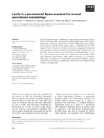
actin or GFP antibody. Figure 1A shows that actin
was specifically immunoprecipitated together with
hhLIM. To demonstrate that endogenous hhLIM and
actin can form a complex in vivo, actin was immuno-
precipitated from C2C12 cell lysates and the immuno-
precipitates were analyzed by western blot using
anti-hhLIM Ig. The data showed that actin was specifi-
cally immunoprecipitated together with endogenous
hhLIM, whereas protein A–agarose did not precipitate
hhLIM. Lysates were immunoprecipitated with
anti-hhLIM Ig and detected by anti-actin Ig, and
results showed the same specific interaction between
endogenous hhLIM and actin, which indicated that the
interaction of these two proteins is not an artifact of
hhLIM overexpression (Fig. 1B). The glutathione
S-transferase (GST) pull-down experiment also demon-
strated a direct interaction between GST–hhLIM and
actin. GST or GST–hhLIM fusion proteins were bound
to glutathione–Sepharose and incubated with purified
rabbit skeletal muscle actin or lysates from hhLIM-
expressing cells. After extensive washing, Sepharose
pellets were immunoblotted with anti-actin Ig to detect
actin in fusion protein or the pellets with anti-GST Ig
to demonstrate equal loading of fusion protein. As
shown in Fig. 1C, both purified actin and endogenous
actin bound to GST–hhLIM but not GST.
hhLIM bundles F-actin directly
In order to identify whether hhLIM and actin interact
directly, we investigated the activities of hhLIM bind-
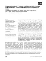
ing to actin using a co-sedimentation assay. Purified
F-actin was incubated with recombinant hhLIM pro-
tein, and pelleted by centrifugation at 10 000 g, which
allows pelleting of heavy, cross-linked F-actin only.
Controls for this series of experiments included
SM22a, a known actin cross-linking protein, and BSA,
which does not interact with or cross-link actin. In the
absence of hhLIM, the majority of actin remained in
the supernatant (S) and only a small amount was
detected in the pellets (P). The addition of hhLIM sig-
nificantly enhanced the amount of actin present in the
pellets (P) compared with samples with actin alone or
with the BSA control (Fig. 2A). These data indicated
that hhLIM binds to and has a bundling effect on
actin. Figure 2B shows that, in the absence of hhLIM,
20% of the total actin was detected in the pellet. By
contrast, in the presence of hhLIM, the amount of
B. Zheng et al. hhLIM binds to F-actin
FEBS Journal 275 (2008) 1568–1578 ª 2008 The Authors Journal compilation ª 2008 FEBS 1569
actin in the pellet increased along with the increment
of the hhLIM, indicating that hhLIM induces F-actin
bundling. Maximum actin bundling occurred when
molar ratios of hhLIM (2 lm) to actin (8 lm) were
> 1 : 4. Indeed, when the concentration of hhLIM
exceeded 4 lm, 60% of total actin was detected in the
pellet (Fig. 2B). Cumulative data from several indepen-
dent experiments demonstrated that the co-sediments
of hhLIM and F-actin was greater than that of actin
alone (Fig. 2C). In order to directly analyze the effect
of GST–hhLIM on actin filament bundling, we per-
formed electron microscopy on negatively stained actin
filaments. As shown in Fig. 2D, in the absence of
hhLIM, actin filaments formed a uniform meshwork
of fine filaments. The inclusion of BSA had no effect
on the ability to bundle actin, however, when actin
was polymerized in the presence of hhLIM, higher
order structures were observed. Although single actin
filaments were still present, most of the actin filaments
were recruited into thick and long actin bundles,
confirming the cross-linking activity of hhLIM. To
determine whether hhLIM also binds to monomeric
(G)-actin, GST pull-down assays were performed with
GST–hhLIM versus GST alone. Although actin was
pulled down with GST–hhLIM, there was no signifi-
cant difference between samples containing GST–
hhLIM and GST alone (Fig. 2E). Thus, this approach
suggests that hhLIM does not bind to monomeric
actin.
hhLIM stabilizes F-actin in C2C12 cells
To further determine whether hhLIM modulates the
actin cytoskeleton in C2C12 cells, we studied the
effects of hhLIM overexpression on the actin stress
fibers. Overexpression of hhLIM induced actin poly-
merization (data not shown). We have established that
overexpression of hhLIM may increase the expression
of actin [7]. The actin fractionation assay showed that
the F-actin fraction (csk) was increased compared with
the G-actin fraction (sol) in cells overexpressing
hhLIM. Silencing of hhLIM expression by siRNA had
the opposite result (Fig. 3A). If the expression of
GFP–hhLIM could increase actin filament bundling,
then GFP–hhLIM would be expected to redistribute
to the Triton X-100-insoluble cytoskeletal fraction. As
shown in Fig. 3B, the insoluble hhLIM fraction
increased with in a dose-related manner. So, we pre-
dicted that hhLIM might participate in F-actin forma-
tion and stabilization of actin filaments. In order to
test whether hhLIM could affect the stability of the
actin cytoskeleton following its ectopic expression in
C2C12 cells, actin depolymerization was induced by
cytochalasin B in hhLIM–GFP–transfected C2C12
cells. The actin cytoskeleton was visualized by TRITC–
phalloidin staining before adding cytochalasin B, and
(a)
A
B
C
(b)
IP with actin Ab
– +
hhLIM
Actin
hhLIM
IP with hhLIM Ab – +
Actin
hhLIM
Actin
pcDNA3-hhLIM + – –
– –
+
pEGFP-actin
+ +
IP with Myc Ab
GFP
Myc
GST GST-hhLIM
1 2
3
4
Actin
GST
Total lysates Total lysates
Fig. 1. Actin interacts with hhLIM in C2C12 cells. Coimmunopre-
cipitation of GFP-tagged actin with myc-tagged hhLIM. Lysates of
C2C12 cells transfected with full-length myc-tagged hhLIM and
GFP-tagged actin was immunoprecipitated (IP) by anti-myc Ig cou-
pled to Sepharose, and interacting proteins were separated by
SDS ⁄ PAGE and blotted with anti-GFP or anti-myc Ig. (B) (a) Cell
lysates of C2C12 cells were immunoprecipitated with anti-actin Ig
or protein A–agarose as indicated. Immunoprecipitates and total
lysates were analyzed by western blotting using anti-actin and anti-
hhLIM Ig; (b) cell lysates were immunoprecipitated with anti-hhLIM
Ig or protein A–agarose and detected using anti-hhLIM and anti-
actin Ig. Whole-cell extracts of each group were harvested as a
control to demonstrate proper expression of each protein. These
experiments were repeated three times. (C) GST pull-down assay.
Purified recombinant GST (lane 1) or GST–hhLIM fusion protein
(lanes 2–4) coupled to glutathione–Sepharose was incubated with
rabbit muscle actin (lane 2) cell extracts from C2C12 cells transfect-
ed with hhLIM expression plasmids (lanes 1 and 3) or transfected
with pcDNA plasmid (lane 4). After extensive washing, Sepharose
beads were analyzed by SDS ⁄ PAGE and immunoblotted using anti-
actin (upper) or anti-GST (lower) Ig. 1, Extracts from C2C12 cells
transfected with hhLIM expression plasmid; 2, rabbit muscle actin
protein; 3, extracts from C2C12 cells transfected with hhLIM
expression plasmid; 4, extracts from C2C12 cells transfected with
pcDNA plasmid.
hhLIM binds to F-actin B. Zheng et al.
1570 FEBS Journal 275 (2008) 1568–1578 ª 2008 The Authors Journal compilation ª 2008 FEBS
10 and 30 min after treatment (Fig. 3C). As early as
10 min after cytochalasin B application, partial
depolymerization of the actin cytoskeleton occurred in
nontransfected cells, whereas hhLIM-expressing cells
showed an unaffected actin network (data not
shown). After 30 min of treatment, most of the non-
transfected cells showed a fully depolymerized actin
cytoskeleton. By contrast, the morphology of hhLIM–
GFP-expressing cells remained normal, indicating that
the cytoskeleton was existent and supported the
S
P
S P S P S P S P
hhLIM + + – – – – – –
Actin + + + + + + + +
SM22α – – – – + + – –
BSA – – – – – – + +
Actin (8 µM)
hhLIM
Actin (8 µ
M)
hhLIM
0 0.25 0.5 1 2 4 8 16 µ
M
[hhLIM]
BSA
Actin
hhLIM
SM22α
(a)
(b) (c)
(d)
0
2
Actin
hhLIM + actin
SM22 + actin
BSA + actin
P/S ratio for actin
*
*
0
0.4
0.8
1.2
1.6
2
0 5 10 15 20
hhLIM
(µM)
hhLIM bound to F-actin
A
ct
in – + – +
Actin
GST
GST GST-hhLIM
A
B
D
C
E
Fig. 2. Functional interaction between hhLIM and F-actin. (A) Coomassie Brilliant Blue stained SDS ⁄ PAGE gel showing typical actin co-sedi-
mentation assay. hhLIM, SM22a or BSA were incubated with actin for 30 min in F-actin buffer containing ATP and Ca
2+
and then centri-
fuged at 10 000 g for 30 min. Proteins in the pellets (P) and supernatants (S) were analyzed by SDS ⁄ PAGE. Densitometry was performed to
determine the actin P ⁄ S ratios of three independent experiments to quantify the effect of hhLIM on actin sedimentation. *P < 0.05, com-
pared with the control. (B) Actin at 8 l
M alone or in the presence of different concentrations of hhLIM (0.25–16 lM) was polymerized and
centrifuged. Proteins in the pellets (P) and supernatants (S) were analyzed by SDS ⁄ PAGE and stained with Coomassie Brilliant Blue. (C)
Quantitation analysis for GST–hhLIM association with F-actin at different concentrations of GST–hhLIM. The F-actin concentration was 8 l
M.
After SDS ⁄ PAGE and staining, gels were scanned and the amount of protein that was present in the pellet and supernatant was quantified.
The concentration of actin-bound hhLIM was plotted against the concentration of free hhLIM. Values are means ± SEM for three indepen-
dent experiments. (D) Electron microscopy morphology of the filaments assembled from the GST–hhLIM-actin complex. Electron microscopy
of negatively stained actin filaments was performed with the following combinations of purified proteins: (a) 8 l
M actin and 2 lM GST–
hhLIM; (b) 8 l
M actin; (c) 8 lM actin and 2 lM SM22a; (d) 8 lM actin and 2 lM BSA. Bar = 70 nm. (E) In vitro binding analysis using nono-
meric (G) actin and GST or GST–hhLIM bound to glutathione agarose beads. Western blot of GST pull-down assay fractions using an actin
antibody showing similar amounts of actin in samples with GST Sepharose versus GST-tagged hhLIM. As expected, no signal was detected
in the absence of G-actin. Similar results were obtained in three independent experiments.
B. Zheng et al. hhLIM binds to F-actin
FEBS Journal 275 (2008) 1568–1578 ª 2008 The Authors Journal compilation ª 2008 FEBS 1571
appearance of the cell (Fig. 3C). Finally, 120 min after
cytochalasin B application, almost all the hhLIM-
expressing cells presented a fully disrupted actin cyto-
skeleton (data not shown). In order to test this further,
C2C12 cells were treated with cytochalasin B and phal-
loidin for 30 min, and the distribution of hhLIM and
actin in the soluble (sol) and cytoskeleton (csk) frac-
tions was determined by western blotting. As shown in
Con hhLIM hhLIM (–)
Actin (sol)
Actin (csk)
hhLIM (sol)
GAPDH
(csk)
hhLIM actin Merged actin
hhLIM actin Merged actin
hhLIM actin Merged actin
hhLIM
Cytochalasin B
Pholloidin
Con
pcDNA-hhLIM
Sol csk sol csk sol csk sol csk
0
2
4
6
8
10
34 56
csk/sol
pcDNA-hhLIM
hhLIM
(sol)
hhLIM
(csk)
Actin (sol)
Actin (csk)
Con CB Phalloidin
A
B
C
(a) (b) (c) (d)
(h)(g)(f)(e)
(i) (j) (k) (l)
D
Fig. 3. hhLIM stabilizes F-actin in C2C12
cells. Extracts from C2C12 cells transfected
with pcDNA, pcDNA
3
–hhLIM or hhLIM
siRNA expression plasmids were separated
into cytosolic soluble (sol) and cytoskeleton-
associated proteins (csk). Equal amounts
were separated by SDS ⁄ PAGE and proteins
in each fraction were detected by immuno-
blotting by using anti-actin or anti-hhLIM Ig.
(B) C2C12 cells transfected with 0.5, 1, or
1.5 lg of hhLIM expression plasmid were
lysed, and cytosolic soluble (sol) and cyto-
skeleton-associated proteins (csk) were sep-
arated for analysis. Left, a representative
result from three independent experiments
is shown. Right, the density of specific band
of csk ⁄ sol was scanned and quantified.
(C) hhLIM delayed the effect of cytochala-
sin B on C2C12 cells. (a–c) C2C12 cells
transfected with pEGFP–hhLIM were trea-
ted with cytochalasin B for 30 min;
(d) C2C12 cells were treated with cytochala-
sin B for 30 min; (e–g) C2C12 cells trans-
fected with pEGFP–hhLIM were treated
with phalloidin for 30 min; (h) C2C12 cells
were treated with phalloidin for 30 min;
(i–k) C2C12 cells transfected with pEGFP–
hhLIM; (l) C2C12 cells transfected with
pEGFP. (D) C2C12 cells were treated with
cytochalasin B or phalloidin for 30 min and
lysed by lysis buffer and separated into
cytosolic soluble (sol) and cytoskeleton-
associated proteins (csk). Equal amount
were separated by SDS ⁄ PAGE and proteins
in each fraction were detected by immuno-
blotting by using anti-actin or anti-hhLIM Ig.
hhLIM binds to F-actin B. Zheng et al.
1572 FEBS Journal 275 (2008) 1568–1578 ª 2008 The Authors Journal compilation ª 2008 FEBS
Fig. 3D, cytochalasin B led to the release of hhLIM
from the insoluble fractions. This was consistent with
the result of immunofluorescence analysis, indicating
that hhLIM participates in actin polymerization
(Fig. 3C). However, pelletable hhLIM from
phalloidin-treated C2C12 cells was increased by 100%.
Together, these results indicate that modulation of
the actin cytoskeleton induces changes in hhLIM
localization.
LIM domain 2 of hhLIM mediates the interaction
between hhLIM and actin
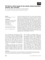
hhLIM has two LIM domains. To identify which
domains or sites of hhLIM interact with actin, a series
of truncated mutants was constructed and a GST pull-
down assay was used. This showed that the F4 region
(amino acids 41–194), which contains the LIM
domain 2, has almost the same activity as full-length
hhLIM. Interestingly, although the F5 region (amino
acids 41–154), with the C-terminus of the F4 region
deleted, is still able to interact with actin, binding
activity is decreased compared with the F4 region. By
contrast, the F3 fragment (amino acids 1–120), with
the C-terminus of hhLIM deleted, is not able to inter-
act with actin. The data suggest that hhLIM binding
to actin requires a functional LIM domain 2 (Fig. 4A).
To further characterize that LIM domain 2 is sufficient
to interact with actin, the LIM domain was function-
ally destroyed by replacing cysteine with serine in
either domain 1 (mLIM1) or 2 (mLIM2), and an
in vitro GST pull-down assay was used. Figure 4B
shows that F-actin was pulled down by full-length
hhLIM and mutant mLIM1, indicating an interaction,
whereas mLIM2 did not pellet with actin. To further
identify the LIM domain that mediates the interaction
of hhLIM with actin, we transfected C2C12 cells with
GFP-tagged full-length hhLIM or GFP-tagged LIM
domain-mutated constructs and detected the distribu-
tion of hhLIM. The results revealed that mLIM2 is
mainly diffused and fuzzily distributed (Fig. 4C). To
characterize further the interaction between hhLIM
mutants and actin, co-sedimentation assays were per-
formed using purified actin and GST–mLIM1or GST–
mLIM2 protein. As shown in Fig. 4D, full-length
hhLIM and hhLIM mutants co-sedimented with
F-actin, but the amount of sedimented actin is lower
in the presence of mLIM2 than in the presence of
mLIM1 or full-length hhLIM. Importantly, mutation
of LIM domain 2 dramatically affected the contraction
of the C2C12 cells compared with cells expressing
hhLIM, which may underlie the dysfunction (Fig. 4E).
Taking these factors together, we determined that tar-
geted disruption of the second LIM domain of hhLIM
destroys the interaction between hhLIM and the con-
tractive ability of C2C12 cells, indicating the important
role that LIM domain 2 plays in controlling assembly
and organization of the actin cytoskeleton.
Discussion
The plasticity of the actin cytoskeleton relies mainly
on the ability of actin filaments to form, branch, bun-
dle, and disassemble within short timeframes in
response to many signals. LIM proteins play a critical
role in the organization of the actin cytoskeleton.
WLIM1 was found both to associate with the actin
cytoskeleton in a very dynamic manner and to circu-
late rapidly throughout the cytoplasm, making it avail-
able wherever and whenever it was needed for new
actin bundle formation [1,14]. WLIM1 protein con-
tains two LIM domains, deletion of one of the
domains reduced significantly, but did not entirely
abolish, the ability of WLIM1 to bind actin filaments.
Variants lacking the C-terminal or inter-LIM domain
were only weakly affected in their F-actin stabilizing
and bundling activities, and trigger the formation of
thick cables containing tightly packed actin filaments
as does the native protein. By contrast, deletion of one
of the two LIM domains negatively impacted both
activities and resulted in the formation of thinner and
wavier cables [13]. Zyxin-related protein 1, which
belongs to a family of LIM-containing proteins that
includes zyxin and lipoma-preferred partner, partici-
pates in the organization of the actin cytoskeleton [15].
FHL2 was observed, along with F-actin, to be
involved in the focal adhesion of C2C12 and H9C2
myotubes [16]. Overexpression of FHL2 promotes
differentiation by binding to b-catenin [17]. FHL3 reg-
ulates a-actinin-mediated actin bundling as an actin-
binding protein [18]. CRP3 (also called muscle LIM
protein–MLP) plays an important role in myogenesis
and in the promotion of myogenic differentiation. This
function has been related to its myofibrillar location in
close vicinity to the Z disk and its interaction with
a-actinin. MLP is highly expressed during differentia-
tion in all types of striated muscle, but its expression
in the adult is restricted to cardiac and slow-twitch
fibers of skeletal muscle [8,19]. Moreover, it has been
reported that targeted deletion of MLP in mice causes
marked disruption of the myocardial cytoarchitecture,
leading to dilated cardiomyopathy and death resulting
from cardiac failure [10,20,21]. Despite the dramatic
consequences associated with loss of MLP expression,
the mechanistic details of CRP function in muscles
remain speculative. The data presented here identify a
B. Zheng et al. hhLIM binds to F-actin
FEBS Journal 275 (2008) 1568–1578 ª 2008 The Authors Journal compilation ª 2008 FEBS 1573
member of the CRP family, hhLIM, as a new F-actin-
binding protein whose targeting of actin filaments
stabilizes the actin cytoskeleton and promotes actin
bundle ⁄ cable formation. We conclude that hhLIMs are
real F-actin-binding protein on the following observa-
tions: (a) hhLIM colocalized with F-actin, (b) hhLIM
showed F-actin-binding activity, and (c) hhLIM co-
sedimented with F-actin. The interaction between
hhLIM and actin filaments was previously believed to
be indirect, requiring intermediary proteins such as
a-actinin or zyxin. However, it is clearly established
that hhLIM and other members of the CRP family
are autonomous F-actin-binding proteins. Our in vitro
investigations provide, for the first time, strong aug-
ments supporting the idea that the LIM domain parti-
cipates in the F-actin binding and bundling activities
displayed by hhLIM.
Confocal analyses showed that hhLIM accumulates
in both the nucleus and the cytoplasm, where it pre-
dominantly associates with the actin cytoskeleton [7].
This dual location is in agreement with that reported
previously for members of the CRP family and other
CRP-related proteins, such as MLP [22]. Although
CRPs were first believed to interact indirectly with the
actin cytoskeleton via intermediary proteins, such as
zyxin and a-actinin, recent studies have shown that
CRP1 and CRP2 are autonomous actin-binding pro-
teins [11,23]. Our in vitro results extend this property
to the hhLIM protein, suggesting that all CRPs and
CRP-related proteins have the ability to associate with
F-actin. Here, we demonstrate the ability of a new LIM
protein to interact with F-actin in a direct manner.
Formation of higher order actin structures, such as
bundles and cables, is crucial to stabilize the organiza-
tion of transvacuolar strands and maintain overall
cellular architecture. As mentioned above, CRP1 may
participate in the formation and ⁄ or maintenance of
long actin cables [12]. Consistent with this hypothesis,
we observed that ectopic expression of hhLIM in
C2C12 cells stabilizes actin filaments ⁄ bundles against
cytochalasin B. In addition, overexpression of hhLIM
in C2C12 cells induces an increase in the overall
amounts of actin and F-actin. This prompted us to
investigate whether hhLIM stabilizes and bundles actin
filaments directly. In vitro cytochalasin B experiments
demonstrated that hhLIM stabilizes F-actin by itself.
In addition, co-sedimentation assays and the direct
observation of in vitro actin filaments that have been
polymerized in the presence of hhLIM demonstrated
that hhLIM bundles actin filaments in an autonomous
manner.
hhLIM consists of two LIM domains. Targeted dis-
ruption of the second LIM domain of hhLIM abol-
ished F-actin-binding activity, indicating the important
role that LIM domain 2 plays in the control of assem-
bly and organization of the actin cytoskeleton.
In conclusion, in vitro results show that hhLIM inter-
acts with filamentous actin in a direct manner. hhLIM
enhances the stability of the actin cytoskeleton and pro-
motes actin bundling. Although the exact contribution
made by hhLIM protein to actin cytoskeleton dynam-
ics ⁄ remodeling remains to be explored, the data pro-
vide strong evidence that hhLIM is an actin
cytoskeleton organizer. An open question is the signifi-
cance of hhLIM in the nucleus. Several LIM proteins
have been shown to shuttle between the cytoplasm and
the nucleus and it has been suggested that they mediate
communication between both compartments. Similar
functions for hhLIM proteins cannot be excluded. Con-
sistent with a nuclear role for hhLIM, it has been
Fig. 4. Relationship between the structure and the activation activity of hhLIM. (A) Requirement of the C-terminal half of hhLIM for association
activity with actin. hhLIM and its various derivatives were constructed into PGEX-3X plasmids. GST–hhLIM and its derivative proteins are sche-
matically depicted on the left. Association activities of hhLIM and its derivatives are represented on the right. Extracts from C2C12 cells were
precleared with GST–Sepharose beads and then incubated with GST–hhLIM Sepharose beads or its derivative proteins. Pellets were washed,
and interacting proteins were separated by SDS ⁄ PAGE and identified by western blotting. (B) Mutation of LIM domain 2 of hhLIM disrupts
the association with actin. Extracts from C2C12 cells were precleared with GST–Sepharose beads and then incubated with GST–hhLIM Sepha-
rose beads, or LIM domain-mutated (mLIM1, GST-mLIM110Cys fi Ser, 13Cys fi Ser, mLIM2, GST-mLIM2120Cys fi Ser, 123Cys fi Ser)
Sepharose beads or GST–Sepharose beads. Pellets were washed, and interacting proteins were separated by SDS ⁄ PAGE and identified
by western blotting. (C) Fluorescence analysis of hhLIM in the C2C12 cells. C2C12 cells were transfected with pEGFP–hhLIM, pEGFP–
mLIM1(10Cys fi Ser, 13Cys fi Ser), pEGFP-mLIM2(120Cys fi Ser, 123Cys fi Ser) or pEGFP. The cells were fixed and examined with an
IX71 fluorescence microscope (Olympus). (D) Actin co-sedimentation assay verified the functional interaction between hhLIM and F-actin.
Purified F-actin was incubated with GST–hhLIM or LIM domain-mutated hhLIM. Cross-linked F-actin was pelleted by centrifugation, separated
by SDS ⁄ PAGE, and stained with Coomassie Brilliant Blue. (E) Densitometry micrograph was obtained of the agonist-induced contraction of
C2C12 cells. C2C12 cells were transfected with pEGFP (control), pEGFP–hhLIM, pEGFP–mLIM1(10Cys fi Ser, 13Cys fi Ser) or pEGFP–
mLIM2(120Cys fi Ser, 123Cys fi Ser) and maintained in physiological rodent saline (138 m
M NaCl, 2.7 mM KCl, 1.8 mM CaCl
2
, 1.06 mM
MgCl
2
, 12.4 mM HEPES, and 5.6 mM glucose, pH 7.3) in a chamber ( 2 mL) mounted on the stage of an inverted microscope. The C2C12 cell
length was modified by acetylcholine stimulation (100 l
M). *P < 0.05, compared with C2C12 cells transfected with pcDNA
3
–hhLIM plasmid.
hhLIM binds to F-actin B. Zheng et al.
1574 FEBS Journal 275 (2008) 1568–1578 ª 2008 The Authors Journal compilation ª 2008 FEBS
reported to activate brain natriuretic factor (BNP) and
atrial natriuretic factor (ANF) gene expression [7,24].
Identification of further regulatory mechanisms that
trigger the translocation of hhLIM between the cyto-
plasm and the nucleus is an important goal for the
future. Perhaps the most fruitful area of future research
in LIM biology will involve dissecting the precise roles
of LIM proteins in both the nuclear and cytoplasmic
1 10 41 120 154 194 aa
Actin
GST
F1 F2 F3 F4 F5 Neg
F1
F2
F3
F4
F5
LIM zinc-binding domain
Neg mLIM1 LIM mLIM2
Actin
GST
P P
P
P
1 10 31 41 95 111 117 120 154 160 194
Zinc finger C2H2 type domain
LIM zinc-binding domain
Protein kinase C phosphorylation site
P
S P S P S P S P S P
mLIM1 + + – – – – – – – –
mLIM2
– – + + – – – – – –
hhLIM – – – – + + – – – –
Actin + + + + + + + + + +
SM222
– – – – – – – – + +
Actin
*
0
1
2
3
con hhLIM mLIM1 mLIM2
cell contraction (um)
A
B
C
D
pEGFP-hhLIM pEGFP-mLIM1 pEGFP-mLIM2 pEGFP
E
B. Zheng et al. hhLIM binds to F-actin
FEBS Journal 275 (2008) 1568–1578 ª 2008 The Authors Journal compilation ª 2008 FEBS 1575
compartments, and deciphering how the role of a LIM
protein that is associated with actin filaments might be
integrated with nuclear functions and vice versa.
Experimental procedures
Cell culture and transfection
The C2C12 mouse myoblast line was maintained with
Dulbecco’s modified Eagle’s medium with 10% fetal bovine
serum. Differentiation was induced in C2C12 cells by
replacing medium with Dulbecco’s modified Eagle’s med-
ium containing 2% horse serum. hhLIM expression plasmid
was gift from KH Chen (National Institute on Aging, Balti-
more, MD, USA). A hhLIM siRNA-expressing plasmid
was constructed using BLOCK-iTÔ U6 RNAi Vector
by subcloning double-stranded oligonucleotides comple-
mentary (5¢-CACCGCAGTGCCATGGAAGGAGTTTC
CACACGAATGTGGAAACTCCTTCCATGGCACTG-3¢)
according to the manufacture’s protocol (Invitrogen, Carls-
bad, CA, USA). Transfections with various DNA con-
structs were performed with lipofectamine 2000 (Invitrogen)
according to the manufacturer’s instructions.
Immunoprecipitation and western blotting
C2C12 cells grown in Dulbecco’s modified Eagle’s medium,
supplemented with 10% fetal bovine serum were transfected
with cDNA constructs using Lipofectamine according to
the manufacturer’s protocol. Forty-eight hours later, cells
were lysed in lysis buffer [20 mm Tris, pH 7.5, 150 mm
NaCl, 1 mm EGTA, 1 mm EDTA, 1% Triton X-100, pro-
tease inhibitor mixture (Sigma, St Louis, MO, USA), and
1 lm Na
3
VO
4
). Lysates were sonicated on ice, and cell deb-
ris was removed by centrifugation. Lysates were precleared
with protein A ⁄ G–agarose beads (Santa Cruz Biotechnolo-
gies, Santa Cruz, CA, USA), and the proteins were immu-
noprecipitated with the appropriate antibody overnight at
4 °C followed by incubation with protein A ⁄ G–agarose for
1 h at 4 °C. Immunoprecipitates were washed three times
with lysis buffer, and proteins were separated on
SDS ⁄ PAGE. Immunoblotting analysis was performed as
described previously [25–28]. Primary antibodies used for
the assays were anti-GST polyclonal Ig (1 : 500; Santa
Cruz), anti-hhLIM polyclonal Ig (gift of KH Cheng,
National Institute on Aging, Baltimore, MD), anti-GFP
polyclonal Ig (1 : 500; Santa Cruz), and anti-(skeletal
a-actin) polyclonal Ig (1 : 500; Santa Cruz).
Site-directed mutagenesis of the LIM domain
of hhLIM
Site-directed mutation of each LIM domain was carried out
by PCR using oligonucleotide primers that coded for the
appropriate point substitutions of amino acids. The reac-
tions were carried out using a QuikChange site-directed
mutagenesis kit (Stratagene, La Jolla, CA, USA). Each
mutation was verified by DNA sequence analysis. PCR
primers used in the site-directed mutagenesis of the LIM
domain of hhLIM introduced two point mutations into each
LIM domain: LIM1(10Cys fi Ser, 13Cys fi Ser):5¢-GGA
GGCGCAAAATCTGGAGCCTCTGAAAAGACCGTCTA
C-3¢;LIM2(120Cysfi Ser, 123Cys fi Ser): 5¢-GAGAGTCC
GAGAAGTCCCCTCGATCT GGCAAGTC AGTCTAT G-3¢.
Actin fractionation
Cells were scraped, washed with NaCl ⁄ P
i
, and lysed in buf-
fer A (20 mm Tris ⁄ HCl, pH 7.5, 1% Triton X-100, 5 mm
EGTA, 1 mm phenylmethylsulfonyl fluoride) on ice for
30 min, and then centrifuged at 12 000 g and 4 °C for
30 min. The supernatants (sol) were harvested. The pellets
(csk) were lysed in buffer B (10 mm Tris ⁄ HCl, pH 7.5,
150 mm NaCl, 1% Triton X-100, 0.1% SDS, 1 mm sodium
deoxycholate, 2 mm EGTA, 1 mm phenylmethylsulfonyl
fluoride) on ice for 30 min, and then centrifuged at
12 000 g for 30 min. The supernatants from the lysed pel-
lets (csk) were harvested. Protein concentration was deter-
mined by a modified Lowry protein assay. Equal amounts
of the supernatant (sol) and pellet (csk) were separated by
10% SDS ⁄ PAGE and stained with an antibody against
hhLIM or actin, with visualization by secondary antibodies
and enhanced chemiluminescence [29,30].
Fluorescence staining
Fluorescence staining was performed as described previ-
ously [12,31]. The cells were stained for 20 min with
TRITC ⁄ phalloidin (1 lgÆmL
)1
) in blocking solution (1%
BSA and 0.1% Triton X-100 in NaCl ⁄ P
i
) in the dark at
room temperature to localize F-actin.
GST pull-down assay
In order to produce GST fusion proteins, full-length and
domain-specific regions of hhLIM were generated in a
pGEX-3X vector inframe with the N-terminal GST tag. All
new constructs were confirmed by restriction digestion fol-
lowed by sequencing. Protein expression was induced by
reaction with 0.2 mm isopropyl thio-b-d-galactoside at
30 °C for 3 h. Bacterial lysates were purified over glutathi-
one–agarose. For the pull-down assay, cell lysate was pre-
pared by lysing the C2C12 cells transiently transfected with
myc-tagged different mutant or site-directed mutagenesis
hhLIM that had been precleared with GST Sepharose
beads. Assay mixtures were then incubated with GST
Sepharose beads or with hhLIM ⁄ GST Sepharose beads.
After centrifugation, the pellets were washed, and the
hhLIM binds to F-actin B. Zheng et al.
1576 FEBS Journal 275 (2008) 1568–1578 ª 2008 The Authors Journal compilation ª 2008 FEBS
interacting proteins were separated by SDS ⁄ PAGE and
identified by western blot with an anti-actin Ig [32].
Assay for low-speed co-sedimentation of hhLIM
with F-actin
G-Actin (Sigma) was polymerized by incubation at room
temperature for 30 min in a polymerization buffer (20 mm
imidazole ⁄ Cl, pH 7.0, 2 mm MgCl
2
,1mm ATP, 0.5 mm
dithiothreitol, 90 mm KCl). The lysates of the hhLIM-
expressing cells were centrifuged at 10 000 g for 30 min,
and the supernatant was used for the assay (F-actin). The
supernatant of the lysates was incubated at room tempera-
ture for 30 min with 0.3 mgÆmL
)1
F-actin in a solution con-
taining 25 mm imidazole ⁄ Cl, pH 7.0, 2 mm MgCl
2
,1mm
ATP, 0.5 mm dithiothreitol, 27 m m KCl and 100 mm NaCl,
and the mixture (50 lL) was placed over a 50 lL cushion
of 30% sucrose in the polymerization buffer. After the sam-
ple was centrifuged at 10 000 g for 20 min, the supernatant
and the pellet were subjected to SDS ⁄ PAGE, followed by
western blot analysis using the anti-hhLIM and anti-actin
Ig [33–35].
Electron microscopy
Actin (8 lm) was polymerized at room temperature. The
actin mixtures were then diluted 1 ⁄ 8 with Mg-ATP buffer
in the presence of purified GST–hhLIM (2 lm) alone or
with BSA in a final reaction volume of 25 lL. These mix-
tures were incubated for 1 h at room temperature. The pro-
tein mixtures were adsorbed onto carbon-coated 400-mesh
grids for 1 min. Actin filaments were negatively stained
with 2% phosphotungstic acid, pH 7.4, for 15 s. Grids were
visualized using transmission electron microscopy (Hitachi
Ltd., Saitama, Japan) at an accelerating voltage of 80 kV
and a nominal magnification of ·100 000 [18].
Measurement of contraction
C2C12 cells were transfected with pcDNA
3
(control),
pcDNA
3
–hhLIM, pcDNA
3
–mLIM1(10Cys fi Ser, 13Cys fi
Ser) or pcDNA
3
–mLIM2(120Cys fi Ser, 123Cys fi Ser)
and maintained in physiological rodent saline (138 mm
NaCl, 2.7 mm KCl, 1.8 mm CaCl
2
, 1.06 mm MgCl
2
,
12.4 mm Hepes, and 5.6 mm glucose, pH 7.3) in a chamber
( 2 mL) mounted on the stage of an inverted microscope
(Olympus, Tokyo, Japan). The C2C12 cell length was
modified by acetylcholine stimulation (100 lm) for 1 min
[25,36,37].
Statistical analysis
To control for day-to-day variations in staining intensity,
untreated cells were always compared with treated cells on
the same microscope slide because cells on the same slide
undergo identical culture, fixation, permeabilization, stain-
ing and microscopy conditions, allowing meaningful com-
parisons between samples. All data are presented as
means ± SE.
Acknowledgements
We thank Dr Da-zhi Wang (University of North Caro-
lina) for helpful discussions and comments on the
manuscript. This work was supported by the Program
for New Century Excellent Talents in University
(No. NCET-05-0261), a Key Project of the Chinese
Ministry of Education (No.206016), the National Nat-
ural Science Foundation of the People’s Republic of
China (No.30300132, 30570661) and the Major State
Basic Research Development Program of China (No.
2005CCA03100).
References
1 Thomas C, Hoffmann C, Dieterle M, Van Troys M,
Ampe C & Steinmetz A (2006) Tobacco WLIM1 is a
novel F-actin binding protein involved in actin cytoskel-
eton remodeling. Plant Cell 18, 194–2206.
2 Kadrmas JL & Beckerle MC (2004) The LIM domain:
from the cytoskeleton to the nucleus. Nat Rev Mol Cell
Biol 5, 920–931.
3 Zhi S, Yao A, Zubair I, Sugishita K, Ritter M, Li F,
Hunter JJ, Chien KR & Barry WH (2001) Effects of
deletion of muscle LIM protein on myocyte function.
Am J Physiol Heart Circ Physiol 280, H2665–H2673.
4 Zheng B, Wen JK & Han M (2003) Factors involved in
the cardiac hypertrophy. Biochemistry Mosc 68, 650–657.
5 Bach I (2000) The LIM domain: regulation by associa-
tion. Mech Dev 91, 5–17.
6 Chen H, Zhou Z, Zhang JF & Zhou AR (2000) Screen
heart-specific growth genes using three-element PCR-
select cDNA subtraction. Chin J Biochem Mol Biol 16,
295–300.
7 Zheng B, Wen JK, Han M & Zhou AR (2004) hhLIM
protein is involved in cardiac hypertrophy. Biochim
Biophys Acta 1690, 1–10.
8 Arber S, Halder G & Caroni P (1994) Muscle LIM pro-
tein, a novel essential regulator of myogenesis, promotes
myogenic differentiation. Cell 79, 221–231.
9 Bourgouin C, Lundgren SE & Thomas IB (1992) Apte-
rousis a Drosophila LIM domain gene required for the
development of a subset of embryonic muscles. Neuron
9, 549–561.
10 Arber S & Caroni P (1996) Specificity of single LIM
motifs in targeting and LIM ⁄ LIM interactions in situ.
Gene Dev 10, 289–300.
B. Zheng et al. hhLIM binds to F-actin
FEBS Journal 275 (2008) 1568–1578 ª 2008 The Authors Journal compilation ª 2008 FEBS 1577
11 Grubinger M & Gimona M (2004) CRP2 is an autono-
mous actin-binding protein. FEBS Lett 557, 88–92.
12 Tran TC, Singleton CA, Fraley TS & Greenwood JA
(2005) Cysteine-rich protein 1 CRP1 regulates actin fila-
ment bundling. BMC Cell Biol 6, 45–58.
13 Thomas C, Moreau F, Dieterle M, Hoffmann C, Gatti S,
Hofmann C, Troys MV, Ampe C & Steinmetz A (2007)
The LIM domains of WLIM1 define a new class of actin
bundling modules. J Biol Chem 282, 33599–33608.
14 Ketelaar T, Anthony RG & Hussey PJ (2004) Green
fluorescent protein–mTalin causes defects in actin orga-
nization and cell expansion in Arabidopsis and inhibits
actin depolymerizing factor’s actin depolymerizing activ-
ity in vitro. Plant Physiol 136, 3990–3998.
15 Sanz-Rodriguez F, Guerrero-Esteo M, Botella LM,
Banville D, Vary CP & Bernabeu C (2004) Endoglin
regulates cytoskeletal organization through binding to
ZRP-1 a member of the Lim family of proteins. J Biol
Chem 279, 32858–32868.
16 Li HY, Kotaka M, Kostin S, Lee SM, Kok LD, Chan
KK, Tsui SK, Schaper J, Zimmermann R, Lee CY
et al. (2001) Translocation of a human focal adhesion
LIM-only protein FHL2 during myofibrillogenesis and
identification of LIM2 as the principal determinant of
FHL2 focal adhesion localization. Cell Motil Cytoskelet
48, 11–23.
17 Martin B, Schneider R, Janetzky S, Waibler Z, Pandur
P, Kuhl M, Behrens J, von der Mark K, Starzinski-Po-
witz A & Wixler V (2002) The LIM-only protein FHL2
interacts with b-catenin and promotes differentiation of
mouse myoblasts. J Cell Biol 159, 113–122.
18 Coghill ID, Brown S, Cottle DL, McGrath MJ, Robin-
son PA, Nandurkar HH, Dyson JM & Mitchell CA
(2003) FHL3 is an actin-binding protein that regulates
a-actinin-mediated actin bundling. J Biol Chem 278,
24139–24152.
19 Schneider AG, Sultan KR & Pette D (1999) Muscle
LIM protein: expressed in slow muscle and induced in
fast muscle by enhanced contractile activity. Am J
Physiol Cell Physiol 276, C900–C906.
20 Arber S, Hunter JJ, Ross JJ, Hongo M, Sansig G, Borg
J, Perriard JC, Chien KR & Caroni P (1997) MLP-defi-
cient mice exhibit a disruption of cardiac cytoarchitec-
tural organization dilated cardiomyopathy and heart
failure. Cell 88, 393–403.
21 Flick MJ & Konieczny SF (2000) The muscle regulatory
and structural protein MLP is a cytoskeletal binding
partner of betaI-spectrin. J Cell Sci 113, 1553–1564.
22 Han M, Wen JK & Zheng B (2005) Roles of LIM pro-
teins in cardiac hypertrophy. Future Cardiol 1, 319–329.
23 Pomies P, Louis HA & Beckerle MC (1997) CRP1 a
LIM domain protein implicated in muscle differentiation
interacts with alpha-actinin. J Cell Biol 139, 157–168.
24 Zheng B, Han M, Wen JK & Zhang R (2008) Human
heart LIM protein activates atrial-natriuretic-factor
gene expression by interacting with the cardiac-
restricted transcription factor Nkx2.5. Biochem J 409,
683–690.
25 Han M, Wen JK, Zheng B, Cheng Y & Zhang C (2006)
Serum deprivation results in redifferentiation of human
umbilical vascular smooth muscle cells. Am J Physiol
Cell Physiol 291, C50–C58.
26 Zheng B, Wen JK & Han M (2006) hhLIM is involved
in cardiomyogenesis of embryonic stem cells. Biochem
Mosc 71, S71–S76.
27 Jiang GJ, Han M, Zheng B & Wen JK (2006) Hyperpla-
sia suppressor gene associates with smooth muscle
alpha-actin and is involved in the redifferentiation of
vascular smooth muscle cells. Heart Vessels 21, 315–320.
28 Gorovoy M, Niu J, Bernard O, Profirovic J, Minshall
R, Neamu R & Voyno-Yasenetskaya T (2005) LIM
kinase 1 coordinates microtubule stability and actin
polymerization in human endothelial cells. J Biol Chem
280, 26533–26542.
29 Posern G, Sotiropoulos A & Treisman R (2002) Mutant
actins demonstrate a role for unpolymerized actin in
control of transcription by serum response factor. Mol
Biol Cell 13, 4167–4178.
30 Ballestrem C, Wehrle-Haller B & Imhof BA (1998)
Actin dynamics in living mammalian cells. J Cell Sci
111, 1649–1658.
31 Jiang GJ, Pan L, Huang XY, Han M, Wen JK & Sun
FZ (2005) Expression of HSG is essential for mouse
blastocyst formation. Biochem Biophys Res Commun
335, 351–355.
32 Akazawa H, Kudoh S, Mochizuki N, Takekoshi N,
Takano H, Nagai T & Komuro I (2004) A novel LIM
protein Cal promotes cardiac differentiation by associa-
tion with CSX ⁄ NKX2-5. J Cell Biol 164, 395–405.
33 Autieri MV, Kelemen SE & Wendt KW (2003) AIF-1 is
an actin-polymerizing and Rac1-activating protein that
promotes vascular smooth muscle cell migration. Circ
Res 92, 1107–1114.
34 Chew CS, Chen XS, Parntte JA, Tarrer S, Okamoto C
& Qin HY (2002) Lasp-1 binds to non-muscle F-actin
in vitro and is localized within multiple sites of dynamic
actin assembly in vivo. J Cell Sci 115, 4787–4799.
35 Cai L, Marhov AM & Bear JM (2007) F-actin binding
is essential for coronin 1B function in vivo. J Cell Sci
120, 1779–1790.
36 Han P, Suizu T, Grounds MD & Bakkerl AJ (2003)
Effect of indomethacin on force responses and sarco-
plasmic reticulum function in skinned skeletal muscle
fibers and cytosolic [Ca
2+
] in myotubes. Am J Physiol
Cell Physiol 285, C881–C890.
37 Hopf FW, Reddy P, Hong J & Steinhardt RA (1996) A
capacitative calcium current in cultured skeletal muscle
cells is mediated by the calcium-specific leak channel
and inhibited by dihydropyridine compounds. J Biol
Chem 271, 22358–22367.
hhLIM binds to F-actin B. Zheng et al.
1578 FEBS Journal 275 (2008) 1568–1578 ª 2008 The Authors Journal compilation ª 2008 FEBS