Báo cáo y học: " Vgf is a novel biomarker associated with muscle weakness in amyotrophic lateral sclerosis (ALS), with a potential role in disease pathogenesis"
Bạn đang xem bản rút gọn của tài liệu. Xem và tải ngay bản đầy đủ của tài liệu tại đây (622.08 KB, 8 trang )
Int. J. Med. Sci. 2008, 5
92
International Journal of Medical Sciences
ISSN 1449-1907 www.medsci.org 2008 5(2):92-99
© Ivyspring International Publisher. All rights reserved
Research Paper
Vgf is a novel biomarker associated with muscle weakness in amyotrophic
lateral sclerosis (ALS), with a potential role in disease pathogenesis
Zhong Zhao
1,2
, Dale J. Lange
1,3
,Lap Ho
1,2
, Sara Bonini
1,2
, Belinda Shao
1,2
, Stephen R. Salton
4,5
, Sunil Tho-
mas
1,2
, and Giulio Maria Pasinetti
1,2,4,5
1. James J. Peters Veterans Affairs Medical Center, Bronx, NY 10468
2. Departments of Psychiatry, Mount Sinai School of Medicine, New York, NY-10029
3. Departments of Neurology, Mount Sinai School of Medicine, New York, NY-10029
4. Departments of Neuroscience, Mount Sinai School of Medicine, New York, NY-10029
5. Departments of Geriatrics, Mount Sinai School of Medicine, New York, NY-10029
Correspondence to: Dr. Giulio Maria Pasinetti, Mount Sinai School of Medicine, Department of Psychiatry, One Gustave L., Levy Place,
Box 1668, New York, NY-10029. Email:
Received: 2008.02.25; Accepted: 2008.04.12; Published: 2008.04.15
Amyotrophic lateral sclerosis (ALS) is a progressive neurodegenerative disease that affects nerve cells in the
brain and the spinal cord. Previous proteomic evidence revealed that the content of certain peptide fragments
including Vgf-derived peptide aa 398-411 (Vgf
398-411
) of the precursor Vgf protein in the cerebral spinal fluid (CSF)
correctly identified patients with ALS from normal and disease controls. Using quantitative ELISA immunoassay
we found that the CSF levels of Vgf decreases with muscle weakness in patients with ALS. In SOD1 G93A
transgenic mice, loss of full-length Vgf content in CSF, serum and in SMI-32 immunopositive spinal cord motor
neurons is noted in asymptomatic animals (approximately 75 days old) and continues to show a progressive de-
cline as animals weaken. In vitro studies show that viral-mediated exogenous Vgf expression in primary mixed
spinal cord neuron cultures attenuates excitotoxic injury. Thus, while Vgf may be a reliable biomarker of pro-
gression of muscle weakness in patients with ALS, restoration of Vgf expression in spinal cord motor neurons
may therapeutically rescue spinal cord motorneurons against excitotoxic injury.
Key words: VGF, ALS, biomarker, excitotoxicity, SOD-1 mice, disease progression
Introduction
Vgf nerve growth factor inducible peptide, a non
acronymic peptide is a member of the chromo-
granin/secretogranin family of proteins. These pro-
teins are stored in large dense core vesicles (LDCVs)
located in neuroendocrine, endocrine and neuronal
cells. LDCVs are released in response to depolarization
and other stimuli. Vgf is widely expressed in the cen-
tral nervous system including spinal cord motorneu-
rons. We previously reported that decreased CSF lev-
els of certain peptide fragments including Vgf
398-411
correctly identify patients with ALS from normal and
disease controls. [1] The biological role of Vgf is in-
completely understood, [2-13] although recent studies
demonstrate significant endocrine, metabolic and
anti-depressant effects of Vgf-derived peptides. [14-17]
The present study suggests that Vgf may be a
useful biomarker to monitor ALS onset and clinical
progression and that therapeutic preservation of Vgf
might neuroprotect spinal cord motorneurons against
excitotoxic injury in ALS.
Methods
Human subjects
CSF from normal subjects (n=21) and ALS pa-
tients (n=17) were used for ELISA. ALS patients were
classified as having either definite or probable ALS
according to the WFN El-Escorial diagnostic criteria.
[18] ALS patients were classified according to number
of segments with clinical weakness, from a total of 3
segments of the central nervous system (cranial, cer-
vical, and lumbar). Clinical weakness identified only in
one segment occurred in 10 patients; weakness in two
segments was identified in 7 patients. The total score
on manual muscle testing (MMT) measured severity of
muscle weakness. Five muscle groups in each of the
four limbs were examined and graded according to the
standard Medical Research Council (MRC) criteria, on
a scale from 0 (no movement) to 5 (full strength against
maximal resistance). The total possible normal score
on this examination is 100. All CSF samples used were
derived from comparable fractions (e.g. 20–25 ml), to
limit variability from rostro-caudal concentration gra-
dients. Following collection, samples were gently
Int. J. Med. Sci. 2008, 5
93
mixed, divided into aliquots, and immediately frozen
in dry-ice and stored at –80° C. Written informed con-
sent, approved by the Mount Sinai School of Medicine
Institutional Review Board (IRB) on November 3, 2004,
was obtained from all subjects.
ELISA assays
hVgf ELISA, microtiter plates were coated with
2-mg/mL goat polyclonal anti-Vgf antibody (R15)
(Santa Cruz Biotech, Santa Cruz, CA), which recog-
nizes the C-terminal epitope of Vgf. Unoccupied
binding sites on the plates were blocked by incubation
with casein. Samples and standards were applied in
duplicate and incubated overnight at 4°C. Following
the Vgf capture phase, the plates were reacted with
rabbit anti-Vgf antibody (#9130 against Vgf
78-340
) [19]
followed by incubation with a reporter antibody
(HRP–conjugated anti–rabbit IgG, Santa Cruz Biotech,
CA). The assay was developed using a stabilized HRP
substrate. All samples were analyzed in the linear
range of the ELISA using over-expressed human Vgf
as a standard.
Assessment of motor function
Mutant G93A SOD-1 transgenic mice were tested
on the accelerating Rotarod (7650 Ugo Basile Biol. Res.
App., Comerio, Italy) as described previously. [20-21]
Mice were tested 3 times a week beginning at ~ 70
days, until the transgenic mice could no longer per-
form the tests. Before testing, mice underwent a
one-week training period wherein they were intro-
duced to the apparatus and handled by the operator
daily. Testing was conducted during the last 4 hours of
the day portion of the light cycle in an environment
with minimal stimuli (noise, movement, changes in
light or temperature) for a maximum time maintained
on the rod by each mouse of 240 seconds.
Western blot and protein expression analysis
Frozen brain and spinal cord samples were first
pulverized on dry ice, homogenized in cell lysis buffer
(20 mM Tris/HCl, pH 7.5, 150 mM NaCl, 1 mM EDTA,
1 mM EGTA, 1% Triton X-100, 2.5 mM sodium pyro-
phosphate, 1 mM β-glycerophosphate, 1 mM Na
3
VO4,
1 μg/ml leupeptin, and 1mM phenylmethyl sulphonyl
fluoride), and sonicated for 2 min at 4ºC. The lysates
were centrifuged at 2,500 x g for 15 min at 4ºC to re-
move nuclei and cell debris. Samples were then sepa-
rated (50-100 μg protein loaded per lane) on 12%
SDS-PAGE, transferred to a PVDF membrane (Bio-
Rad), and detected with rabbit anti-Vgf antibody
(Ab5901, 1:1000).[22] On the same membrane,
β-tubulin (rat anti-β tubulin, 1:5000, Sigma, St. Louis,
MO) was used to control sample loading and to nor-
malize Vgf immunoreactivity.
Immunocytochemistry
For semiquantitative assessment of Vgf in spinal
cord motorneurons, immunohistochemistry was per-
formed as previously reported [21] with minor modi-
fications. Briefly, spinal cord tissue sections
were
treated with an antibody against Vgf (rabbit anti rat
monoclonal D20, 1:1000, Santa Cruz, CA) or against
SMI-32 (rabbit polyclonal,
1:200 dilution; Santa Cruz
Biotechnology, Inc.) labeled with
Zenon Alexa Fluor
594 (mouse IgG labeling kit; Molecular Probes
Inc.) at
25°C for 1 h. Furthermore, the sections were treated
with the monoclonal antibody against Vgf (rat mono-
clonal,
1:100 dilution; Santa Cruz Biotechnology, Inc.)
labeled with
Zenon Alexa Fluor 488 (mouse IgG la-
beling kit; Molecular Probes
Inc.) at 25°C for 1 h. The
fluorescence emitted was observed
through each ap-
propriate filter on a fluorescence microscope
(BX51;
Olympus) and digitally photographed using a cooled
charge-coupled-device camera (model VB-6000/6010;
Keyence Co.). In control studies run in parallel, tissue
sections were also stained with anti- glial fibrillary
acidic protein (GFAP), a glial marker, or and anti
NeuN, a neuronal marker, as previously described.[21]
Stereology of SMI-32 immunopositive neurons
For stereological assessment of SMI-32 (a
non-phosphorylated neurofilament epitope) immu-
noreactive spinal cord motorneurons, 10 serial coronal
sections (12 μm thick) were cut 350 μm apart through
the lumbar (L3 to L5) spinal cord of each animal. The
sections were mounted onto positively charged glass
slides (Superfrost Plus, Fisher Scientific) and immu-
nostained using a commercially available rabbit
anti-rat SMI-32 antibody (D20, 1:1000, Santa Cruz,
CA). SMI-32 immunopositive neurons were counted
from digitised images (200X) within the ventral horns
under fluoresce microscopy. These counts were within
a homogenous structure, making the tenets of
stereology valid. SMI-32-stained neurons were
counted using the Neurolucida system at a magnifica-
tion of 250X in both ventral horn areas from six L3-L5
tissue sections of the spinal cord of each mouse. All
SMI-32 immunoreactive neurons were counted from
within the ventral horn below a lateral line across the
spinal cord from the central canal. Correction for tissue
section thickness was made in all specimens.
For stereological analysis, 10 serial coronal sec-
tions (12μm thick) were cut 350μm apart through the
lumbar (L3 to L5) spinal cord of each animal (WT con-
trols; n=6).
Semiquantitative Vgf quantification in spinal cord
motorneurons
The immunostaining densities were digitized
with a high-resolution fluorescence
charge-coupled--
Int. J. Med. Sci. 2008, 5
94
device camera (Sony, Tokyo, Japan) and
semi-quantified
using Bioquant computer-assisted
densitometry (Biometrics, Nashville, TN). Fluores-
cence camera aperture and
focus were adjusted to
provide an optimal image. The overall
illumination
was also adjusted so that the distribution of relative
values fell within the limits of the system
avoiding a
floor
or ceiling effect. Once established, the setting
remained constant
for all the images acquired for all
the ICC experiments.
Therefore, when all the parame-
ters were fixed, only tissue staining intensities
influ-
enced the measured values. Average value density
measurements from individual Vgf immunoreactive
dorsal spinal cord neurons,
reflecting immunostaining
intensity, were made on digitized images
by delimiting
the cellular area of interest free hand, using
predeter-
mined criteria to define the region of interest. The
immune intensity of the cellular Vgf encompassing the
L3-L5 regions of spinal cord were semi-quantified
from approximately 6 to 8 frames per spinal cord tis-
sue sections
; about 3 to 10
neurons per frame were
randomly quantified. The technician who
performed
these measurements had no knowledge of the study
groups. To normalize any unevenness in lighting
across the field
of view; background values were de-
termined over the white
matter area of each individual
tissue section and automatically subtracted
from the
values over unstained spinal cord motorneurons. The
optical density of cellular Vgf immunostaining in spi-
nal cord dorsal neurons was expressed using arbitrary
units.
Mouse Vgf radio immuno assay (RIA)
C-terminal specific Vgf antibody (ab5901) was
used in RIA analysis as previously described [22] to
detect full-length Vgf and processed Vgf peptides
containing the C-terminus. AQEE30 peptide was ra-
diolabelled with I
125
at ~2000 Ci/mmol specificity by
GE-Healthcare (Woburn, MA). Briefly, samples or
standard AQEE30 peptide, from 30-3000 fmol, were
incubated with anti-Vgf (AQEE30) antibody (1:3000
dilution) in 200 µl RIA buffer (50mM Tris-Cl, 0.1%
BSA, 0.1% Triton-X100, 0.1% Gelatin, 0.02% Sodium
Azide) at 4°C overnight. After adding 100 µl of
I
125
-AQEE30 tracer (10,000 cpm) at 4°C overnight, the
antibody complex was precipitated with 100 µl of goat
anti rabbit IgG and 10 µl of normal rabbit serum
(Peninsula Laboratories Inc., San Carlos, CA) dis-
solved in RIA buffer. After incubating at room tem-
perature for 1.5 hr, the reactions were stopped by ad-
dition of 250 µl ice-cold termination buffer (50mM
Tris-HCl, 0.1% Triton-X100, 0.02% sodium azide). The
supernatants were aspirated after centrifugation at
3700 x g for 20 min. Vgf-specific radioactivity was
quantified using a CobraII Auto γ Counter (PerkinEl-
mer, Wellesley, MA).
Adeno-Vgf viral constructs
The replication-defective recombi-
nant-adeno-expression virus was generated using the
Adeno-X expression system following the manufac-
turer’s procedure (Clontech, CA). Briefly, mouse Vgf
cDNA (Salton, unpublished data) was isolated via Xba
I-Apa I restriction cleavage, and cloned into the
NheI-ApaI sites of a pShuttle vector to generate the
expression cassette under regulation of the cytomega-
lovirus (CMV) promoter. The cDNA constructs were
sequence-confirmed, and the expression cassette was
then transferred from the pShuttle construct into puri-
fied Adeno-X viral DNA via I-CeuI and PI-SceI sites.
The recombinant viral cDNA construct was con-
firmed by nucleotide sequencing, and the recombinant
virus was packaged by infecting the PacI linearized
recombinant viral DNA into human embryonic kidney
(HEK)-293 cells (Clontech, CA). The resulting recom-
binant virus was further propagated in HEK 293 cells,
and a viral titer was determined by tissue culture in-
fectious dose 50 (TCID50). [23] A control Lac-Z re-
combinant adenovirus expressing the β-galactosidase
gene product was prepared using an identical strategy.
HEK 293 cells were cultured in DMEM medium sup-
plemented with 10% fetal calf serum (FBS) and 100
U/ml penicillin-streptomycin (Gibco, NY).
Spinal cord neuron cultures
Mixed spinal cord cultures were prepared ac-
cording to Zhao et al.[21]. Briefly, spinal cord cultures
were prepared from E14 embryos dissected from
pregnant wild type females that had been mated with
SOD1-G93A transgenic males. Each spinal tube was
dissected, removed from the meninges, and incubated
for 10 min in 0.25% trypsin/EDTA at 37°C and then
dissociated by gentle trituration with a fire-polished
Pasteur pipette. The cell suspension was plated in
D-MEM/F12 supplemented with 10% FBS on a poly D
lysine-coated 96 well plate at a density of 10
5
cells/well. After 30 min, the medium was replaced
with Neurobasal media supplemented with 2% B-27,
0.5 mM glutamine, and 1% penicillin/streptomycin.
Cultures were maintained under standard conditions
as previously reported [21].
Excitotoxicity studies in vitro
In viral expression studies, 5 day-old cultures
were replaced with fresh Neurobasal medium con-
taining Adeno-LacZ or Adeno-Vgf constructs, at a
multiplicity of infection (MOI) of 5, and culture me-
dium was replaced again 3 days thereafter. For exci-
totoxicity studies ~ 8-day-old spinal cord cultures were
challenged with the glutamate receptor agonists
AMPA (5 µM) and NMDA (20 µM) for 48 hours.
Int. J. Med. Sci. 2008, 5
95
Neurotoxicty was assessed by LDH assay kit according
the manufacturer’s instructions (Promega Corp
Madison, WI), or using an MTT
[3-(4,5-dimethylthiazol-2-yl)-2,5-diphenyltetrazolium
bromide] assay.[24]
Statistical analysis
Statistical analyses were performed using Sig-
maStat (version 3.0, SPSS Inc., Chicago, IL). Inde-
pendently measured t-tests were used to compare
endpoints between control and experimental groups in
either clinical or preclinical studies. ANOVA tests
among groups were followed, when significant, by the
Student-Newman-Keuls multiple comparison tests.
The Student-Newman-Keuls test was chosen for
post-hoc multiple comparisons due to its generally
higher sensitivity compared to the Bonferroni test. In
all tests, results with probability values less than 0.05
were considered statistically significant. Presented
data are shown as mean ± SEM, unless otherwise
noted. The receiver operating characteristic (ROC)
curve was used to relate “sensitivity” and “specific-
ity,” or sensitivity at a given specificity for providing
cut-off values, as previously described [1]. The null
hypothesis was rejected at the 0.05 level in all analyses.
Results
CSF Levels of Vgf correctly diagnose ALS and as-
sociates
with clinical severity
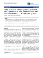
Quantitative ELISA assay revealed that the de-
creased CSF levels of total full-length Vgf (P<0.05),
correctly diagnosed ALS patients with 77% sensitivity
and 87% specificity based on receiving operating
characteristic (ROC) analysis (Figure 1A). Vgf CSF
content decreased as a function of progression of
muscle weakness characterized by an increasing
number of affected muscle (segments) assessed by
manual muscle testing (P<0.05) (Figure 1B).
Figure 1. Full length Vgf content in CSF in ALS. In A, full-length Vgf was assessed by quantitative ELISA assays; in B, Vgf
content decreased as a function of progression of muscle weakness assessed by manual muscle testing revealing an increased number
of affected muscle (segments). Quantitative muscle testing was based on the MRC clinical grading system, out of a total of 100
possible points. ROC analysis was carried out to determine the sensitivity and specificity of Vgf in dissecting control vs. ALS
subjects. Values are expressed as percent of control level (mean ± SEM; * 2-tailed t-test, p<0.05). Inset, Vgf protein sequence used to
raise Vgf antibodies for ELISA assays (see Materials and Methods for more information).
Decreased Vgf content In CSF and serum precedes
onset of ALS-type muscle weakness assessed by
rotarod-assays.
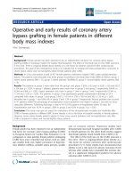
In our laboratory setting, G93A mutant
SOD-1ALS mice develop muscle weakness by ~90
days of age (Figure 2A). The severity of motor im-
pairment progresses to paralysis by ~130 days of age,
followed by sacrifice.[1] No detectable change in Vgf
content in CSF and serum of G93A SOD-1 ALS mice
was found in ~35 days old G93A SOD-1 ALS mice,
relative to age-, gender-, and strain-matched wild-type
littermates (Figure 2B,C).
Reduction in Vgf content in the CSF (F
1,7793
=4.913,
P=0.0288 for age, F
7,23660
=2.131
,
P=0.0466 for Vgf con-
tent) and in the serum (F
1,19840
=5.4573
,
P=0.0345 for age,
F
7,34510
=3.945, P=0.0487 for Vgf content) in ~75 days old
G93A SOD-1 mice was found to precede the onset of
muscle weakness assessed by rotarod assay that nor-
mally occurs at ~90 days, relative to age-, gender-, and
strain-matched wild-type littermates.
Serum and CSF content of Vgf continued to de-
crease up to ~130 days of age when G93A-SOD1 ALS
mice are characterized by near complete paralysis,
relative to gender-matched WT littermates (Figure 2B,
C).
Int. J. Med. Sci. 2008, 5
96
Vgf immunoreactive material in the lumbar (L3 to
L5) spinal cord colocalizes with SMI-32 im-
munopositive motorneurons and decreases as a
function of age progression in SOD-1 ALS mice.
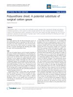
In the lumbar L3 to L5 region of spinal cord the
distribution of Vgf immunoreactive material colocal-
ized with SMI-32 immunoreactive spinal cord neurons
(Figure 3A, panels 2-4). No detectable Vgf im-
munopositive signal was found to co-localize with
NeuN immunoreactive neurons in the same spinal
cord region (Figure 3B, panels 2-4), nor was there Vgf
immunoreactive signal that co-localized with GFAP
immunopositive astrocytes (data not shown).
Figure 2. Decreased Vgf content in the CSF and serum precedes ALS-type motor impairment assessed by rotarod assay. In
A, ALS-type muscle weakness in mutant G93A SOD-1 as a function of clinical progression (age). In B,C, decreased Vgf levels in
CSF and in serum respectively precedes ALS-type muscle weakness in ~90 day-old symptomatic mutant G93A-SOD-1 mice and
continue to decline as a function of progression of ALS-type clinical disease. Values are expressed as mean ± SEM; n=4-5 per group;
2-way ANOVA. No detectable muscle weakness was found in age-gender matched WT controls at any time examined (not shown).
Figure 3. Vgf immunoreactive material in the lumbar spinal cord co-localizes with SMI-32 immunopositive motorneurons and
decreases as a function of age progression of SOD-1 ALS mice. In A, Vgf immunoreactive material is selectively localized within
the nuclear region of SMI-32 immunoreactive spinal cord neurons. In B, no detectable Vgf co-localization with NeuN immunore-
active neurons. In C, Vgf immunoreactive material in spinal cord motorneurons as a function of age. In D, SMI-32 spinal cord
motorneurons in the L3-L5 region of spinal cord in ~130 days old mutant G93A-SOD1 ALS mice. Values are expressed as mean ±
SEM; n=4-5 per group; In C,*2-way ANOVA; in D, *p<0.05 by 2-tailed t-test