Báo cáo khoa học: Analysis of the regulatory motifs in eukaryotic initiation factor 4E-binding protein 1 pot
Bạn đang xem bản rút gọn của tài liệu. Xem và tải ngay bản đầy đủ của tài liệu tại đây (801.14 KB, 15 trang )
Analysis of the regulatory motifs in eukaryotic initiation
factor 4E-binding protein 1
Vivian H. Y. Lee
1
, Timothy Healy
1
, Bruno D. Fonseca
1
, Amanda Hayashi
2
and
Christopher G. Proud
1
1 Department of Biochemistry and Molecular Biology, University of British Columbia, Vancouver, Canada
2 Institute of Food Nutrition and Human Health, Massey University and Food, Metabolism and Microbiology, AgResearch Limited,
Palmerston North, New Zealand
Signalling through the mammalian target of rapamycin
complex 1 (mTORC1) plays a key role in the control
of a number of cellular functions [1,2]. These roles
have largely been revealed through the use of rapamy-
cin, an immunosuppressant drug that interferes with
signalling through mTORC1.
mTORC1 is a complex comprising several proteins.
These include mammalian target of rapamycin
(mTOR), a multidomain protein that possesses a pro-
tein kinase domain related to lipid kinases, and raptor,
a scaffold protein that interacts with proteins that are
phosphorylated by mTOR [3–8]. mTORC1 also com-
prises Rheb, a small G-protein that appears to activate
mTOR when it is in its GTP-bound form [9,10].
Signalling from cell surface receptors, such as those
for insulin, growth factors and mitogens, activates
mTORC1 through the inactivation of the tuberous
sclerosis complex (TSC), which comprises TSC1 and
TSC2 [11–15]. In association with TSC1, TSC2 acts as
a GTPase activator protein (GAP) which converts
Keywords
4E-BP1; mTOR; mTORC1; RAIP motif; TOS
motif
Correspondence
C. G. Proud, Department of Biochemistry
and Molecular Biology, University of British
Columbia, Life Sciences Centre, 2350
Health Sciences Mall, Vancouver V6T 1Z3,
BC, Canada
Fax: +1 604 822 5227
Tel: +1 604 827 3923
E-mail:
Website: />fac_research/faculty/proud.html
(Received 10 December 2007, revised 22
February 2008, accepted 3 March 2008)
doi:10.1111/j.1742-4658.2008.06372.x
Mammalian target of rapamycin complex 1 (mTORC1) phosphorylates
proteins such as eukaryotic initiation factor 4E-binding protein 1 (4E-BP1)
and the S6 kinases. These substrates contain short sequences, termed TOR
signalling (TOS) motifs, which interact with the mTORC1 component rap-
tor. Phosphorylation of 4E-BP1 requires an additional feature, termed the
RAIP motif (Arg–Ala–Ile–Pro). We have analysed the interaction of
4E-BP1 with raptor and the amino acid residues required for functional
RAIP and TOS motifs, as assessed by raptor binding and the phosphoryla-
tion of 4E-BP1 in human cells. Binding of 4E-BP1 to raptor strongly
depends on an intact TOS motif, but the RAIP motif and additional
C-terminal features of 4E-BP1 also contribute to this interaction. Muta-
tional analysis of 4E-BP1 reveals that isoleucine is a key feature of the
RAIP motif, that proline is also very important and that there is greater
tolerance for substitution of the first two residues. Within the TOS motif,
the first position (phenylalanine in the known motifs) is most critical,
whereas a wider range of residues function in other positions (although an
uncharged aliphatic residue is preferred at position three). These data
provide important information on the structural requirements for efficient
signalling downstream of mTORC1.
Abbreviations
4E-BP1, eukaryotic initiation factor 4E-binding protein 1; ECL, enhanced chemiluminescence; eIF, eukaryotic initiation factor; GAP, GTPase
activator protein; GST, glutathione S-transferase; HIF1a, hypoxia-inducible factor 1a; mTOR, mammalian target of rapamycin; mTORC1,
mTOR complex 1; PKB, protein kinase B (also termed Akt); PKC, protein kinase C; PRAS40, proline-rich Akt-substrate 40 kDa; PVDF,
poly(vinylidene difluoride); RAIP motif, Arg–Ala–Ile–Pro motif; S6K, S6 kinase; TOS motif, TOR signalling motif; TSC, tuberous sclerosis
complex.
FEBS Journal 275 (2008) 2185–2199 ª 2008 The Authors Journal compilation ª 2008 FEBS 2185
Rheb
.
GTP to its inactive GDP-bound form. For exam-
ple, agents that activate protein kinase B (PKB, also
termed Akt) induce the phosphorylation of TSC2. This
is believed to inactivate its GAP function [9,16],
thereby allowing Rheb to accumulate in its GTP-
bound form and to switch on mTORC1. Recent data
have suggested that RhebÆGTP activates mTORC1 by
bringing about the release of FKBP38, an inhibitor of
mTORC1 activity [17].
Raptor appears to promote signalling downstream
of mTORC1 by binding to short TOR signalling
(TOS) motifs found in proteins whose phosphorylation
is positively regulated by mTORC1 [4,5,7,18,19]. The
first proteins shown to contain functional TOS motifs
were the ribosomal protein S6 kinases (S6Ks) and the
eukaryotic initiation factor (eIF) 4E-binding proteins
(4E-BPs; Fig. 1A), each of which is subject to rapamy-
cin-sensitive phosphorylation at multiple sites. The
interaction of these proteins with raptor, via their TOS
motifs, promotes their phosphorylation by mTOR
in vitro. Both of these types of protein are implicated
in controlling the translational machinery [20].
mTORC1 also controls other cellular functions,
although the mTORC1 targets involved in these effects
largely remain to be identified [1]. Very recently, whilst
our manuscript was in preparation, two further pro-
teins were shown to contain TOS motifs: hypoxia-
inducible factor 1a (HIF1a [21]) and the proline-rich
Akt-substrate 40 kDa (PRAS40 [22–24]).
Although the TOS motifs in these proteins resemble
one another, there are a number of differences between
them, and it is not clear what are the real requirements
for a functional TOS motif. Defining a ‘consensus’
TOS motif would help to identify such motifs in other
proteins that may be controlled by mTORC1 and reg-
ulate cellular functions in addition to mRNA transla-
tion. It is also not clear whether the TOS motif is
sufficient for the interaction with raptor, or whether
other features are also required.
It is of particular interest that the in vivo phosphory-
lation of 4E-BP1, the best-understood 4E-BP, requires
an additional motif with the sequence Arg–Ala–Ile–
Pro (hence ‘RAIP motif’ [25]; Fig. 1A). The phosphor-
ylation of the two N-terminal sites in 4E-BP1
(Thr37 ⁄ 46 in the human protein; Thr36 ⁄ 45 in rat
4E-BP1) requires the RAIP motif [19], and their phos-
phorylation is needed for the subsequent modification
of two sites (Thr70 ⁄ Ser65) close to the eIF4E-binding
motif [19,26–29]. The mTOR-dependent control of
4E-BP1 is thus an example of hierarchical phosphory-
lation. It is the phosphorylation of Thr70 ⁄ Ser65 that
controls the binding of 4E-BP1 to eIF4E, and thus the
availability of eIF4E to form functional translation
initiation complexes (as 4E-BP1 competes with the
scaffolding factor eIF4G for binding to eIF4E [30]).
Our earlier work revealed that the RAIP and TOS
motifs play distinct roles in regulating the phosphory-
lation of 4E-BP1 within cells. The phosphorylation of
4E-BP1 is regulated by amino acids and by stimuli
such as insulin. The RAIP motif appears to mediate
the amino acid input [25,29] that promotes the phos-
phorylation of the N-terminal threonines in both
4E-BP1 and 4E-BP2 (which is not very prone to inhi-
bition by rapamycin). In contrast, the TOS motif is
required for the insulin-induced phosphorylation of
Ser65 (and, in some cell types, Thr70). Phosphoryla-
tion of Ser65 is generally completely blocked by rapa-
mycin. Although TOS motifs have now been identified
in a number of proteins, no systematic analysis of the
sequence requirements for a functional TOS motif has
been performed.
Similarly, the (sequence) requirements for a func-
tional RAIP motif remain to be defined. The roles of
the RAIP and TOS motifs in the interaction of
4E-BP1 with raptor also remain incompletely under-
stood. In this article, we address these issues and the
requirements for a functional TOS motif. We show
that several regions of 4E-BP1, including both the
TOS and RAIP motifs, plus other features, play roles
in its binding to raptor. We also analyse the amino
acid sequence requirements for functional TOS and
RAIP motifs in 4E-BP1.
Results and Discussion
Regions of 4E-BP1 involved in binding to raptor
The two known regulatory motifs in 4E-BP1 are
located at opposite ends of the polypeptide chain
(Fig. 1A). We have previously reported that the
extreme C-terminus of 4E-BP1 (the final 20 amino
acids) can bind raptor in an overlay (far-western)
assay, whereas the N-terminal portion cannot [19], sug-
gesting that the RAIP motif does not itself bind rap-
tor. In contrast, another study [31] found that,
although wild-type 4E-BP1 could be coimmunoprecipi-
tated with raptor, variants with mutations in the TOS,
RAIP or both motifs could not. This implies a role for
the RAIP motif in binding to raptor. [It should be
noted, however, that neither protocol definitively dem-
onstrates that raptor binds directly to any part of
4E-BP1, as raptor is expressed in mammalian cells, and
the interaction could be mediated by another (mamma-
lian) protein. For simplicity, we refer to the binding
seen as ‘raptor binding’.] Because of substantial prob-
lems of nonspecific binding, we have been unable to
Regulatory motifs in 4E-BP1 V. H. Y. Lee et al.
2186 FEBS Journal 275 (2008) 2185–2199 ª 2008 The Authors Journal compilation ª 2008 FEBS
successfully use coimmunoprecipitation approaches to
study raptor–4E-BP1 binding (A. Beugnet, B. D. Fonseca
& C. G. Proud, unpublished data; see also [19]).
Previous work has shown that mutation of the phen-
ylalanine to alanine in the TOS motif eliminates the
binding of raptor to the C-terminal fragment of human
4E-BP1 in the overlay assay [19] (see also Fig. 1B). We
have also observed no binding of raptor to a truncated
4E-BP1 molecule lacking the final six residues that
harbour the TOS motif (D6; Fig. 1B). This confirms
A
B
D
E
C
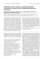
Fig. 1. Analysis of the binding of raptor to
variants based on 4E-BP1. (A) Schematic
diagram of 4E-BP1 showing the RAIP and
TOS motifs, the region that binds eIF4E and
the four phosphorylation sites discussed in
this report. Numbering is based on human
4E-BP1; for the rodent proteins, adjust by
)1. Schematic diagram is not to scale.
(B–D) Binding of raptor to wild-type 4E-BP1
or variants, assessed using the overlay (far-
western) assay (see Experimental proce-
dures). The top sections of each panel show
the blots for Myc-tagged raptor; the bottom
sections show the blots with anti-GST to
allow a comparison of the amounts of GST
fusion proteins used in each case. Some
degradation of the GST fusion proteins is
evident from the presence of products run-
ning at the position of GST itself. (E) Binding
of raptor to different amounts of wild-type
4E-BP1 or the AAAA mutant, assessed
using the overlay (far-western) assay. The
graph shows the quantification of the data
from three independent experiments. Error
bars indicate the standard deviation. Stu-
dent’s t-test (two-sample unequal variance,
two-tailed distribution) was used to deter-
mine the probability that raptor binds wild-
type 4E-BP1 and AAAA mutant equally. In
all instances, the P-value was 0.01 or lower
(*0.002; §0.002; ‡0.01; †0.002; #0.00004).
V. H. Y. Lee et al. Regulatory motifs in 4E-BP1
FEBS Journal 275 (2008) 2185–2199 ª 2008 The Authors Journal compilation ª 2008 FEBS 2187
that the TOS motif is essential for detectable stable
binding of raptor to 4E-BP1, but does not tell us
whether it is sufficient.
To assess the contribution of other regions of
4E-BP1 to raptor binding, we created a series of N-ter-
minally truncated mutants. We reasoned that such
truncations cannot perturb the higher order structure
in 4E-BP1, as 4E-BP1 is apparently unstructured in
solution (as assessed by NMR spectroscopy [32]). A
second potential concern is that, in this type of ‘far-
western’ analysis, 4E-BP1 is denatured (by SDS). This
concern is also lessened by the fact that 4E-BP1 lacks
a folded structure.
We created variants in which the first 17, 37, 57,
77 or 97 residues of 4E-BP1 were removed. The first
of these, ‘4E-BP1 (18–117)’, already lacks the RAIP
motif. As shown in Fig. 1C, each of these truncated
proteins bound to raptor less efficiently than full-
length wild-type 4E-BP1 (1–117) in the overlay assay.
Reproducibly, two regions appeared to be involved in
assisting the binding to raptor: the first 17 amino
acids [compare the signal for full-length 4E-BP1 (1–
117) with that for the ‘18–117’ variant] and sections
of the C-terminal half of 4E-BP1 [compare, for exam-
ple, the 4E-BP1 (98–117) variant with full-length
4E-BP1 (1–117)], in agreement with our earlier data
[19]. This suggests that the N-terminus, containing
the RAIP motif, and a more C-terminal region (out-
side the final 20 residues, i.e. other than the TOS
motif) are involved in binding to raptor. Although
the TOS motifs in 4E-BP1 and 4E-BP2 are identical,
other parts of their C-terminal regions are poorly
conserved, and it is not obvious which other features
contribute to raptor binding. We have not therefore
attempted to define further the features in the C-ter-
minus of 4E-BP1 that are involved in its binding to
raptor. The data for the other truncation mutants
shown in Fig. 1C indicate that other regions of
4E-BP1 also contribute to stable binding to raptor.
The first 17 residues of 4E-BP1 contain the RAIP
motif. To assess whether removal of the RAIP motif
accounts for the reduced binding of raptor to the 18–
117 fragment, we compared the binding of raptor to
this truncated protein and to full-length 4E-BP1 in
which the RAIP motif was altered to AAAA. The
phosphorylation of this mutant within cells was
severely impaired ([25]; see also Fig. 2A). The binding
of raptor to these two variants was similar (Fig. 1D),
implying that the loss of raptor binding on removal of
the first 17 residues may be accounted for simply by
the loss of the RAIP motif. We therefore also tested
the binding of raptor to full-length 4E-BP1 and to the
RAIP ⁄ AAAA variant. A marked and reproducible
decrease was seen for the RAIP ⁄ AAAA mutant, when
compared with wild-type 4E-BP1 (Fig. 1E). The RAIP
motif clearly makes a substantial contribution to the
binding of 4E-BP1 to raptor. However, in contrast
with the TOS motif, it is not essential for this interac-
tion (compare with the D6 truncation in Fig. 1B, which
displays no binding to raptor). The finding that the
RAIP motif is important for the binding of 4E-BP1 to
raptor is consistent with earlier observations showing
that an intact RAIP motif is required for the efficient
in vitro phosphorylation of the N-terminal threonines
in 4E-BP1 by mTOR raptor [5].
Taken together, these data show the following: (a)
that the TOS motif plays a critical role in binding rap-
tor; (b) that the region containing the RAIP motif also
contributes to this interaction, but is not absolutely
required; and (c) that other regions of 4E-BP1 are also
involved in binding raptor. Interestingly, as noted
above, mutating the RAIP and TOS motifs separately
has qualitatively distinct effects on the phosphorylation
of 4E-BP1 within cells [19], revealing that they serve
different, rather than additive, functions. Interestingly,
Eguchi et al. [31] have shown that the introduction of
acidic residues at the positions of the phosphorylation
sites in 4E-BP1 decreases the interaction of 4E-BP1
with raptor. This implies that the regions of 4E-BP1
containing these residues also influence the interaction
with raptor, and is in accordance with our data
(Fig. 1C), which indicate that it is not only the TOS
and (to a lesser extent) RAIP motifs that are needed
for raptor–4E-BP1 binding.
Further definition of the RAIP motif in the
N-terminus of 4E-BP1
So far, very little information is available on what
actually constitutes a RAIP-type motif, i.e. what are
the sequence requirements. To learn more about the
nature of the RAIP motif and, in particular, to define
better what residues constitute this type of motif, we
created a range of further mutations in this region of
4E-BP1. It is important to note that, in the vector used
here, the Myc tag is at the C-terminus, i.e. at the
opposite end from the RAIP motif, to avoid any possi-
ble interference with the function of the N-terminal
RAIP motif. The vector encodes rat 4E-BP1, which
was used extensively in our earlier studies to define the
RAIP motif [25]. The use of the rat protein also has
the advantage that there is no cross-reactivity of the
(P)Ser64 antibody with other sites, which is a compli-
cating feature of the human protein (in which this anti-
serum recognizes both Ser65 and another site, Ser101
[33]). We have shown previously that the behaviour of
Regulatory motifs in 4E-BP1 V. H. Y. Lee et al.
2188 FEBS Journal 275 (2008) 2185–2199 ª 2008 The Authors Journal compilation ª 2008 FEBS
the rat and human 4E-BP1 proteins expressed in
HEK293 cells is very similar [33].
To assess the functional consequences of mutations
in the RAIP motif, we studied the phosphorylation of
4E-BP1 in HEK293 cells, focusing on Thr36 ⁄ 45, as
these sites are involved earlier in the hierarchy of
phosphorylation and depend absolutely on the RAIP
motif [25,26]. Clearly, making a full range of substitu-
tions, even within a four-residue motif, would be an
enormous undertaking. We therefore created and
tested a set of mutants, selected as described below.
Given the diversity of mutants tested, we are unable
to show data for each one relative to all relevant
variants within the same panel in Fig. 2; however,
each panel contains wild-type 4E-BP1 (‘RAIP’) as a
reference.
Our earlier data [25] indicated that isoleucine within
the RAIP motif (Ile15) plays a particularly important
role in the phosphorylation of 4E-BP1 in HEK293
cells [25]. This is also clearly seen in the data in Fig. 2,
where the phosphorylation of the RAAP variant
(Fig. 2A) is more severely reduced relative to wild-type
4E-BP1 than the phosphorylation of either the AAIP
(Fig. 2A) or RAIA (Fig. 2B) variants. This is espe-
cially true for the basal phosphorylation at Thr36 ⁄ 45,
which is maintained by the amino acids in the medium
A
B
C
D
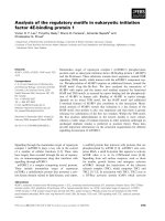
Fig. 2. Assessment of the phosphorylation of 4E-BP1 mutants containing variants of the RAIP motif. (A–D) Wild-type 4E-BP1 (RAIP) or the
indicated mutants were expressed in HEK293 cells. Twenty-four hours following transfection, the cells were starved of serum for 16 h and,
where indicated, treated with 100 n
M insulin for 25 min. The top sections of each panel show the results from western blots using the
phosphospecific antibody for Thr36 ⁄ 45; the bottom sections show the data from anti-Myc blots (to assess the relative levels of expression
of the 4E-BP1 variants). With this gel system, 4E-BP1 runs as up to three bands (a–c, in order of increasing phosphorylation) as indicated.
V. H. Y. Lee et al. Regulatory motifs in 4E-BP1
FEBS Journal 275 (2008) 2185–2199 ª 2008 The Authors Journal compilation ª 2008 FEBS 2189
[29], but is also true for the increased phosphorylation
induced by insulin.
We therefore first replaced the isoleucine by the
other branched-chain residues, valine and leucine.
These 4E-BP1 variants were expressed in HEK293
cells. Their phosphorylation was analysed using a
phosphospecific antiserum that recognizes both
(P)Thr36 and (P)Thr45 in rat 4E-BP1. 4E-BP1
migrates as three distinct species (a–c) under these
conditions of SDS-PAGE. The slowest moving species
(c) is the most highly phosphorylated form, and is only
evident after insulin stimulation. This is because insulin
induces the phosphorylation of additional sites (nota-
bly Ser64, see below), which causes the protein to run
as the c species.
As shown in Fig. 2A, the basal phosphorylation of
Thr36 ⁄ 45 in the Ile15Val (RAVP) variant was identical
to that of the wild-type protein and, likewise, was only
slightly stimulated by insulin. In contrast, replacement
of Ile15 by leucine caused a very marked decrease in
basal phosphorylation at Thr36 ⁄ 45 and impaired the
insulin-stimulated phosphorylation of these sites.
Next, we studied the importance of the arginine and
proline residues within the RAIP motif. In order to
help us discern the effects of the substitutions more
clearly, we used a 4E-BP1 mutant (AAIP) which
already contained one mutation in the RAIP motif,
the rationale being that using a mutant with a partially
defective RAIP motif would probably enhance any
effects of other mutations. Thus, we tested the impor-
tance of the proline residue in a variant of 4E-BP1 in
which the arginine was mutated to alanine (AAIP,
which shows modestly decreased basal and insulin-
stimulated phosphorylation relative to wild-type
4E-BP1; Fig. 2A,D). Proline is an imino, not an
amino, acid: arguably the most closely related amino
acid is valine. Although the AAIP mutant showed sub-
stantial basal phosphorylation at Thr36 ⁄ 45 (which was
increased somewhat by insulin; Fig. 2A,D), the AAIV
mutant did not undergo any detectable phosphoryla-
tion at Thr36 ⁄ 45 under basal or insulin-stimulated
conditions (Fig. 2B,D). As even the relatively conser-
vative replacement of proline by valine almost com-
pletely abolished the phosphorylation of 4E-BP1
(compared with the AAIP variant; Fig. 2A,D), we did
not test any other mutations at this position in this
study. Earlier work has shown that mutating the pro-
line to alanine (to give the RAIA mutant) causes a
defect in the basal and insulin-stimulated phosphoryla-
tion of 4E-BP1 [25]. We also tested the proline to
valine mutation in wild-type 4E-BP1. The phosphory-
lation of the resulting RAIV mutant was more severely
impaired than that of the RAIA variant (Fig. 2D).
We then turned our attention to the arginine residue
within the RAIP motif, making mutations at this posi-
tion within the RAIA variant, which already shows a
reduction in basal and insulin-stimulated phosphoryla-
tion at Thr36 ⁄ 45 (Fig. 2A). Mutation of the arginine
to lysine in the RAIA variant (to create KAIA) did
not discernibly affect the basal or insulin-stimulated
phosphorylation of Thr36 ⁄ 45 (Fig. 2A). Mutation of
the arginine to methionine (no charge, bulky side-chain
similar to arginine; Fig. 2B) also did not impair the
phosphorylation of Thr36 ⁄ 45. Mutation to glutamate
(negative charge; Fig. 2C) diminished the basal level of
phosphorylation, but still permitted some induction of
phosphorylation by insulin. Mutation of the arginine
to glutamine (QAIP; Fig. 2B), threonine or asparagine
(both Fig. 2C) in wild-type 4E-BP1 had similar partial
effects. It therefore appears that Arg13 is less impor-
tant than Pro16 for the function of the RAIP motif,
and that several different types of residue can be toler-
ated here with only small, if any, effects on 4E-BP1
phosphorylation. For reasons that remain to be clari-
fied, such deficits are often more apparent for basal
than for insulin-induced phosphorylation. One possible
explanation is that, when the function of the RAIP
motif is impaired, the phosphorylation of Thr36 ⁄ 45
becomes more dependent on the rapamycin-sensitive
input provided by the TOS motif.
Lastly, we tested the effect of selected mutations of
the alanine residue in the RAIP motif. Mutation to
valine markedly reduced the basal phosphorylation of
4E-BP1 and slightly impaired the effect of insulin
(Fig. 2B), whereas replacement by a negatively charged
residue, aspartate, had no effect on basal phosphoryla-
tion (Fig. 2B).
Overall, these data indicate that isoleucine is the
most important single residue within the RAIP motif.
This is in accordance with our earlier data [25]: the
present findings extend those observations by demon-
strating that replacing this residue with valine, but not
leucine, permits retention of RAIP motif function.
Defining what constitutes a functional TOS motif
The data presented above (Fig. 1B) further confirm the
key role played by the TOS motif in the binding of
4E-BP1 to raptor in the far-western analysis employed
here. Two further proteins were described as contain-
ing TOS motifs whilst this paper was in the final stages
of preparation (HIF1a [21] and PRAS40 [22–24];
Table 1). However, so far, no detailed analysis has
been performed to define which residues are actually
required for a functional TOS motif: such data would
be helpful in identifying potential TOS motifs in other
Regulatory motifs in 4E-BP1 V. H. Y. Lee et al.
2190 FEBS Journal 275 (2008) 2185–2199 ª 2008 The Authors Journal compilation ª 2008 FEBS
proteins. Here, we employed two approaches to study
this: (a) the ability of 4E-BP1 variants to bind to rap-
tor; and (b) the ability of a given TOS-like motif to
promote the phosphorylation of 4E-BP1 in cells.
The first type of analysis could, in principle, be per-
formed using the TOS motif segment alone, provided
that this motif is sufficient to confer binding to raptor.
To test this, we added the sequence FEMDI (the TOS
motif found in the C-termini of mammalian 4E-BP1–
3) to the C-terminus of glutathione S-transferase
(GST). To obviate possible issues of steric hindrance,
we provided a spacer (four alanine residues) between
the C-terminus of GST and the TOS motif, to create
‘GST-Ala
4
-TOS’. As shown in Fig. 3A, the addition of
the TOS motif to GST did not allow raptor binding.
Thus, the five-residue TOS motif is incapable, by itself,
of binding raptor in this assay. This is consistent with
the data in Fig. 1 and [19], which show that additional
features in 4E-BP1 are required for raptor binding
(but that the TOS motif is nonetheless essential).
We therefore elected to examine the effects of
altering the TOS motif in 4E-BP1. Phosphorylation of
4E-BP1 involves multiple sites and a rather complex
hierarchy [19,26,27,33,34]. To assess the effects of alter-
ing the TOS motif, we mainly examined the phosphory-
lation state of Ser64, as this site is late in the hierarchy,
and hence ‘integrates’ the effects of phosphorylation of
other sites in 4E-BP1. In HEK293 cells, phosphoryla-
tion at this site is stimulated by insulin [19,29], and this
is entirely dependent on the TOS motif [19,25]
(Fig. 4A). The level of phosphorylation of Ser64 in
insulin-treated cells is therefore especially informative.
We have shown previously that mutation of Phe113
to alanine in the 4E-BP1 TOS motif markedly impairs
the phosphorylation of Ser64 [19]. The present data
also showed that this mutation (which yields the AE-
MDI mutant) completely blocks the ability of 4E-BP1
to bind raptor in a far-western blot (Fig. 3A) and
almost eliminates the phosphorylation of 4E-BP1 at
Ser64 [19] (Fig. 4A,B). As reported previously [19], this
mutation can decrease the basal level of phosphoryla-
tion of the N-terminal threonines in 4E-BP1 in
HEK293 cells. This mutation also impairs the in vitro
phosphorylation of Thr36 ⁄ 45 by mTOR [5]. The phen-
ylalanine to alanine change is clearly major, and we
therefore tested whether the more conservative muta-
tion of the aromatic phenylalanine to a bulky aliphatic
residue (leucine) also affected function. The LEMDI
mutant underwent insulin-stimulated phosphorylation
at Ser64 to a similar degree to the wild-type protein
(Fig. 4C): in this and all other cases, this phosphoryla-
tion was blocked by rapamycin, confirming that it
requires mTORC1. However, the LEMDI variant
failed to bind raptor in the far-western assay (Fig. 3B).
The simplest explanation for this is that the mutation
weakens the TOS–raptor interaction to the extent that
it is insufficiently stable to ‘survive’ the washes of the
far-western procedure, but can still support an interac-
tion in vivo. These data imply that merely examining
raptor binding in, for example, a far-western method
does not indicate what constitutes a functional TOS
motif. In contrast with the LEMDI variant, the
IEMDI mutant underwent only a small degree of
phosphorylation at Ser64 (Fig. 4B). This variant did
not bind to raptor in the overlay assay (Fig. 3A). It is
notable that all the currently known TOS motifs have
phenylalanine in the first position (Table 1).
We then created a systematic set of other variants
based on the FEMDI sequence found in the 4E-BPs.
Mutation of the second residue (glutamate) to another
acidic residue (aspartate) had no effect on raptor bind-
ing (FDMDI; Fig. 3B), and we did not therefore
examine its effect on the phosphorylation of 4E-BP1.
Changing the second residue to valine (FVMDI;
Fig. 4D) or alanine (FAMDI; Fig. 4D) did not dis-
cernibly affect the phosphorylation of 4E-BP1 in
HEK293 cells. Replacement by proline slightly
impaired the phosphorylation of Ser64 (FPMDI;
Fig. 4E). Mutation to arginine (carries positive charge,
FRMDI; Fig. 4F) substantially decreased the phos-
phorylation of 4E-BP1 when compared with the wild-
type protein. Raptor binding to all of these variants
was similar to that of the wild-type protein
(Fig. 3B,C). Thus, although an acidic residue is present
at this position in both the 4E-BPs (glutamate) and
S6Ks (aspartate) (Table 1), this feature does not actu-
ally appear to be very important for the regulation of
Table 1. Known potential TOS motifs in selected proteins (italics
indicate putative TOS motifs; the other TOS motifs have been
shown to function in their respective proteins).
Protein (all
Homo sapiens) Sequence
Residue
numbers
S6K1 FDIDL 5–9
a
S6K2 FDLDL 5–9
a
4E-BP1 FEMDI 114–118
4E-BP2 FEMDI 116–120
4E-BP3 FEMDI 86–90
HIF1a FVMVL 99–103
PRAS40 FVMDE 129–133
PKCd FVMEF 425–429
PKCe FVMEY 484–488
STAT3 FPMEL
FDMDL
26–30
756–760
a
Numbering is based on the shorter splice variants of these
proteins.
V. H. Y. Lee et al. Regulatory motifs in 4E-BP1
FEBS Journal 275 (2008) 2185–2199 ª 2008 The Authors Journal compilation ª 2008 FEBS 2191
4E-BP1 or for raptor binding. Interestingly, the TOS-
like motifs in PRAS40 and HIF1a each lack an acidic
residue at the second position (FVMDE and FVMVL,
respectively [23,24]). They have valine in this position
instead, which is clearly as effective as an acidic resi-
due in promoting the phosphorylation of 4E-BP1 at
Ser64 (Fig. 4D).
In contrast with the tolerance for variations in the sec-
ond position, mutation of the third residue (methionine:
an uncharged, relatively nonpolar amino acid) to ala-
nine or glutamate abolished raptor binding (Fig. 3C).
The methionine to alanine mutation also strongly
decreased the phosphorylation of Ser64 (FEADI;
Fig. 4F), and the phosphorylation of Ser64 was also
decreased by placing glutamate or, to a lesser extent,
arginine at this position (Fig. 4G). Mutation of the
methionine to isoleucine (also a nonpolar, aliphatic
residue) maintained Ser64 phosphorylation at wild-type
A
C
D
B
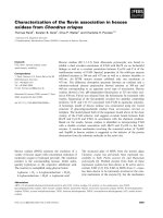
Fig. 3. Binding of raptor to the TOS motif in wild-type 4E-BP1 (FEMDI) and variants thereof. (A) An overlay assay (see Experimental proce-
dures) was used to assess the binding of raptor to the TOS motif (FEMDI) tagged at its N-terminus with GST and also containing a four ala-
nine spacer (Ala
4
) between the GST tag and the TOS motif. Wild-type GST–4E-BP1 and GST–4E-BP1 (AEMDI) served as positive and
negative controls, respectively. The top sections of each panel show raptor overlays, developed with anti-Myc. The bottom sections show
western blots for GST to assess the levels of each protein. (B,C) The overlay assay was used to detect binding of raptor to wild-type GST–
4E-BP1 (FEMDI) or mutants with the indicated sequences in place of the TOS motif. GST and GST–4E-BP1 (AEMDI) served as negative con-
trols. The top sections of each panel show the Myc-tagged raptor overlay. The bottom sections show western blots with anti-GST to assess
the amounts of each protein used. Arrowheads with asterisks denote degradation products (cleaved at the C-terminus) that react with anti-
GST (but do not bind raptor). (D) The binding of bacterially expressed native wild-type GST-4E-BP1 or variants to raptor was tested using a
dot blot as described in Experimental procedures.
Regulatory motifs in 4E-BP1 V. H. Y. Lee et al.
2192 FEBS Journal 275 (2008) 2185–2199 ª 2008 The Authors Journal compilation ª 2008 FEBS
levels (Fig. 4E). It seems probable that the presence of an
aliphatic residue with a side-chain larger than a methyl
group is required for function. This is in accordance
with the sequences of known TOS motifs (Table 1),
which have methionine (4E-BPs; PRAS40; HIF1a),
isoleucine (S6K1) or leucine (S6K2) at this position.
A
BC
DE
FG
HI
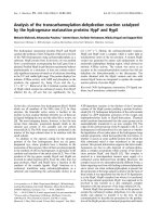
Fig. 4. Phosphorylation of 4E-BP1 variants expressed in HEK293 cells. (A–I) Wild-type Myc-tagged 4E-BP1 or variants of 4E-BP1 were
expressed in HEK293 cells. Twenty-four hours following transfection, the cells were starved of serum for 16 h and, in some instances, sub-
sequently treated with 100 n
M insulin for 25 min. Where indicated, cells were also incubated with 100 nM rapamycin for 30 min prior to insu-
lin stimulation (see Experimental procedures for details). Samples were analysed using the indicated phosphospecific antibodies for 4E-BP1
(top sections of each panel) or anti-Myc (bottom section in each panel; to assess the expression levels of 4E-BP1 variants). The differentially
phosphorylated a–c species are indicated.
V. H. Y. Lee et al. Regulatory motifs in 4E-BP1
FEBS Journal 275 (2008) 2185–2199 ª 2008 The Authors Journal compilation ª 2008 FEBS 2193
The fourth residue in the 4E-BP1 TOS motif is
acidic: aspartate. This was mutated to alanine
(Fig. 4H), asparagine (Fig. 4H) or arginine (Fig. 4I).
The substitution by alanine very substantially
decreased the phosphorylation at Ser64, but the phos-
phorylation of the FEMRI variant was similar to that
of wild-type 4E-BP1, and that of the FEMNI protein
was intermediate between the other two mutants
(Fig. 4H). None of these three variants was able to
bind raptor (Fig. 3C). Thus, although the first TOS
motifs to be discovered contained a negatively charged
aspartate at this position (4E-BPs; S6K1 and S6K2),
and the recently reported TOS motif in PRAS40 simi-
larly has a glutamate at this position, other residues
are tolerated, even if, as for arginine, they carry a posi-
tive charge. The latest reported TOS motif, in HIF1a,
has an aliphatic, nonpolar residue in the fourth posi-
tion (valine; Table 1).
Mutation of the final residue from isoleucine to
arginine or alanine reduced raptor binding (FEMDR ⁄
FEMDA; Fig. 3C), but had little effect on Ser64
phosphorylation (Fig. 4C,I). We also tested the effect
of an acidic residue at this position, i.e. the FEMDE
variant. Phosphorylation of this mutant at Ser64 was
slightly reduced compared with the wild-type protein
(Fig. 4D). It was still able to bind raptor, albeit less
well than wild-type 4E-BP1 (Fig. 3C). In view of this
tolerance for a variety of residues at position five, we
did not create further mutations here. Although
almost all of the known TOS motifs have either leu-
cine or isoleucine at this position, residues that are not
branched-chain amino acids can clearly function in
this position.
It seems surprising that several variants failed to
bind raptor in the overlay assay, but still underwent
substantial phosphorylation within HEK293 cells
(e.g. the FEMRI and FEMDR variants). It is possi-
ble that the use of denatured 4E-BP1 in the far-wes-
tern assay led to misleading results (although this
does not seem likely, as 4E-BP1 reportedly has little
if any folded structure [32]). Therefore, we also
performed dot blot overlay assays in which GST–
4E-BP1 was applied to the membrane without prior
denaturation on an SDS-polyacrylamide gel. This
yielded very similar results to the far-western analy-
ses, i.e. all of the variants that were negative in that
assay (including the two just mentioned) were also
negative in the dot blot assay, whereas wild-type
4E-BP1 and FAMDI variants bound raptor in both
assays (Fig. 3C,D). It should be noted that, although
the other 4E-BP1 variants appear to interact weakly
with raptor in the ‘dot blot far-western’ assay
(Fig. 3D), they do so to an identical extent to GST
itself, indicating that this residual binding is
nonspecific.
Analysis of TOS-like motifs from other proteins
reported to be controlled by mTOR signalling
A number of other proteins have been reported to be
regulated in a rapamycin-sensitive manner. The phos-
phorylation of STAT3 has been reported to be con-
trolled by mTOR [35,36], as has the phosphorylation
of the atypical protein kinase C isoforms (PKCd ⁄ e)
[37,38]. As shown in Table 1, there are two putative
TOS motifs in STAT3, i.e. FDMDL and (with less
similarity to the known TOS motifs) FPMEL. PKCd
(one of the forms studied by Parekh et al. [37,38]) has
the motif FVMEF. Interestingly, the classical PKC iso-
form, PKCc, also contains a similar motif, FVMEY.
To test whether motifs with these sequences could
actually bind raptor, and to learn more about the
requirements for raptor binding, we decided to intro-
duce these motifs into 4E-BP1 (in place of its own
TOS motif), as the mTOR regulation of 4E-BP1 is
much better characterized than the control of STAT3
or PKC isoforms. We therefore created a range of
mutants of 4E-BP1, in both the GST fusion protein
(to test raptor binding) and the vector for mammalian
expression (to check their effect on the phosphoryla-
tion of 4E-BP1).
As shown in Fig. 5A, in the far-western assay, all of
these variant 4E-BP1 proteins, except one, bound rap-
tor to a similar extent to wild-type 4E-BP1. (The
exception is the variant with the FVMEY motif, which
did bind raptor, but less well than the others). As each
variant contains at least two changes from the wild-
type FEMDI sequence, it is inappropriate to try to
interpret these data in terms of the roles of individual
residues, except to say that placing a tyrosine in the
last position has a deleterious effect on raptor binding
(compare FVMEY with FVMEF in Fig. 5A).
These findings suggested that it was probable that
these motifs would support the phosphorylation of
4E-BP1 when the variant proteins were expressed in
HEK293 cells. Indeed, all but one of the variants
underwent substantial insulin-induced phosphorylation
at all the sites tested (Thr36 ⁄ 45 ⁄ 69 and Ser64)
(Fig. 5B,C). The exception, surprisingly, in view of its
good ability to bind raptor (Fig. 5A), was the FVMEF
motif (from PKCd). Conversely, although the FVMEY
variant bound poorly to raptor (Fig. 5A), it became
quite strongly phosphorylated in response to insulin
(Fig. 5B). As already observed for other variants tested
here, there is imperfect correspondence between raptor
binding (in the far-western assay) and function in
Regulatory motifs in 4E-BP1 V. H. Y. Lee et al.
2194 FEBS Journal 275 (2008) 2185–2199 ª 2008 The Authors Journal compilation ª 2008 FEBS
promoting phosphorylation of 4E-BP1 within cells.
The latter approach is probably more informative in
terms of motif function.
Conclusions
This study represents the first systematic attempt to
define the sequence requirements of the TOS and
RAIP motifs. It provides new information on the fea-
tures of 4E-BP1 that are required for its interaction
with raptor and⁄ or for its phosphorylation at different
sites in living cells. Firstly, our findings demonstrate
that, although the TOS motif is essential for interac-
tion with raptor and for phosphorylation of specific
sites in cells, other features of 4E-BP1 are necessary
for this interaction. Indeed, the five-amino-acid TOS
motif is not sufficient to confer binding to raptor. Our
findings show that the RAIP motif also plays a role in
binding raptor (although it is not essential for this),
and that other regions of the 4E-BP1 polypeptide are
involved in this interaction (especially parts of the
C-terminal half of the molecule). The present data help
to resolve earlier discrepancies concerning the role of
the RAIP motif: although this motif is not sufficient
by itself to bind stably to raptor [19], it plays an
‘accessory’ role in raptor binding, provided that a TOS
motif is also present [5,31]. This interpretation is con-
sistent with the recent observation that short interfer-
ing RNA (siRNA)-mediated knock-down of raptor
expression impairs the phosphorylation of Thr37 ⁄ 46 in
4E-BP1 (which depends on the RAIP motif) [24].
Our analysis of the TOS motif demonstrates that its
ability to bind raptor in vitro is not a reliable index of
function: a number of variants that failed to bind rap-
tor in the far-western assay were able to support phos-
phorylation of 4E-BP1 within cells. Although mutants
that bind raptor in vitro effectively support phosphory-
lation within cells, the converse is not true: for exam-
ple, the LEMDI mutant does not bind raptor but is as
effective as the wild-type sequence at facilitating phos-
phorylation (Figs 3B and 4C). Studying the phosphor-
ylation of 4E-BP1 is thus a more reliable method than
in vitro raptor binding to assess TOS motif function.
The first position in the TOS motif is critical: muta-
tion to another closely similar residue (isoleucine)
almost abolishes the phosphorylation of Ser64
(Fig. 4B). Consistent with this, all the known TOS
motifs have phenylalanine at this position. Our data
indicate that the nature of the second residue in the
motif is less critical: although the first motifs to be
identified (in S6Ks and 4E-BPs) contained acidic resi-
dues here, a range of other residues support the phos-
phorylation of 4E-BP1 in cells. The diversity of these
A
B
C
Fig. 5. Ability of (putative) TOS motifs from other proteins to
support the binding of 4E-BP1 to raptor or its phosphorylation at
Ser64. (A) Wild-type 4E-BP1 or variants containing the indicated
(putative) TOS motifs from other proteins (as noted) were
expressed as GST fusions in Escherichia coli. Binding to raptor
was assessed using the overlay assay (top section). The bottom
section shows the amount of each protein used, as assessed by
western blot with anti-GST. Arrowheads with asterisks denote
degradation products (cleaved at the C-terminus) that react with
anti-GST (but do not bind raptor). GST and GST-tagged wild-type
4E-BP1 served as negative and positive controls, respectively.
(B, C) Wild-type Myc-tagged 4E-BP1 or the indicated variants
were expressed in HEK293 cells. Twenty-four hours following
transfection, the cells were starved of serum for 16 h and then
treated with 100 n
M insulin for 25 min, where indicated (see
Experimental procedures for details). Samples were analysed
with the indicated phosphospecific antibody for Ser64 (top
section of each panel) or anti-Myc (bottom section of each panel;
to assess the expression levels of 4E-BP1 variants). The differen-
tially phosphorylated a–c species are indicated. Only the slowest
migrating species is phosphorylated at Ser64.
V. H. Y. Lee et al. Regulatory motifs in 4E-BP1
FEBS Journal 275 (2008) 2185–2199 ª 2008 The Authors Journal compilation ª 2008 FEBS 2195
‘functional’ residues (valine, alanine, proline) indicates
that a range of side-chains are tolerated here. Interest-
ingly, the least effective residue tested (arginine) is a
positively charged residue, which is not found in any
known TOS motif. Indeed, the known TOS motifs
have either an acidic residue or valine at this position
(which works well in 4E-BP1; Fig. 4D).
In the third position (methionine in 4E-BPs, HIF1a
and PRAS40; Table 1), we tested another aliphatic resi-
due (isoleucine), and positively or negatively charged
residues (arginine, glutamate). Isoleucine worked as
well as methionine (Fig. 4E), consistent with the fact
that this residue is isoleucine in S6K1, and suggesting
that a bulky side-chain is needed here. It is therefore
interesting that the only other known TOS motif, in
S6K2, has a similar residue (leucine) at this position
(Table 1). Although almost all the known TOS motifs
have an acidic residue (glutamate or aspartate; Table 1)
in the fourth position, arginine (which carries a positive
charge) also functions well in this position (FEMRI;
Fig. 4I). The only exception (HIF1a; Table 1; reported
very recently) has valine at this position.
Finally, in the fifth position (leucine or isoleucine in
all known TOS motifs except one, PRAS40; Table 1),
alanine or arginine worked well (Fig. 4C,I). Glutamate
was less effective, although the PRAS40 motif contains
glutamate at this position (Table 1). The observation
that the FEMDE motif was not fully effective in
4E-BP1 may reflect the fact that the final glutamate is
also the C-terminal residue of the entire mutant protein,
and thus actually carries two negatively charged car-
boxyl groups, whereas, in PRAS40, this is not the C-ter-
minal residue and thus carries only one negative charge.
Our data thus suggest that the nature of the first
and third residues in the motif is particularly impor-
tant, at least for the phosphorylation of 4E-BP1:
hydrophobic residues are required at both positions
(phenylalanine or leucine in the first position, and
methionine, isoleucine or probably similar residues in
the third). The requirements at the other positions are
less strict: in the second and fourth positions, a variety
of residues seem to be tolerated; based on our data
and the recent information from newly discovered
TOS motifs, there is no strict need for an acidic resi-
due (as found in the ‘original’ TOS motifs). Although
a positively charged residue seems to be detrimental at
position two (FRMDI; Fig. 4F), this is not so at
position four (FEMRI; Fig. 4I). The situation for the
final residue is less clear, as an acidic residue is detri-
mental in 4E-BP1, but functions in PRAS40 (as noted
already) [22–24].
Mutational analysis within the RAIP motif demon-
strates that the isoleucine residue is the most important:
even its replacement by a very closely related amino
acid, leucine, substantially impairs the intracellular
phosphorylation of 4E-BP1. Proline also plays an
important role: its replacement by the most similar
amino acid (valine) abolishes phosphorylation (although
alanine is tolerated slightly better). The substitution of
alanine by other residues with relatively small side-
chains (valine or aspartate) markedly impairs phosphor-
ylation. In contrast, a number of different residues can
be tolerated at the first position: in particular, methio-
nine allows phosphorylation to at least the same extent
as arginine (Fig. 2B). This shows that a positive charge
is not required here: rather, the important factor may be
a bulky polar side-chain, although even alanine works
reasonably well at this position.
Our study is consistent with the concept that the
sequence [bulky side-chain]–[Ala]–[Ile ⁄ Val]–[Pro] pro-
vides an efficiently functional motif. There are cur-
rently no other known examples of proteins that
contain functional RAIP-type motifs (apart from
4E-BP2, which contains an identical sequence that
also functions to mediate the amino acid input to the
protein’s phosphorylation [29]). Therefore, we cannot
draw upon further information (as for the TOS motif)
to help define the functional requirements. As pointed
out above, further mutational analysis may help to fur-
ther refine them. Interestingly, although 4E-BP3 con-
tains a RAIP-like motif (CPIP), it is phosphorylated
only modestly even after treatment of cells with insulin
[25]. Replacing its N-terminus with the N-terminal part
of 4E-BP1 led to a marked increase in its phosphoryla-
tion (even in the absence of insulin). Thus, it seems
that the CPIP motif is inferior to the RAIP motif in
supporting phosphorylation in vivo, perhaps because
the first residue is not a large aliphatic residue. Further
work is needed to study this.
The rather ‘tolerant’ nature of the functional
requirements for the TOS and RAIP motifs will proba-
bly make it difficult to identify proteins containing
such motifs by computational methods, such as blast
searches, alone. Functional approaches, such as that
recently described by Oshiro et al. [23] (which identi-
fied PRAS40 as a target for mTORC1), may be much
more useful for this. The present data will nonetheless
be valuable in identifying potential TOS and RAIP
motifs in proteins linked to mTORC1 signalling.
Experimental procedures
Chemicals and other reagents
General laboratory chemicals were obtained from Sigma-
Aldrich (Oakville, Canada), Fisher Scientific (Ottawa,
Regulatory motifs in 4E-BP1 V. H. Y. Lee et al.
2196 FEBS Journal 275 (2008) 2185–2199 ª 2008 The Authors Journal compilation ª 2008 FEBS
Canada) or EMD Chemicals ⁄ Calbiochem (La Jolla, CA,
USA). Rapamycin and recombinant human insulin were
purchased from Calbiochem and Sigma-Aldrich, respec-
tively. Protein G-Sepharose and glutathione-Sepharose 4B
were purchased from Amersham (GE Healthcare, Piscat-
away, NJ, USA). Tissue culture reagents were purchased
from Invitrogen (Burlington, Canada).
Vectors, cloning and site-directed mutagenesis
pRK5 Myc-tagged human raptor was a generous gift from
D. Sabatini (Massachusetts Institute of Technology, Bos-
ton, MA, USA). pcDNA3.1 Myc ⁄ His-tagged rat 4E-BP1
for mammalian expression and the pGEX-3X human
4E-BP1 for bacterial expression have been described previo-
usly [19,25]. N-terminally truncated human 4E-BP1 mutants
were generated by PCR amplification using the forward
primers (5¢-CGGGATCCCCCCAGGGGTCACTAGCCC
TAC-3¢;5¢-CGGGATCCCCCTGATGGAGTGTCGGA
ACTC-3¢;5¢-CGGGATCCCCGGCGGCACGCTCTTCA
GC-3¢;5¢-CGGGATCCCCCGCCGCGTAGCCCTCGG-3¢)
and reverse primer (5¢-GATGAATTCTAAATGTCCAT
CTCAAACTGTG-3¢). 4E-BP1 PCR fragments were cloned
into the pGEX-3X vector (BamHI, EcoRI) for bacterial
expression. Site-directed mutagenesis was carried out using
the Stratagene (La Jolla, CA, USA) Quikchange
Ò
system,
according to the manufacturer’s guidelines.
Sources of Antisera
Antisera specific to Myc and GST were purchased from
Sigma-Aldrich and Roche Applied Science (Laval, Canada),
respectively. Antisera specific to phosphorylated 4E-BP1
(Thr36 ⁄ 45, Thr69 and Ser64) were purchased from Cell
Signaling (Danvers, MA, USA).
Cell culture, transfections, lysis, immuno-
precipitations and related procedures
HEK293 cells were grown in Dulbecco’s modified Eagle’s
medium (DMEM) supplemented with 10% (v ⁄ v) fetal
bovine serum, 2 mml-glutamine, 100 lgÆmL
)1
streptomy-
cin sulfate and 100 UÆmL
)1
penicillin G. Transient trans-
fections were carried out by calcium phosphate
precipitation, as detailed previously [39]. Cells were
starved of serum for 16 h and, in some instances, of
amino acids for 90 min, as detailed previously [24]. Where
indicated, cells were also treated with 100 nm rapamycin
for 30 min, followed by stimulation with 100 nm insulin
for 25 min. Cells were lysed in 400 lL of extraction buffer
containing 50 mm b-glycerophosphate (pH 7.5), 1 mm
EGTA, 1 mm EDTA, 1% (v ⁄ v) Triton X-100, 1 mm
Na
3
VO
4
, 0.1% (v ⁄ v) b-mercaptoethanol, protease inhibitors
(leupeptin, pepstatin and antipain, each 1 lgÆmL
)1
) and
phenylmethylsulfonyl fluoride (200 lm). Lysates were pre-
cleared by centrifugation at 13 000 g for 10 min at 4 °C.
Typically, 20 lg of total protein lysate was used for analysis
by SDS-PAGE ⁄ western blotting. Myc immunoprecipitates
were prepared by incubating 1 mg of total lysate with anti-
Myc and 50 lL of protein G-Sepharose 50% (w ⁄ v) slurry for
3 h at 4 °C.
Expression and purification of GST fusion
proteins in Escherichia coli
GST-tagged versions of wild-type 4E-BP1 and various
mutants were expressed in E. coli and purified on glutathi-
one-Sepharose 4B as described previously [40].
SDS-PAGE and western blotting
SDS-PAGE and western blotting were carried out as
described previously [19,33], with the modification that, for
all experiments using 4E-BP1, proteins were cross-linked to
the poly(vinylidene difluoride) (PVDF) membrane using
0.2% (v ⁄ v) glutaraldehyde in NaCl ⁄ P
i
containing 0.02%
(v ⁄ v) Tween-20. Cross-linking was carried out (after trans-
fer but prior to blocking) for 30 min at room temperature
with constant agitation. Blots were visualized by enhanced
chemiluminescence (ECL).
Far-western analysis of raptor binding
This was performed as described previously using lysates
from HEK293 cells expressing Myc-tagged raptor [24].
Blots were developed with anti-Myc and visualized by
ECL.
Dot blot far-western analysis of raptor binding
Dot blots were performed by spotting 0.8 lg of bacteri-
ally expressed, GST-tagged, wild-type 4E-BP1 (or 4E-BP1
mutants) on nitrocellulose membrane (0.45 lm) from Bio-
Rad Laboratories (Hercules, CA, USA). The membranes
were blocked with 5% (w ⁄ v) fat-free milk in NaCl ⁄ P
i
–
Tween-20 for 1 h at room temperature, and subsequently
incubated with lysates from HEK293 cells
expressing Myc-tagged raptor, as detailed previously [24].
Dot blots were developed with anti-Myc and visualized
by ECL.
Acknowledgements
This work was funded through support from the
Wellcome Trust (UK), the Canadian Institutes for
Health Research and the University of British
Columbia. BDF acknowledges generous support from
the University of Dundee (School of Life Sciences)
Alumni Fund.
V. H. Y. Lee et al. Regulatory motifs in 4E-BP1
FEBS Journal 275 (2008) 2185–2199 ª 2008 The Authors Journal compilation ª 2008 FEBS 2197
References
1 Wullschleger S, Loewith R & Hall MN (2006) TOR
signaling in growth and metabolism. Cell 124, 471–
484.
2 Sabatini DM (2006) mTOR and cancer: insights into a
complex relationship. Nat Rev Cancer 6, 729–734.
3 Kim DH, Sarbassov D, Ali SM, King JE, Latek RR,
Erdjument-Bromage H, Tempst P & Sabatini DM
(2002) mTOR interacts with raptor to form a nutrient-
sensitive complex that signals to the cell growth machin-
ery. Cell 110, 163–175.
4 Hara K, Maruki Y, Long X, Yoshino K, Oshiro N,
Hidayat S, Tokunaga C, Avruch J & Yonezawa K
(2002) Raptor, a binding partner of target of rapamycin
(TOR), mediates TOR action. Cell 110, 177–189.
5 Choi K-M, McMahon LP & Lawrence JC (2003) Two
motifs in the translational repressor PHAS-I required
for efficient phosphorylation by mammalian target of
rapamycin and for recognition by raptor. J Biol Chem
278, 19667–19673.
6 Nojima H, Tokunaga C, Eguchi S, Oshiro N, Hidayat S,
Yoshino K, Hara K, Tanaka J, Avruch J & Yonezawa K
(2003) The mTOR partner, raptor, binds the mTOR
substrates, p70 S6 kinase and 4E-BP1, through their
TOS (TOR signaling) motifs. J Biol Chem 278, 15461–
15464.
7 Schalm SS, Fingar DC, Sabatini DM & Blenis J (2003)
TOS motif-mediated raptor binding regulates 4E-BP1
multisite phosphorylation and function. Curr Biol 13,
797–806.
8 Kim DH & Sabatini DM (2004) Raptor and mTOR:
subunits of a nutrient-sensitive complex. Curr Top
Microbiol Immunol 279, 259–270.
9 Manning BD & Cantley LC (2003) Rheb fills a GAP
between TSC and TOR. Trends Biochem Sci 28, 573–
576.
10 Long X, Lin Y, Ortiz-Vega S, Yonezawa K & Avruch J
(2005) Rheb binds and regulates the mTOR kinase.
Curr Biol 15, 702–713.
11 McManus EJ & Alessi DR (2002) TSC1–TSC2: a com-
plex tale of PKB-mediated S6K regulation. Nat Cell
Biol 4, E214–E216.
12 Tee AR, Fingar DC, Manning BD, Kwiatkowski DJ,
Cantley LC & Blenis J (2002) Tuberous sclerosis com-
plex-1 and -2 gene products function together to inhibit
mammalian target of rapamycin (mTOR)-mediated
downstream signaling. Proc Natl Acad Sci USA 99,
13571–13576.
13 Inoki K, Li Y, Xu T & Guan KL (2003) Rheb GTPase
is a direct target of TSC2 GAP activity and regulates
mTOR signaling. Genes Dev 17, 1829–1834.
14 Tee AR, Anjum R & Blenis J (2003) Inactivation of the
tuberous sclerosis complex-1 and -2 gene products
occurs by phosphoinositide 3-kinase ⁄ Akt-dependent
and -independent phosphorylation of tuberin. J Biol
Chem 278, 37288–37296.
15 Zhang Y, Gao X, Saucedo LJ, Ru B, Edgar BA & Pan D
(2003) Rheb is a direct target of the tuberous sclerosis
tumour suppressor proteins. Nat Cell Biol 5, 578–581.
16 Cai SL, Tee AR, Short JD, Bergeron JM, Kim J, Shen
J, Guo R, Johnson CL, Kiguchi K & Walker CL (2006)
Activity of TSC2 is inhibited by AKT-mediated phos-
phorylation and membrane partitioning. J Cell Biol
173, 279–289.
17 Bai X, Ma D, Liu A, Shen X, Wang QJ, Liu Y & Jiang
Y (2007) Rheb activates mTOR by antagonizing its
endogenous inhibitor, FKBP38. Science 318, 977–980.
18 Schalm SS & Blenis J (2002) Identification of a con-
served motif required for mTOR signaling.
Curr Biol
12, 632–639.
19 Beugnet A, Wang X & Proud CG (2003) The
TOR-signaling and RAIP motifs play distinct roles in
the mTOR-dependent phosphorylation of initiation
factor 4E-binding protein 1 in vivo. J Biol Chem 278,
40717–40722.
20 Wang X & Proud CG (2006) The mTOR pathway in
the control of protein synthesis. Physiology (Bethesda)
21, 362–369.
21 Land SC & Tee AR (2007) Hypoxia inducible factor
1alpha is regulated by the mammalian target of rapa-
mycin (mTOR) via an mTOR-signalling motif. J Biol
Chem 282, 20534–20543.
22 Wang L, Harris TE, Roth RA & Lawrence JC (2007)
PRAS40 regulates mTORC1 kinase activity by func-
tioning as a direct inhibitor of substrate binding. J Biol
Chem 282, 20036–20044.
23 Oshiro N, Takahashi R, Yoshino KI, Tanimura K,
Nakashima A, Eguchi S, Miyamoto T, Hara K,
Takehana K, Avruch J et al. (2007) The proline-rich
Akt substrate of 40 kDa (PRAS40) is a physiological
substrate of mTOR complex 1. J Biol Chem 282,
20329–20339.
24 Fonseca BD, Smith EM, Lee VH, MacKintosh C &
Proud CG (2007) PRAS40 is a target for mammalian
target of rapamycin complex 1 and is required for sig-
naling downstream of this complex. J Biol Chem 282,
24514–24524.
25 Tee AR & Proud CG (2002) Caspase cleavage of initia-
tion factor 4E-binding protein 1 yields a dominant
inhibitor of cap-dependent translation and reveals a
novel regulatory motif. Mol Cell Biol 22, 1674–1683.
26 Gingras A-C, Gygi SP, Raught B, Polakiewicz RD,
Abraham RT, Hoekstra MF, Aebersold R & Sonenberg
N (1999) Regulation of 4E-BP1 phosphorylation: a
novel two-step mechanism. Genes Dev 13, 1422–1437.
27 Mothe-Satney I, Yang D, Fadden P, Haystead TAJ &
Lawrence JC (2000) Multiple mechanisms control phos-
phorylation of PHAS-I in five (S ⁄ T)P sites that govern
translational repression. Mol Cell Biol 20, 3558–3567.
Regulatory motifs in 4E-BP1 V. H. Y. Lee et al.
2198 FEBS Journal 275 (2008) 2185–2199 ª 2008 The Authors Journal compilation ª 2008 FEBS
28 Mothe-Satney I, Brunn GJ, McMahon LP, Capaldo
CT, Abraham RT & Lawrence JC (2000) Mamma-
lian target of rapamycin-dependent phosphorylation
of PHAS-1 in four (S ⁄ T)P sites detected by
phospho-specific antibodies. J Biol Chem 275, 33836–
33843.
29 Wang X, Beugnet A, Murakami M, Yamanaka S &
Proud CG (2005) Distinct signaling events downstream
of mTOR cooperate to mediate the effects of amino
acids and insulin on initiation factor 4E-binding
proteins. Mol Cell Biol 25, 2558–2572.
30 Mader S, Lee H, Pause A & Sonenberg N (1995) The
translation initiation factor eIF-4E binds to a common
motif shared by the translation factor eIF-4gamma and
the translational repressors 4E-binding proteins. Mol
Cell Biol 15, 4990–4997.
31 Eguchi S, Tokunaga C, Hidayat S, Oshiro N, Yoshino
K, Kikkawa U & Yonezawa K (2006) Different roles
for the TOS and RAIP motifs of the translational regu-
lator protein 4E-BP1 in the association with raptor and
phosphorylation by mTOR in the regulation of cell size.
Genes Cells 11, 757–766.
32 Fletcher CM, McGuire AM, Gingras A-C, Li H,
Matsuo H, Sonenberg N & Wagner G (1998) 4E
binding proteins inhibit the translation factor eIF4E
without folded structure. Biochemistry 37, 9–15.
33 Wang X, Li W, Parra J-L, Beugnet A & Proud CG
(2003) The C-terminus of initiation factor 4E-binding
protein 1 contains multiple regulatory features that
influence its function and phosphorylation. Mol Cell
Biol 23, 1546–1557.
34 Gingras A-C, Raught B, Gygi SP, Niedzwieka A,
Miron M, Burley SK, Polakiewicz RD, Wyslouch-Cie-
czyska A, Aebersold R & Sonenberg N (2001) Hierar-
chical phosphorylation of the translation inhibitor
4E-BP1. Genes Dev 15, 2852–2864.
35 Yokogami K, Wakisaka S, Avruch J & Reeves SA
(2000) Serine phosphorylation and maximal activation
of STAT3 during CNTF signaling is mediated by the
rapamycin target mTOR. Curr Biol 10, 47–50.
36 Fang P, Hwa V & Rosenfeld RG (2006) Interferon-
gamma-induced dephosphorylation of STAT3 and
apoptosis are dependent on the mTOR pathway. Exp
Cell Res 312, 1229–1239.
37 Le Good JA, Ziegler WH, Parekh DB, Alessi DR,
Cohen P & Parker PJ (1998) Protein kinase C isotypes
controlled by phosphoinositide 3-kinase through the
protein kinase PDK1. Science 281, 2042–2045.
38 Parekh D, Ziegler W, Yonezawa K, Hara K & Parker
PJ (1999) Mammalian TOR controls one of two kinase
pathways acting upon PKCdelta and epsilon. J Biol
Chem 274, 34758–34764.
39 Scheper GC, Parra J-L, Wilson ML, van Kollenburg B,
Vertegaal ACO, Han Z-G & Proud CG (2003) The N
and C termini of the splice variants of the human mito-
gen-activated protein kinase-interacting kinase Mnk2
determine activity and localization. Mol Cell Biol 23,
5692–5705.
40 Waskiewicz AJ, Flynn A, Proud CG & Cooper JA
(1997) Mitogen-activated kinases activate the ser-
ine ⁄ threonine kinases Mnk1 and Mnk2. EMBO J 16,
1909–1920.
V. H. Y. Lee et al. Regulatory motifs in 4E-BP1
FEBS Journal 275 (2008) 2185–2199 ª 2008 The Authors Journal compilation ª 2008 FEBS 2199