Báo cáo khoa học: Trehalose synthase converts glycogen to trehalose pptx
Bạn đang xem bản rút gọn của tài liệu. Xem và tải ngay bản đầy đủ của tài liệu tại đây (310.87 KB, 13 trang )
Trehalose synthase converts glycogen to trehalose
Yuan-Tseng Pan
1
,J.D.Carroll
2
,NaokiAsano
3
, Irena Pastuszak
1
, Vineetha K. Edavana
1
andAlanD.Elbein
1
1 Department of Biochemistry and Molecular Biology, University of Arkansas for Medical Sciences, Little Rock, AR, USA
2 Department of Microbiology and Immunology, University of Arkansas for Medical Sciences, Little Rock, AR, USA
3 Faculty of Pharmaceutical Sciences, Hokurika University, Kanagawa-machi, Kanazawa, Japan
Trehalose is a nonreducing disaccharide of d-glucose
in which the two glucoses are linked in an a,a-1,
1-glycosidic linkage [1,2]. Trehalose can play a number
of different roles in biological systems, including serv-
ing as a reservoir of glucose for energy and ⁄ or carbon
[3]; functioning as a stabilizer or protectant of proteins
Keywords
amylase; glycogen levels; growth on
trehalose; trehalose mutants; validoxylamine
Correspondence
A. D. Elbein, Department of Biochemistry
and Molecular Biology, UAMS, 4301 West
Markham Street, Slot 516, Little Rock, AR
72205, USA
Fax: +1 501 686 8169
Tel: +1 501 686 5176
E-mail:
(Received 5 March 2008, revised 11 April
2008, accepted 30 April 2008)
doi:10.1111/j.1742-4658.2008.06491.x
Trehalose (a,a-1,1-glucosyl-glucose) is essential for the growth of mycobac-
teria, and these organisms have three different pathways that can produce
trehalose. One pathway involves the enzyme described in the present study,
trehalose synthase (TreS), which interconverts trehalose and maltose. We
show that TreS from Mycobacterium smegmatis, as well as recombinant
TreS produced in Escherichia coli, has amylase activity in addition to the
maltose M trehalose interconverting activity (referred to as MTase). Both
activities were present in the enzyme purified to apparent homogeneity
from extracts of Mycobacterium smegmatis, and also in the recombinant
enzyme produced in E. coli from either the M. smegmatis or the Mycobac-
terium tuberculosis gene. Furthermore, when either purified or recombinant
TreS was chromatographed on a Sephacryl S-200 column, both MTase and
amylase activities were present in the same fractions across the peak, and
the ratio of these two activities remained constant in these fractions. In
addition, crystals of TreS also contained both amylase and MTase activi-
ties. TreS produced both radioactive maltose and radioactive trehalose
when incubated with [
3
H]glycogen, and also converted maltooligosaccha-
rides, such as maltoheptaose, to both maltose and trehalose. The amylase
activity was stimulated by addition of Ca
2+
, but this cation inhibited the
MTase activity. In addition, MTase activity, but not amylase activity, was
strongly inhibited, and in a competitive manner, by validoxylamine. On the
other hand, amylase, but not MTase activity, was inhibited by the known
transition-state amylase inhibitor, acarbose, suggesting the possibility of
two different active sites. Our data suggest that TreS represents another
pathway for the production of trehalose from glycogen, involving maltose
as an intermediate. In addition, the wild-type organism or mutants blocked
in other trehalose biosynthetic pathways, but still having active TreS, accu-
mulate 10- to 20-fold more glycogen when grown in high concentrations
(‡ 2% or more) of trehalose, but not in glucose or other sugars. Further-
more, trehalose mutants that are missing TreS do not accumulate glycogen
in high concentrations of trehalose or other sugars. These data indicate
that trehalose and TreS are both involved in the production of glycogen,
and that the metabolism of trehalose and glycogen is interconnected.
Abbreviations
MTase, maltose M trehalose interconverting activity; TPP [OtsB], trehalose phosphate phosphatase; TPS [OtsA], trehalose phosphate
synthase; TreS, trehalose synthase; TreY, maltooligosyl trehalose synthase; TreZ, maltooligosyl trehalose trehalohydrolase.
3408 FEBS Journal 275 (2008) 3408–3420 ª 2008 The Authors Journal compilation ª 2008 FEBS
and membranes during times of stress [4]; acting as a
regulatory molecule in the control of glucose metabo-
lism [5]; serving as a transcriptional regulator [6]; and
playing a structural and functional role as a compo-
nent of various cell wall glycolipids in mycobacteria
and related organisms [7].
In Mycobacterium smegmatis and related organisms,
there are at least three different pathways that can give
rise to trehalose [1,8]. The best known and most wide-
spread pathway in many biological systems is referred
to as the TPS ⁄ TPP or OtsA ⁄ OtsB pathway, which
involves two enzymes. The first enzyme, trehalose
phosphate synthase (TPS or OtsA), transfers glucose
from UDP-glucose to glucose 6-phosphate to form
trehalose phosphate and UDP [9]. The second
enzyme is a highly specific phosphatase, trehalose-
phosphate phosphatase (TPP or OtsB), that removes
the phosphate to produce free trehalose plus inorganic
phosphate [10]. A second pathway of more limited
scope in biological systems also involves two enzymes
that convert glycogen to trehalose [11]. The first
enzyme of this pathway is maltooligosyl trehalose syn-
thase (TreY), which changes the a1-4 linkage at the
reducing end of bacterial glycogen to the a,a,1,1-link-
age of trehalose. The second enzyme, maltooligosyl
trehalose trehalohydrolase (TreZ), cleaves the a1,4-gly-
cosidic linkage to which the newly-formed trehalose is
attached, producing free trehalose and leaving a glyco-
gen chain minus two glucoses [12]. The third pathway
involves a single enzyme, trehalose synthase (TreS),
which catalyzes the interconversion of maltose and
trehalose [13,14]. Although TreS can produce trehalose
from maltose, it has been postulated that its real role,
at least in corynebacteria, is to control intracellular
levels of trehalose by converting excess trehalose to
maltose, which can then be converted by a-glucosidas-
es to glucose [15,16]. By contrast, mycobacteria have a
potent trehalase [17], whereas corynebacteria do not.
Therefore, the TreS of mycobacteria may have a differ-
ent and more significant role in the synthesis of treha-
lose from maltose. However, until now, it has not been
clear where mycobacteria could obtain the maltose to
transform into trehalose because M. smegmatis grows
very poorly on maltose.
Our preliminary experiments suggested that TreS
was somehow involved in glycogen synthesis and deg-
radation. Thus, it was important to determine how the
presence of TreS affects the levels of glycogen and tre-
halose in cells. Accordingly, mutants of M. smegmatis
that were missing TreS or one of the other trehalose
biosynthetic pathways were prepared (for designation
of mutants, see Table 1) and the levels of glycogen and
trehalose were compared in these cells. In addition,
either recombinant TreS made in Escherichia coli,or
TreS purified from the wild-type M. smegmatis, was
assayed to determine its substrate specificity, and its
sensitivity to various inhibitors of trehalose or glyco-
gen metabolism. These studies demonstrated that TreS
contains amylase activity, in addition to its malt-
ose M trehalose interconverting activity (referred to as
MTase). These experiments also show that all of the
M. smegmatis stains that contain TreS accumulate
large amounts of glycogen when grown in high concen-
trations of trehalose, but mutants missing TreS activity
do not accumulate glycogen, regardless of the amount
of trehalose in the media. The results obtained indicate
that TreS plays an key role in the utilization of treha-
lose for the production of glycogen. We hypothesize
that TreS acts as a sensor or regulator of trehalose
levels in these cells by catalyzing the conversion of gly-
cogen to trehalose when cytoplasmic trehalose levels
are low, but this enzyme also can expedite or promote
the conversion of trehalose to glycogen when cytoplas-
mic trehalose levels become too high.
Results
Purification and demonstration of two activities
TreS was initially purified to near homogeneity from
extracts of M. smegmatis as previously described [14].
The final preparation showed one major band on SDS
gels with a molecular mass of approximately 68 kDa.
This activity of TreS, referred to here as MTase, cata-
lyzed the conversion of trehalose to maltose as mea-
sured by the reducing sugar method, or by the
formation of maltose on the Dionex carbohydrate ana-
lyzer [14]. MTase also catalyzed the reverse reaction
(i.e. the conversion of maltose to trehalose). Studies on
the substrate specificity of TreS showed that the puri-
fied enzyme could also produce maltose from either
glycogen or maltooligosaccharides (amylase activity).
This second activity was of considerable interest
because it suggested that at least one function of TreS
Table 1. Enzymatic profiles of various mycobacterial trehalose bio-
synthetic mutants.
Mutant
designation
Enzyme(s) missing
(trehalose biosynthesis)
Trehalose biosynthetic
pathways (active)
Wild-type None All (i.e. TPS ⁄ TPP;
TreS TreY ⁄ TreZ)
#47 TPP TreS; TreY ⁄ TreZ
#74 TPS, TPP, TreY TreS
#91 TreS TPS ⁄ TPP; TreY ⁄ TreZ
#80 TPS ⁄ TPP, TreS, TreY None
Y. T. Pan et al. TreS converts glycogen to trehalose
FEBS Journal 275 (2008) 3408–3420 ª 2008 The Authors Journal compilation ª 2008 FEBS 3409
could be to convert glycogen to trehalose by a series
of reactions: glycogen fi maltose M trehalose. Tre-
halose has been shown to be essential for the growth
of mycobacteria [18,19]; therefore, TreS could have an
important function under certain conditions, such as
when cytoplasmic trehalose levels are low, where this
enzyme could provide the essential trehalose from
glycogen.
The TreS gene from both M. smegmatis and
M. tuberculosis was cloned and expressed in E. coli
with a (His)
6
tag at the amino terminus, and active
enzyme was produced in good yield. The expressed
proteins were applied to a Ni column and the 100 mm
imidazole eluate of the column containing the purified
TreS was concentrated on the Amicon filtration appa-
ratus (Millipore, Billerica, MA, USA) several times to
remove imidazole. Both recombinant TreS prepara-
tions made from either the M. tuberculosis or the
M. smegmatis gene, as well as TreS purified directly
from extracts of M. smegmatis, undergo a self-induced
or autocatalytic proteolysis upon long-term storage on
ice, during which time the 68 kDa protein is slowly
converted to a 58 kDa protein. This transformation is
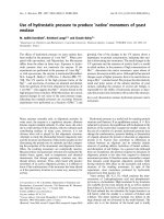
shown in Fig. 1. In this experiment, recombinant
M. smegmatis TreS, purified on the Ni column, was
kept on ice for 43 days and, at various times, samples
were removed and subjected to SDS ⁄ PAGE and also
assayed for MTase and amylase activities. The MTase
activity increased as the protein was degraded and was
approximately two-fold higher in the 58 kDa protein
as in the 68 kDa MTase. On the other hand, the amy-
lase activity remained constant during this change, but
it was present in all of the intermediate proteins, as
well as in the 58 kDa protein. The 58 kDa band was
eluted from the gel and subjected to tryptic digestion
and Q-TOF MS to identify the peptides. These data
indicated that the 58 kDa protein was identical to the
68 kDa TreS, except for the loss of approximately
10 kDa of peptide from the carboxy terminus. Thus,
these data indicate that the MTase activity is increased
by the loss of the carboxy-terminal region of the pro-
tein, but the amylase activity remains at the same level
in the various intermediate forms of the enzyme.
Additional evidence that both MTase and amylase
activities reside in the same protein is demonstrated by
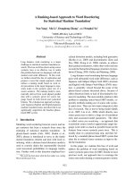
the experiment shown in Fig. 2. In this case, recombi-
nant M. smegmatis TreS was purified on a Ni column
and, after removal of imidazole, the protein was
allowed to remain in an ice bath for several weeks
until most of the protein had been converted to the
Fig. 1. Time course of conversion of 68 kDa TreS to 58 kDa TreS.
M. smegmatis TreS gene was cloned and expressed in E. coli with
a (His)
6
tag at the amino terminus. TreS was isolated on a Ni
column and enzyme was eluted with 100 m
M imidazole. An aliquot
of the purified TreS was subjected to SDS (0.1%) ⁄ PAGE (0 time),
and also was assayed for MTase and amylase activities. The TreS
elution from imidazole was stored on ice and aliquots were
removed at the times shown in the figure, and subjected to
SDS ⁄ PAGE and also tested to determine the activities of MTase
and amylase. The final protein product at 43 days was mostly com-
prised of the 58 kDa band, which had both MTase and amylase
activities. The following protein standards (STD) were run on the
gels to determine the molecular weight of the TreS: rabbit muscle
myosin, 200 kDa; ß-galactosidase, 116 kDa; phosphorylase B,
97 kDa; serum albumin, 66 kDa; ovalbumin, 45 kDa; carbonic anhy-
drase, 31 kDa.
Fraction A
41–50
0.5
0.8
51–60
0.5
1.1
61–70
8
4.4
71–80
16
7.8
81–90
9.7
3.8
91–100
6.7
2.5
101–110
4.5
1.8
BC DE F GSTDs
Tubes Pooled
MTase
Amylase
Fig. 2. Gel filtration profile of 58 kDa TreS-evidence for both activi-
ties in one protein. Purified recombinant TreS prepared from the
M. smegmatis or M. tuberculosis gene was stored for several
weeks on ice to produce the 58 kDa TreS protein. This protein was
chromatographed on a 1.5 · 120 cm Sephacryl S-200 column, and
the column was eluted with 10 m
M potassium phosphate buffer
(pH 6.8), containing 1
M KCl. Fractions were collected and starting
at tube number 41, fractions were pooled in batches of ten tubes
(i.e. tubes 41–50 = fraction A; tubes 51–60 = fraction B; tubes
61–70 = fraction C; tubes 71–80 = fraction D; and so on). Fractions
were concentrated on an Amicon concentrator and an aliquot of
each fraction was subjected to SDS ⁄ PAGE to identify and quanti-
tate the amount of protein, whereas another aliquot was assayed
to determine the amount of MTase and amylase activity, and the
ratios of the two. The activity of these enzymes and the ratio is
shown. Standard proteins (STDs) are as reported in Fig. 1.
TreS converts glycogen to trehalose Y. T. Pan et al.
3410 FEBS Journal 275 (2008) 3408–3420 ª 2008 The Authors Journal compilation ª 2008 FEBS
58 kDa form. This protein preparation was then
applied to a Sephacryl S-200 column (GE Healthcare,
Uppsala, Sweden), and fractions from the column were
collected. Starting at tube number 41, every 10 tubes
were pooled to give seven fractions as follows:
A = 41–50; B = 51––60; C = 61–70; D = 71–80;
E = 81–90; F = 91–100; G = 101–110; and
H = 111–120. An aliquot of each pooled fraction was
subjected to SDS ⁄ PAGE (Fig. 2) and MTase activity
and amylase activity were also assayed in each of these
fractions. Figure 2 shows that the 58 kDa protein was
clearly evident on SDS ⁄ PAGE gels in fractions C to
G, but was present in highest amounts in fractions D
and E. In addition, both MTase and amylase activities
were present in fractions B to H but, more impor-
tantly, the ratio of MTase to amylase remained fairly
constant in fractions C to F (Fig. 2, bottom). These
data strongly suggest that these two activities reside in
the same protein. As a control for these experiments,
we prepared a cell-free extract of the untransfected
vector and put it through the same purification proce-
dure. In this case, we did not find any amylase activity
in the imidazole elutions of the Ni column.
Finally, as further proof that amylase and MTase
activities reside in the same protein, we demonstrated
the presence of both activities in crystals of TreS.
These crystals had both MTase activity for converting
trehalose to maltose and amylase activity that con-
verted either glycogen or maltoheptaose to maltose
(Table 2). The amylase activity was better with malto-
heptaose as a substrate than with glycogen. A second
set of crystals was also isolated and tested in the same
way and showed both activities, although at slightly
different levels.
Demonstration of amylase activity
As described in the Experimental procedures, the
Dionex analyzer readily separates trehalose, maltose
and glucose from each other and quantifies the amount
of each sugar using an amperometric detection system.
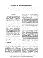
Figure 3A shows that the amount of maltose produced
from glycogen by the recombinant TreS was linear
with time of incubation for up to 24 h, and was also
proportional to the amount of enzyme added
(Fig. 3B), for up to at least 3 lg of protein. These data
also indicate that the amylase activity was quite stable
at 37 °C in the presence of glycogen because the rate
of production of maltose remained linear for at least
24 h of incubation. In these experiments, very little tre-
halose was detected at early times, probably because
the K
m
of MTase for maltose is approximately 10 mm
[14] and, therefore, even at 6 h of incubation, the
amount of maltose produced is far below the K
m
.
However, the production of trehalose from glycogen
could be demonstrated using radioactive glycogen as
the substrate, as described below.
The production of maltose from glycogen, as well as
the production of trehalose, could be demonstrated
Table 2. Enzymatic activities of MTase and amylase in crystals of
TreS. ND, not determined.
Time of
incubation
(min)
Amylase activity on [amount
of maltose (lg)]:
MTase activity
[maltose
produced (lg)]
Glycogen Maltoheptaose
5 ND ND 100
10 ND ND 260
15 ND ND 288
60 1.2 2.8 ND
120 2.5 4.1 ND
480 4.0 8.6 ND
1440 0.9 14.2 ND
A
B
Fig. 3. Effect of (A) time of incubation and (B) amount of enzyme
on the production of maltose from glycogen by TreS (i.e. amylase
activity). Incubations were as described in the text and contained
0.5 mg of glycogen in 100 lLof40m
M potassium phosphate buf-
fer (pH 6.0), containing 10 m
M CaCl
2
and various amounts of TreS.
The production of maltose was determined and quantitated on the
Dionex HPLC carbohydrate analyzer.
Y. T. Pan et al. TreS converts glycogen to trehalose
FEBS Journal 275 (2008) 3408–3420 ª 2008 The Authors Journal compilation ª 2008 FEBS 3411
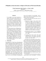
using the Dionex carbohydrate analyzer (Fig. 4).
[
3
H]glycogen was incubated either with the purified
TreS (lower profile), or with a commercial preparation
of pancreatic amylase to serve as a control (upper pro-
file). After incubation for 6 h, the reaction mixtures
were passed through a column of Biogel P-4, and those
column fractions representing the monosaccharide to
tetrasaccharide elution region of the column were
pooled, concentrated, and the radioactive sugars were
identified on the Dionex HPLC by analyzing an ali-
quot of each fraction for its radioactive content. The
upper profile shows that the pancreatic amylase gener-
ated a large peak of [
3
H]maltose and a smaller peak of
[
3
H]glucose, but no radioactive trehalose was produced
by this enzyme. By contrast, incubation with the TreS
generated a large peak of radioactive maltose as well
as a substantial peak of radioactive trehalose and a
small peak of [
3
H]glucose. The radioactive peak corre-
sponding to trehalose was completely susceptible to
digestion by a specific recombinant trehalase produced
in E. coli, and this digestion resulted in the production
of radioactive glucose as the only product (data not
shown). That maltose is the initial product produced
from glycogen was previously demonstrated by the
experiment shown in Fig. 3A when the time course
fractions were analyzed on the Dionex and essentially
no trehalose was observed at the early time points, but
was clearly evident at later times of incubation. Thus,
TreS not only has MTase activity, but also it has
amylase activity that produces the initial maltose.
Properties of the M. smegmatis amylase activity
As indicated in Fig. 3, the production of maltose from
glycogen by TreS increased in a linear fashion with
increasing time of incubation and with increasing
amounts of protein. The pH requirement for the con-
version of glycogen to maltose was determined and the
pH optimum was found to be in the range 6.0–6.2
(data not shown). Interestingly, the pH optimum for
the MTase activity (conversion of trehalose to maltose)
of TreS was previously determined to be 7.0 [14].
TreS can also use maltooligosaccharides as sub-
strates to produce maltose and then trehalose. A com-
parison of the activity of TreS on glycogen and on
maltoheptaose is presented in Table 3. Maltoheptaose
Fig. 4. Production of radioactive maltose and trehalose from
[
3
H]glycogen by TreS. [
3
H]Glycogen was incubated with either com-
mercial porcine pancreatic a-amylase (upper profile) or with purified
TreS (lower profile) for 24 h in 40 m
M potassium phosphate buffer
(pH 6.0), containing 10 m
M CaCl
2
. Reactions were terminated by
heating and each mixture was passed through a 1.5 · 200 cm col-
umn of Biogel P-4. Fractions emerging in the monosaccharide
through tetrasaccharide region of the column were pooled, concen-
trated to a small volume, deionized with mixed-bed ion-exchange
resin (Dowex-1-CO
3
2)
and Dowex-50-H
+
) and analyzed on the Dio-
nex carbohydrate analyzer. The HPLC was equipped with a splitter
so that the fractions of the effluent could be withdrawn for deter-
mination of their radioactive content. The position of elution of the
standards glucose, maltose and trehalose are indicated on each
chromatogram and the amount of radioactivity in each area is
plotted as shown.
Table 3. Comparison of maltoheptaose and glycogen as substrates
for TreS.
Amount of substrate
(lg added to incubation)
Maltose (lg) produced from:
Maltoheptaose Glycogen
20 1.12 0.57
50 1.84 1.13
100 2.44 2.07
250 2.94 1.54
500 5.00 1.70
TreS converts glycogen to trehalose Y. T. Pan et al.
3412 FEBS Journal 275 (2008) 3408–3420 ª 2008 The Authors Journal compilation ª 2008 FEBS
was a good substrate for the production of maltose
and the rate of maltose formation increased with
increasing amounts of substrate. In the presence of
higher amounts of maltoheptaose and with longer
incubations, trehalose was also identified in these incu-
bations. The production of maltose was measured by
determining the area of the maltose peak on the Dio-
nex analyzer. It is not possible to directly compare the
effectiveness of maltoheptaose to glycogen because the
commercial glycogen is a mixture of glucose polymers
of different molecular masses, and probably different
degrees of branching. However, this experiment dem-
onstrates that TreS can use various maltooligosaccha-
rides, as well as glycogen, to produce maltose and
trehalose.
The amylase activity of TreS was stimulated by the
addition of Ca
2+
(Fig. 5). Although amylase activity
was not completely dependent on the presence of this
divalent cation, the activity was stimulated by as much
as four-fold in the presence of 14 mm Ca
2+
. Other
metal ions such as Mg
2+
,Mn
2+
, and Co
2+
were also
somewhat effective but Hg
2+
,Cu
2+
,Ni
2+
and Zn
2+
were inhibitory. The requirement for Ca
2+
for activity,
and its ability to stabilize this group of glucosylhydro-
lases, has been demonstrated for a number of a-amy-
lases [20–22]. Whereas the amylase activity of TreS is
stimulated by calcium ions, the MTase is inhibited by
divalent cations. This effect is also shown in Fig. 5.
These data show that MTase not only does not require
Ca
2+
, but also that this activity is strongly inhibited
by calcium concentrations of 5 mm or higher. Other
cations were also inhibitory to MTase activity.
Selective inhibition of MTase and amylase
activities
Two inhibitors have been identified that selectively
inhibit either the amylase activity or the MTase
activity, suggesting the possibility of two different
active sites in the TreS. Validoxylamine is a known
inhibitor of trehalases [23] and has a structure that
mimics a number of known glycosidase inhibitors
[24]. Figure 6A shows that validoxylamine is a potent
inhibitor of the MTase activity of TreS, with a K
i
of
approximately 25 nm . However, validoxylamine had
no effect on the amylase activity, even at concentra-
tions of 2 lm. Figure 6B shows a Lineweaver–Burk
plot of the effect of trehalose concentration on
MTase activity at two different concentrations of
validoxylamine. These data clearly demonstrate that
validoxylamine is a competitive inhibitor of MTase
with respect to trehalose. Although the amylase
activity of TreS is not inhibited by validoxylamine, it
is inhibited by the known amylase inhibitor, acarbose
[24–27]. These experiments with acarbose (Fig. 7)
demonstrate that the amylase activity is strongly
inhibited by acarbose, with 50% inhibition occurring
at a concentration of approximately 5 lgÆmL
)1
.On
the other hand, MTase activity was not susceptible
to inhibition by acarbose, even at concentrations of
100 lgÆmL
)1
. Other studies on the crystal structure
of pancreatic amylase demonstrated that acarbose
binds at the active site of this enzyme [26,27]. Addi-
tional studies with acarbose have suggested that it
acts as a transition state inhibitor with amylase-like
enzymes, also indicating that it binds at the active
sites of these enzymes.
Additional support for the hypothesis that TreS has
two different binding sites is provided by the finding
that adding glycogen to incubations of MTase with
trehalose does not inhibit the conversion of trehalose
to maltose. In this experiment, increasing amounts of
glycogen were added to incubation mixtures containing
buffer, trehalose and TreS (Table 4). Following incu-
bation for 15 min, reactions were stopped by heating,
and the amount of maltose was determined by the
reducing sugar method. A series of control incubations
were also run that contained the same amounts of gly-
cogen, buffer and enzyme, but trehalose was omitted
from these incubations. Glycogen had no effect on the
MTase activity because the amount of reducing sugar
did not change in these incubations containing treha-
lose, regardless of the amount of glycogen added
Fig. 5. Effect of calcium ion concentration on the activity of a-amy-
lase or MTase (designated as TreS) activities of TreS. TreS (0.2 lg)
was incubated with glycogen (
) in the presence of 40 mM sodium
acetate buffer and various amounts of calcium for 10 h, and then
the production of maltose was determined on the Dionex carbohy-
drate analyzer. To determine the effect of calcium on MTase activ-
ity, 50 m
M trehalose ( ) was incubated with 0.2 lg MTase for
10 min in 40 m
M potassium phosphate (pH 6.8) with various
amounts of calcium as shown. In this case, maltose was deter-
mined by the reducing sugar determination.
Y. T. Pan et al. TreS converts glycogen to trehalose
FEBS Journal 275 (2008) 3408–3420 ª 2008 The Authors Journal compilation ª 2008 FEBS 3413
(Table 4). The product from several of these incuba-
tions was also examined on the Dionex analyzer, and
trehalose and maltose were the only oligosaccharides
detected. On the other hand, no reducing sugar or
maltose was detected in the control incubations of
glycogen and enzyme, but without trehalose. It should
be noted that incubations were only 15 min in length;
therefore, the amylase activity on the added glycogen
was not sufficient to produce detectable amounts of
maltose. Thus, the MTase site of TreS appears to be
distinct from the amylase site.
Importance of TreS in homeostasis of
mycobacteria
Although the exact function of TreS is not known,
and mutants lacking TreS can still grow if trehalose is
added to the medium, this enzyme does appear to play
a key role in the interactions between glycogen and
trehalose. Thus, under some circumstances, such as
low levels of cytoplasmic trehalose, it is likely that the
cells would degrade glycogen to maltose, and this
maltose would then be converted to trehalose to raise
trehalose levels. Interestingly, as shown in the present
Fig. 7. Effect of acarbose on the amylase and MTase (designated
as TreS) activities of TreS. Incubations were as described previ-
ously but contained various amounts of acarbose (0–10 lg per incu-
bation mixture). The amount of maltose produced from glycogen in
these incubations (h) was determined on the Dionex carbohydrate
analyzer, whereas the amount of maltose from trehalose (,) was
measured by the reducing sugar test.
100
A
75
50
10 20 30 40
Amount of validoxylamine A
(ng per assay)
Enzyme activity
(% of control)
50
Amylase
TreS
5000
25
0
0.6
0.5
0.4
0.3
0.2
0.1
0
020–20
1 / S
1 / V
40 60 80
100
B
Fig. 6. Effect of validoxylamine (upper graph) on the MTase (,)
and amylase (h) activities of TreS. (A) Incubations of MTase (desig-
nated as TreS) with trehalose were as described in the Experimen-
tal procedures, but contained various amounts of validoxylamine
(0–500 ng per incubation mixture). Each incubation contained
0.2 lg of purified and recombinant TreS. The amount of maltose
produced was determined by the reducing sugar method. Validoxyl-
amine is also shown to have no effect on the amylase activity (h)
of TreS. These incubations were as described in the Experimental
procedures, except that increasing amounts of validoxylamine were
added up to 500 ng per incubation. The formation of maltose from
glycogen was determined by HPLC. (B) Incubations contained
increasing amounts of trehalose plus buffer and 0.2 lg of purified
TreS. One set of tubes served as the control (r) to determine the
K
m
for the substrate, the second set was identical, except that
each tube also had 5 ng of the inhibitor, validoxylamine (
), and
the third set contained the same components as the second set,
except that it had 10 ng per incubation (
) of validoxylamine. Again,
the amount of maltose produced from trehalose was determined
by the reducing sugar method, and the data was plotted by the
method of Lineweaver and Burk.
Table 4. Effect of glycogen on MTase activity (trehalose fi malt-
ose). Incubations with trehalose as substrate were as described in
the text. The amount of maltose formed was determined by the
reducing sugar test.
Amount of glycogen
added to incubations (lg)
Reducing
sugar (A
620
)
0 0.96
100 0.98
200 1.04
400 0.98
800 0.91
TreS converts glycogen to trehalose Y. T. Pan et al.
3414 FEBS Journal 275 (2008) 3408–3420 ª 2008 The Authors Journal compilation ª 2008 FEBS
study, high levels of trehalose in the cytoplasm also
appear to cause ⁄ or stimulate the accumulation of gly-
cogen in these cells. This effect is shown by the data
presented in Table 5, where the levels of cytoplasmic
glycogen are compared in wild-type M. smegmatis,or
in various trehalose mutants (for identification
of mutants, see Table 1) grown in a mineral salts med-
ium with low (0.1%) or high (2% or 4%) amounts of
trehalose. Those flasks containing 0.1% trehalose also
had 1.9% glucose. With 2% or higher concentrations
of trehalose in the media, cells containing TreS (wild-
type and mutants #47 and #74; Table 1) had 10 to
30-fold more glycogen than cells grown in low treha-
lose, or in cells lacking TreS (mutants #80 and #91)
(Table 5). Furthermore, Table 6 shows that this
increase in glycogen levels only occurred when treha-
lose was in the media at concentrations of 25 mm
( 1%) or higher, but did not occur in the presence of
high levels of glucose, or other sugars such as sucrose
or lactose (not shown). The level of cytoplasmic treha-
lose in wild-type M. smegmatis was not significantly
altered by high (100 mm) concentrations of trehalose or
glucose in the media (Table 6), suggesting that the level
of intracellular trehalose is carefully regulated. There is
some evidence from other systems that high intracellu-
lar concentrations of trehalose may be toxic to cells.
Discussion
TreS is a 68 kDa protein that is present in a number
of bacteria, including mycobacteria, corynebacteria,
nocardia and streptomyces, as well as arthrobacter,
sulfolobus and rhizobium [8,11–13]. TreS has been
purified to near homogeneity from M. smegmatis, and
the gene for this protein was cloned and expressed in
E. coli [14]. The expressed protein had a subunit
molecular mass of 68 kDa on SDS gels, but active
enzyme eluted as a 390 kDa protein upon gel filtration,
suggesting that active TreS is a hexamer of six identi-
cal subunits. TreS catalyzes the reversible interconver-
sion of trehalose and maltose. The reaction kinetics
favor the conversion of maltose to trehalose, with a
K
m
for maltose of approximately 10 mm, whereas the
K
m
for trehalose is approximately 90 mm.
In Corynebacterium glutamicum , TreS has been pro-
posed to function as a substitute for a trehalase to
control intracellular levels of trehalose because no
ORF homologous to known trehalase genes have been
identified, nor has any trehalase activity been demon-
strated in this organism [15]. However, M. smegmatis
does have a highly specific and active trehalase [17], in
addition to the TreS described above [14]. Another
report on the TreS of C. glutamicum suggests that this
enzyme is only involved in trehalose biosynthesis when
these organisms are growing on maltose. Thus, a criti-
cal question with regard to the production of trehalose
by TreS remains. What is the possible source of
maltose that TreS could use as a substrate to produce
trehalose?
Exogenous maltose is not a likely source of maltose
for M. smegmatis because this organism grows very
poorly on maltose. However, the results obtained in
the present study indicate that endogenous maltose
can be produced from glycogen by the amylase activity
of TreS, and that this maltose is readily converted to
trehalose by the MTase activity of TreS.
The present study provides evidence indicating that
both activities reside in the same protein. First, TreS,
purified from M. smegmatis as well as recombinant
TreS produced in E. coli, had both MTase activity and
amylase activity. Second, the 68 kDa TreS undergoes
auto-proteolysis to give a 58 kDa protein, which also
contains both MTase and amylase activity. Third, the
58 kDa protein was subjected to gel filtration and frac-
tions were collected. Six fractions across the protein
peak had variable amounts of MTase activity with the
Table 5. Effect of exogenous trehalose on accumulation of glyco-
gen. In all cases, the sugar content of the media was 2% or higher
(i.e. 0.1% trehalose + 1.9% glucose, etc.).
Amount of trehalose in
media (% ⁄ weight)
Amount of glycogen in cells
(nmol glycogen as glucoseÆmg
)1
dry cells)
in the following wild-type or mutant:
B11 (wild) 47 74 80 91
0.1 (+1.9 glucose) 14.6 30.9 13.4 14.5 14.6
2.0 340.4 187.4 315.1 4.2 12.9
4.0 513.9 369.5 316.5 6.8 11.1
Presence of TreS
in cells
+++))
Table 6. Effect of trehalose concentration in the media on levels
of glycogen and trehalose in cells of M. smegmatis. All experi-
ments were performed with the wild-type organism (i.e. B11).
Sugar in the
media (glucose or
trehalose) (m
M)
Amount of trehalose
in cytoplasm
(lgÆmg
)1
cells)
Amount of glycogen
in (nmol as
glucose ⁄ mg cells)
On
glucose
On
trehalose
On
glucose
On
trehalose
0.125 7.1 10.2 20.0 25.1
1.25 12.5 15.8 6.6 6.8
25.0 4.4 6.6 17.0 150.1
50.0 5.4 4.6 10.3 303.7
100.0 13.4 3.4 47.5 372.0
Y. T. Pan et al. TreS converts glycogen to trehalose
FEBS Journal 275 (2008) 3408–3420 ª 2008 The Authors Journal compilation ª 2008 FEBS 3415
highest activity corresponding to fractions showing the
most 58 kDa protein by SDS ⁄ PAGE. Importantly, the
ratio of MTase ⁄ amylase, but not the absolute activity,
remained fairly constant in fractions having different
amounts of the 58 kDa protein. Finally, crystals of
TreS were obtained, and these isolated crystals have
both amylase activity and MTase activity.
These results strongly indicate that the MTase act-
ivity and the amylase activity are in the same protein,
and suggest that this multifunctional protein has the
capacity to convert glycogen to trehalose.
The partial amino acid sequence of TreS from
M. smegmatis allowed us to locate the ORF for this
protein and a blastp search indicated that it had
approximately 83% identity to a gene (Rv 0126) for a
hypothetical a-amylase in the M. tuberculosis genome
[28]. It also has 72% identity to a putative TreS from
Streptomyces avermitilis, 69% identity in C. glutami-
cum, and 61% identity to the putative TreS from Pseu-
domonas sp. Because there are no reports on the
isolation or characterization of these TreS proteins, it
is not known whether they also have amylase activity,
but it will be interesting to determine whether the TreS
of corynebacteria also shares this activity. It will also
be important to determine ways to test this amylase
activity for function in vivo to establish whether it can
really act in collaboration with the TreS activity to
convert glycogen glucoses into cytoplasmic trehalose.
We propose that TreS has two distinct active sites:
one catalyzing the interconversion of maltose and tre-
halose (referred to here as MTase activity) and the
other catalyzing the breakdown of glycogen to maltose
(amylase activity).
The present study provides evidence supporting the
existence of the two sites. First, the amylase site is acti-
vated by Ca
2+
whereas the MTase activity is inhibited
by Ca
2+
and other cations. Second, we have identified
two inhibitors each of which competitively inhibits one
activity and not the other. Thus, validoxylamine com-
petitively inhibits MTase but not amylase, whereas
acarbose competitively inhibits amylase but not
MTase. Third, glycogen, which is a substrate for the
amylase activity of TreS, has no effect on the MTase
activity of TreS. That is, incubations of MTase with
trehalose produce the same amount of maltose, even in
the presence of high amounts of glycogen.
These data suggest that these two activities reside in
different sites on the protein. However, it will require
site-directed mutagenesis studies, or deletions of vari-
ous parts of the protein, to conclusively prove that
there are indeed two sites. Once we have identified
active site amino acids for each catalytic activity, it will
be possible to perform site-directed mutagenesis to
modify one activity and not the other. We have been
able to obtain small-sized crystals of TreS but they do
not have sufficiently high resolution for structural
analysis. Attempts to improve the resolution of these
crystals is in progress.
Our hypothesis on the function of TreS is that it
serves as a sensor and ⁄ or controller of the cellular tre-
halose levels in mycobacteria and perhaps other organ-
isms. The present studies show that TreS can mediate
the formation of trehalose from glycogen. In addition,
growth studies with the wild-type M. smegmatis show
that, when this organism is grown in a mineral salts
medium with high concentrations (1–4%) of trehalose
as the major carbon source, these cells contain 10- to
30-fold higher amounts of glycogen than cells grown
in the same concentration of glucose or other sugars.
Furthermore, additional studies with a number of tre-
halose mutants that are missing one, two or all three
of the trehalose biosynthetic pathways (Table 1) dem-
onstrate that any of the mutants still containing TreS
(including the mutant that only has TreS) show this
accumulation of glycogen in the presence of high tre-
halose, but any mutants that are missing TreS do not
accumulate glycogen at any level of trehalose, or any
other sugar. Thus, TreS not only is involved in the
production of trehalose from glycogen, but also
appears to play an essential role in the formation,
and ⁄ or accumulation, of glycogen. This accumulation
somehow involves the utilization of trehalose as the
carbon source, but the mechanism of this conversion is
not known. We propose that when high levels of treha-
lose are produced in the cell, perhaps as a result of
exposure to stress, TreS may function to convert this
trehalose to maltose and then to glycogen when the
stress is removed. Removal of trehalose is probably
essential because high levels of trehalose may be toxic.
On the other hand, if trehalose falls to a dangerously
low level, TreS may function to convert glycogen to
maltose and then to trehalose. Ongoing studies are
attempting to determine how trehalose is involved in
the formation of glycogen, and how TreS functions as
a sensor or regulator of trehalose and ⁄ or glycogen
levels in these cells.
Experimental procedures
Bacterial strains and culture conditions
M. smegmatis was obtained from the American Type Cul-
ture Collection (ATCC 14468). It was maintained on slants
of Trypticase Soy Agar and was grown at 37 °Cin2L
Erlenmeyer flasks containing 1 L of Trypticase Soy Broth
(Becton Dickinson, Franklin Lakes, NJ, USA). The E. coli
TreS converts glycogen to trehalose Y. T. Pan et al.
3416 FEBS Journal 275 (2008) 3408–3420 ª 2008 The Authors Journal compilation ª 2008 FEBS
strains DH5a and HMS-F were used for cloning and
expression studies, respectively. HMS-F is a derivative of
the expression strain HMS174(DE-3) (Novagen, Madison,
WI, USA). HMS174(DE-3) contains a chromosomal iso-
propyl thio-b-d-galactoside-inducible T7 RNA pol gene.
HMS-F contains an additional copy of the lac repressor
lacI
q
on an F episome, which was transferred from the
E. coli cloning strain XL-1 (Stratagene, La Jolla, CA,
USA). This addition essentially represses expression from
the T7 promoter on the E. coli expression vector pET15b
(Novagen) in the absence of isopropyl thio-b-d-galactoside.
HMS-F was routinely cultured in the presence of tetracy-
cline (10 lgÆmL
)1
) to maintain carriage of the F episome.
E. coli strains were cultured in LB-broth and on LB-agar
supplemented with 100 lgÆmL
)1
ampicillin, 20 lgÆmL
)1
kanamycin or 10 lgÆmL
)1
tetracycline, individually or in
combination, where applicable.
Preparation of mutant strains of M. smegmatis
missing various trehalose synthetic pathways
Mutants were prepared by allele replacement mutagenesis.
Target genes were PCR amplified using gene specific
primers from M. smegmatis genomic DNA. The cloned
target gene was mutagenized by generating an internal
deletion in the target ORF. The deletion was confirmed by
sequencing the mutagenized allele. The PCR product was
ligated into the plasmid pMAR1, a mycobacterial suicide
vector constructed by introducing a unique PacI restriction
site and a wild-type allele of M. smegmatis rspL [29] into
the E. coli cloning vector pSP72 (Promega, Madison, WI,
USA). A PacI-ended selection cassette, containing a posi-
tive selector hyg (hygromycin resistant), the reporter gene
lacZ and the negative selector sacB (each driven by sepa-
rate mycobacterial promotors), was inserted into the
pMAR1 PacI site [30]. The resulting plasmid was then
transformed into the wild-type M. smegmatis [31]. Duplica-
tion insertions, resulting from homologous recombination
between the plasmid-borne mutant allele and the chromo-
somal wild-type target gene, were recovered on medium
containing hygromycin and X-Gal. These transformants
were also streptomycin-sensitive, as a result of acquisition
of the plasmid-borne rspL
wt
gene. Wild-type rspL-mediated
streptomycin sensitivity (Str
s
) is dominant over mutant
rspL-mediated streptomycin resistance [32]. The presence of
both wild-type and mutant alleles were confirmed by PCR.
Resolution of the duplication and loss of one of the target
gene alleles by homologous recombination was selected for
by growing the culture in nonselective medium and plating
on medium containing streptomycin and X-Gal. Loss of
the integrated pMAR1 plasmid restored streptomycin resis-
tance (Str
r
). Resultant Str
r
Lac
–
colonies were screened by
PCR for the carriage of the wilt-type or mutant allele. As
this strategy generates unmarked deletion mutants that are
fully amenable to further rounds of mutagenesis, multiple
pathways were mutagenized in the same M. smegmatis
strain [30]. The mutants used are shown in Table 1 and
comprise: mutant #47 missing TPP and the TPS ⁄ TPP path-
way, but having functional TreS and TreY ⁄ TreZ pathways;
mutant #74 missing TPS, TPP and TreY and the TPS ⁄ TPP
and TreY ⁄ TreZ pathways, but having a functional TreS
pathway; mutant #91 missing TreS and the TreS pathway
but having functional TPS ⁄ TPP and TreY ⁄ TreZ pathways;
mutant #80 missing TPS, TPP, TreS and TreY and having
no trehalose biosynthetic pathways. mutant #80 absolutely
requires trehalose in the media for growth.
Materials and reagents
Trehalose, maltose, isomaltose, malto-oligosaccharides and
other sugars were purchased from Sigma Chemical Co.
(St Louis, MO, USA). DEAE-cellulose and various other
chromatographic resins for protein purification, molecular
markers for gel filtration and buffers were also obtained
from Sigma. Bio-Rad protein reagent and DE-52 were from
Bio-Rad Laboratories Inc. (Hercules, CA, USA). Trypti-
case soy broth was from Becton Dickinson, and LB broth
was from Fisher Scientific Co. (Pittsburgh, PA, USA).
Radioactive glycogen was made by growing M. smegmatis
in high-specific activity [
3
H]glucose in a mineral salts
medium for 48 h. The glycogen was isolated and purified as
previously described [33]. One hundred to five hundred lCi
of [U-
3
H]-glucose was added to Trypticase Soy Broth that
did not contain any unlabeled glucose. The flasks contain-
ing this radioactivity were inoculated with a small innocu-
lum of a growing culture of M. smegmatis and the cultures
were grown for 2 days at 37 °C on a recriprocal shaker. At
the end of this time, cells were harvested by centrifugation,
washed with NaCl ⁄ P
i
and sonicated in water. The cell deb-
ris was removed by high-speed centrifugation, and the
supernatant (cytosolic) fraction was cooled and cold tri-
chloroacetic acid was added with stirring to a final concen-
tration of 5% to precipitate the protein. The precipitated
protein was removed by centrifugation and discarded, and
the supernatant liquid was placed in a large separatory fun-
nel and extracted four times with large volumes of ethyl
ether to remove the trichloroacetic acid. The aqueous frac-
tion from these extractions was concentrated to a smaller
volume and three volumes of ice cold methanol was added
with stirring to precipitate the glycogen. The precipitate
was isolated by centrifugation, dissolved in water and sub-
jected to descending paper chromatography on 3MM paper
(Whatman, Clifton, NJ, USA) in n-butanol ⁄ pyridine ⁄ water
(4:3:4, v⁄ v⁄ v). As glycogen is a large molecule, it
remains at the origin, but monosaccharides and oligosac-
charides migrate down the paper and away from the origin.
The papers were dried and the glycogen at the origins was
eluted with water and passed through a column of Biogel
P-4 (Bio-Rad Laboratories Inc.). A large symmetrical peak
of radioactive glycogen emerged from the P-4 column at
Y. T. Pan et al. TreS converts glycogen to trehalose
FEBS Journal 275 (2008) 3408–3420 ª 2008 The Authors Journal compilation ª 2008 FEBS 3417
the void volume of the column. This fraction was used as
the glycogen substrate. It reacted with iodine and this
complex gave a spectrum in the same visible range as that
produced by authentic glycogen.
Purification of TreS from M. smegmatis
TreS was purified from cell free extracts of M. smegmatis
as previously described [14]. This included ammonium
sulfate fractionation, gel filtration, chromatography on
columns of DEAE-cellulose, hydroxyapatite, aminohexyl-
agarose and then phenyl-sepharose. At the final stage of
purification, the enzyme preparation showed one major
protein band of 68 kDa on SDS gels, which had both
MTase activity and amylase activity (see Results).
Crystals of TreS were obtained using the crystallization
kits, and these crystals were tested for the presence of both
MTase activity and amylase activity. Although these crys-
tals were small and not of high enough resolution for struc-
tural studies, they were isolated by centrifugation in a
microfuge tube, washed several times with the same fresh
crystallization fluid, and then dissolved in the assay buffer.
Both MTase activity and amylase activity were measured in
these dissolved crystals using the assay methods described
below. The results of those assays are presented in Table 2.
Assay of TreS activities
The MTase activity of TreS was measured by determining
the formation of reducing sugar resulting from the forma-
tion of maltose, when the enzyme was incubated with tre-
halose. Assays were performed in a final volume of
100 lL containing 40 mm potassium phosphate buffer
(pH 6.8), various amounts of trehalose (usually
50–100 mm) and an appropriate amount of enzyme. After
incubation at 37 °C for various time periods, the mixture
was heated in a boiling water bath for several minutes to
stop the reaction, and the amount of maltose produced
was determined by the reducing sugar method [34]. The
production of maltose could also be determined by
subjecting the heated reaction mixture to HPLC on the
Dionex carbohydrate analyzer (Dionex, Sunnyvale, CA,
USA). In addition, the activity could also be assayed in
the opposite direction by measuring the formation of
trehalose when TreS (MTase) was incubated with maltose.
This was best perfomred using the Dionex carbohydrate
analyzer that readily separates trehalose from maltose, glu-
cose and other sugars (Fig. 4). As shown in the present
study, TreS can also convert glycogen to maltose and tre-
halose. For assay of these reactions, incubations contained
0.5 mg of glycogen in 100 lLof40mm potassium phos-
phate buffer (pH 6.0) or sodium acetate buffer (pH 6.0),
10 mm CaCl
2
, and various amounts of enzyme. After
incubation as described above, the reaction mixtures were
subjected to HPLC on the Dionex carbohydrate analyzer
and the amounts of maltose and trehalose produced from
glycogen were measured.
Separation and identification of sugars
Sugars were separated and identified using high-perfor-
mance anion-exchange chromatography on the Dionex
carbohydrate analyzer. Eluents were distilled water (E1)
and 400 nm NaOH (E2). Appropriate aliquots (0–3 nmol)
from each sample were injected into a CarboPac PA-1 col-
umn equilibrated with a mixture of E1 and E2
(E1 ⁄ E2 = 98 ⁄ 2). The elution and resolution of the carbo-
hydrate mixtures was performed as follows: T
0
–
T
20 min
=2% E2 (v⁄ v); T
20 min
– T
30 min
= gradient 2%
E2 to 100% E2 (v ⁄ v); T
30 min
–T
¥
= 100% E2 (v ⁄ v). Each
constituent was detected by pulse amperometry as recom-
mended by the manufacturer (Dionex, technical note, 20
March 1989) at a range setting of 300 K. In some cases, an
aliquot of the elution fraction was subjected to liquid scin-
tillation counting to determine the radioactive content of
each peak. These aliquots were mixed with scintillation
fluid and counted in a Beckman scintillation counter (Beck-
man Coulter Inc., Fullerton, CA, USA).
Other methods
Protein was measured with the Bio-Rad protein reagent
using BSA as the standard. Sugars were analyzed using
the Dionex carbohydrate analyzer to separate maltose, tre-
halose and other sugars. Reducing sugars were measured
and quantified by the copper colorimetric method of
Nelson [34]. SDS ⁄ PAGE was performed according to
Laemmli in 10% polyacrylamide gel using 0.1% SDS
[35]. The gels were stained with 0.5% Coomassie blue in
10% acetic acid.
Acknowledgements
We thank Dr Alan Tackett (Department of Biochemi-
stry and Molecular Biology, University of Arkansas
for Medical Sciences) for sequencing the 58 kDa TreS
and comparing this sequence to that of the 68 kDa
TreS. We also thank Drs Reha Celikel and Kottayil
Varughese (Department of Physiology and Biophysics,
University of Arkansas for Mediucal Sciences) for
obtaining the crystals of TreS.
References
1 Elbein AD, Pan YT, Pastuszak I & Carroll JD (2003)
New insights on trehalose: a multifunctional molecule.
Glycobiology 13, 17R–27R.
2 Elbein AD (1974) The metabolism of a,a-trehalose. Adv
Carbohydrate Chem Biochem 30, 227–256.
TreS converts glycogen to trehalose Y. T. Pan et al.
3418 FEBS Journal 275 (2008) 3408–3420 ª 2008 The Authors Journal compilation ª 2008 FEBS
3 Sussman AS & Lingappa BT (1959) Role of trehalose
in ascospores of Neurospora tetrasperma. Science 130,
1343–1344.
4 Arguillis JC (2000) Physiological roles of trehalose in
bacteria and yeast: a comparative analysis. Arch Micro-
biol 174, 217–224.
5 Thevelein JM & Hohmann S (1995) Trehalose synthase:
guard to the gate of glycolysis in yeast? Trends Biochem
Sci 20, 3–10.
6 Burklen L, Schock F & Dahl MK (1998) Molecular
analysis of the interaction between the Bacillus trehalose
repressor TreR and the tre operator. Mol Gen Genetics
260, 48–55.
7 Brennan PJ & Nikaido H (1995) The envelope of myco-
bacteria. Annu Rev Biochem 64, 29–63.
8 DeSmet KAL, Weston A, Brown IN, Young DB &
Robertson BD (2000) Three pathways for trehalose bio-
synthesis in mycobacteria. Microbiology 146, 199–208.
9 Cabib E & Leloir LF (1958) The biosynthesis of treha-
lose-phosphate. J Biol Chem 231, 259–275.
10 Klutts S, Pastuszak I, Korath-Edavana V, Thampi P,
Pan YT, Abraham E, Carroll JD & Elbein AD (2003)
Purification cloning expression and properties of myco-
bacterial trehalose-phosphate phosphatase. J Biol Chem
278, 2093–2100.
11 Maruta K, Mitsuzumi H, Nakada T, Kubota M, Chaen
H, Fukuda S, Sugimoto T & Kurimoto M (1996) Clon-
ing and sequencing of a cluster of genes encoding novel
enzymes of trehalose biosynthesis from thermophilic
archaebacterium Sulfolobus acidocaldarius. Biochim Bio-
phys Acta 1291, 177–181.
12 Nakada T, Ikegama S, Chaen H, Kubata M, Fukuda S,
Sugimoto T, Kurimoto M & Tsujisaka Y (1996) Purifi-
cation and characterization of thermostable maltooligo-
syl trehalose trehalohydrolase from the thermophilic
archaebacterium Sulfolobus acidocaldarius. Biosci
Biotechnol Biochem 60, 267–270.
13 Nishimoto T, Nakano M, Nakada T, Chaen H, Fukuda
S, Sugimoto T, Kurimoto M & Tsujisaka Y (1995)
Purification and properties of a novel enzyme trehalose
synthase from Pimelobacter sp R48. Biosci Biotechnol
Biochem 60, 640–644.
14 Pan YT, Korath-Edavana V, Jourdian WJ, Edmondson
R, Carroll JD & Elbein AD (2004) Trehalose synthase
of Mycobacterium smegmatis: purification cloning
expression and properties of the enzyme. Eur J Biochem
271, 4259–4269.
15 Wolf A, Kramer R & Morbach S (2003) Three
pathways for trehalose metabolism in Corynebacterium
glutamicum ATCC13032 and their significance in
response to osmotic stress. Mol Microbiol 49, 1119–
1134.
16 Tzetkov M, Klopprogge C, Zelder O & Liebl W
(2003) Genetic dissection of trehalose biosynthesis in
Corynebacterium glutamicum: inactivation of trehalose
production leads to impaired growth and an altered
cell wall lipid composition. Microbiology
149, 1659–
1673.
17 Carroll JD, Pastuszak I, Korath-Edavana V, Pan YT &
Elbein AD (2007) A novel trehalase from Mycobacte-
rium smegmatis: purification properties and require-
ments. FEBS J 274, 1701–1714.
18 Tropis M, Meniche X, Wolf A, Gebhardt H, Strekov S,
Chami M, Schomburg D, Kramer R, Morbach S &
Daffe M (2005) The crucial role of trehalose and struc-
turally related oligosaccharides in the biosynthesis and
transfer of mycolic acids in Corynebacterineae. J Biol
Chem 280, 26573–26585.
19 Woodruff PJ, Carlson BL, Siridechadilok B, Pratt MR,
Senaratne RH, Mougous JD, Riley LW, Williams SJ &
Bertozzi CR (2004) Trehalose is required for growth of
Mycobacterium smegmatis. J Biol Chem 279, 28835–
28843.
20 Vallee BL, Stein EA, Sumerwell WN & Fischer EH
(1959) Metal content of a-amylases of various origins.
J Biol Chem 234, 2901–2909.
21 Buisson G, Duee E, Haser R & Payan F (1987) Three
dimensional structure of porcine pancreatic a-amylase
at 2.9 angstrom resolution. Role of calcium in structure
and activity. EMBO J 6, 3909–3916.
22 Bush DS, Sticher L, van Huystee R, Wagner D & Jones
RJ (1989) The calcium requirement for stability and
enzymatic activity of two isoforms of barley aleurone
a-amylase. J Biol Chem 264, 19392–19398.
23 Kameda Y, Asano N, Yamaguchi T & Mutsui K (1987)
Validoxylamines as trehalase inhibitors. J Antibiot 40,
563–565.
24 Asano N (2003) Glycosidase inhibitors: update and per-
spectives on practical use. Glycobiology 13, 93R–104R.
25 Schmidt DD, Frommer W, Muller L, Junge B, Wingen-
der W & Truscheit E (1977) a-Glucosidase inhibitors.
New complex oligosaccharides of microbial origin.
Naturwissenschaften 64, 535–536.
26 Gilles C, Astier J-P, Marchis-Mouren G, Cambillau C
& Payan F (1996) Crystal structure of pig pancreatic
a-amylase isozyme II, in complex with the carbohydrate
inhibitor acarbose. Eur J Biochem 238, 561–569.
27 Qian M, Haser R, Buisson G, Duee E & Payan F
(1994) The active center of a mammalian a-amylase.
Structure of the complex of a pancreatic amylase with a
carbohydrate inhibitor refined to 2.2-A resolution. Bio-
chemistry 33, 6284–6294.
28 Fleischmann RD, Alland D, Eisen JA, Carpenter L,
White O, Petersen J, DeBoy RT, Dodson R, Gwinn
ML, Haft EK et al. (2002) Whole-genome comparison
of Mycobacterium tubersulosis: clinical and laboratory
strains. J Bacteriol 184, 5479–5490.
29 Kenney TJ & Churchward G (1994) Cloning and
sequence analysis of the rspL and rspG
genes of Myco-
bacterium smegmatis and characterization of mutations
Y. T. Pan et al. TreS converts glycogen to trehalose
FEBS Journal 275 (2008) 3408–3420 ª 2008 The Authors Journal compilation ª 2008 FEBS 3419
causing resistance to streptomycin. J Bacteriol 176,
6153–6156.
30 Parish T & Stoker NG (2000) Use of a flexible cassette
method to generate a double unmarked Mycobacterium
tuberculosis tlyAplcABC mutant by gene replacement.
Microbiology 146, 1969–1975.
31 Kenny TJ & Churchward G (1996) Genetic analysis of
the Mycobacterium smegmatis rspL promoter. J Bacte-
riol 178, 3564–3571.
32 Snapper SB, Melton RE, Mustafa S, Kieser T & Jacobs
WRJ (1990) Isolation and characterization of efficient
plasmid transformation mutants of Mycobacterium
smegmatis. Mol Microbiol 4, 1911–1919.
33 Preiss J (1984) Bacterial glycogen synthesis and its regu-
lation. Annu Rev Microbiol 38, 419–458.
34 Nelson N (1944) A photometric adaptation of the
Somogyi method for the determination of glucose.
J Biol Chem 153, 375–380.
35 Laemmli UK (1970) Cleavage of structural proteins
during the assembly of the head of bacteriophage T4.
Nature 227, 680–685.
TreS converts glycogen to trehalose Y. T. Pan et al.
3420 FEBS Journal 275 (2008) 3408–3420 ª 2008 The Authors Journal compilation ª 2008 FEBS