Báo cáo khoa học: Biosynthesis of isoprenoids – studies on the mechanism of 2C-methyl-D-erythritol-4-phosphate synthase pdf
Bạn đang xem bản rút gọn của tài liệu. Xem và tải ngay bản đầy đủ của tài liệu tại đây (1.89 MB, 14 trang )
Biosynthesis of isoprenoids – studies on the mechanism
of 2C-methyl-
D-erythritol-4-phosphate synthase
Susan Lauw, Victoria Illarionova, Adelbert Bacher, Felix Rohdich and Wolfgang Eisenreich
Center for Integrated Protein Research, Lehrstuhl fu
¨
r Biochemie, Department Chemie, Technische Universita
¨
tMu
¨
nchen, Garching, Germany
Terpenes are the largest group of natural products,
comprising more than 35 000 compounds [1]. They are
all biosynthesized from two simple precursors, isopen-
tenyl diphosphate and dimethylallyl diphosphate.
These universal precursors were initially believed to be
biosynthesized exclusively via the mevalonate pathway
[2–4], but more recent studies have shown the existence
of a second pathway via 1-deoxy-d-xylulose 5-phos-
phate (1) and 2C-methyl-d-erythritol 4-phosphate ( 3)
(Fig. 1) [5–9]. This pathway is now known to supply
the precursors for the isoprenoids of apicomplexan
protozoa and of many eubacteria, as well as for the
majority of isoprenoids from plants [10–14].
2C-Methyl-d-erythritol-4-phosphate synthase (IspC),
encoded by the ispC gene (also designated dxr), cata-
lyzes the first committed step in the nonmevalonate
pathway [15] and has been shown to be the molecular
target of fosmidomycin [16,17], an antibiotic from
Keywords
deoxyxylulose; dimethylallyl diphosphate;
isopentenyl diphosphate; terpene
Correspondence
W. Eisenreich, Center for Integrated Protein
Research, Lehrstuhl fu
¨
r Biochemie,
Department Chemie, Technische Universita
¨
t
Mu
¨
nchen, Lichtenbergstr. 4, D-85747
Garching, Germany
Fax: +49 89 289 13363
Tel: +49 89 289 13336
E-mail:
F. Rohdich, Center for Integrated Protein
Research, Lehrstuhl fu
¨
r Biochemie,
Department Chemie, Technische Universita
¨
t
Mu
¨
nchen, Lichtenbergstr. 4, D-85747
Garching, Germany
Fax: +49 89 289 13363
Tel: +49 89 289 13336
E-mail:
(Received 11 March 2008, revised 8 June
2008, accepted 11 June 2008)
doi:10.1111/j.1742-4658.2008.06547.x
2C-Methyl-d-erythritol-4-phosphate synthase, encoded by the ispC gene
(also designated dxr), catalyzes the first committed step in the nonmevalo-
nate isoprenoid biosynthetic pathway. The reaction involves the isomeriza-
tion of 1-deoxy-d-xylulose 5-phosphate, giving a branched-chain aldose
derivative that is subsequently reduced to 2C-methyl-d-erythritol 4-phos-
phate. The isomerization step has been proposed to proceed as an intramo-
lecular rearrangement or a retroaldol–aldol sequence. We report the
preparation of
13
C-labeled substrate isotopologs that were designed to opti-
mize the detection of an exchange of putative cleavage products that might
occur in the hypothetical retroaldol–aldol reaction sequence. In reaction
mixtures containing large amounts of 2C-methyl-d-erythritol-4-phosphate
synthase from Escherichia coli, Mycobacterium tuberculosis or Arabidop-
sis thaliana, and a mixture of [1-
13
C
1
]-2C-methyl-d-erythritol 4-phosphate
and [3-
13
C
1
]2C-methyl-d-erythritol 4-phosphate, the reversible reaction
could be followed over thousands of reaction cycles. No fragment exchange
could be detected by NMR spectroscopy, and the frequency of exchange, if
any, is less than 5 p.p.m. per catalytic cycle. Hydroxyacetone, the putative
second fragment expected from the retroaldol cleavage, was not incorpo-
rated into the enzyme product. In contrast to other reports, IspC did not
catalyze the isomerisation of 1-deoxy-d-xylulose 5-phosphate to give
1-deoxy-l-ribulose 5-phosphate under any conditions tested. However, we
could show that the isomerization reaction proceeds at room temperature
without a requirement for enzyme catalysis. Although a retroaldol–aldol
mechanism cannot be ruled out conclusively, the data show that a retrol-
dol–aldol reaction sequence would have to proceed with very stringent
fragment containment that would apply to the enzymes from three geneti-
cally distant organisms.
Abbreviation
IspC, 2C-methyl-
D-erythritol-4-phosphate synthase.
4060 FEBS Journal 275 (2008) 4060–4073 ª 2008 The Authors Journal compilation ª 2008 FEBS
Streptomyces lavendulae [18,19]. The development of
that compound as an antibiotic drug was aborted in
the 1980s, but recent work has shown activity against
various Plasmodium spp., including Plasmodium fal-
ciparum, a major human pathogen [17,20–22]. These
studies have validated IspC as a target for the develop-
ment of novel antimalarial agents, which are urgently
needed in light of the enormous death toll of malaria
[23] and the rapid dissemination of variants with resis-
tance against currently available drugs [24]. Moreover,
IspC and the consecutive enzymes of the pathway are
believed to be potential targets for the chemotherapy
of infections by a variety of eubacterial pathogens,
most notably Mycobacterium tuberculosis [14,25–27].
The first step of the reaction catalyzed by IspC has
been shown to give the branched aldose derivative,
2C-methyl-d-erythrose 4-phosphate, which is subse-
quently reduced to 2C-methyl-d-erythritol 4-phosphate
[28]. The reductive reaction step has been shown to
involve the transfer of a hydride ion from the pro-S
position at C-4 of NADPH to the RE position of C-1
of reaction intermediate 2 (Fig. 1) [29,30]. The forma-
tion of 2 from the linear deoxyketose-type substrate
has been shown to proceed by cleavage of the bond
between C-3 and C-4 and the generation of a novel
bond between C-1 and C-3 of the substrate [31,32]. A
sigmatropic rearrangement and a retroaldol–aldol reac-
tion sequence are both compatible with the presently
available data (Fig. 2) [28,31–34], whereas a hydride
shift mechanism has been ruled out by isotope labeling
studies [35]. Recently, the formation of 1-deoxy-l-ribu-
lose 5-phosphate, an epimer of 1, was reported in an
IspC-catalyzed reaction without NADPH and Mg
2+
or Mn
2+
, and this observation was interpreted as
evidence for a retroaldol mechanism of the reaction
catalyzed by IspC [36]. Here, we report on extensive
stable isotope experiments aimed at discrimination
between a sigmatropic rearrangement and a retro-
aldol–aldol mechanism. Additional mechanistic infor-
mation on the enzyme-catalyzed reaction could benefit
the development of novel inhibitors for use as anti-
infective drugs.
Results
IspCs of Escherichia coli, M. tuberculosis and Arabid-
opsis thaliana were selected for parallel enzyme studies,
after a phylogenetic analysis of 31 IspC amino acid
Fig. 1. Reactions catalyzed by IspC: 1,
1-deoxy-
D-xylulose 5-phosphate; 2,
2C-methyl-
D-erythrose 4-phosphate; 3,
2C-methyl-
D-erythritol 4-phosphate.
Fig. 2. Hypothetical mechanism of the
enzymatic reaction catalyzed by IspC: 1,1-
deoxy-
D-xylulose 5-phosphate; 2,2C-methyl-
D-erythrose 4-phosphate; 4, glycolaldehyde
phosphate; 5, enolate of hydroxyacetone.
S. Lauw et al. Mechanism of IspC protein
FEBS Journal 275 (2008) 4060–4073 ª 2008 The Authors Journal compilation ª 2008 FEBS 4061
sequences from prokaryotic and eukaryotic species had
shown the genetic distance between these three enzyme
species to be relatively large (Fig. 3). The degrees of
sequence identity of the enzyme from E. coli with the
orthologous enzymes from M. tuberculosis and A. tha-
liana are 40% and 43%, respectively. Notably, the
study organisms are located in three different branches
of the dendrogram.
As opposed to a sigmatropic rearrangement, a retro-
aldol–aldol reaction sequence can involve the exchange
of fragments between different substrate molecules,
unless the reaction proceeds in strict containment in a
reaction cavity that does not permit the escape and
reutilization of reaction intermediates. The hypotheti-
cal retroaldol cleavage of 2C-methyl-d-erythritol
4-phosphate (3), according to Fig. 4, should give
glycolaldehyde phosphate (4) and hydroxyacetone (5)
as reaction intermediates [33]. In order to measure the
frequency of any potential intermediate exchange, we
prepared two substrate isotopomers of 3 that were
specifically designed to maximize the sensitivity for
the diagnosis of fragment exchange by
13
C-NMR
spectroscopy. Specifically, [1-
13
C
1
]2C-methyl-d-erythri-
tol 4-phosphate and [3-
13
C
1
]2C-methyl-d-erythritol
4-phosphate (3a and 3b, respectively, Fig. 4) were
obtained from [3,4-
13
C
2
]glucose and [2,5-
13
C
2
]glucose,
respectively, by the enzyme-assisted one-pot reaction
strategy described previously [37]. An enzyme-mediated
recombination of fragments 4, 5a, 4a and 5 generated
from a mixture of [1-
13
C
1
]2C-methyl-d-erythrose
4-phosphate and [3-
13
C
1
]2C-methyl-d-erythrose 4-phos-
phate (2a and 2b, respectively) via the proposed
retroaldol–aldol mechanism should result in the forma-
tion of four isotopolog species of 1-deoxy-d-xylulose
5-phosphate (1a–1d, Fig. 4). Notably, the enzyme-
mediated recombination of [1-
13
C
1
]glycolaldehyde (4a)
and the enolate of [1-
13
C
1
]hydroxyacetone (5a) could
then give [3,4-
13
C
2
]1-deoxy-d-xylulose 5-phosphate
Fig. 3. Phylogenetic tree of IspCs from vari-
ous organisms. The consensus cladogram
was constructed by neighbor-joining analysis
from an alignment of IspC amino acid
sequences from six plant species, one cya-
nobacterium (Synechocystis sp.), one protist
(P. falciparum), and 23 eubacteria represent-
ing different families. Gaps were removed
from the alignment, and the total number of
positions taken into account was 327. The
numbers at the nodes are the statistical
confidence estimates computed by the
bootstrap procedure. The bar represents
0.134 percent accepted mutation distance.
Mechanism of IspC protein S. Lauw et al.
4062 FEBS Journal 275 (2008) 4060–4073 ª 2008 The Authors Journal compilation ª 2008 FEBS
B
C
D
A
p.p.m.
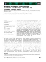
Fig. 4. NMR analysis of IspC assays using [1-
13
C
1
]2C-methyl-D-erythritol 4-phosphate and [3-
13
C
1
]2C-methyl-D-erythritol 4-phosphate (3a and
3b, respectively) as initial substrates. 1a, [3-
13
C
1
]1-deoxy-D-xylulose 5-phosphate; 1b, [4-
13
C
1
]1-deoxy-D-xylulose 5-phosphate; 1c, [3,4-
13
C
2
]1-
deoxy-
D-xylulose 5-phosphate; 2a, [1-
13
C
1
]2C-methyl-D-erythrose 4-phosphate; 2b, [3-
13
C
1
]2C-methyl-D-erythrose 4-phosphate, 4, protonated
glycolaldehyde phosphate (unlabeled); 4a, protonated [1-
13
C
1
]glycolaldehyde phosphate; 5, enolate of hydroxyacetone (unlabeled); 5a, enolate
of [1-
13
C
1
]hydroxyacetone.
13
C-NMR signals of an incubation mixture without enzyme (A), and with IspC from E. coli (B), M. tuberculosis (C)
and A. thaliana (D), respectively. The asterisks denote signals due to impurities.
S. Lauw et al. Mechanism of IspC protein
FEBS Journal 275 (2008) 4060–4073 ª 2008 The Authors Journal compilation ª 2008 FEBS 4063
(1c). This double-labeled species would be detected via
satellite lines in the
13
C-NMR spectrum due to
13
C
13
C
coupling.
In order to maximize the diagnostic sensitivity, we
decided to conduct the experiments under steady-state
conditions where reactants 1 and 3 are present in simi-
lar amounts at thermodynamic equilibrium. For that
purpose, reaction mixtures containing 5 mm 3a,5mm
3b, 215 mm NADP
+
and 0.15–0.25 mm IspC from
E. coli, M. tuberculosis or A. thaliana were incubated
at pH 8 and 37 °C for 24 h and were monitored by
13
C-NMR spectroscopy. The partial reduction of
NADP
+
by the enzyme rapidly resulted in steady-state
conditions where the steady-state concentrations of 1
and 3 were approximately equal (Fig. 4). Conse-
quently, the forward and the reverse reaction rate
under equilibrium condition were also bound to be
approximately equal. Notably, the IspC enzymes were
present in very high (near-stoichiometric) concentra-
tions. Under these conditions, the substrate molecules
should be engaged by enzyme molecules on a near-
permanent basis.
The residual enzyme activity after 24 h of incubation
was measured after massive dilution of an aliquot of
the reaction mixture, using 1 as substrate. The decrease
in activity during the 24 h incubation period was in
the range 27–37% for the three different enzymes
under study.
From the starting conditions and the enzyme stabil-
ity measurements under our reaction conditions, it
follows that an average substrate molecule should
have passed through approximately 8800, 12 100 and
2400 forward–reverse cycles in the experiments with
enzymes from E. coli, M. tuberculosis and A. thaliana,
respectively (supplementary Table S4).
For the equilibrium constant of the reaction cata-
lyzed by IspC as defined by Eqn (1), we obtained a
value of (2.8 ± 0.2) · 10
)10
m at pH 8.0 and 37 °C.
This is well in line with a value of (4.6 ±
0.5) · 10
)10
m at pH 7.7 and 37 °C that had been
reported earlier [28].
K ¼
½NADPH
eq
Á½1
eq
Á½H
þ
½NADP
þ
eq
Á½3
eq
ð1Þ
Figure 4 shows
13
C-NMR signals of the reaction
mixtures prior to the addition of enzyme (Fig. 4A) and
after incubation with enzymes from E. coli (Fig. 4B),
M. tuberculosis (Fig. 4C) and A. thaliana (Fig. 4D),
respectively. Reaction mixtures treated with enzymes
from the three different organisms studied showed very
similar results.
The crucial observation is the absence of any detect-
able excess of the
13
C
13
C coupling satellites beyond the
natural abundance level for the signals of C-3 and C-4
of a hypothetical product 1c. The hypothetical posi-
tions of the
13
C
13
C coupling satellites expected in the
spectrum of 1c are marked by arrows in Fig. 4B–D. In
each case, the integrals of the satellite signals are in
the range of 1% as compared to the central signal.
Signals of that size would be expected in the complete
absence of fragment exchange, where they reflect the
presence of about 1.1%
13
C in those carbon atoms of
the reactant that were not labeled.
On the basis of the quantitative evaluation of the
13
C-NMR signal intensities and coupling satellites in
experiments with
13
C-labeled substrates, it can be esti-
mated that fewer than one fragment exchange has
occurred during more than 100 000 reaction cycles.
Although these data are not sufficient to rule out a ret-
roaldol–aldol reaction sequence, they do show that a
hypothetical retroaldol–aldol sequence would require
extremely tight confinement of the intermediary molec-
ular fragments at the active site of the enzyme. The
limit for escape and reutilization of a retroaldol frag-
ment would be fewer than once in 100 000 forward–
reverse cycles. In this context, it is also worth noting
that the branched intermediate 2C-methyl-d-erythrose
4-phosphate (2) (Fig. 1) can be used as substrate by
the enzyme at a rate that is comparable with the con-
version rate of substrate 1 [28]; thus, strict confinement
seems at least not to apply to that intermediate.
In a second set of experiments, we checked whether
exogenous hydroxyacetone, whose enolate is the pre-
dicted intermediate of the hypothetical retroaldol–aldol
mechanism, can be incorporated into reactants 1 and 3
by fragment exchange. Preliminary experiments had
shown that hydroxyacetone does not significantly
change the catalytic rate of 2C-methyl-d-erythritol
4-phosphate synthase when present in concentrations
up to 2% (v ⁄ v). The reaction mixtures contained
10 mm [1,3,4-
13
C
3
]2C-methyl-d-erythritol 4-phosphate
(3c, Fig. 5), 215 mm NADP
+
, 243 mm (2%, v ⁄ v)
hydroxyacetone, 100 mm Tris ⁄ HCl (pH 8.0), and
0.23–0.25 mm IspC from E. coli, M. tuberculosis or
A. thaliana. They were incubated for 8 h at 37 °C and
were then analyzed by NMR spectroscopy.
As described above, these initial conditions were
rapidly conducive to steady conditions where 1 and 3
were present in very similar concentrations, and the
rates of the forward reaction (conversion of 1 to 3)
and the backward reaction (conversion of 3 into 1)
were also essentially the same. Any ‘wash-in’ of
unlabeled hydroxyacetone (6) should give the iso-
topolog 1f, which carries only two
13
C atoms. This
Mechanism of IspC protein S. Lauw et al.
4064 FEBS Journal 275 (2008) 4060–4073 ª 2008 The Authors Journal compilation ª 2008 FEBS
A
B
C
D
p.p.m.
Fig. 5. NMR analysis of IspC assays using [1,3,4-
13
C
3
]2C-methyl-D-erythritol 4-phosphate and unlabeled hydroxyacetone (3c and 6) as initial
substrates. 1e, [3,4,5-
13
C
3
]1-deoxy-D-xylulose 5-phosphate; 1f, [4,5-
13
C
2
]1-deoxy-D-xylulose 5-phosphate; 3d, [3,4-
13
C
2
]2C-methyl-D-erythritol
4-phosphate; 4b, protonated [1,2-
13
C
2
]glycolaldehyde phosphate; 5, enolate of hydroxyacetone (unlabeled); 5a, enolate of [1-
13
C
1
]hydroxyace-
tone; 6, hydroxyacetone (unlabeled).
13
C-NMR signals of an incubation mixture without enzyme (A), and with IspC from E. coli (B), M. tuber-
culosis (C) and A. thaliana (D), respectively. The asterisks denote signals due to impurities.
S. Lauw et al. Mechanism of IspC protein
FEBS Journal 275 (2008) 4060–4073 ª 2008 The Authors Journal compilation ª 2008 FEBS 4065
isotopolog would be diagnosed easily in the
13
C-NMR
spectra by a distinctive double doublet signature of
C-4 that would be caused by
13
C
13
C coupling and
13
C
31
P coupling (predicted signal positions are indi-
cated by arrows in Fig. 5B–D).
Figure 5A–D shows
13
C-NMR signals detected in the
exchange experiment with unlabeled hydroxyacetone.
Signal intensities showed a steady-state ratio of 59 : 41
for 1 and 3c (supplementary Table S6). The crucial
double doublets as expected for a retroaldol–aldol
mechanism (Fig. 5B–D) were absent. The
13
C NMR
data of 1e and 3c are shown in supplementary Table S5.
In the following set of experiments, we investigated
whether glycolaldehyde phosphate and hydroxyacetone
can serve as direct substrates for IspC from different
organisms to form 2C-methyl-d-erythritol 4-phosphate,
as shown in the hypothetical retroaldol–aldol reaction
sequence illustrated in Fig. 5. Specifically, the reaction
mixtures contained 2 mm [1,2-
13
C
2
]glycolaldehyde
phosphate (4b), 243 mm (2%, v ⁄ v) hydroxyacetone (5),
Tris ⁄ HCl (pH 8.0), 3 mm NADPH, and 0.21–0.34 mm
IspC from E. coli, M. tuberculosis or A. thaliana.
The
13
C-NMR spectra obtained after incubation
periods of 1.5 and 3 h, respectively, showed only dou-
ble doublet signals at 89.2 p.p.m. due to the presence
of the hydrate of 4b (Fig. 6B–D). As shown in Fig. 6,
no evidence for the formation of [3,4-
13
C
2
]2C-methyl-
d-erythritol 4-phosphate (3d) could be obtained. Nota-
bly, it would have been possible to detect any 3d by the
specific double doublet signature of C-3, as confirmed
by a titration experiment with [1,3,4-
13
C
3
]2C-methyl-d-
erythritol 4-phosphate (3c) (Fig. 6E). It should be
noted that these experiments were conducted with very
high concentrations of enzymes (almost in the millimo-
lar range) and with a very high concentration of
243 mm (2%, v ⁄ v) of hydroxyacetone, which had been
shown to be tolerated by the enzyme without significant
reduction in the rate for the IspC reaction measured
with 1e as substrate (see also supplementary Table S7).
Wong & Cox [36] reported the formation of
1-deoxy-l-ribulose 5-phosphate (7b, Fig. 7), an epimer
of 1, in an IspC reaction mixture in the absence of
NADPH and of divalent metal ions. Specifically, they
observed a new
13
C-NMR signal at 71.6 p.p.m., which
A
B
C
D
E
p
.
p
.m.
Fig. 6. NMR analysis of IspC assays using
protonated [1,2-
13
C
2
]glycolaldehyde phos-
phate and the enolate of hydroxacetone (4b
and 5a, respectively) as initial substrates.
2c, [3,4-
13
C
2
]2C-methyl-D-erythrose 4-phos-
phate; 3d, [3,4-
13
C
2
]2C-methyl-D-erythritol
4-phosphate. (A)–(E) are
13
C-NMR spectra
obtained from IspC reactions using
[1,2-
13
C
2
]glycolaldehyde phosphate (4b) and
hydroxacetone (5a) as substrates.
13
C-NMR
signals of an incubation mixture without
enzyme (A), and with IspC from E. coli (B),
M. tuberculosis (C) and A. thaliana (D) and
with the addition of [1,3,4-
13
C
3
]2C-methyl-D-
erythritol 4-phosphate after 3 h of incubation
of the reaction mixture B (E). The asterisks
denote signals due to impurities.
Mechanism of IspC protein S. Lauw et al.
4066 FEBS Journal 275 (2008) 4060–4073 ª 2008 The Authors Journal compilation ª 2008 FEBS
was assigned to C-4 of 7. When we repeated that
experiment with [3,4,5-
13
C
3
]-1 as substrate, we found
that a signal was already present at 71.6 p.p.m., even
prior to incubation of the reaction mixture (Fig. 8,
lane A), and the intensity of that signal increased by a
factor of about 2 during the subsequent incubation.
Notably, the same phenomenon was observed in sam-
ples without IspC. This unexpected finding prompted a
more detailed investigation, which revealed that 1 is
subject to spontaneous isomerization to give 7. The
details are described below.
Specifically, we prepared [3,4,5-
13
C
3
]-1 by an enzy-
matic procedure starting from [U-
13
C
6
]glucose [37].
Despite the absence of IspC, the formation of the
target compound 1 was accompanied by the formation
of a compound characterized by a
13
C resonance at
71.6 p.p.m., albeit at a much lower rate. Specifically,
after incubation for 1 h, the yield of 1 was about 50%,
and the relative yield, based on 1, of the compound
resonating at 71.6 p.p.m. was 1%. Even after the
removal of all proteins by ultrafiltration, the relative
amount of that contaminant continued to increase over
a period of about 1 week; the final ratio of the two
compounds, believed to represent a state of equilib-
rium, was about 4 : 1. The apparent rate constant for
the formation of 7 from 1 was 3 · 10
)7
s
)1
, and, the
equilibrium constant was calculated to be 3.45 (supple-
mentary Fig. S1). In parallel experiments with and
without addition of IspC, the
13
C signal at 71.6 p.p.m.
increased at the same rate. More specifically, reaction
mixtures containing 100 mm Tris ⁄ HCl (pH 8), 10 mm
1, and 0.1 mm (5 mgÆmL
)1
) IspC when required, were
incubated at 37 °C. The component resonating at
71.6 p.p.m. increased from 3% to 5% (based on the
concentration of 1) during a period of 24 h at 37 °C,
irrespective of the presence or absence of IspC.
Using an equilibrium mixture of [U-
13
C
5
]-1 and of
the component resonating at 71.6 p.p.m., we could
assign all
13
C signals of the latter on the basis of
13
C
13
C and
13
C
31
P coupling in one-dimensional
13
C-
NMR spectra (supplementary Fig. S2). All
1
H-NMR
signals of the newly formed compound were then
assigned by HMQC spectroscopy (supplementary
Table S8). The NMR data were in close correspon-
dence with those reported earlier for a chemically
synthesized sample of 1-deoxy-l-ribulose 5-phosphate
(7b, Fig. 7) [38]. However, it should be noted that
13
C-NMR is unable to discriminate between the
d-enantiomer and l-enantiomer under the experimental
conditions used, and enantiomer assignment of the
1-deoxyribulose 5-phosphate formed by spontaneous
isomerization of 1-deoxy-d-xylulose 5-phosphate (1)is
not possible from our experimental data.
Discussion
The main part of the present study was a search,
under conditions of maximal stringency, for fragment
exchange that could be the hallmark of the hypotheti-
cal retroaldol–aldol mechanism. The essentials of that
high-stringency strategy can be summarized as fol-
lows: (a) the experiments shown in Figs 4 and 5 were
conducted under steady-state conditions (at thermody-
namic equilibrium), thus enabling each molecule to
pass through thousands of forward–backward reaction
cycles; (b) enzymes were used at near-stoichiometric
concentrations, in order to engage substrate molecules
on a near-permanent basis; (c) multiple
13
C labeling
was used in order to optimize detection of the crucial
molecular species that would have resulted from frag-
ment exchange by the utilization of
13
C
13
C coupling;
(d) substrates used included not only the natural sub-
strate of the reaction, 1-deoxy-d-xylulose 5-phosphate
(1), but also the hypothetical fragments that would be
expected to result from a retroaldol fragmentation,
i.e. hydroxyacetone and glycolaldehyde phosphate –
one of these substrates (glycolaldehyde phosphate, 4b)
was double-labeled with
13
C, and the other was used
at an unusually high concentration (in the decimolar
Fig. 7. Stereoisomers of 1-deoxy-D-xylulose 5-phosphate: 1,
1-deoxy-
D-xylulose 5-phosphate; 7a, 1-deoxy-D-ribulose 5-phos-
phate; 7b, 1-deoxy-
L-ribulose 5-phosphate; 8, 1-deoxy-L-xylulose
5-phosphate.
S. Lauw et al. Mechanism of IspC protein
FEBS Journal 275 (2008) 4060–4073 ª 2008 The Authors Journal compilation ª 2008 FEBS 4067
range) in order to maximize diagnostic sensitivity; (e)
three IspC orthologs from genetically distant sources
were used, with the expectation that all orthologs
would not necessarily confine fragments with the same
degree of stringency – as it is unlikely that strong
selective pressure specifically enforced very high
degrees of stringency, it would not appear implausible
that different taxa might have enzymes with different
stringencies.
On the basis of these results, the limit on observed
fragment escape and fragment reutilization is fewer
than one in many thousands of forward–reverse
cycles.
The active site of IspC is located close to the sur-
face. A flexible loop at the active site (amino acids
206–216) [39–42] is able to fold into at least three dif-
ferent conformations. Specifically, in the apoenzyme
structure, this loop is unordered, whereas the structure
with bound NADPH, and especially the complex with
bound NADPH as well as 1-deoxy-d-xylulose 5-phos-
phate (1), showed this loop to be well ordered and to
be closing the active site region of the enzyme (Fig. 9).
On the other hand, it has also been demonstrated that
the branched intermediate 2 can access the active site
cavity from the bulk solvent and can then serve as a
substrate, obviously without hindrance from the said
loop [28]. The available structural data are not a suffi-
cient basis to support a claim of absolute confinement
of the active site. Notably, both hypothetical fragments
resulting from retroaldol cleavage would be small by
comparison with the branched aldose intermediate 2;
as the active site is even accessible to 2, one would
expect that the hypothetical intermediates 4b and 5,
which are both small by comparison, should be able to
exchange with the bulk solvent.
3-Deoxy-1 and 4-deoxy-1 have been shown to act as
weak inhibitors, but not as substrates of IspC [28,38].
Had these investigations resulted in the demonstration
of any (even low) substrate activity for the 4-deoxy
compound, that would have ruled out the retroaldol
mechanism. Clearly, however, the reverse argument
would be a logical fallacy; the failure of the 4-deoxy
compound to act as a substrate could be due to a
wide variety of reasons, and does not determine the
mechanism.
The claimed conversion of 1 into the epimer 7 by
IspC in the absence of pyridine nucleotides and diva-
lent metal ions could have been construed as support
for a retroaldol mechanism. Unfortunately, our results
suggest that the formation of 7 in those experiments
was incorrectly ascribed to the catalytic action of IspC,
and reflected, in reality, the spontaneous, uncatalyzed
epimerization of 1.
In summary, our data are all consistent with a sig-
matropic rearrangement, albeit they do not constitute
definite proof. However, it appears safe to say that the
present experiments extend the degree of stringency to
the limits of experimental feasibility as ultimately
defined by the long-term chemical stability of the
proteins, substrates and coenzymes involved.
p
.
p
.m.
Fig. 8.
13
C-NMR spectra of 1-deoxy-D-xylu-
lose 5-phosphate and its diastereomer.
Spectra were recorded in time intervals of
2 days at 37 °C. (A) Day 0. (B) Day 2. (C)
Day 4. (D) Day 6. (E) Day 8. The signals of
[3,4,5-
13
C
3
]1-deoxy-D-xylulose 5-phosphate
(1e) are shown in orange, and those of
[3,4,5-
13
C
3
] 1-deoxy-L-ribulose 5-phosphate
(7b) in green.
Mechanism of IspC protein S. Lauw et al.
4068 FEBS Journal 275 (2008) 4060–4073 ª 2008 The Authors Journal compilation ª 2008 FEBS
Experimental procedures
Materials
[U-
13
C
6
]Glucose was purchased from Isotec, Miami Town-
ship, OH; [3,4-
13
C
2
]-glucose and [2,5-
13
C
2
]glucose were
from Omicron Inc., South Bend, OH. [3,4,5-
13
C
3
]1-deoxy-
d-xylulose 5-phosphate, [U-
13
C
5
]1-deoxy-d-xylulose 5-phos-
phate and [1,3,4,-
13
C
3
]2C-methyl-d-erythrose 4-phosphate
were synthesized as described previously [37,43,44].
Hexokinase from yeast, triosephosphate isomerase from
rabbit muscle, glutamate dehydrogenase from bovine liver
and glucose dehydrogenase from Thermoplasma acido-
philum were from Sigma. 1-Deoxy-d-xylulose-5-phosphate
synthase from Bacillus subtilis and 2C-methyl-d-erythritol
4-phosphate synthase (IspC) from E. coli were prepared
by published procedures [37,43,44]. The preparation of
IspC from A. thaliana, fructose-1,6-biphosphate aldolase,
phosphofructokinase, glucose-6-phosphate isomerase from
E. coli, 6-phosphogluconate dehydrogenase and glucose-6-
phosphate dehydrogenase from B. subtilis is described
elsewhere [44–47]. The recombinant proteins used for
substrate synthesis and enzyme assays are listed in supple-
mentary Table S3.
Construction of a recombinant strain for
hyperexpression of the M. tuberculosis ispC gene
The ispC gene of M. tuberculosis (accession no.
gb BX842581.1) was amplified by PCR using the oligonucle-
otides ispCMycSacivo and ispCMycPstIhi as primers and
chromosomal M. tuberculosis DNA as template. The amplifi-
cate was digested with the restriction endonucleases SacI and
PstI, and the resulting fragment was ligated into the expres-
sion vector pQE30, which had been digested with the same
restriction enzymes. The ligation mixture was electroporated
into E. coli XL1-Blue [48] cells, giving the recombinant
strains XL1-pQEispCMyco and M15-pQEispCMyco.
Bacterial strains, plasmids and oligonucleotides used in this
study are listed in supplementary Tables S1 and S2.
Sequence determination
DNA sequencing was performed by the automated dide-
oxynucleotide method. N-terminal peptide sequences were
obtained by pulsed-liquid mode.
Expression of recombinant IspC from
M. tuberculosis
The recombinant E. coli strain XL1-pQEispCMyco was
grown in LB broth containing ampicillin (180 mgÆL
)1
)as
appropriate. Cultures were incubated at 37 °C with shaking.
At an attenuance of 0.7 (600 nm), isopropylthiogalactoside
was added to a final concentration of 0.5 mm, and the
cultures were incubated overnight at 30 °C. The cells were
harvested by centrifugation at 5000 g for 30 min at 4°Con
an SLA-3000 rotor (Sorvall, Du Pont, Newton, CT), washed
with 0.9% (w ⁄ v) sodium chloride, and stored at )20 °C.
Preparation of recombinant IspC from
M. tuberculosis
The frozen cell mass (40 g) of the recombinant E. coli
strain XL1-pQEispCMyco was thawed in 200 mL of
100 mm Tris ⁄ HCl (pH 8.0), containing 0.5 m sodium
chloride, 20 mm imidazole hydrochloride, and 10% (v ⁄ v)
glycerol. The cells were disrupted using a French press,
and the suspension was centrifuged at 15 000 g for 30 min
at 40°C on an SS-34 rotor (Sorvall). The supernatant was
applied to a column of Ni-chelating Sepharose FF (column
volume, 34 mL) that had been equilibrated with 100 mm
Tris ⁄ HCl (pH 8.0) containing 0.5 m sodium chloride,
20 mm imidazole hydrochloride, and 10% (v ⁄ v) glycerol
(flow rate, 3 mLÆmin
)1
). The column was washed with
100 mm Tris ⁄ HCl (pH 8.0) containing 0.5 m sodium
A
B
Fig. 9. Crystal structures of monomeric IspC from E. coli. (A)
Apoenzyme (Protein Data Bank file 1K5H [41]). (B) Enzyme in com-
plex with NADPH (orange) and 1-deoxy-
D-xylulose 5-phosphate
(blue) (Protein Data Bank file 1Q0Q [39]). The flexible loop (residues
206–216) in both structures is shown in magenta.
S. Lauw et al. Mechanism of IspC protein
FEBS Journal 275 (2008) 4060–4073 ª 2008 The Authors Journal compilation ª 2008 FEBS 4069
chloride, 20 mm imidazole hydrochloride, and 10% (v ⁄ v)
glycerol, and was then developed with a gradient of
20–500 mm imidazole in 50 mm Tris ⁄ HCl (pH 8.0)
containing 0.5 m sodium chloride and 10% glycerol (v ⁄ v;
total volume, 200 mL). Fractions were combined,
concentrated by ultrafiltration (15 kDa), desalted with a
HIPREP 26 ⁄ 10 column, and stored at )80 °C.
Synthesis of [1-
13
C
1
]2C-methyl-D-erythritol
4-phosphate and [3-
13
C
1
]2C-methyl-D-erythritol
4-phosphate
A solution containing 18 mm [3,4-
13
C
2
]glucose or
[2,5-
13
C
2
]glucose, 36 mm potassium phosphoenolpyruvate,
1mm thiamine pyrophosphate, 0.7 mm ATP, 10 mm
MgCl
2
,5mm dithiothreitol and 150 mm Tris ⁄ HCl was
adjusted to pH 8.0 by the addition of 5 m NaOH (final vol-
ume, 10 mL). Hexokinase (300 lg, 27 units), phosphofruc-
tokinase (400 lg, 2 units), fructose-1,6-biphosphate aldolase
(540 lg, 2 units), triose phosphate isomerase (4 lg,
21 units), glucose-6-phosphate isomerase (18 lg, 9 units)
and 1-deoxy-d-xylulose-5-phosphate synthase from
B. subtilis (690 lg, 2 units) were added, and the pH was
adjusted to 8.0. The reaction mixture was incubated at
37 °C and monitored by
13
C-NMR spectroscopy. After 3 h,
protein was removed by ultrafiltration (10 kDa cutoff).
NADP
+
(8.4 mg, 10 lmol), glucose dehydrogenase
(57 lg, 12 units), d-glucose (75 mg) and IspC from E. coli
(800 lg) were added, and the pH was adjusted to 8.0 by
the addition of 5 m NaOH (final volume, 11 mL). The reac-
tion was controlled by
13
C-NMR spectroscopy. After 3 h,
proteins were removed by ultrafiltration (10 kDa cutoff).
The solution was lyophilized.
The residue was dissolved in 3 mL of a solution contain-
ing 40% (v ⁄ v) methanol and 20% (v ⁄ v) isopropanol. The
solution was applied to a column of microcrystalline cellu-
lose (volume, 40 mL) that had been equilibrated with the
same solution. The column was developed with the same
solution. Fractions were analyzed by TLC and
13
C-NMR
spectroscopy, and were then combined and evaporated to a
small volume under reduced pressure. The solution was
lyophilized. The residue was dissolved in H
2
O and stored
at )80 °C.
Preparation of [1,2-
13
C
2
]glycolaldehyde
phosphate
[U-
13
C
5
]Ribulose 5-phosphate was prepared according to
published procedures [49,50] with slight modifications. A
solution (30 mL) containing 100 mm Tris ⁄ HCl, 10 mm
MgCl
2
, 18.5 mm [U-
13
C
6
]glucose, 80 mm dithiothreitol,
30 mm ATP, 81 mm ammonium acetate, 81 mm a-ketogluta-
rate and 1.6 mm NADP
+
was adjusted to pH 8.0 by
addition of 1 m NaOH, and 300 U of hexokinase, 25 U of
6-phosphogluconate dehydrogenase, 62 U of glucose-6-phos-
phate dehydrogenase and 100 U of glutamate dehydrogenase
were added. The mixture was incubated at 37 °C, and the
reaction was monitored by
13
C-NMR spectroscopy. After
1 h, protein was removed by ultrafiltration (3 kDa cutoff).
The solution was lyophilized. The residue was dissolved in
6 mL of water, and the pH was adjusted to 6. Sodium
metaperiodate was added to a final concentration of 333 lm,
and the mixture was incubated at room temperature and
monitored by
13
C-NMR. After 10 min, the excess of perio-
date was quenched by the addition of glycerol to a final con-
centration of 1 mm, and [1,2-
13
C
2
]glycolaldehyde phosphate
was purified on Dowex (Cl
)
-form, 3 g; volume, 10 mL).
Spectrophotometric assay of the forward
reaction catalyzed by IspC
Assay mixtures contained 100 mm Tris ⁄ HCl (pH 8.0),
10 mm MgCl
2
,1mm NADPH, 30 lg of BSA, 4 mm dith-
iothreitol, 2 lg of IspC and 3.5 mm 1 in a volume of
500 lL. They were incubated at 37 °C, and the reaction
was monitored photometrically at 340 nm.
Spectrophotometric assay of the backward
reaction catalyzed by IspC
Assay mixtures contained 100 mm Tris ⁄ HCl (pH 8.0),
10 mm MgCl
2
,5mm NADP
+
,4mm dithiothreitol, 10 mm
2, 30 lg of BSA and 2 lg of IspC in a volume of 500 lL.
They were incubated at 37 °C, and the reaction was
monitored photometrically at 340 nm.
NMR assay of the IspC reaction using [1-
13
C
1
]2C-
methyl-
D-erythritol 4-phosphate and [3-
13
C
1
]2C-
methyl-
D-erythritol 4-phosphate as initial
substrates
Assay mixtures contained 100 mm Tris ⁄ HCl (pH 8.0),
10 mm MgCl
2
, 215 mm NADP
+
,5mm [1-
13
C
1
]-2,5mm
[3-
13
C
1
]-2, 10% (v ⁄ v) D
2
O and IspC (from E. coli,
M. tuberculosis and A. thaliana, respectively) in a volume of
500 lL. The mixtures were incubated at 37 °C and analyzed
by
13
C-NMR spectroscopy after 1.5 and 24 h, respectively.
NMR assay of the IspC reaction using
[1,3,4-
13
C
3
]2C-methyl-D-erythritol 4-phosphate
as substrate
Assay mixtures contained 100 mm Tris ⁄ HCl (pH 8.0),
10 mm MgCl
2
, 215 mm NADP
+
,10mm [1,3,4-
13
C
3
]-2,
10% (v ⁄ v) D
2
O, 243 mm hydroxyacetone and IspC (5 mg
of E. coli protein, 5 mg of M. tuberculosis protein and 6 mg
of A. thaliana protein, respectively) in a volume of 500 lL.
Mechanism of IspC protein S. Lauw et al.
4070 FEBS Journal 275 (2008) 4060–4073 ª 2008 The Authors Journal compilation ª 2008 FEBS
The mixtures were incubated at 37 °C and analyzed by
13
C-NMR spectroscopy after 2 and 8 h.
NMR assay of the IspC reaction using
glycolaldehyde phosphate and hydroxyacetone
as putative substrates
Assay mixtures contained 100 mm Tris ⁄ HCl (pH 8.0),
10 mm MgCl
2
,3mm NADPH, 2 mm [1,2-
13
C
2
]glycolalde-
hyde phosphate, 10% (v ⁄ v) D
2
O, 243 mm hydroxyacetone
and IspC (7.5 mg of E. coli protein, 5.9 mg of M. tuberculo-
sis protein, and 5.1 mg of A. thaliana protein, respectively) in
a volume of 500 lL. The mixtures were incubated at 37 °C
and analyzed by
13
C-NMR spectroscopy after 1.5 and 3 h.
NMR spectroscopy
1
H-NMR and
1
H-decoupled
13
C-NMR spectra were recorded
at 500.1 and 125.5 MHz, respectively. Chemical shifts were
referenced to external trimethyl silylpropane sulfonate.
Samples were dissolved in 150 mm Tris ⁄ HCl (pH 8.0).
Acknowledgements
We thank the Fonds der Chemischen Industrie and the
Hans Fischer Gesellschaft for support. We thank
Katrin Ga
¨
rtner for skillful assistance and Fritz
Wendling for expert help with the preparation of the
manuscript.
References
1 Sacchettini JC & Poulter CD (1997) Creating isoprenoid
diversity. Science 277, 1788–1789.
2 Qureshi N & Porter JW (1981) Conversion of acetyl-
coenzyme A to isopentenyl pyrophosphate. In Biosynthe-
sis of Isoprenoid Compounds (Porter JW & Spurgeon SL,
eds), pp. 47–94. John Wiley, New York, NY.
3 Bloch K (1992) Sterol molecule: structure, biosynthesis,
and function. Steroid 57, 378–383.
4 Bochar DA, Friesen JA, Stauffacher C & Rodwell VW
(1999) Biosynthesis of mevalonic acid from acetyl-CoA.
In Comprehensive Natural Product Chemistry (Cane DE,
ed.), pp. 15–44. Pergamon, Oxford.
5 Rohmer M, Knani M, Simonin P, Sutter B & Sahm H
(1993) Isoprenoid biosynthesis in bacteria: a novel path-
way for the early steps leading to isopentenyl
diphosphate. Biochem J 295, 517–524.
6 Schwarz MK. (1994) Terpen-Biosynthese in Ginkgo
biloba: Eine u
¨
berraschende Geschichte. ETH Zu
¨
rich,
Zu
¨
rich.
7 Broers STJ. (1994) U
¨
ber die fru
¨
hen Vorstufen der
Biosynthese von Isoprenoiden in Escherichia coli.
ETH Zu
¨
rich, Zu
¨
rich.
8 Eisenreich W, Menhard B, Hylands PJ, Zenk MH &
Bacher A (1996) Studies on the biosynthesis of taxol:
the taxane carbon skeleton is not of mevalonoid origin.
Proc Natl Acad Sci USA 93, 6431–6436.
9 Lichtenthaler HK, Schwender J, Disch A & Rohmer M
(1997) Biosynthesis of isoprenoids in higher plant
chloroplasts proceeds via a mevalonate-independent
pathway. FEBS Lett 400, 271–274.
10 Schwarz MK & Arigoni D. (1999) Ginkgolide biosyn-
thesis. In Comprehensive Natural Product Chemistry
(Cane DE, ed.), pp. 367–399. Pergamon, Oxford.
11 Rohmer M. (1999) A mevalonate-independent rout to
isopentenyl diphosphate. In Comprehensive Natural
Product Chemistry (Cane DE, ed.), pp. 45–68. Perg-
amon, Oxford.
12 Eisenreich W, Rohdich F & Bacher A (2001) Deoxyxy-
lulose phosphate pathway to terpenoids. Trends Plant
Sci 6, 78–84.
13 Eisenreich W, Bacher A, Arigoni D & Rohdich F
(2004) Biosynthesis of isoprenoids via the non-mevalo-
nate pathway. Cell Mol Life Sci 61, 1401–1426.
14 Rohmer M, Grosdemange-Billiard C, Seemann M &
Tritsch D (2004) Isoprenoid biosynthesis as a novel
target for antibacterial and antiparasitic drugs. Curr
Opin Investig Drugs 5, 154–162.
15 Takahashi S, Kuzuyama T, Watanabe H & Seto H
(1998) A 1-deoxy-D-xylulose 5-phosphate reductoisom-
erase catalyzing the formation of 2-C-methyl-D-erythri-
tol 4-phosphate in an alternative nonmevalonate
pathway for terpenoid biosynthesis. Proc Natl Acad Sci
USA 95, 9879–9884.
16 Kuzuyama T, Shimizu T, Takahashi S & Seto H (1998)
Fosmidomycin, a specific inhibitor of 1-deoxy-D-xylu-
lose 5-phosphate reductoisomerase in the nonmevalo-
nate pathway for terpenoid biosynthesis. Tetrahedron
Lett
39, 7913–7916.
17 Jomaa H, Wiesner J, Sanderbrand S, Altincicek B,
Weidemeyer C, Hintz M, Turbachova I, Eberl M,
Zeidler J, Lichtenthaler HK et al. (1999) Inhibitors of
the nonmevalonate pathway of isoprenoid biosynthesis
as antimalarial drugs. Science 285 , 1573–1576.
18 Okuhara M, Kuroda Y, Goto T, Okamoto M, Terano
H, Kohsaka M, Aoki H & Imanaka H (1980) Studies
on new phosphonic acid antibiotics. III. Isolation and
characterization of FR-31564, FR-32863 and FR-33289.
J Antibiot (Tokyo) 33, 24–28.
19 Kuemmerle HP, Murakawa T & De Santis F (1987)
Pharmacokinetic evaluation of fosmidomycin, a
new phosphonic acid antibiotic. Chemioterapia 6, 113–
119.
20 Missinou MA, Borrmann S, Schindler A, Issifou S,
Adegnika AA, Matsiegui PB, Binder R, Lell B, Wiesner
J, Baranek T et al. (2002) Fosmidomycin for malaria.
Lancet 360, 1941–1942.
S. Lauw et al. Mechanism of IspC protein
FEBS Journal 275 (2008) 4060–4073 ª 2008 The Authors Journal compilation ª 2008 FEBS 4071
21 Borrmann S, Issifou S, Esser G, Adegnika AA, Ramhar-
ter M, Matsiegui PB, Oyakhirome S, Mawili-Mboumba
DP, Missinou MA, Kun JF et al. (2004) Fosmido-
mycin–clindamycin for the treatment of Plasmodium
falciparum malaria. J Infect Dis 190, 1534–1540.
22 Borrmann S, Lundgren I, Oyakhirome S, Impouma B,
Matsiegui PB, Adegnika AA, Issifou S, Kun JF, Hutch-
inson D, Wiesner J et al. (2006) Fosmidomycin plus
clindamycin for treatment of pediatric patients aged 1
to 14 years with Plasmodium falciparum malaria.
Antimicrob Agents Chemother 50, 2713–2718.
23 Hoffman SL, Subramanian GM, Collins FH & Venter
JC (2002) Plasmodium, human and Anopheles genomics
and malaria. Nature 415, 702–709.
24 Morlais I, Mori A, Schneider JR & Severson DW
(2003) A targeted approach to the identification of can-
didate genes determining susceptibility to Plasmodium
gallinaceum in Aedes aegypti. Mol Genet Genomics 269,
753–764.
25 Rohdich F, Bacher A & Eisenreich W (2004) Perspec-
tives in anti-infective drug design. The late steps in the
biosynthesis of the universal terpenoid precursors, iso-
pentenyl diphosphate and dimethylallyl diphosphate.
Bioorg Chem 32, 292–308.
26 Rohdich F, Bacher A & Eisenreich W (2005) Isoprenoid
biosynthetic pathways as anti-infective drug targets.
Biochem Soc Trans 33, 785–791.
27 Proteau PJ (2004) 1-Deoxy-D-xylulose 5-phosphate
reductoisomerase: an overview. Bioorg Chem 32, 483–
493.
28 Hoeffler JF, Tritsch D, Grosdemange-Billiard C &
Rohmer M (2002) Isoprenoid biosynthesis via the
methylerythritol phosphate pathway. Mechanistic
investigations of the 1-deoxy-D-xylulose 5-phosphate
reductoisomerase. Eur J Biochem 269, 4446–4457.
29 Proteau PJ, Woo YH, Williamson RT & Phaosiri C
(1999) Stereochemistry of the reduction step mediated
by recombinant 1-deoxy-D-xylulose 5-phosphate
isomeroreductase. Org Lett 1, 921–923.
30 Radykewicz T, Rohdich F, Wungsintaweekul J, Herz S,
Kis K, Eisenreich W, Bacher A, Zenk MH & Arigoni
D (2000) Biosynthesis of terpenoids: 1-deoxy-D-xylu-
lose-5-phosphate reductoisomerase from Escherichia
coli is a class B dehydrogenase. FEBS Lett 465, 157–
160.
31 Putra SR, Lois LM, Campos N, Boronat A & Rohmer
M (1998) Incorporation of [2,3-
13
C
2
]- and [2,4-
13
C
2
]-D-
1-deoxyxylulose into ubiquinone of Escherichia coli via
the mevalonate-independent pathway for isoprenoid
biosynthesis. Tetrahedron Lett 39, 23–26.
32 Arigoni D, Sagner S, Latzel C, Eisenreich W, Bacher A
& Zenk MH (1997) Terpenoid biosynthesis from
1-deoxy-D-xylulose in higher plants by intramolecular
skeletal rearrangement. Proc Natl Acad Sci USA 94,
10600–10605.
33 Fox DT & Poulter CD (2005) Mechanistic studies with
2-C-methyl-d-erythritol 4-phosphate synthase from
Escherichia coli. Biochemistry 44, 8360–8368.
34 Fox DT & Poulter CD (2005) Synthesis and evaluation
of 1-deoxy-D-xylulose 5-phosphoric acid analogues as
alternate substrates for methylerythritol phosphate
synthase. J Org Chem 70, 1978–1985.
35 Argyrou A & Blanchard JS (2004) Kinetic and chemical
mechanism of Mycobacterium tuberculosis 1-deoxy-D-
xylulose-5-phosphate isomeroreductase. Biochemistry
43, 4375–4384.
36 Wong U & Cox RJ (2007) The chemical mechanism of
D-1-deoxyxylulose 5-phosphate reductoisomerase from
Escherichia coli. Angew Chem Int Ed Engl 46, 4926–
4929.
37 Hecht S, Kis K, Eisenreich W, Amslinger S, Wungsinta-
weekul J, Herz S, Rohdich F & Bacher A (2001)
Enzyme-assisted preparation of isotope-labelled
1-deoxy-d-xylulose 5-phosphate. J Org Chem 66,
3948–3952.
38 Phaosiri C & Proteau PJ (2004) Substrate analogs for
the investigation of deoxyxylulose 5-phosphate
reductoisomerase inhibition: synthesis and evaluation.
Bioorg Med Chem Lett 14 , 5309–5312.
39 MacSweeney A, Lange R, Fernandes RP, Schulz H,
Dale GE, Douangamath A, Proteau PJ & Oefner C
(2005) The crystal structure of E. coli 1-deoxy-D-xylu-
lose-5-phosphate reductoisomerase in a ternary
complex with the antimalarial compound fosmido-
mycin and NADPH reveals a tight-binding closed
enzyme conformation. J Mol Biol 345, 115–127.
40 Steinbacher S, Kaiser J, Eisenreich W, Huber R, Bacher
A & Rohdich F (2003) Structural basis of fosmidomy-
cin action revealed by the complex with 2-C-methyl-D-
erythritol 4-phosphate synthase (IspC). Implications for
the catalytic mechanism and anti-malaria drug develop-
ment. J Biol Chem 278, 18401–18407.
41 Reuter K, Sanderbrand S, Jomaa H, Wiesner J, Steinb-
recher I, Beck E, Hintz M, Klebe G & Stubbs MT
(2002) Crystal structure of 1-deoxy-D-xylulose-5-
phosphate reductoisomerase, a crucial enzyme in the
non-mevalonate pathway of isoprenoid biosynthesis.
J Biol Chem 277, 5378–5384.
42 Yajima S, Hara K, Iino D, Sasaki Y, Kuzuyama T,
Ohsawa K & Seto H (2007) Structure of 1-deoxy-
D-xylulose 5-phosphate reductoisomerase in a
quaternary complex with a magnesium ion, NADPH
and the antimalarial drug fosmidomycin. Acta
Crystallogr F Struct Biol Cryst Commun 63,
466–470.
43 Hecht S, Wungsintaweekul J, Rohdich F, Kis K, Rad-
ykewicz T, Schuhr CA, Eisenreich W, Richter G &
Bacher A (2001) Biosynthesis of terpenoids: efficient
multistep biotransformation procedures affording iso-
tope-labelled 2C-methyl-D-erythritol 4-phosphate using
Mechanism of IspC protein S. Lauw et al.
4072 FEBS Journal 275 (2008) 4060–4073 ª 2008 The Authors Journal compilation ª 2008 FEBS
recombinant 2C-methyl-D-erythritol 4-phosphate
synthase. J Org Chem 66, 7770–7775.
44 Illarionova V, Kaiser J, Ostrojenkova E, Bacher A,
Eisenreich W & Rohdich F (2006) Non-mevalonate
terpene biosynthesis enzymes as antiinfective drug
targets. Substrate synthesis and high throughput
screening methods. J Org Chem 71, 8824–8834.
45 Rohdich F, Lauw S, Kaiser J, Feicht R, Ko
¨
hler P,
Bacher A & Eisenreich W (2006) Isoprenoid biosynthe-
sis in plants: 2C-methyl-D-erythritol 4-phosphate
synthase (IspC protein) of Arabidopsis thaliana. FEBS J
273, 4446–4458.
46 Ro
¨
misch W (2000) Synthese Isotopmarkierter RNA.TU
Mu
¨
nchen, Mu
¨
nchen.
47 Ro
¨
misch W (2005) Coenzym-Isotopologe als spektrosk-
opische Sonden.TUMu
¨
nchen, Mu
¨
nchen.
48 Bullock WO, Fernandez JM & Short JM (1987) XL1-
Blue: a high efficiency plasmid transforming recA
Escherichia coli strain with beta-galactosidase selection.
Biotechniques 5, 376–379.
49 Rieder C, Eisenreich W, O’Brien J, Richter G, Go
¨
tze E,
Boyle P, Blanchard S, Bacher A & Simon H (1998)
Rearrangement reactions in the biosynthesis of molyb-
dopterin – an NMR study with multiply
13
C ⁄
15
N
labelled precursors. Eur J Biochem 255, 24–36.
50 Volk R & Bacher A (1991) Biosynthesis of riboflavin.
Studies on the mechanism of L-3,4-dihydroxy-2-buta-
none 4-phosphate synthase. J Biol Chem 266, 20610–
20618.
Supplementary material
The following supplementary material is available
online:
Fig. S1. Spontaneous conversion of 1-deoxy-d-xylulose
5-phosphate (1) into 1-deoxy-l-ribulose 5-phosphate
(7b).
Fig. S2.
13
C-NMR signals of [U-
13
C]1-deoxy-d-xylu-
lose 5-phosphate ( 1) and 1-deoxy-l-ribulose 5-phos-
phate (7b).
Table S1. Bacterial strains and plasmids used in this
study.
Table S2. Oligonucleotides used in this study.
Table S3. List of recombinant proteins used for
substrate synthesis and enzyme assays.
Table S4. Conversion ratios and equilibrium constants
for the IspC reaction.
Table S5.
13
C-NMR data of [3,4,5-
13
C
3
]1-deoxy-d-
xylulose 5-phosphate (1e) and [1,3,4-
13
C
3
]2C-methyl-d-
erythritol 4-phosphate (3c).
Table S6. Conversion ratios and cycles for the IspC
reaction containing hydroxyacetone.
Table S7. Calculated hypothetical cycles for the IspC
reaction containing [1,2-
13
C
2
]glycolaldehyde phosphate
(4b) and hydroxyacetone (5).
Table S8.
13
C-NMR data of [3,4,5-
13
C
3
]1-deoxy-d-
xylulose 5-phosphate (1e) and [1,3,4-
13
C
3
]2C-methyl-d-
erythritol 4-phosphate (3c).
This material is available as part of the online article
from
Please note: Blackwell Publishing is not responsible
for the content or functionality of any supplementary
materials supplied by the authors. Any queries (other
than missing material) should be directed to the corre-
sponding author for the article.
S. Lauw et al. Mechanism of IspC protein
FEBS Journal 275 (2008) 4060–4073 ª 2008 The Authors Journal compilation ª 2008 FEBS 4073