Báo cáo khoa học: Intracellular degradation of somatostatin-14 following somatostatin-receptor 3-mediated endocytosis in rat insulinoma cells doc
Bạn đang xem bản rút gọn của tài liệu. Xem và tải ngay bản đầy đủ của tài liệu tại đây (557.43 KB, 12 trang )
Intracellular degradation of somatostatin-14 following
somatostatin-receptor 3-mediated endocytosis in rat
insulinoma cells
Dirk Roosterman
1
, Nicole E. I. Brune
2
, Oliver J. Kreuzer
2
, Micha Feld
1
, Sylvia Pauser
1
, Kim Zarse
2
,
Martin Steinhoff
1
and Wolfgang Meyerhof
2
1 Department of Dermatology, IZKF Mu
¨
nster and Ludwig Boltzmann Institute for Cell and Immunobiology of the Skin, Germany
2 Department of Molecular Genetics, German Institute of Human Nutrition Potsdam-Rehbruecke, Nuthetal, Germany
Somatostatin is a cyclic peptide that is widely
expressed throughout the central nervous system,
endocrine tissue, skin and gastrointestinal tract [1].
Somatostatin exerts a wide range of important biolo-
gical effects, including inhibition of secretion of growth
hormone, insulin, glucagon and gastrin as well as other
hormones secreted from the pituitary, skin and gastro-
intestinal tract [2].
Among other actions, somatostatin elicits strong
antiproliferative effects in in vivo as well as in vitro
models of cancer [3–5]. Somatostatin analogs are there-
fore being used in the diagnosis and therapy of various
tumors, in particular neuroendocrine tumors, which
express somatostatin receptors (SSTRs) [3–5]. SSTR
scintigraphy (SRS), a widely used imaging technique,
is employed to detect and localize such tumors. The
Keywords
endocytosis; G-protein-coupled receptor;
neuropeptide; proteolysis; somatostatin
Correspondence
D. Roosterman, Department of Dermatology
and IZKF Mu
¨
nster, Von-Esmarch-Strasse 58,
D-48148 Mu
¨
nster, Germany
Fax: +49 0251 8357452
Tel: +49 0251 8352932
E-mail:
(Received 27 March 2008, revised 20 June
2008, accepted 23 July 2008)
doi:10.1111/j.1742-4658.2008.06606.x
Somatostatin receptor (SSTR) endocytosis influences cellular responsiveness
to agonist stimulation and somatostatin receptor scintigraphy, a common
diagnostic imaging technique. Recently, we have shown that SSTR1 is dif-
ferentially regulated in the endocytic and recycling pathway of pancreatic
cells after agonist stimulation. Additionally, SSTR1 accumulates and
releases internalized somatostatin-14 (SST-14) as an intact and biologically
active ligand. We also demonstrated that SSTR2A was sequestered into
early endosomes, whereas internalized SST-14 was degraded by endosomal
peptidases and not routed into lysosomal degradation. Here, we examined
the fate of peptide agonists in rat insulinoma cells expressing SSTR3 by
biochemical methods and confocal laser scanning microscopy. We found
that [
125
I]Tyr11-SST-14 rapidly accumulated in intracellular vesicles, where
it was degraded in an ammonium chloride-sensitive manner. In contrast,
[
125
I]Tyr1-octreotide accumulated and was released as an intact peptide.
Rhodamine-B-labeled SST-14, however, was rapidly internalized into endo-
some-like vesicles, and fluorescence signals colocalized with the lysosomal
marker protein cathepsin D. Our data show that SST-14 was cointernalized
with SSTR3, was uncoupled from the receptor, and was sorted into an
endocytic degradation pathway, whereas octreotide was recycled as an
intact peptide. Chronic stimulation of SSTR3 also induced time-dependent
downregulation of the receptor. Thus, the intracellular processing of inter-
nalized SST-14 and the regulation of SSTR3 markedly differ from the
events mediated by the other SSTR subtypes.
Abbreviations
EGFP, enhanced green fluorescent protein; FITC, fluorescein isothiocyanate; HSV, herpes simplex virus glycoprotein D; RIN, rat insulinoma;
SRS, somatostatin receptor scintigraphy; SST-14, somatostatin-14; SSTR, somatostatin receptor.
4728 FEBS Journal 275 (2008) 4728–4739 ª 2008 The Authors Journal compilation ª 2008 FEBS
success of SRS is based on specific interactions of sta-
ble radiolabeled somatostatin analogs injected into
patients, causing them to bind to SSTRs expressed by
tumor cells [4,6]. These interactions are not restricted
to the binding of the peptide agonist to its cognate
receptor, but also lead to agonists accumulating in the
tumor cell after internalization of the receptor–agonist
complex [7–9]. Understanding the internalization of
somatostatin–receptor complexes and their intracellular
fate is therefore of considerable interest in tumor diag-
nostics and therapy as well as neuroinflammation.
Somatostatin binds to and activates six different
G-protein-coupled SSTR subtypes: SSTR1, SSTR2A,
SSTR2B, SSTR3, SSTR4 and SSTR5. The various
SSTR subtypes show distinct internalization pathways
[10,11]. In human embryonic kidney 293 cells and
neuroendocrine pancreatic b-cells, rat SSTR1,
SSTR2A, SSTR3 and SSTR5 but not SSTR4 were
internalized upon stimulation by somatostatin. Further
investigation of SSTR1 and SSTR2A clearly indicated
that the fate of internalized somatostatin-14 (SST-14)
strongly depends on the receptor subtype, although the
particular pathways are not yet fully explored. We
recently demonstrated that chronic stimulation of
SSTR1 induced accumulation of SST-14 in cells via a
dynamic process of internalization, recycling and rein-
ternalization of the ligand [11]. In contrast, stimulation
of SSTR2A with SST-14 or its stable analog octreotide
induced prolonged sequestration of the receptor–ligand
complex into early endosomes that was dependent on
arrestins [12,13]. Subsequently, the endosomal
peptidase endothelin-converting enzyme-1 cleaves inter-
nalized SST-14 between positions Asn5-Phe6 and
Thr10-Phe11, leading to release of internalized SST-14
as SST-14(6–10) (FFWKT) and SST-14(1–5) ⁄ (11–14)
(AGCLN ⁄ FTSC) [13].
Further analysis of SSTR3, an SSTR subtype of
particular importance in human thymoma [14], shows
that agonist-mediated internalization of SSTR3 is criti-
cally dependent on phosphorylation of the C-terminal
tail [15]. As the phosphorylation sites do not corre-
spond to consensus sequences for second messenger-
regulated protein kinases, protein kinase A or protein
kinase C, it was suggested that specific G-protein-
coupled receptor kinases were involved [16]. Moreover,
colocalization studies and the use of dominant-nega-
tive mutants of arrestin-2 demonstrated that internali-
zation of SSTR3 involves arrestin-2, the adaptor
protein-2 complex, and proceeds via clathrin-coated
pits and vesicles [16]. In contrast to the trafficking
process of the receptor, the fate of the peptide agonist
has not thus far been analyzed after cointernalization
with SSTR3.
Therefore, we examined the fate of SST-14 and
octreotide cointernalized with SSTR3 in transfected
rat insulinoma (RIN) cells by biochemical methods
and confocal laser scanning microscopy. We show
that SST-14 endocytosed with SSTR3, uncoupled
from the receptor and proceeded to lysosomal degra-
dation, whereas octreotide endocytosed with SSTR3
but was released as an intact peptide from the cells.
Moreover, chronic stimulation of SSTR3 with SST-
14 induced time-dependent downregulation of the
receptor. Our results demonstrate that SSTR3 traf-
ficking and ligand processing differ markedly from
the mechanisms observed for either SSTR1 or
SSTR2A.
Results
Reduction of cell surface binding sites
To examine the time course of SST-14-induced loss of
SSTR3-specified cell surface binding sites, RIN-SSTR3
cells were stimulated with SST-14 for 0–120 min
(Fig. 1A). Incubation was stopped by placing cells on
ice, and surface binding sites were determined. Stimu-
lation of the SSTR3-expressing cells with SST-14
induced a relatively slow reduction in the number of
surface binding sites as compared to SSTR1 [11]. Sixty
minutes after chronic stimulation with SST-14, the
number of binding sites was decreased to 50% of
the original density, and it remained at this level for
another 60 min.
Recovery of cell surface binding sites
Next, we determined whether or not cell surface binding
recovered following removal of the stimulus. RIN-
SSTR3 cells were first stimulated with SST-14 for
120 min. Cell surface-bound ligand was washed off, and
cells were incubated for another 0–120 min. The recov-
ery of surface binding sites was determined as described
above. Interestingly, during the first 15 min of incuba-
tion, surface binding recovered to 76%. However, incu-
bation of the cells for a period of up to 120 min did not
lead to recovery of surface binding beyond this value
(Fig. 1B). This result indicates that prolonged incuba-
tion of SSTR3-expressing cells with SST-14 induced
marked downregulation of the receptor. We therefore
determined the time dependence of SSTR3 downregula-
tion by chronic stimulation. Therefore, RIN-SSTR3
cells were stimulated for 0–1300 min with SST-14 at
37 °C. Subsequently, the agonist peptide was washed
off, and cells were incubated for recovery of surface
binding sites (Fig. 1C). Chronic stimulation resulted in
D. Roosterman et al. Intracellular degradation of somatostatin
FEBS Journal 275 (2008) 4728–4739 ª 2008 The Authors Journal compilation ª 2008 FEBS 4729
time-dependent downregulation of the receptor. After a
period of 1300 min of stimulation and 120 min of recov-
ery, cell surface binding was reduced to 43 ± 5%.
The time-dependent loss of surface binding sites
during stimulation with SST-14 suggests that the recep-
tor was internalized. We determined the ratio between
cell surface-located receptors and internalized receptors
after 1 h of incubation with SST-14 (Fig. 1D) by mea-
suring cell surface binding and total cellular binding in
the presence of saponin [17]. Incubation of untreated
cells with saponin did not significantly change the
number of binding sites. After stimulation with SST-14,
cell surface binding decreased to 52 ± 3% of that of
untreated cells, whereas total binding remained at 91 ±
4% of that of untreated cells. Thus, after peptide stimu-
lation, approximately 40% of the SSTR3 receptors were
localized in intracellular compartments, and the data
clearly indicate that SSTR3 was internalized during stimu-
lation. These data are in line with the fluorescence micros-
copy quantifi cation of intracellularly located SSTR3 [18].
The loss of cell surface binding sites during chronic
stimulation suggests that the receptor is sequestered
AB
CD
EF
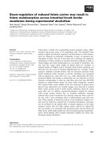
Fig. 1. Loss and recovery of SST-14 binding sites. (A) SST-14-mediated reduction of cell surface binding sites. RIN-SSTR3 cells were stimu-
lated with SST-14 at 37 °C for the indicated times. Cells were washed with acidic buffer, and surface binding sites were determined by incu-
bation with [
125
I]SST-14 at 4 °C. (B) Recovery of cell surface binding sites. RIN-SSTR3 cells were stimulated for 120 min with SST-14 at
37 °C. Cells were washed with acidic buffer and incubated for the indicated times, and cell surface binding sites were determined as
described above. (C) Downregulation of surface binding sites. RIN-SSTR3 cells were stimulated with SST-14 for the indicated times, washed
with acidic buffer, and incubated for 120 min at 37 °C. Surface binding sites were determined as described above. (D) Determination of cell
surface and total binding after stimulation with SST-14. RIN-SSTR3 cells were stimulated with SST-14 in the absence or presence of sapo-
nin. Binding was measured as described above. The data are expressed at mean ± SEM values from three independent experiments. (E, F)
Distribution of SSTR3–HSV in control cells and under conditions of receptor downregulation. RIN cells expressing SSTR3–HSV were
stimulated (F) or not stimulated (E) with SST-14 for 1300 min. Then, the peptide was removed and cells were allowed to recover for 90 min.
Thereafter, the epitope-tagged SSTR3 was visualized by indirect immunfluorescence. (F) Localization of SSTR3 after chronic stimulation
with SST-14. Chronic stimulation of SSTR3 with SST-14 mediates downregulation of SSTR3. The immunofluorescence signal of SSTR3 is
concentrated in one area of the cell and not equally distributed in the cell membrane (arrows).
Intracellular degradation of somatostatin D. Roosterman et al.
4730 FEBS Journal 275 (2008) 4728–4739 ª 2008 The Authors Journal compilation ª 2008 FEBS
within the cells or that it is downregulated by degrada-
tion. To distinguish between the two possibilities, we
determined the localization of SSTR3 after 1300 min
of stimulation with SST-14 and 90 min of recovery. In
untreated cells, SSTR3 showed a bright immunofluo-
rescence signal at the cell membrane (Fig. 1E, arrows).
In cells chronically stimulated with SST-14, the fluores-
cence signal was weaker than the signal seen in
untreated cells. Moreover, in all of these cells, the
SSTR3 immunofluorescence signal was locally concen-
trated in only one area of the cell surface and not
evenly distributed over the plasma membrane (Fig. 1F,
arrows).
Together, the results indicate that stimulation of
SSTR3 with SST-14 induced internalization of the
receptor. Chronic stimulation with SST-14 mediated
downregulation of SSTR3.
Uptake of [
125
I]Tyr11-SST-14 and [
125
I]Tyr1-octre-
otide in SSTR3-expressing rat insulinoma cells
To examine the fate of SST-14 cointernalized with
SSTR3, we measured the uptake of [
125
I]Tyr11-SST-
14 in RIN-SSTR3 cells in the absence (Fig. 2A,
diamonds) or presence (Fig. 2A, triangles) of ammo-
nium chloride. Treatment of the cells with
AB
CD
EF
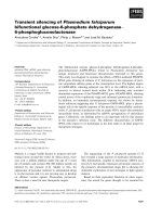
Fig. 2. SSTR3-mediated uptake of
125
I-labeled peptides. (A) SSTR3-mediated uptake of [
125
I]Tyr11-SST-14. RIN-SSTR3 cells were incubated
with [
125
I]SST-14 in the presence (triangle) or absence (diamonds) of ammonium chloride. Cell surface [
125
I]Tyr11-SST-14 was washed off,
and the amount of cell-associated radioactivity was determined. (B) SSTR3-mediated uptake of [
125
I]Tyr1-octreotide. RIN-SSTR3 cells were
incubated with [
125
I]Tyr1-octreotide. Cell surface [
125
I]Tyr1-octreotide was washed off, and the amount of cell-associated radioactivity was
determined. (C) HPLC separation of cell-associated, agonist-bound internalized radioactivity in the presence of ammonium chloride. Cells
were pretreated with ammonium chloride, incubated with [
125
I]Tyr11-SST-14 at 37 °C for 30 min in the presence of ammonium chloride,
washed with acidic buffer, and analyzed by HPLC. (D) HPLC separation of the cell supernatant incubated for 30 min with [
125
I]Tyr11-SST-14.
(E) Time course of SSTR3-mediated uptake of [
125
I]Tyr11-SST-14. RIN- SSTR3 cells were stimulated for 0–15 min with [
125
I]Tyr11-SST-14,
washed with acidic buffer, and incubated for 0–90 min. Cell-associated (triangles) and released (diamonds) radioactivity was determined by
HPLC. (F) Time course of SSTR3-mediated uptake of [
125
I]Tyr1-octreotide. RIN- SSTR3 cells were incubated for 0–15 min with [
125
I]Tyr1-
octreotide, washed with acidic buffer, and incubated for 0–90 min at 37 °C. Cell-associated radioactivity and radioactivity from the superna-
tants was determined by HPLC. The data are expressed as mean ± SEM values from three independent experiments.
D. Roosterman et al. Intracellular degradation of somatostatin
FEBS Journal 275 (2008) 4728–4739 ª 2008 The Authors Journal compilation ª 2008 FEBS 4731
ammonium chloride neutralized acidic cellular com-
partments [19]. Within 10–15 min at 37 °C, the cellu-
lar uptake of [
125
I]Tyr11-SST-14 reached maximal
levels in the absence of ammonium chloride corre-
sponding to 80% of the amount of the cell sur-
face-bound peptide. The amount of intracellular
radioactivity then quickly declined over a period of
15 min to very low levels corresponding to about
30% of the amount of cell surface-bound peptide.
These levels slowly decreased over the next 90 min
to < 20% of the initial value. These observations
are best explained by assuming that specific intracel-
lular degradation destroys the radiolabeled peptide,
and the degradation products are then released from
the cells. The low level of radioactivity observed to
be cell-associated between 60 and 120 min probably
reflects the steady-state level of [
125
I]Tyr11-SST-14
determined by receptor-mediated uptake and degra-
dation. The receptor population engaged in agonist
uptake appears to be largely diminished at these
times, due to receptor internalization and desensitiza-
tion [20]. Notably, the amount of the internalized
peptide corresponds to 80% of the cell surface-bound
peptide, suggesting that most of the agonist-occupied
receptors were engaged in endocytosis in the presence
of subnanomolar concentrations of agonist. When the
experiment was carried out in the presence of ammo-
nium chloride (Fig. 2A, triangles), radioactivity accu-
mulated with a similar kinetic during the first 10 min of
incubation but reached a plateau corresponding to
almost 100% of cell surface-bound [
125
I]Tyr11-SST-14.
Thus, under conditions in which the vesicular pH is
neutral [19], almost all cell surface-bound radioactivity
accumulated and remained in the cells.
Next, we determined the SSTR3-mediated uptake of
[
125
I]octreotide (Fig. 2B). Chronic stimulation of
SSTR3-expressing cells with octreotide induced contin-
uous uptake of the ligand. During the first 30 min of
incubation, 118% of surface-bound octreotide was
found to be cell-associated. Further incubation induced
a linear accumulation of radioactivity within the cells.
After 4 h of incubation, the amount of internalized
radioactivity was equivalent to 256% of cell surface-
bound octreotide.
In order to distinguish between radioactivity corre-
sponding to degraded or intact peptide, we examined
the cell-associated radioactivity by HPLC. We found
that [
125
I]Tyr eluted in fractions 1–5, degraded peptide
fragments in fractions 9–11, and intact [
125
I]SST-14
in fractions 14–17 (Fig. 2C,D). Figure 2C shows a
radiogram of the cell-associated radioactivity after
30 min of stimulation with [
125
I]SST-14 in the presence
of ammonium chloride. This treatment blocked the
degradation of [
125
I]SST-14. For instance, more than
95% of the cell-associated radioactivity eluted as
intact [
125
I]SST-14 in fraction 16. Minor amounts of
peptide fragments of [
125
I]SST-14 were observed in
fractions 9–11, suggesting modest degradation of
[
125
I]SST-14 by peptidases. The degradation of
[
125
I]SST-14 to [
125
I]Tyr was completely blocked.
Together, these results suggest that internalized
[
125
I]Tyr11-SST-14 was targeted in a degradation
pathway that is sensitive to ammonium chloride.
In contrast, a representative HPLC chromatogram of
the supernatant collected 30 min after stimulation of the
cells incubated with [
125
I]Tyr11-SST-14 shows that 97%
of the radioactivity was [
125
I]Tyr (Fig. 2D) [21,22]. This
result suggests that SST-14 was completely degraded to
its amino acids, which were subsequently released into
the supernatant. Similar HPLC analyses of the radio-
active degradation products found in the supernatant of
stimulated SSTR1 cells have demonstrated that SST-14
is relatively stable in the supernatant and is only slowly
degraded by phosphoamidon-sensitive peptidase [19].
Thus, we conclude that [
125
I]SST-14 is processed to
amino acids within the cells.
Figure 2E shows the time courses of association of
[
125
I]Tyr11-SST-14 (triangles) with the cells and the
accumulation of its degradation product [
125
I]Tyr (dia-
monds) in the extracellular medium, as assayed by
HPLC analyses of the fractions. During the first 15 min
of stimulation, [
125
I]Tyr11-SST-14 accumulated within
the cells. After removal of the peptide agonist by wash-
ing and further incubation at 37 °C, the amount of cell-
associated [
125
I]Tyr11-SST-14 rapidly decreased. Ninety
minutes after stimulation, the amount of cell-associated
[
125
I]Tyr11-SST-14 was reduced to 6%. This decay was
paralleled by accumulation of [
125
I]Tyr (Fig. 2E,
diamonds) in the medium. Thus, all of the internalized
peptide was degraded to its amino acids.
Next, we analyzed whether or not octreotide was
degraded during the internalization process. In accor-
dance with Fig. 2B,F (filled circles) shows that
[
125
I]Tyr1-octreotide was rapidly internalized by RIN-
SSTR3 cells during the 15 min stimulation period,
i.e. as long as the peptide was present. However,
when the stimulus was removed, the cells released
the endocytosed peptide into the supernatatant. Inter-
estingly, HPLC analysis of the cell lysate demon-
strated that octreotide was not degraded (data not
shown). Both the radioactivity determined in the
supernatant and the cell-associated radioactivity
eluted with a retention time identical to that of
[
125
I]Tyr1-octreotide. Thus, SSTR3 mediates ligand-
specific processing. Whereas SST-14 is sorted into an
ammonium-sensitive degradation pathway, octreotide
Intracellular degradation of somatostatin D. Roosterman et al.
4732 FEBS Journal 275 (2008) 4728–4739 ª 2008 The Authors Journal compilation ª 2008 FEBS
bypasses degradation, accumulates in the cell and is
released as intact ligand from the cells in the sur-
rounding medium.
SSTR3-mediated uptake of fluorescein
isothiocyanate (FITC)–SST-14
To directly visualize the receptor-mediated uptake of
the peptide agonist, internalization of FITC-labeled
SST-14 was examined in RIN-SSTR3 cells by confocal
laser scanning microscopy. The fluorescent peptide was
colocalized with herpes simplex virus glycoprotein D
tagged SSTR3 (SSTR3–HSV), as detected by indirect
immunofluorescence. Cells were incubated with FITC–
SST-14 at 4 °C. After removal of unbound peptide, a
temperature shift to 37 °C induced internalization of
cell surface-bound agonist for 2, 30 or 60 min. At the
beginning of the observation period at 2 min, fluores-
cence signals of FITC–SST-14 were barely visible. How-
ever, a few discrete zones were labeled at the cell surface
(Fig. 3A, green arrows, top left) that colocalized with
SSTR3–HSV (Fig. 3A, red arrows, top middle panel,
yellow arrows in the overlay). After 30 min, internalized
FITC–SST-14 and SSTR3–HSV frequently colocalized
in intracellular vesicles (Fig. 3A, middle panels, yellow
arrows). However, vesicles that appear only in red or
green suggest that some FITC–SST-14 (Fig. 3A, green
arrow) dissociated from SSTR3–HSV (Fig. 3A, red
arrow) and that SSTR3 and the agonist peptide were
sorted into different cell pathways. After 60 min of
stimulation, most of the receptors were recycled to the
plasma membrane (Fig. 3A, red arrows, bottom pan-
els), whereas the fluorescence signal of the ligand was
still observed within intracellular vesicular structures.
Traces of SSTR3–HSV were also detected within intra-
cellular vesicular compartments, where it colocalized
with SST-14 (Fig. 3A, yellow arrowheads).
Agonist-induced mobilization of arrestin-2–
enhanced green fluorescent protein (EGFP)
HPLC analysis of internalized SST-14 and octreotide
demonstrated that SST-14 but not octreotide was
metabolized after internalization. We reasoned that the
integrity of the ligand could influence the association
of arrestins with the internalized receptor. Therefore,
we analyzed the localization of arrestin-2–EGFP and
SSTR3–HSV after stimulation with 1 lm SST-14 or
octreotide for 15 min (Fig. 3B). In unstimulated cells,
arrestin-2–EGFP was diffusely located within the cells
and SSTR3–HSV was primarily located at the plasma
membrane (Fig. 3B, top panels). Stimulation with
either of the two agonists induced internalization of
the receptor (Fig. 3B, red arrows, middle panel).
Accordingly, arrestin-2 was mobilized and transported
from the cytosol to the cell membrane (Fig. 3B, green
arrows, middle panel). Interestingly, 15 min after stim-
ulation, arrestin-2 was only partially associated with
internalized SSTR3–HSV, indicating that arrestin-2
A
B
Fig. 3. Agonist-induced internalization of SSTR3–HSV. (A) SSTR3-
mediated uptake of FITC–SST-14. RIN-SSTR3 cells were incubated
for 60 min with FITC–SST-14 at 0 °C. Cells were washed and incu-
bated for 2, 30 and 60 min at 37 °C. FITC–SST-14 was detected
using the FITC label (shown in green). SSTR3 was detected using
antibody directed against the HSV tag (shown in red). (B) Agonist-
induced mobilization of arrestin-2–EGFP. RIN-SSTR3 cells were
transiently transfected with arrestin-2–EGFP. Cells were stimulated
with SST-14 (1 l
M) or octreotide (1 lM) for 15 min at 37 °C.
SSTR3–HSV was localized using an antibody against HSV and arres-
tin-2-EGFP by EGFP. The experiment was performed three times,
with similar results.
D. Roosterman et al. Intracellular degradation of somatostatin
FEBS Journal 275 (2008) 4728–4739 ª 2008 The Authors Journal compilation ª 2008 FEBS 4733
dissociated from the internalized receptor at or close
to the plasma membrane. Virtually no differences
could be determined in the localization of arrestin-2–
EGFP after stimulation with SST-14 or octreotide.
Thus, our data indicate that the stable ligand, octreo-
tide, did not induce stronger association of arrestin-2–
EGFP with the internalized SSTR3 than did SST-14.
Internalized SST-14 is transported to lysosomes
The complete intracellular degradation of internalized
SST-14 suggested that the ligand was processed into
the lysosomal degradation pathway in RIN-SSTR3
cells. To examine whether fluorescent dye-labeled
SST-14 was sorted to lysosomes, experiments on colo-
calization of rhodamine-B–SST-14 with cathepsin D, a
lysosomal protease [23], were performed (Fig. 4).
Therefore, we incubated RIN-SSTR3 cells with rhoda-
mine–SST-14 at 4 °C. Under these conditions, the fluo-
rescence signals of the peptide were predominantly
observed as a punctate pattern at the cell surface,
whereas the fluorescence signals of the lysosomal pro-
tease appeared to be scattered within the cytoplasm
(Fig. 4, top panels). Warming the cells to 37 °C
induced the internalization of rhodamine–SST-14 and
aggregation of lysosomes (Fig. 4, middle panels). After
30 min of stimulation, clear colocalization of cathe-
psin D and rhodamine-B–SST-14 was observed, indi-
cating that the peptide was routed to a lysosomal
degradation pathway (Fig. 4, middle panel, yellow
arrowhead). After 60 min, both fluorescence signals
still colocalized within these compartments (Fig. 4,
middle panel, yellow arrowhead). The observation that
endocytosed SST-14 colocalized with cathepsin D
agrees with our data obtained using biochemical
assays, demonstrating complete degradation of inter-
nalized SST-14 in RIN-SSTR3 cells.
Discussion
Recent studies provided clear evidence that the SSTR
subtypes (SSTR1, SSTR2, SSTR3 and SSTR5) inter-
nalize to similar extents after stimulation with SST-14,
somatostatin-28, and synthetic agonists [16,18,20,
24,25]. However, detailed analyses of the endocytic
processes and the pathways of trafficking of the SSTR
subtypes revealed explicit differences.
For example, SSTR1 did not mobilize arrestin-2
during internalization, whereas SSTR3 interacted tran-
siently with arrestin-2, and internalized SSTR2A
formed a stable complex with arrestin-2 [11,16,25]. The
differences between the receptor subtypes in their inter-
action with arrestins indicate the existence of internali-
zation and trafficking pathways that are specific for
the SSTR subtypes.
Determining the fate of the internalized ligand
revealed three different pathways of receptor traffic-
king and agonist processing. SSTR1 mediates accumu-
lation and release of intact SST-14. This phenomenon
was accomplished by a dynamic process of internaliza-
tion, recycling and reinternalization of the peptide, a
pathway consistent with the role of SSTR1 as an auto-
receptor [11]. In contrast, SSTR2A induced sequestra-
tion of the receptor–ligand complex within early
endosomes. SSTR2A did not recycle within a period of
2 h after agonist stimulation. SST-14, endocytosed
with SSTR2A, was degraded by endothelin-converting
enzyme-1 and other peptidases and was not routed
into lysosomal degradation. This strong association of
arrestins with the internalized receptor and the seques-
tration of the receptor in early endosomes is indicative
of a class B receptor [13,26]. Stimulation of SSTR2A
with octreotide induced long-lasting sequestration of
the intact ligand into early endosomes.
Here, we investigated the intracellular trafficking of
SSTR3 and the processing of internalized [
125
I]Tyr11-
SST-14 and [
125
I]Tyr1-octreotide. Our data show
that SSTR3 transiently interacts with arrestins and
Fig. 4. Rhodamine-B–SST-14 is transported to lysosomes. RIN-
SSTR3 cells were incubated for 2 h with rhodamine-B–SST-14 at
4 °C, washed, and incubated for 0, 30 and 60 min at 37 °C. Rhoda-
mine-B–SST-14 (red) was detected using rhodamine-B fluores-
cence; lysosomes (green) were detected using an antibody against
cathepsin D. The experiment was performed three times, with sim-
ilar results.
Intracellular degradation of somatostatin D. Roosterman et al.
4734 FEBS Journal 275 (2008) 4728–4739 ª 2008 The Authors Journal compilation ª 2008 FEBS
directs SST-14 to lysosomal degradation. This
transient interaction of the receptor with arrestins is
indicative of a class A receptor, and our data are in
line with data in [16] and [25].
When we analyzed the fate of the ligand, we demon-
strated that SSTR3 routed internalized SST-14 towards
a lysosomal degradation pathway. Internalized
[
125
I]Tyr11-SST-14 was rapidly degraded, and [
125
I]Tyr
was released into the cell supernatant. Confocal laser
scanning microscopic analyses of internalized fluores-
cent dye-labeled SST-14 showed strong colocalization of
the fluorescence signal with cathepsin D, a specific mar-
ker protein for lysosomes. The persistence of the fluores-
cence signal within lysosomes as compared to the rapid
degradation of the radioligand within 30 min most
likely reflects the stability of the fluorophore and not
that of the peptide moiety. It is known that internalized
ligands that are routed to lysosomes are degraded by
acidic proteases [27]. To address the question of whether
somatostatin is also degraded by acidic proteases, we
interfered with the acidification of endocytic vesicles by
incubating the cells in the presence of ammonium chlo-
ride [19]. Under these conditions, the degradation of the
internalized radioligand and the continued endocytosis
of the peptide ligand were markedly blocked, suggesting
that receptor trafficking proceeds via acidic vesicles and
that the degradation of somatostatin is accomplished by
acidic proteases when endocytosed with SSTR3. Inter-
estingly, the endosomal degradation of internalized
SST-14, observed after internalization through
SSTR2A, was partially inhibited by neutralization of
acidic cell compartments [13], suggesting that different
peptidases are involved in the degradation process of
SST-14, depending on the coendocytosed SSTR sub-
type, i.e. either SSTR2A or SSTR3.
We also analyzed the intracellular processing of
octreotide in SSTR3-expressing cells. Octreotide is a
synthetic SST-14 analog that binds to SSTR2A as well
as SSTR3. Octreotide is resistant to degradation by
endosomal peptidases [13]. Interestingly, octreotide
was also stable when it was internalized via SSTR3,
suggesting that the synthetic agonist is also resistant to
lysosomal degradation or, alternatively, that it was not
routed to the lysosomes. Chronic stimulation of the
cells with octreotide induced continuous accumulation
of the intact peptide within these cells. After 4 h of
chronic stimulation, 256% of surface-bound octreotide
was observed to be cell-associated, indicating that
SSTR3 was continuously recycled to the cell mem-
brane and reinternalized during chronic stimulation,
thereby mediating the accumulation of octreotide in
the cells. A similar observation was described for the
SSTR1-mediated accumulation of SST-14 [11].
Interestingly, chronic stimulation of SSTR3 with
SST-14 induced time-dependent downregulation of
SSTR3. One hundred and twenty minutes after stimu-
lation, SSTR3 was recycled up to 75%, whereas it was
recycled up to only 43% after 1300 min. In contrast,
SSTR1, which does not direct SST-14 to lysosomal
degradation, recovered up to 100% under the same
conditions [11,20]. This observation underlines our
finding that SSTR3 continuously recycles and is
re-endocytosed under chronic stimulation. One hour
after stimulation of the cells with SST-14, immunofluo-
rescence signals of SSTR3–HSV still colocalized with
the fluorescence signal of the internalized ligand. At
this time point, the ligand was simultaneously detected
within lysosomes in SSTR3-expressing cells. The data
suggest that lysosomal targeting of SSTR3 is responsi-
ble for the downregulation of the receptor.
Taken together, our results show that: (a) SSTR3
continuously internalizes, recycles and reinternalizes
under chronic agonist stimulation; (b) the internalized
SST-14 is routed to lysosomal degradation, where
internalized [
125
I]Tyr11-SST-14 is degraded to
[
125
I]Tyr; (c) internalized octreotide is resistant to deg-
radation, but is accumulated within cells as an intact
ligand; and (d) chronic stimulation of SSTR3 with
SST-14 induces time-dependent downregulation of the
receptor, probably through lysosomal degradation of
SSTR3.
At least two conclusions may be drawn from these
observations. First, agonist-induced SSTR internaliza-
tion is a complex process depending on the receptor
subtype and the nature of the stimulating agonist.
Besides the above-described differences in the regula-
tion of receptor internalization, trafficking and recy-
cling, further functional differences among SSTR
subtypes may be postulated through interactions with
distinct SSTR-binding proteins. In fact, such binding
proteins that specifically associate with SSTR subtypes
have been recently identified [28,29].
Radiolabeled or fluorescent dye-labeled somatostatin
analogs accumulating in certain cancer cells are used
with the diagnostic method SRS, and conjugates of
stable somatostatin analogs with toxic compounds or
radioisotopes have been used for chemotherapy in
certain tumors [30]. Therefore, detailed knowledge of
the mechanisms underlying agonist-induced endocyto-
sis and trafficking of the SSTR subtypes is of great
clinical importance, and cancer patients may benefit
from it in the future.
Our results indicate that future drugs should be
tested for all known aspects of agonist-induced traf-
ficking. They also indicate that the considerable knowl-
edge about the interaction of octreotide with SSTR2A
D. Roosterman et al. Intracellular degradation of somatostatin
FEBS Journal 275 (2008) 4728–4739 ª 2008 The Authors Journal compilation ª 2008 FEBS 4735
cannot be generalized to other SSTR subtypes and
ligands without experimental proof. The receptor
subtype-specific transport of SSTR2A and the ligand-
specific processing of octreotide go well with the use of
octreotide in SSTR2A scintigraphy [13]. On the other
hand, these advantages adversely affect the use of
octreotide in tumor treatment, because this peptide
leads to extensive sequestration of SSTR2A and desen-
sitization of the targeted tumor cells for prolonged
periods [13]. Our results also show that octreotide
recycled during the SSTR3-mediated transport.
Because it did not remain sequestered in the cell when
internalized with SSTR3, labeled octreotide appears
not to be suitable for detecting SSTR3 in receptor
scintigraphy.
Experimental procedures
Materials
SST-14 and octreotide were obtained from Bachem (Weil
am Rhein, Germany), [
125
I]Tyr11-SST-14 (2000 CiÆmmol
)1
)
was from Amersham (Braunschweig, Germany), and
[
125
I]Tyr1-octreotide was from Anawa (Wangen, Switzer-
land). FITC–SST-14 was from Advanced Bioconcept
(Derry, NH, USA). FITC-conjugated anti-rabbit IgG,
Texas Red-conjugated anti-mouse IgG, paraformaldehyde,
glycerol ⁄ gelatin solution and BSA (fraction IV) were pur-
chased from Sigma (Taufkirchen, Germany). The poly-
clonal antiserum against cathepsin D was a generous gift
from A. Hille-Rehfeld (Goettingen, Germany), and has
been described in detail elsewhere [23].
Generation of cDNA constructs and cell line
The construct with arrestin-2 tagged with EGFP has been
described previously [31]. Generation of the neuroendocrine
RIN 1046-38 cell line stably expressing the C-terminal HSV
epitope-tagged rat SSTR3 tagged with the HSV glycopro-
tein D epitope at the C-terminus (SSTR3–HSV) has been
described previously, and it has been demonstrated to
possess a maximal binding capacity of 1660 (± 350) fmol
per 2 · 10
4
cells for [
125
I]Tyr11-SST-14 [20].
Synthesis of rhodamine-B-labeled SST-14
SST-14 was generated on a LIPS vario multiple peptide
synthesizer using the robot’s standard protocol following
the F moc strategy ( peptides&elephants, Potsdam, Germany).
The rhodamine-B label was attached by deprotection of the
N-terminal a-amino function. The rhodamine-B was acti-
vated using ByBop (Novabiochem, Darmstadt, Germany)
and N-methylmorpholine as a base. Rhodamine-B was
added in a four-fold surplus to the a-amino function.
The rhodamine ⁄ ByBop ⁄ N-methylmorpholine ratio was
1 : 0.9 : 2 in 1 mL of dimethylformamide. The coupling
reaction was performed two times for 3 h. The resin was
washed with dimethylformamide until the washing solution
was colorless. After this, the resin was washed with
dichloromethane and dried overnight. The next day, the
peptide was cleaved and deprotected using reagent K
[tri-isopropylsilan (5%), water (2.5%), trifluoroacetic acid
(92.5%)]. The cleavage was performed for 2.5 h at room
temperature. After this, the peptide was precipitated with
diethyl ether and washed three times with ice-cold diethyl
ether. Cyclization was performed following the protocol of
Bodansky and Bodansky [32].
The peptide was further purified by HPLC, and the
identity was confirmed by MALDI-TOF MS. Rhodamine-
B–SST-14 has a 10-fold lower affinity for SSTR3 than
unlabeled SST-14 [24].
Reduction of cell surface binding
Cells grown in 24-well dishes were stimulated with 1 lm
SST-14 in RPMI-1640 (0.1% BSA) for 0–120 min at 37 °C.
Cells were placed on ice, washed three times with chilled
acidic buffer, and incubated with 100 000 c.p.m. per
0.3 mL of [
125
I]Tyr11-SST-14, 0.01 nm SST-14, and 0.1%
BSA in RPMI-1640, at 4 °C for 90 min. Bound [
125
I]Tyr11-
SST-14 was collected after lysing of the cells in 1 mL of
1 m NaOH and determined in a c-counter (Canberra Pack-
ard, Dreieich, Germany). Calculations and graphical
presentations were carried out using ms-excel and adobe
photoshop. Unspecific binding was determined in the
presence of 0.1 mm SST-14 [20].
Recovery of cell surface binding
Cells grown in 24-well dishes were stimulated with SST-14
(1 lm) in RPMI-1640 (0.1% BSA) for 120 min at 37 °C.
Surface-bound SST-14 was removed by three acidic washes
with Hank’s buffered saline (HBS) (acetic acid, pH 4.8) and
incubated for the indicated times in RPMI-1640 (0.1%
BSA). The cells were placed on ice and incubated with
100 000 c.p.m. per 0.3 mL of [
125
I]Tyr11-SST-14, 0.01 nm
SST-14, and 0.1% BSA in RPMI-1640, at 4 °C for 90 min.
Bound [
125
I]Tyr11-SST-14 was collected after lysing of the
cells in 1 mL of 1 m NaOH and determined in a c-counter
(Canberra Packard) [20].
Determination of cell surface and total binding
RIN-SSTR3 cells grown in 24-well dishes were stimulated
with SST-14 (1 lm) in RPMI-1640 (0.1% BSA) at 37 °C
for 60 min. Cells were washed three times with HBS (acetic
acid, pH 4.8) in the presence or absence of 0.1% saponin.
The cells were washed with RPMI-1640 to adjust the pH.
Intracellular degradation of somatostatin D. Roosterman et al.
4736 FEBS Journal 275 (2008) 4728–4739 ª 2008 The Authors Journal compilation ª 2008 FEBS
Surface binding sites were determined by incubation with
100 000 c.p.m. per 0.3 mL of [
125
I]Tyr11-SST-14, 0.01 nm
SST-14, and 0.1% BSA in RPMI-1640, at 4 °C for 90 min.
Total binding was determined by incubation with
100 000 c.p.m. per 0.3 mL of [
125
I]Tyr11-SST-14, 0.01 nm
SST-14, 0.1% BSA and 0.1% saponin in RPMI-1640, at
4 °C for 90 min. Bound [
125
I]Tyr11-SST-14 was collected
after lysing of the cells in 1 mL of 1 m NaOH and deter-
mined in a c-counter (Canberra Packard) [20].
Downregulation of cell surface binding sites
RIN-SSTR3 cells grown in 24-well dishes were stimulated
with SST-14 (1 lm) in RPMI-1640 (0.1% BSA) for the
indicated times at 37 °C. Surface-bound SST-14 was
removed by three acidic washes with HBS (acetic acid,
pH 4.8) and incubated for 120 min in RPMI-1640 (0.1%
BSA). The cells were placed on ice and incubated with
100 000 c.p.m. per 0.3 mL of [
125
I]Tyr11-SST-14, 0.01 nm
SST-14, and 0.1% BSA in RPMI-1640, at 4 °C for 90 min.
Bound [
125
I]Tyr11-SST-14 was collected after lysing of the
cells in 1 mL of 1 m NaOH and determined in a c-counter
(Canberra Packard) [20].
Uptake of [
125
I]Tyr-labeled ligand
RIN 1046-38 cells transfected with SSTR3–HSV cDNA
were seeded in 24-well microplates and grown to 75% con-
fluence. The culture medium was replaced by serum-free
medium containing [
125
I]Tyr11-SST-14 or [
125
I]Tyr1-octreo-
tide (100 000 c.p.m., 2000 CiÆmmol
)1
) and 0.1% BSA pre-
warmed to 37 °C, and incubated at this temperature for the
indicated times. Cells were then washed at acidic pH to
remove all cell surface-bound peptide [33], and cell-associ-
ated radioactivity was determined in a c-counter (LKB
Wallac, Ontario, Canada) following lysis of the cells in 1 m
NaOH. In parallel experiments, cell surface binding was
determined at 0 °C [20]. Cell-associated radioactivity was
expressed as percentage of total cell surface-bound radio-
activity. In addition, the experiment was carried out in the
presence of 40 mm NH
4
Cl [11].
HPLC analysis of internalized and released
[
125
I]Tyr-labeled ligand
RIN-SSTR3 cells grown in 24-well dishes were stimulated
for 0–30 min with [
125
I]Tyr11-SST-14 or [
125
I]Tyr1-octreo-
tide (100 000 c.p.m.) in RPMI-1640 (0.1% BSA) in the
presence or absence of 40 mm NH
4
Cl. The cells were
washed in acidic buffer and incubated for 0–90 min in
RPMI-1640 (0.1% BSA). The supernatants were collected,
and acidified by adding 10 lL of trifluoroacetic acid. The
supernatants were centrifuged (5 min, 13 000 g ) and sub-
jected to HPLC separation. Cell-associated radioactivity
was determined by adding 0.5 mL of HPLC buffer A.
Lysed cells were centrifuged (5 min, 13 000 g) and subjected
to HPLC separation. HPLC was performed on a reverse-
phase C-18 column (2 · 25 mm). A separating gradient of
0–40% acetonitrile ⁄ 0.08% trifluoroacetic acid for 25 min at
a flow rate of 1 mLÆmin
)1
was used with an HPLC-Akta
(General Healthcare, Munich, Germany). The HPLC
gradient was fractionated every minute, and the eluted
radioactivity was determined in a c -counter (LKB Wallac).
The radioactivity of each fraction was divided by the initial
amount of cell-associated radioactivity determined after
15 min of incubation with 100 000 c.p.m.Æ mL
)1
radioacti-
vity [11].
Microscopy and immunofluorescence
Cells were incubated with SST-14 (1 lm) for 1300 min,
washed, and incubated for 90 min at 37 °C. The cells were
fixed with paraformaldehyde 4%, washed, and incubated for
30 min in NaCl ⁄ P
i
(0.05% saponin, 5% normal goat serum).
SSTR3–HSV was detected using mouse antibody against
glycoprotein D (1 : 10 000) and Texas Red-conjugated anti-
mouse IgG (1 : 200). In other experiments, cells were incu-
bated with FITC–SST-14 or rhodamime-B–SST-14 on ice in
RPMI-1640 and 0.1% BSA. Unbound ligand was washed
off, and the cells were incubated for the indicated times at
37 °C, washed with HBS ⁄ acetic acid (pH 4.75) at 4 °C, fixed,
and permeabilized for 30 min in HBS, 5% normal goat
serum, and 0.05% saponin. SST-14 was detected using the
fluorescence dye, cathepsin D was detected using polyclonal
antiserum against cathepsin D, and SSTR3–HSV was
detected by using mouse antibody against glycoprotein D
(1 : 10 000, overnight incubation at 4 °C). FITC-conjugated
or Texas Red-conjugated goat anti-(mouse IgG) or goat
anti-(rabbit IgG) were used as secondary antibodies (1 : 200,
1 h, room temperature). Cells were embedded in Vectashield
mounting medium (Vector, Burlingame, CA, USA) and
observed with confocal microscopy [20,32].
Acknowledgements
This work was supported by grants from IZKF
(STEI2 ⁄ 076 ⁄ 06), SFB 293 (A14), SFB 492 (B13),
DFG STE 1014 ⁄ 2-2 (to M. Steinhoff), the Rosacea
Foundation (to M. Steinhoff and D. Roosterman) and
IMF Mu
¨
nster (RO 120611) (to D. Roosterman).
References
1 Olias G, Viollet C, Kusserow H, Epelbaum J & Meyer-
hof W (2004) Regulation and function of somatostatin
receptors. J Neurochem 89, 1057–1091.
2 Csaba Z & Dournaud P (2001) Cellular biology of
somatostatin receptors. Neuropeptides 35, 1–23.
D. Roosterman et al. Intracellular degradation of somatostatin
FEBS Journal 275 (2008) 4728–4739 ª 2008 The Authors Journal compilation ª 2008 FEBS 4737
3 Reubi JC & Laissue JA (1995) Multiple actions of
somatostatin in neoplastic disease. Trends Pharmacol
Sci 16, 110–115.
4 de Herder WW, Hofland LJ, van der Lely AJ &
Lamberts SW (2003) Somatostatin receptors in gastro-
entero-pancreatic neuroendocrine tumours. Endocr
Relat Cancer 10, 451–458.
5 Resmini E, Dadati P, Ravetti JL, Zona G, Spaziante R,
Saveanu A, Jaquet P, Culler MD, Bianchi F, Rebora A
et al. (2007) Rapid pituitary tumor shrinkage with dis-
sociation between antiproliferative and antisecretory
effects of a long-acting octreotide in an acromegalic
patient. J Clin Endocrinol Metab 92, 1592–1599.
6 Moulik PK, Varma TR, Vora JP & Vinjamuri S (2002)
The role of somatostatin receptor scintigraphy in the
management of pituitary tumours. Nucl Med Commun
23, 117–120.
7 Hofland LJ, Lamberts SW, van Hagen PM, Reubi JC,
Schaeffer J, Waaijers M, van Koetsveld PM, Srinivasan
A, Krenning EP & Breeman WA (2003) Crucial role for
somatostatin receptor subtype 2 in determining the
uptake of [111In-DTPA-D-Phe1]octreotide in somato-
statin receptor-positive organs. J Nucl Med 44, 1315–
1321.
8 Ginj M, Zhang H, Waser B, Cescato R, Wild D, Wang
X, Erchegyi J, Rivier J, Macke HR & Reubi JC (2006)
Radiolabeled somatostatin receptor antagonists are
preferable to agonists for in vivo peptide receptor
targeting of tumors. Proc Natl Acad Sci USA 103,
16436–16441.
9 Storch D, Behe M, Walter MA, Chen J, Powell P,
Mikolajczak R & Macke HR (2005) Evaluation of
[99mTc ⁄ EDDA ⁄ HYNIC0]octreotide derivatives com-
pared with [111In-DOTA0,Tyr3, Thr8]octreotide and
[111In-DTPA0]octreotide: does tumor or pancreas
uptake correlate with the rate of internalization? J Nucl
Med 46, 1561–1569.
10 Hukovic N, Panetta R, Kumar U & Patel YC (1996)
Agonist-dependent regulation of cloned human somato-
statin receptor types 1–5 (hSSTR1-5): subtype selective
internalization or upregulation. Endocrinology 137,
4046–4049.
11 Roosterman D, Kreuzer OJ, Brune N, Cottrell GS,
Bunnett NW, Meyerhof W & Steinhoff M (2007)
Agonist-induced endocytosis of rat somatostatin
receptor 1. Endocrinology 148, 1050–1058.
12 Liu Q, Cescato R, Dewi DA, Rivier J, Reubi JC &
Schonbrunn A (2005) Receptor signaling and endocyto-
sis are differentially regulated by somatostatin analogs.
Mol Pharmacol 68, 90–101.
13 Roosterman D, Kempkes C, Cottrell GS, Padilla BE,
Bunnett NW, Turck CW & Steinhoff M (2008) Endo-
thelin-converting enzyme-1 degrades internalized
SST-14. Endocrinology 149, 2200–2207.
14 Ferone D, van Hagen MP, Kwekkeboom DJ, van
Koetsveld PM, Mooy DM, Lichtenauer-Kaligis E,
Schonbrunn A, Colao A, Lamberts SW & Hofland LJ
(2000) Somatostatin receptor subtypes in human thy-
moma and inhibition of cell proliferation by octreotide
in vitro. J Clin Endocrinol Metab 85, 1719–1726.
15 Roth A, Kreienkamp HJ, Meyerhof W & Richter D
(1997) Phosphorylation of four amino acid residues in
the carboxyl terminus of the rat somatostatin receptor
subtype 3 is crucial for its desensitization and internali-
zation. J Biol Chem 272, 23769–23774.
16 Tulipano G, Stumm R, Pfeiffer M, Kreienkamp HJ,
Hollt V & Schulz S (2004) Differential beta-arrestin
trafficking and endosomal sorting of somatostatin
receptor subtypes. J Biol Chem 279, 21374–21382.
17 Roosterman D, Cottrell GS, Padilla BE, Muller L,
Eckman CB, Bunnett NW & Steinhoff M (2007) Endo-
thelin-converting enzyme 1 degrades neuropeptides in
endosomes to control receptor recycling. Proc Natl
Acad Sci USA 104, 11838–11843.
18 Cescato R, Schulz S, Waser B, Eltschinger V, Rivier JE,
Wester HJ, Culler M, Ginj M, Liu Q, Schonbrunn A
et al. (2006) Internalization of sst2, sst3, and sst5 recep-
tors: effects of somatostatin agonists and antagonists.
J Nucl Med 47, 502–511.
19 Presky DH & Schonbrunn A (1986) Receptor-bound
somatostatin and epidermal growth factor are processed
differently in GH4C1 rat pituitary cells. J Cell Biol 102,
878–888.
20 Roosterman D, Roth A, Kreienkamp HJ, Richter D &
Meyerhof W (1997) Distinct agonist-mediated endocyto-
sis of cloned rat somatostatin receptor subtypes expressed
in insulinoma cells. J Neuroendocrinol 9, 741–751.
21 Lucius R & Mentlein R (1991) Degradation of the
neuropeptide somatostatin by cultivated neuronal and
glial cells. J Biol Chem 266, 18907–18913.
22 Mentlein R & Dahms P (1994) Endopeptidases 24.16
and 24.15 are responsible for the degradation of
somatostatin, neurotensin, and other neuropeptides by
cultivated rat cortical astrocytes. J Neurochem 62, 27–
36.
23 Delbruck R, Desel C, von Figura K & Hille-Rehfeld A
(1994) Proteolytic processing of cathepsin D in prelysos-
omal organelles. Eur J Cell Biol 64, 7–14.
24 Roth A, Kreienkamp HJ, Nehring RB, Roosterman D,
Meyerhof W & Richter D (1997) Endocytosis of the rat
somatostatin receptors: subtype discrimination, ligand
specificity, and delineation of carboxy-terminal positive
and negative sequence motifs. DNA Cell Biol 16, 111–
119.
25 Kreuzer OJ, Krisch B, Dery O, Bunnett NW & Meyerhof
W (2001) Agonist-mediated endocytosis of rat somato-
statin receptor subtype 3 involves beta-arrestin and clath-
rin coated vesicles. J Neuroendocrinol 13, 279–287.
Intracellular degradation of somatostatin D. Roosterman et al.
4738 FEBS Journal 275 (2008) 4728–4739 ª 2008 The Authors Journal compilation ª 2008 FEBS
26 Padilla B, Cottrell GS, Roosterman D, Pikios S, Muller
L, Steinhoff M & Bunnett NW (2007) Endothelin-con-
verting enzyme-1 regulates endosomal sorting of calcito-
nin receptor-like receptor and beta-arrestins. J Cell Biol
179, 981–997.
27 Mellman I (1996) Endocytosis and molecular sorting.
Annu Rev Cell Dev Biol 12, 575–625.
28 Zitzer H, Honck HH, Bachner D, Richter D & Kre-
ienkamp HJ (1999) Somatostatin receptor interacting
protein defines a novel family of multidomain proteins
present in human and rodent brain. J Biol Chem 274,
32997–33001.
29 Wente W, Stroh T, Beaudet A, Richter D & Kreienk-
amp HJ (2005) Interactions with PDZ domain proteins
PIST ⁄ GOPC and PDZK1 regulate intracellular sorting
of the somatostatin receptor subtype 5. J Biol Chem
280, 32419–32425.
30 Lamberts SW, Krenning EP & Reubi JC (1991) The
role of somatostatin and its analogs in the diagnosis
and treatment of tumors. Endocr Rev 12, 450–482.
31 Roosterman D, Cottrell G, Schmidlin F, Steinhoff M &
Bunnett NW (2004) Recycling and resensitization of
the neurokinin 1 receptor: influence of agonist concen-
tration and RAB GTPases. J Biol Chem 279, 30670–
30679.
32 Bodansky M & Bodansky A (1994) The practice of
peptide synthesis, 2nd edn. Springer-Verlay, New York,
NY.
33 Presky DH & Schonbrunn A (1988) Somatostatin
pretreatment increases the number of somatostatin
receptors in GH4C1 pituitary cells and does not reduce
cellular responsiveness to somatostatin. J Biol Chem
263, 714–721.
D. Roosterman et al. Intracellular degradation of somatostatin
FEBS Journal 275 (2008) 4728–4739 ª 2008 The Authors Journal compilation ª 2008 FEBS 4739