Báo cáo khoa học: Modulation of aryl hydrocarbon receptor transactivation by carbaryl, a nonconventional ligand pptx
Bạn đang xem bản rút gọn của tài liệu. Xem và tải ngay bản đầy đủ của tài liệu tại đây (741.87 KB, 13 trang )
Modulation of aryl hydrocarbon receptor transactivation
by carbaryl, a nonconventional ligand
´
˜
Susanna Boronat1, Susana Casado2, Jose M. Navas2 and Benjamin Pina1
´
1 Institut de Biologia Molecular de Barcelona, Consejo Superior de Investigaciones Cientıficas, Barcelona, Spain
´
2 Department of Environment, Instituto Nacional de Investigacion y Tecnolog’a Agraria y Alimentaria (INIA), Madrid, Spain
Keywords
bioassays; dioxin-like; endocrine disruptors;
recombinant yeast assays; transcriptional
response
Correspondence
B. Pina, IBMB-CSIC, Jordi Girona,
˜
18, 08034 Barcelona, Spain
Fax: +34 93 204 59 04
Tel: +34 93 400 61 57
E-mail:
(Received 2 March 2007, revised 2 May
2007, accepted 3 May 2007)
doi:10.1111/j.1742-4658.2007.05867.x
Carbaryl (1-naphthyl-N-methylcarbamate), a widely used carbamate insecticide, induces cytochrome P450 1A gene expression in mammalian cells.
This activity is usually mediated by the interaction of the compound with
the aryl hydrocarbon receptor. However, it has been proposed that this
mechanism does not apply to carbaryl because its structure differs from
that of typical aryl hydrocarbon receptor ligands. We show here that carbaryl promotes activation of target genes in a yeast-based bioassay expressing both aryl hydrocarbon receptor and aryl hydrocarbon receptor nuclear
translocator. By contrast, carbaryl acted as a competitive inhibitor, rather
than as an agonist, in a simplified yeast system, in which aryl hydrocarbon
receptor nuclear translocator function is bypassed by fusing aryl hydrocarbon receptor to a heterologous DNA binding domain. This dual action of
carbaryl, agonist and partial antagonist, was also observed by comparing
carbaryl response in two vertebrate cell lines. A yeast two-hybrid assay
showed that the mammalian coactivator cAMP response element-binding
protein readily interacts with aryl hydrocarbon receptor bound to its
canonical ligand b-naphthoflavone, but not with the carbaryl–aryl hydrocarbon receptor complex. We propose that carbaryl interacts with aryl
hydrocarbon receptor, but that its peculiar structure imposes a substandard
configuration on the aryl hydrocarbon receptor ligand-binding domain that
prevents interaction with key coactivators and activates transcription without the need for aryl hydrocarbon receptor nuclear translocator. This effect
may be relevant in explaining its physiological effects in exposed animals,
and may help to predict its effects, and that of similar compounds, in
humans. Our data also identify the aryl hydrocarbon receptor ⁄ cAMP
response element-binding protein interaction as a molecular target for
the identification and development of new aryl hydrocarbon receptor
antagonists.
The known or suspected deleterious effects of global
pollution by different chemical species, ranging from
industrial by-products to pesticides, has developed into
a major public concern in recent decades. Each year,
thousands of new chemicals are released into the environment at a pace that makes impossible the precise
characterization of their acute and ⁄ or chronic impact,
both on human health and on exposed ecosystems.
Abbreviations
AhR, aryl hydrocarbon receptor; ARNT, AhR nuclear translocator; BNF, b-naphthoflavone; CBP, cAMP response element-binding protein;
DBD, DNA binding domain; GUS, b-glucuronidase; HAT, histone acetyltransferase; LBD, ligand-binding domain; LOEC, lowest observed
effect concentration; RTL, rainbow trout liver; RYA, recombinant yeast assay; TCDD, 2,3,7,8-tetrachlorodibenzo(p)dioxin; XRE, xenobiotic
responsive element.
FEBS Journal 274 (2007) 3327–3339 ª 2007 The Authors Journal compilation ª 2007 FEBS
3327
Modulation of AhR transactivation by carbaryl
S. Boronat et al.
Among the different types of pollutants, those interacting with cell receptors constitute an even greater risk
as they become toxic at very low concentrations, sometimes at, or under, the limits of detection by conventional analytical procedures [1].
The aryl hydrocarbon receptor (AhR) belongs to
the basic helix-loop-helix-PAS family of transcription
regulators [2]. This family roots itself on the prokaryotic kingdom; however, the capacity to bind specific
ligands and to modulate the transcriptional activity
according to this binding has apparently only evolved
in chordates [3]. The physiological role of AhR in
vertebrates has not yet been completely elucidated,
but it is known to regulate specific phase I and II
metabolic enzymes, among others [4,5]. Ectopic activation of AhR constitutes an initial step leading to
toxic effects of a variety of harmful pollutants, such
as 2,3,7,8-tetrachlorodibenzo(p)dioxin (TCDD) and
benzo[a]pyrene [6,7], which include immune dysfunction, endocrine disruption, reproductive toxicity,
developmental defects, and cancer in vertebrates
[8–12].
The use of yeast systems to monitor the interaction
of different chemicals with vertebrate receptors has
become a common tool to detect the presence of receptor-binding activity in the environment [13–16]. These
yeast-based bioassays, known as recombinant yeast
assays (RYAs), have been used to correlate the presence of suspected or bona-fide endocrine disruptors
and estrogenic activity in environmental samples [17–
19], and to establish relationships between chemical
structures and affinity for vertebrate hormone receptors [20–24].
It is generally accepted that ligand-free AhR molecules are mainly cytoplasmic, and that binding to the
ligand triggers the translocation of the receptor–ligand
complex to the cell nucleus. During this process, the
receptor–ligand complex binds to an auxiliary cofactor,
the AhR nuclear translocator (ARNT), to form a ternary complex, which is capable of recognizing specific
DNA sequences (xenobiotic responsive elements;
XRE) in the promoter of target genes, increasing their
transcription rates [25]. Both AhR and ARNT by
themselves are capable of triggering transcription when
tethered to upstream regions of reporter genes by
heterologous DNA binding domains (DBD) [26,27].
More specifically, the ligand-binding domain (LBD) of
AhR, when fused to an heterologous DBD, produces
a ligand-dependent, ARNT-independent activator
maintaining most pharmacological features of the
AhR ⁄ ARNT ⁄ XRE system [27]. These chimeric systems
have been used mostly in yeast, but they also work in
mammalian cell lines [28].
3328
The mechanisms by which transcriptional activation
occurs upon binding of the ligand ⁄ AhR ⁄ ARNT complex to XRE are still unclear, but they probably include
recruiting of different coactivators and general transcription factors, which ultimately promote transcription initiation by interacting with the RNA
polymerase II [29]. A key component of this mechanism
is the cAMP response element-binding protein
(CBP) ⁄ p300 complex, which is assumed to have a major
role on the transcriptional activation by AhR ⁄ ARNT
in mammals by interacting with histone acetyltransferases (HATs) [29,30]. In yeast, a key coactivator for
ligand-dependent transcriptional activation by AhR is
the Spt-Ada-Gcn5-acetyltransferase (SAGA) complex
[31], a HAT complex required for function in yeast of
many, but not all, transcriptional activators, including
Gcn4p and VP16 [32,33]. It is also required for liganddependent activation mediated in yeast by several vertebrate receptors, including the glucocorticoid, estrogen
and retinoic acid receptors [34–36].
Carbaryl (1-naphthyl-N-methylcarbamate) is a widespectrum carbamate insecticide that has been applied
for approximately 40 years as a contact and ingestion
insecticide on a wide variety of crops, as well as on
poultry, livestock and pets. It is also used as acaricide
and as molluscicide in aquaculture facilities. It has been
reported that this compound is an inducer of cytochrome P450 1A gene expression [37,38], a biomarker
of ectopic activation of the AhR receptor by exogenous
ligands present in the environment [11,39]. However,
carbaryl differs structurally from typical AhR ligands,
which are aromatic compounds with two or more
rings in the same plane that can be accommodated
within a rectangular binding site of approximately
˚
˚
˚
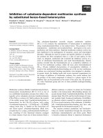
14 A · 12 A · 5 A [40]. Carbaryl does not fit easily
into these structural constraints (Fig. 1). Nevertheless,
both activation of the AhR system by carbaryl in cultured mammalian cells and specific binding in vitro of
carbaryl to AhR has been demonstrated [41]. The
present study intended to further characterize the interaction of carbaryl with AhR by using a combination of
Fig. 1. Chemical structures of b-naphthoflavone (left) and carbaryl
(right).
FEBS Journal 274 (2007) 3327–3339 ª 2007 The Authors Journal compilation ª 2007 FEBS
S. Boronat et al.
mammalian cell culture and yeast-based systems. The
data obtained suggest that the peculiar structure of
carbaryl imposes a nonstandard structure of the AhRLBD, which in turns modulates the capacity of the
complex to interact with CBP ⁄ p300 or SAGA. This
property of carbaryl may be relevant in the explanation
of its physiological effects, and provide an explanation
for the largely contradictory current data on the effects
of carbaryl in different cell lines and tissues.
Results
Differential response of pLMAX and YCM
systems to carbaryl
Addition of increasing concentrations of carbaryl to
YCM cells resulted in a bell-shaped activation ⁄ toxicity
curve, as described for many receptor agonists that
become toxic or inhibitory at high concentrations [22]
(Fig. 2). The calculated EC50 value for carbaryl in
YCM cells was 124.3 ± 9.6 lm (Table 1), which corresponds to a weak agonist. At higher concentrations,
carbaryl becomes an inhibitor, with an apparent IC50
value of 578.0 ± 36.2 lm (Table 1). By contrast, carbaryl was unable to activate the LMAX-RYA system at
any concentration (not shown). In this system, carbaryl
acted as an antagonist because simultaneous addition
of 1 lm of a typical AhR ligand, b-naphthoflavone
(BNF), and increasing concentrations of carbaryl resulted in a typical inhibition curve, with an apparent IC50
of 256.3 ± 38.2 lm (Fig. 2 and Table 1; see BNF and
carbaryl structures in Fig. 1). Therefore, the response
Modulation of AhR transactivation by carbaryl
Table 1. Adjustments to the different activation ⁄ inhibition models.
a 95% confidence margins.
Model
System
Equation
EC50 ⁄ IC50 (lM)a
Activation
Inhibition
Inhibition
Competitive inhibition
Irreversible inhibition
Toxicity
YCM-RYA
YCM-RYA
LMAX-RYA
LMAX-RYA
LMAX-RYA
Gal-GUS
Eqn
Eqn
Eqn
Eqn
Eqn
Eqn
124.3
578.0
256.3
77.3
461.5
459.8
(1)
(1)
(1)
(2)
(3)
(3)
±
±
±
±
±
±
9.6
36.2
38.2
15.3
102.3
74.3
to carbaryl depended on the RYA system used and,
presumably, on the transcriptional activation mechanism predominant in each of them.
Carbaryl as a competitive inhibitor of AhR
in yeast
To elucidate the mechanisms causing inhibition of
carbaryl in LMAX-RYA, a number of dose–response
assays with increasing concentrations of BNF were performed in the presence of different concentrations of
carbaryl. As shown in Fig. 3A,B, the presence of carbaryl affected both the maximal activation at saturating
concentrations of BNF and the position of the sigmoidal curve. Whereas the latter is consistent with a competition for binding to AhR-LBD by carbaryl and
BNF, the decrease of the maximal activation value is
more consistent with a noncompetitive inhibition, either
reversible or irreversible. IC50 values were obtained for
both effects separately (Table 1) from the analysis of
the experimental data shown in Fig. 3A,B. Figure 4A
shows the adjustment of the apparent EC50 values
at different carbaryl concentrations to a competitive
inhibition model [Eqn (2)]. An IC50 value of
77.3 ± 15.3 lm can be calculated from the slope of the
regression line (Table 1). Similarly, the decrease of
maximal activation at increasing concentrations of
carbaryl can be adjusted to a noncompetitive binding
model [Eqn (3); Fig. 4B]. In this case, the corresponding IC50 value obtained from the slope of the regression line was significantly higher, 461.5 ± 102.3 lm
(Table 1), which is compatible to the toxic effect
observed in YCM-RYA. Therefore, we conclude that
the behaviour of carbaryl in both RYA systems is similar, with a binding constant of approximately 100 lm
and a toxic effect at concentrations higher than 400 lm.
Analysis of carbaryl toxicity in yeast
Fig. 2. Dose–response curve for carbaryl in YCM-RYA (s) and in
LMAX-RYA with simultaneous addition of 1 lM BNF (d). Data are
the average of four independent determinations; bars represent
standard errors.
Irreversible inhibition can also be explained as a consequence of cell inactivation by the inhibitory ligand. In
this case, the phenomenon would not be related to the
FEBS Journal 274 (2007) 3327–3339 ª 2007 The Authors Journal compilation ª 2007 FEBS
3329
Modulation of AhR transactivation by carbaryl
S. Boronat et al.
A
B
Fig. 4. Data adjustment for the competitive-reversible (A) and irreversible (B) models for the experiments in Fig. 2; IC50 values were
calculated from the slopes of the regression lines (- - -).
Fig. 3. Dose–response of BNF in the presence of increasing concentrations of carbaryl in pLMAX-RYA. (A) Showing data relative to
the maximal activity of BNF in the absence of carbaryl. Data were
adjusted to the maximal concentration in each series (relative
expression). (B) Showing unadjusted data (GUS arbitrary units).
Dots represent replicas for each series; curves are calculated from
the observed EC50 for each carbaryl concentration (all replicas combined).
characteristics of the receptor, but to the sensitivity of
the particular cell strain used in the assay. This effect
can be monitored by measuring the effect of the compound to the activation of galactose-responsive genes,
an endogenous yeast activation mechanism completely
unrelated to AhR [42]. Figure 5 shows the decrease of
3330
Fig. 5. Inhibition of galactose response by carbaryl in GAL-GUS system. The discontinuous curve represents a nonlinear fitting to a
logistic function.
cell response to galactose in the presence of increasing
concentrations of carbaryl. The decrease follows a sigmoidal curve with an IC50 value of 459 ± 74 lm for
carbaryl, similar to the Ki value obtained for the
FEBS Journal 274 (2007) 3327–3339 ª 2007 The Authors Journal compilation ª 2007 FEBS
S. Boronat et al.
Modulation of AhR transactivation by carbaryl
negative effect at high concentrations of carbaryl in
both RYA systems (irreversible model; Table 1).
Therefore, we consider that the noncompetitive ⁄
irreversible component of carbaryl inhibition in LMAXRYA was likely due to cytotoxicity rather than to a
putative second site for carbaryl binding in the AhR.
Carbaryl as a competitive inhibitor of AhR
in vertebrate cell cultures
Two vertebrate cell lines were tested for their sensitivity to the presence of carbaryl in dose–response assays
using BNF as agonist. The CALUXÒ cell line, commonly used for testing AhR agonists, showed essentially identical dose–response curves in the presence
and absence of 200 lm carbaryl, with EC50 values of
8.40 ± 1.32 lm and 9.04 ± 1.34 lm for BNF, respectively (Fig. 6). By contrast, the rainbow trout liver
(RTL) cell line showed an EC50 for BNF of
0.75 ± 0.28 lm, approximately one tenth of the
corresponding value for CALUXÒ. These values
increased to 4.25 ± 0.87 lm when the dose–response
curve was performed in the presence of 200 lm carbaryl, indicating an antagonistic effect in these cells similar to the one observed for the yeast YCM-RYA
system (Fig. 6; compare with Fig. 3).
Modulation of the interaction of AhR-LBD with
CBP by AhR ligands
AhR activation is at least partially mediated by the
recruitment of CBP to the target promoters [29]. To
assess the influence of different ligands in this interaction, we performed two-hybrid assays in yeast, using
the pLMAX as a DNA binding domain and CBPGal4AD as activation domain. Addition of different
concentrations of BNF to the triple-transformant
resulted in a significant, 50% increase of maximal transcription level relative to an isogenic strain lacking the
CBP-Gal4AD plasmid (Fig. 7A). This effect was minimal, if any, when carbaryl was added to the same
strain, indicating that the interaction between carbarylloaded AhR-LBD and CBP did not occur (Fig. 7B).
The effect of the presence of CBP-Gal4AD in the
two-hybrid system was obscured by the strong activation signal of pLMAX-RYA in the presence of BNF.
This activity can be strongly reduced by the disruption
of the endogenous gene ADA2 in yeast [32] (Fig. 7C,
compare fluorescence units with Fig. 7A). Dada2 strains
expressing pLMAX showed a limited response to the
presence of BNF, and no response whatsoever to carbaryl (Fig. 7C,D). In this specific genetic background,
the presence of the CBP-Gal4AD construct increased
Fig. 6. Dose–response curves for BNF in the presence (s) and
absence (r) of 200 lM carbaryl in (A) RTL cells and (B) DR-CALUXÒ system. Values are average of three independent determinations; bars represent standard deviations.
transcriptional response to BNF by four to five-fold,
whereas no significant response was observed when
carbaryl was added (Fig. 7C,D). As the activation
potential of Gal4p activation domain present in the
CBP-Gal4AD chimera is completely unrelated to the
presence of AhR ligands, we conclude that the lack of
response to carbaryl in the two-hybrid system was due
to the inability of the carbaryl–AhR complex to interact
with CBP. At this point, it should be remembered that
deletion of ADA2 affects very little the transcriptional
activation by Gal4AD [43]. Co-expression of CBPGal4AD In Dada2 strains did not increase expression
of LexA-AhR-LBD. The amount of LexA-AhR-LBD
mRNA in Dada2 cells was calculated at 1.9 · 108 ±
8.5 · 107 copies per cell (an average of 12 independent
FEBS Journal 274 (2007) 3327–3339 ª 2007 The Authors Journal compilation ª 2007 FEBS
3331
Modulation of AhR transactivation by carbaryl
S. Boronat et al.
Fig. 7. Dose–response curves for (A,C) BNF and (B,D) carbaryl in yeast strains transformed with pLMAX (—) or with pLMAX and pGADT7mCBP plasmids (- - - ), using pRB1155 plasmid as a reporter. (A,B) Wild-type yeast strains; (C,D), Dada2 strains.
measurements). The corresponding figure for Dada2 cotransformed with pLMAX and pGADT7-mCBP was
1.3 · 108 ± 7.1 · 107 mRNA copies per cell (12 determinations). These two values were not statistically
different (P > 0.05, Student’s t-test); therefore, we
attributed the increased response to BNF in CBPGal4AD expressing Dada2 strains to a higher efficiency
to promote transcription of the LexA-AhR-LBD ⁄ CBPGal4AD complex relative to the LexA-AhR-LBD
alone, rather than to a differential expression of the
LexA-AhR-LBD fusion protein. From these data, we
conclude that there is ligand-dependent interaction
between AhR and CBP upon addition of BNF, and
that this interaction did not occur when carbaryl,
instead of BNF, was added to the medium.
Discussion
The molecular mechanisms underlying the activation of
genes under the control of XREs by carbaryl have
been object of controversy, especially because of
contradictory reports on its ability to bind AhR
[37,44–46]. However, some recent determinations using
computer modeling, together with experimental data
from cell culture assays with DR-CALUXÒ (dioxin
3332
responsive-chemically activated luciferase) cells and
from an immunoassay detecting activated AhR complexes, demonstrated that carbaryl can interact with the
AhR and trigger transcriptional activation [41]. The
data presented here intend to further elucidate this
mechanism by using a combination of mammalian cell
culture and yeast-based systems, allowing the dissection
of transcriptional activation pathways by genetic tools.
Carbaryl appears to be a better activator in YCMRYA [lowest observed effect concentration (LOEC),
approximately 20 lm) than in the vertebrate DR-CALUXÒ system, with LOEC values of 100 lm [41]. By
contrast, carbaryl acted as a competitive antagonist,
instead of as an agonist, in LMAX-RYA. A similar
antagonistic effect of carbaryl was observed in the
mammalian RTL cell line, but not in the DR-CALUXÒ system, in which it is know to act as an agonist
[41]. We propose the peculiar structure of carbaryl as
the main reason of this dual role as agonist and antagonist in yeast and cell culture mammalian systems.
In silico studies showed that carbaryl adopts preferentially nonplanar conformations, which, in principle,
are less likely to interact with AhR, whereas even the
most stable planar conformations are energetically
slightly less favourable (less than 7 kJỈmol)1) than
FEBS Journal 274 (2007) 3327–3339 ª 2007 The Authors Journal compilation ª 2007 FEBS
S. Boronat et al.
Modulation of AhR transactivation by carbaryl
noncoplanar ones [41]. Assuming that interaction of
carbaryl (as other AhR ligands) with AhR-LBD
should occur through planar and close-to planarity
conformers [41,47], the structural constraints of the
resulting complexes could modify key surfaces of interaction with coactivators and preclude transcriptional
activation. This specific configuration of the AhRLBD would allow the translocation of the receptorligand complex to the nucleus and its interaction with
ARNT, but not the interaction of AhR-LBD with
transcriptional coactivators, including CBP, which are
required for transcriptional activation. In YCM-RYA,
ARNT would provide for the missing interactions and
therefore the system behaves as an agonist; in LMAXRYA these additional interactions would be missing
and the resulting effect is competitive inhibition. A
simplified scheme of this model is depicted in Fig. 8.
There are several reports in the literature of antagonists of AhR, including flavonoids [16,48,49] and several phenolic compounds, like resveratrol [50], either
by inhibiting translocation of AhR to the nucleus and
to stabilize the inactive AhR ⁄ hsp90 complex [48,49]
or by inducing a inactive configuration to the
ligand ⁄ AhR ⁄ ARNT ⁄ XRE complex [50]. This latter
mechanism may partially apply to carbaryl, with the
difference that its binding to AhR may result in agonistic or antagonistic effects depending on the cofactors
prevalent in each cell type, as illustrated by the different effects on RTL and DR-CALUXÒ cell lines. The
model proposed here is similar to the one proposed for
some partial agonists of the estrogen receptor, such as
tamoxifen [51] but, to our knowledge, ours is the first
report indicating that it may also apply to AhR antagonists. It is also the first one on proposing a specific
AhR ⁄ coactivator interaction (CBP) as a target for
AhR inactivation by a ligand.
The results presented here, together with other available data concerning gene activation by carbaryl in cell
lines and in test animals, are relevant in predicting the
effects of carbaryl when it is released into the environment. Carbaryl exposure will likely result in the ectopic
activation of the P450 system in vertebrates, although
with less potency than other known pollutants. However, this effect may vary in different tissues, and perhaps in different organisms, as the activation potential
of activators may depend on the relative importance of
key coactivators in different cell systems. As the ectopic activation of P450 systems is considered to be detrimental in many biological systems [11], this argues
for a stringent control of the release of carbaryl into
the environment.
Experimental procedures
Chemicals
Carbaryl (Riedel-de Haen, Seelze, Germany) was obtained
ă
at a purity of 99.7% and BNF (used as positive control
and considered to be a model ligand compound of the
AhR) was obtained from Sigma (St Louis, MO, USA) at a
minimum purity of 95%. Stock solutions of both compounds were prepared by dissolving them in dimethyl sulfoxide (Sigma).
Plasmids
PLMAX
Plasmid pLMAX contains a fusion construct between the
LexA protein DNA binding domain (amino acids 1–202)
and the 1914 bp EcoRI-XhoI fragment of the mouse AhR
(amino acids 167–805) in the expression plasmid pLexA202
from Clontech (BD Biosciences, Palo Alto, CA, USA).
YCM
LMAX
TCDD/βNF
AhR
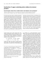
Fig. 8. Model of transcriptional activation for
TCDD ⁄ BNF (upper) and carbaryl (lower) in
YCM-RYA (left) and LMAX-RYA (right). Note
the difference on DNA binding domains and
DNA sequences between both systems as
well as the absence of ARNT in LMAX-RYA.
The proposed differential conformation of
TCDD ⁄ BNF and carbaryl complexes with
the AhR-LBD is also shown.
ARNT
LexA-AhR LBD
ARNT
LexA-AhR LBD
Carbaryl
AhR
FEBS Journal 274 (2007) 3327–3339 ª 2007 The Authors Journal compilation ª 2007 FEBS
3333
Modulation of AhR transactivation by carbaryl
S. Boronat et al.
Plasmid pGADT7-mCBP
Recombinant yeast assay
Plasmid pGADT7-mCBP was kindly provided by H. Jiang
[52]. It contains the N-terminus of mouse CBP (amino acids
1–464) fused at the C-terminus of the GAL4 protein activation domain in the yeast expression vector pGADT7 from
Clontech.
Yeast strains were grown overnight in minimal medium
(6.7 gỈL)1 yeast nitrogen base without amino acids plus
ammonium sulfate; DIFCO, Basel, Switzerland) supplemented with 0.1 gỈL)1 of prototrophic markers as required and
with either glucose or galactose as a carbon source. When
cells were at the appropriate attenuance (0.1–0.2) they were
mixed with carbaryl or with BNF dissolved in dimethyl
sulfoxide. Some 50–100 lL of this mix were added in triplicates in a 96-well siliconized polypropylene microtiter plate
(NUNCTM, Roskilde, Denmark) and further diluted in the
same plate in wells containing cell culture without the
chemical. Cells were incubated for 4 h at 30 °C under mild
shaking. Permeabilization of yeast cells and fluorogenic
quantitation of either lacZ or GUS activity was performed
as described [16]. EC50 values were calculated by fitting the
data to a noncooperative version of the Hill equation using
SPSS for Windows package (version 11.01, SPSS Inc. Chicago, IL, USA), as described in [16]. For general toxicity
testing, the GAL-GUS system strain was grown overnight
in minimal medium supplemented with 0.1 gỈL)1 of prototrophic markers as required and with raffinose as a carbon
source. When cells were at the appropriate attenuance
(0.1–0.2), 2% galactose was added and they were mixed
with carbaryl or BNF and treated as described above.
Plasmid pRB1155
Plasmid pRB1155 is a high copy number yeast reporter
plasmid encompassing lexA-binding sites driving the expression of the lacZ reporter gene [53].
Yeast strains and RYA systems
AhR ⁄ ARNT system (YCM-RYA)
Strain YCM4 was a generous gift from C. A. Miller (Tulane
University, New Orleans, LA, USA) [54]. This strain is a
derivative of W303a (MATa, ade2-1, can1-100, his3-11, 15,
leu2-3, 112, trp1-1, ura3-1), which harbours two foreign genetic elements: one of them is chromosomally integrated and
coexpresses human aryl hydrocarbon receptor and ARNT
genes under the GAL1-10 promoter. The second construct is
the pDRE23-Z reporter, encompassing three XRE5 sequence
and the CYC1-lacZ fusion (more information in the original
paper [54]). To perform the RYA assay, YCM4 and YCM4
derived cells were grown in galactose overnight to express
both AhR and ARNT.
LMAX-system (pLMAX-RYA)
YSB7 (MATa, leu2, his3, met 15, URA3::lexA-GUS) is a
derivative of strain BY4741 (Euroscarf, Frankfurt,
Germany) and contains the 2l plasmid pLMAX and
eight copies of the LexA DNA recognition sequence in
front of the b-glucuronidase (GUS) reporter gene integrated into the genome. Strains YSB37 and YSB39 were
constructed by transforming BY4741 and Y04282
(MATa, leu2, his3, met 15, ura3, ada2::kanMX4),
obtained from Euroscarf, with plasmids pLMAX and
pRB1155. Yeast strains YSB52 and YSB53 were obtained
by transformation of YSB37 and YSB39 with plasmid
pGADT7.
RNA extraction and real time RT-PCR
Total RNA was extracted using the MasterPureTM-Yeast
RNA Purification kit from Epicentre Biotechnologies
(Madison, WI, USA) and used according to the manufacturer’s instructions. cDNAs were prepared with OmniscriptTM Reverse Transcriptase (Qiagen, Valencia, CA,
USA) using oligo-dT primers and 2 lg of total RNA as
template. 1 lL of each cDNA and further 1 : 10 dilutions
were used for real time RT-PCR using SYBRÒGREEN
PCR Master Mix (Applied Biosystems, Warrington, UK)
with 300 nm of each primer in a final volume of 20 lL.
PCR was monitored in an ABIPrismTM 7000 Sequence
Detection System (Applied Biosystems), using the following
primers: 5¢-AGTTTTCCGGCTTCTTGCAA-3¢ (forward)
and 5¢-TTGGACTGGACCCACCTCC-3¢ (reverse), from
Roche (Basel, Switzerland). LexA-mAhR-LBD mRNA
copy numbers were calculated by interpolation in a standard curve using plasmid pLMAX as standard.
GAL-GUS system
A yeast reporter strain was constructed, in which GUS
transcription was controlled by the GAL1-10 promoter [42].
Briefly, yeast strain BY474 was transformed by one-step
double homologous recombination using two overlapping
PCR fragments that allowed both GUS integration at the
GAL1,10 site and nourseothricin selection. Details about
the strategy and the characterization of the strain are provided elsewhere [55].
3334
Vertebrate cell lines culture and enzymatic
measurements
The rainbow trout liver cell line, RTL-W1, was grown as
outlined in the original description of this cell line [56].
Cells were grown in Leibovitz’s L-15 cell culture medium
(Cambrex, North Brunswick, NJ, USA) supplemented with
FEBS Journal 274 (2007) 3327–3339 ª 2007 The Authors Journal compilation ª 2007 FEBS
S. Boronat et al.
5% fetal bovine serum (Cambrex) and penicillin–streptomycin (20 mL)1 to 20 lgỈmL)1, respectively, Cambrex) in
75 cm2 NUNCTM tissue-culture flasks (Nalgene Nunc International, Rochester, NY, USA) at 19 °C. Cells were
detached from confluent flasks using trypsin (Sigma), and
then, seeded in 96 well Falcon plates (Becton Dickinson,
Oxnard, CA, USA) at a density of 20 000 cells in 200 lL
of culture medium per well and allowed to grow to confluency for 1 day. Subsequently, medium was substituted and
new medium with the corresponding concentrations of
BNF (0.2–100 lm) and carbaryl (100 lm) was added. The
maximal concentration of dimethyl sulfoxide in the culture
medium was 0.2%. Control cells received only solvent.
After 48 h of treatment, medium was removed, cells washed
with phosphate buffered saline (pH 7.5) and the plates frozen in liquid nitrogen. They were maintained at )80 °C
until analysis of ethoxyresorufin-O-deethylase activity and
protein following the methodology previously described
[57,58].
The DR-CALUXÒ bioassay performed in this study is
based on the use of a rat hepatoma (H4IIE) cell line stably
transfected with a construct containing the luciferase reporter gene under direct control of DRE (Dioxin Responsive
Element) (BioDetection Systems, Amsterdam, the Netherlands).
Cells were maintained in aMEM (Cambrex) with phenol red and supplemented with 10% fetal bovine serum
(Cambrex), 1% 2 mm l-glutamine (Cambrex) and penicillin–streptomycin (10 mL)1 to 10 lgỈmL)1, respectively,
Cambrex). Cells were grown at 37 °C with 5% CO2 in a
humidified incubator. For the assay, cells grown in
bottles were trypsinized and plated in 96 well plates at a
density of 2.5 · 104 cells per well. After 24 h, the cells
were cotreated with different concentrations of BNF
(0.3–100 lm) and a fixed concentration of carbaryl
(200 lm).
Carbaryl or BNF stock solutions were diluted in culture
medium at a maximal solvent concentration of 0.2%.
Control cells received the maximal dimethyl sulfoxide concentration used in the treated cells. Cells were exposed to
the xenobiotics for 48 h. Subsequently, culture plates were
washed with phosphate buffer and the luminescence emitted by the cells was quantified by means of the SteadyGlo Luciferase assay System from Promega (Madison,
WI, USA) following the manufacturer’s instructions in a
Tecan Genios (Maennedorf, Switzerland) luminescence
detector.
Mathematical modelling
The equations and definitions used in this work are
derived from standard ligand-receptor mathematical models, as previously described [59]. A more detailed description of the models can be found in the Supplementary
material.
Modulation of AhR transactivation by carbaryl
Interaction of a receptor with a single ligand
The simplest model to describe dose ⁄ response curves
assumes an equilibrium between hormone-free and hormone-loaded hormone receptor molecules in solution:
Kd
R ỵ h1 Rh1
where R represents the concentration of hormone-free
receptor molecules, h is the hormone concentration, Rh is
the concentration of the hormone-loaded receptor molecule,
and Kd is the dissociation constant. The model assumes a
single agonist molecule binding to each receptor molecule,
and an hormone concentration much larger than the receptor concentration. The fraction of receptor bound to the
hormone Fr can be described by the Hill equation:
Ur ẳ
ẵRh
1
ẳ
Ro
1 ỵ Kd
ẵh
ẵ1
In which Ro is the total receptor concentration (bound
and free). From Eqn (1), Kd can be calculated as the ligand concentration at which 50% of receptor molecules
are occupied, which in turn coincides with EC50, the hormone concentration at which the physiological effect (i.e.
the reporter activity in our case) reaches 50% of its maximal value at saturating hormone concentration. When
applied to inhibitory effects, such as a decrease on transcription rates upon addition of a compound, the equilibrium constant is usually denominated as Ki and its value
coincides with IC50, the effector concentration at which
the measured physiological activity is reduced to 50%.
Interaction of a receptor with two ligands
Below, we considered three mechanisms of mutually interaction among a pure agonistic ligand (h1, in our case,
BNF) and an inhibitor (h2, in our case carbaryl).
Reversible binding, competitive inhibition
This model proposes an equilibrium between free receptor, R,
and two ligands that bind alternatively to a single site of the
receptor molecule, with dissociation constants Kd1 and Kd2 :
Kd1
Kd2
R ỵ h1 Rh1 ; R þ h2 Rh2
At any given concentrations of h1 and h2, any target gene
would show a fraction of its maximal activation at saturating concentration of h1 A ⁄ Amax that could be expressed as:
A
ẵRh1
ẳ
ẳ
Amax
Ro
1
Kd ẵh
1ỵ
1
Kd1 ỵ Kd 2
2
ẵh1
In this variant of the Hill equation, Amax is independent
from h2, whereas the apparent EC50 for h1 (EC50app) equals
FEBS Journal 274 (2007) 3327–3339 ª 2007 The Authors Journal compilation ª 2007 FEBS
3335
Modulation of AhR transactivation by carbaryl
S. Boronat et al.
to Kd1 only when h2 ¼ 0. Kd2 (identical to IC50) can be calculated by measuring EC50app at different concentrations of
h2 following the equation:
EC50h2 ị
ẵh2
ẳ1ỵ
Kd2
EC50h2 ẳ0ị
Rh2
1
ẳ
K ;
Ro
1 ỵ ẵh2i
R ẳ Ro À Rh2
In this model, the maximal activity at saturating concentrations of h1 depends on the concentration of h2, as follows:
ẵ2
h1 ! 1;
in which EC50h2 ị and EC50h2 ẳ0ị correspond to the EC50 for
h1 in the presence and in the absence of a given concentration of h2.
Noncompetitive, reversible inhibition
This model postulates the binding of h1 and h2 to two independent binding sites in the receptor, and that binding of
h2 allows binding of h1 but precludes transcriptional activation. The model predicts three ligand-receptor complexes:
Amax; h2 ẳ0 Ro
ẵh2
ẳ1ỵ
ẳ
Ki
Amax; h2
R
Ki is therefore equivalent to IC50. This equation is identical
to Eqn (3), and therefore Ki can be calculated as Kd2 in the
previous model.
Acknowledgements
This work has been supported by the Spanish Ministry
for Science and Technology (BIO2005-00840) and
INIA (RTA2006-00022-00-00). The contribution of the
`
Centre de Referencia en Biotecnologia de la Generalitat de Catalunya is also acknowledged.
References
The fraction of the active complex relative to the total
amount of receptor molecules, Ro can be calculated as:
ẵRh1
ẳ
Ro
1
ẵh2
1 ỵ Kd2 ỵ
ẵh
2
Kd1 1ỵKd ị
2
ẵh1
The model predicts that the apparent EC50 for h1 is independent from the concentration of h2, and therefore
identical to the calculated Kd1 in the absence of h2.
However, at saturating concentrations of h1, the maximal
response given by the system, Amax, depends solely on
the concentration of h2 and of Kd2 , following the equation:
Amax;h2 ẳ0
ẵh2
ẳ1ỵ
Kd2
Amax;h2
ẵ3
In which Amax, h2¼0 corresponds to the maximal activation
in the absence of h2 and Kd2 equals to IC50.
Noncompetitive, irreversible inhibition
This model proposes that binding to h2 irreversibly inactivates the receptor, reducing the amount of available receptor molecules for binding to h1. Assuming that
the proportion of receptor molecules becoming inactivated
follows a typical logistic function with an inhibitory constant Ki:
3336
1 Colborn T (1995) Environmental estrogens: health
implications for humans and wildlife. Environ Health
Perspect 103, 135–136.
2 Hahn ME, Karchner SI, Shapiro MA & Perera SA
(1997) Molecular evolution of two vertebrate aryl
hydrocarbon (dioxin) receptors (AHR1 and AHR2) and
the PAS family. Proc Natl Acad Sci USA 94, 13743–
13748.
3 Hahn ME (2002) Aryl hydrocarbon receptors: diversity
and evolution. Chem Biol Interact 141, 131–160.
4 Nebert DW, Puga A & Vasiliou V (1993) Role of the Ah
receptor and the dioxin-inducible toxicity, cancer, and
signal transduction. Ann NY Acad Sci 685, 624–640.
5 Sutter TR, Tang YM, Hayes CL, Wo YY, Jabs EW,
Li X, Yin H, Cody CW & Greenlee WF (1994) Complete cDNA sequence of a human dioxin-inducible
mRNA identifies a new gene subfamily of cytochrome
P450 that maps to chromosome 2. J Biol Chem 269,
13092–13099.
6 Gonzalez FJ & Fernandez-Salguero P (1998) The aryl
hydrocarbon receptor: studies using the AHR-null mice.
Drug Metab Dispos 26, 1194–1198.
7 Shimizu Y, Nakatsuru Y, Ichinose M, Takahashi Y,
Kume H, Mimura J, Fujii-Kuriyama Y & Ishikawa T
(2000) Benzo[a]pyrene carcinogenicity is lost in mice
lacking the aryl hydrocarbon receptor. Proc Natl Acad
Sci USA 97, 779–782.
8 Abbott BD, Perdew GH & Birnbaum LS (1994) Ah
receptor in embryonic mouse palate and expression.
Toxicol Appl Pharmacol 126, 16–25.
9 Poland A & Knutson JC (1982) 2,3,7,8-tetrachlorodibenzo-p-dioxin and related hydrocarbons: examination
FEBS Journal 274 (2007) 3327–3339 ª 2007 The Authors Journal compilation ª 2007 FEBS
S. Boronat et al.
10
11
12
13
14
15
16
17
18
19
20
21
22
of the mechanism of toxicity. Annu Rev Pharmacol
Toxicol 22, 517–554.
Safe S, Wang F, Porter W, Duan R & McDougal A
(1998) Ah receptor agonists as endocrine disruptors:
antiestrogenic activity and mechanisms. Toxicol Lett
102–103, 343–347.
Mandal PK (2005) Dioxin: a review of its environmental effects and its aryl hydrocarbon receptor biology.
J Comp Physiol [B] 175, 221–230.
Colborn T, Dumanoski D & Myers J (1997) Our Stolen
Future, 2nd edn. Plume, New York, NY.
´
Garcia-Reyero N, Grau E, Castillo M, Lopez de Alda
´
MJ, Barcelo D & Pina B (2001) Monitoring of endo˜
crine disruptors in surface waters by the yeast recombinant assay. Environ Toxicol Chem 20, 1152–1158.
Andersen H, Andersson A, Arnold S, Autrup H, Barfoed M, Beresford N, Bjerregaard P, Christiansen L,
Gissel B, Hummel R et al. (1999) Comparison of Shortterm estrogenicity tests for identification of hormonedisrupting chemicals. Environ Health Perspect 107,
89–108.
Bovee T, Heskamp H, Hamers A, Hoogenboom R &
Nielen M (2005) Validation of a rapid yeast estrogen
bioassay, based on the expression of green fluorescent
protein, for the screening of estrogenic activity in calf
urine. Anal Chem Acta 529, 57–64.
´
Noguerol T, Boronat S, Jarque S, Barcelo D & Pina B
˜
(2006) Detection of hormone receptor ligands in yeast
by fluorogenic methods. TALANTA 69, 359–358.
Feldman D & Krishnan A (1995) Estrogens in unexpected places: possible implications for researchers and
consumers. Environ Health Perspect 105, 129–133.
Thomas K, Hurst M, Matthiessen P & Waldock M
(2001) Characterization of estrogenic compounds in
water samples collected from United Kingdom estuaries.
Environ Toxicol Chem 20, 2165–2170.
´
´
Quiros L, Cespedes R, Lacorte S, Viana P, Raldua D,
´
´
Barcelo D & Pina B (2005) Detection and evaluation
˜
of endocrine-disruption activity in water samples from
Portuguese rivers. Environ Toxicol Chem 24, 389–395.
Gaido KW, Leonard LS, Lovell S, Gould JC, Babai
D, Portier CJ & McDonnell DP (1997) Evaluation
of chemicals with endocrine modulating activity in
a yeast-based steroid hormone receptor gene
transcription assay. Toxicol Appl Pharmacol 143,
205–212.
Nishihara T, Nishikawa J, Kanayama T, Dakeyama F,
Saito K, Imagawa M, Takatori S, Kitagawa Y, Hori S
& Utsumi H (2000) Estrogenic activities of 517 chemicals by yeast two-hybrid assay. J Health Sci 46,
282–298.
Garcia-Reyero N, Requena V, Petrovic M, Fischer B,
´
Hansen P, Barcelo D & Pina B (2004) Estrogenic poten˜
tial of halogenated derivatives of nonylphenol ethoxylates
and carboxylates. Environ Toxicol Chem 23, 705–711.
Modulation of AhR transactivation by carbaryl
23 Arulmozhiraja S, Shiraishi F, Okumura T, Iida M,
Takigami H, Edmonds J & Morita M (2005) Structural
requirements for the interaction of 91 hydroxylated
polychlorinated biphenyls with estrogen and thyroid
hormone receptors. Toxicol Sci 84, 49–62.
´
24 Noguerol T, Boronat S, Casado M, Raldua D, Barcelo
´
D & Pina B (2006) Evaluation of vertebrate receptor
˜
interaction with persisten pollutants and antifouling pesticides by recombinant yeast assays. Anal Bioanal Chem
285, 1012–1019.
25 Swanson HI (2002) DNA binding and protein interactions of the AHR ⁄ ARNT heterodimer that facilitate
gene activation. Chem Biol Interact 141, 63–76.
26 Whitelaw ML, Gustafsson JA & Poellinger L (1994)
Identification of transactivation and repression functions
of the dioxin receptor and its basic helix-loop-helix ⁄ PAS
partner factor Arnt: inducible versus constitutive modes
of regulation. Mol Cell Biol 14, 8343–8355.
27 Carver LA, Jackiw V & Bradfield CA (1994) The
90-kDa heat shock protein is essential for Ah receptor
signaling in a yeast expression system. J Biol Chem 269,
30109–30112.
28 Backlund M & Ingelman-Sundberg M (2004) Different
structural requirements of the ligand binding domain of
the aryl hydrocarbon receptor for high- and low-affinity
ligand binding and receptor activation. Mol Pharmacol
65, 416–425.
29 Hankinson O (2005) Role of coactivators in transcriptional activation by the aryl hydrocarbon receptor. Arch
Biochem Biophys 433, 379–386.
30 Kobayashi A, Numayama-Tsuruta K, Sogawa K &
Fujii-Kuriyama Y (1997) CBP ⁄ p300 functions as a
possible transcriptional coactivator of Ah receptor
nuclear translocator (Arnt). J Biochem (Tokyo) 122,
703–710.
31 Yao G, Craven M, Drinkwater N & Bradfield CA
(2004) Interaction networks in yeast define and enumerate the signaling steps of the vertebrate aryl hydrocarbon receptor. PLoS Biol 2, E65.
32 Berger SL, Pina B, Silverman N, Marcus GA, Agapite
˜
J, Regier JL, Triezenberg SJ & Guarente L (1992) Genetic isolation of ADA2: a potential transcriptional adaptor required for function of certain acidic activation
domains. Cell 70, 251–265.
33 Sterner DE & Berger SL (2000) Acetylation of histones
and transcription-related factors. Microbiol Mol Biol
Rev 64, 435–459.
34 vom Baur E, Harbers M, Um SJ, Benecke A, Chambon
P & Losson R (1998) The yeast Ada complex mediates
the ligand-dependent activation function AF-2 of retinoid X and estrogen receptors. Genes Dev 12, 1278–
1289.
35 Wallberg AE, Neely KE, Gustafsson JA, Workman JL,
Wright AP & Grant PA (1999) Histone acetyltransferase
complexes can mediate transcriptional activation by the
FEBS Journal 274 (2007) 3327–3339 ª 2007 The Authors Journal compilation ª 2007 FEBS
3337
Modulation of AhR transactivation by carbaryl
36
37
38
39
40
41
42
43
44
45
S. Boronat et al.
major glucocorticoid receptor activation domain. Mol
Cell Biol 19, 5952–5959.
Anafi M, Yang YF, Barlev NA, Govindan MV, Berger
SL, Butt TR & Walfish PG (2000) GCN5 and ADA
adaptor proteins regulate triiodothyronine ⁄ GRIP1 and
SRC-1 coactivator-dependent gene activation by the
human thyroid hormone receptor. Mol Endocrinol 14,
718–732.
Ledirac N, Delescluse C, de Sousa G, Pralavorio M,
Lesca P, Amichot M, Berge JB & Rahmani R (1997)
Carbaryl induces CYP1A1 gene expression in HepG2
and HaCaT cells but is not a ligand of the human
hepatic Ah receptor. Toxicol Appl Pharmacol 144,
177–182.
Delescluse C, Ledirac N, Li R, Piechocki MP,
Hines RN, Gidrol X & Rahmani R (2001) Induction of
cytochrome P450 1A1 gene expression, oxidative stress,
and genotoxicity by carbaryl and thiabendazole in transfected human HepG2 and lymphoblastoid cells. Biochem
Pharmacol 61, 399–407.
Arukwe A (2002) Complementary DNA cloning,
sequence analysis and differential organ expression of
beta-naphthoflavone-inducible cytochrome P4501A in
Atlantic salmon (Salmo salar). Comp Biochem Physiol C
Toxicol Pharmacol 133, 613–624.
Waller CL & McKinney JD (1995) Three-dimensional
quantitative structure-activity relationships of dioxins
and dioxin-like compounds: model validation and Ah
receptor characterization. Chem Res Toxicol 8, 847–858.
´
Casado S, Alonso M, Herradon B, Tarazona J & Navas
J (2006) Activation of the aryl hydrocarbon receptor by
carbaryl: computacional evidence of the ability of carbaryl to assume a planar coformation. Env Toxicol Chem
25, 3141–3147.
Johnston M & Carlson M (1992) Regulation of carbon
and phosphate utilization. In The Molecular and Cellular Biology of the Yeast Saccharomyces. Gene Expression. (Jones EW, Pringle JR & Broach JR, eds), pp.
193–282. Cold Spring Harbor Laboratory Press, Cold
Spring Harbor, NY.
Pina B, Berger S, Marcus GA, Silverman N, Agapite J
˜
& Guarente L (1993) ADA3: a gene, identified by resistance to GAL4-VP16, with properties similar to and different from those of ADA2. Mol Cell Biol 13, 5981–
5989.
Sandoz C, Lesca P, Carpy A, Laguionie M & Narbonne
J (1998) Effects of carbaryl and naphthalene on rat hepatic CYP1A1 ⁄ 2: Potential binding to Ah receptor and
4S benzo(a)pyrene-binding protein. Int J Mol Medical 2,
615–623.
Denison MS, Phelan D, Winter GM & Ziccardi MH
(1998) Carbaryl, a carbamate insecticide, is a ligand for
the hepatic Ah (dioxin) receptor. Toxicol Appl Pharmacol 152, 406–414.
3338
46 Sandoz C, Lesca P, Narbonne JF & Carpy A (2000)
Molecular characteristics of carbaryl, a CYP1A1 gene
inducer. Arch Biochem Biophys 373, 275–280.
47 Denison MS & Nagy SR (2003) Activation of the aryl
hydrocarbon receptor by exogenous and endogenous
chemicals. Annu Rev Pharmacol Toxicol 43, 309–334.
48 Quadri SA, Qadri AN, Hahn ME, Mann KK & Sherr
DH (2000) The bioflavonoid galangin blocks aryl
hydrocarbon receptor activation and polycyclic aromatic
hydrocarbon-induced pre-B cell apoptosis. Mol Pharmacol 58, 515–525.
49 Zhou JG, Henry EC, Palermo CM, Dertinger SD &
Gasiewicz TA (2003) Species-specific transcriptional
activity of synthetic flavonoids in guinea pig and mouse
cells as a result of differential activation of the aryl
hydrocarbon receptor to interact with dioxin-responsive
elements. Mol Pharmacol 63, 915–924.
50 Casper RF, Quesne M, Rogers IM, Shirota T, Jolivet
A, Milgrom E & Savouret JF (1999) Resveratrol has
antagonist activity on the aryl hydrocarbon receptor:
implications for prevention of dioxin toxicity. Mol Pharmacol 56, 784–790.
51 Edwards DP (2000) The role of coactivators and corepressors in the biology and mechanism of action of
steroid hormone receptors. J Mammary Gland Biol
Neoplasia 5, 307–324.
52 Chen Q, Chen J, Sun T, Shen J, Shen X & Jiang H
(2004) A yeast two-hybrid technology-based system for
the discovery of PPARg agonist and antagonist. Anal
Biochem 335, 253–259.
53 Brent R & Ptashne M (1985) A eukaryotic transcriptional activator bearing the DNA specificity of a prokaryotic repressor. Cell 43, 729–736.
54 Miller C (1999) A human aryl hydrocarbon receptor
signaling pathway constructed in yeast displays additive
responses to ligand mixtures. Toxicol Appl Pharmacol
160, 297–303.
55 Boronat S & Pina B (2006) Development of RNR3- and
˜
RAD54-GUS reporters for testing genotoxicity in
Saccharomyces cerevisiae. Anal Bioanal Chem 386,
1625–1632.
56 Lee LE, Clemons JH, Bechtel DG, Caldwell SJ, Han
KB, Pasitschniak-Arts M, Mosser DD & Bols NC
(1993) Development and characterization of a rainbow
trout liver cell line expressing cytochrome P450-dependent monooxygenase activity. Cell Biol Toxicol 9,
279–294.
57 Bols NC, Schirmer K, Joyce EM, Dixon DG, Greenberg
BM & Whyte JJ (1999) Ability of polycyclic aromatic
hydrocarbons to induce 7-ethoxyresorufino-deethylase
activity in a trout liver cell line. Ecotoxicol Environ Saf
44, 118–128.
58 Behrens A, Schirmer K, Bols N & Segner H (1998)
Microassay for rapid measurement of 7-ethoxyresorufin-
FEBS Journal 274 (2007) 3327–3339 ª 2007 The Authors Journal compilation ª 2007 FEBS
S. Boronat et al.
O-deethylase activity in intact fish hepatocytes. Mar
Environ Res 46, 369–373.
59 Neubig RR, Spedding M, Kenakin T & Christopoulos
A (2003). International Union of Pharmacology Comittee
on Receptor Nomenclature and Drug Classification.
XXXVIII. Update on Terms and Symbols in Quantitative
Pharmacology. IUPAHR, Kansas City, KS.
Supplementary material
Modulation of AhR transactivation by carbaryl
mathematical models described in [59] and references
therein.
This material is available as part of the online article
from
Please note: Blackwell Publishing is not responsible
for the content or functionality of any supplementary
materials supplied by the authors. Any queries (other
than missing material) should be directed to the corresponding author for the article.
The following supplementary material is available online:
Doc S1. The equations and definitions used in this
work are derived from standard ligand-receptor
FEBS Journal 274 (2007) 3327–3339 ª 2007 The Authors Journal compilation ª 2007 FEBS
3339