Báo cáo khoa học: Transcriptional regulation of the desferrioxamine gene cluster of Streptomyces coelicolor is mediated by binding of DmdR1 to an iron box in the promoter of the desA gene doc
Bạn đang xem bản rút gọn của tài liệu. Xem và tải ngay bản đầy đủ của tài liệu tại đây (427.01 KB, 13 trang )
Transcriptional regulation of the desferrioxamine gene
cluster of Streptomyces coelicolor is mediated by binding
of DmdR1 to an iron box in the promoter of the desA gene
Sedef Tunca
1
, Carlos Barreiro
1
, Alberto Sola-Landa
1
, Juan Jose
´
R. Coque
1,2
and Juan F. Martı
´
n
1,2
1 Instituto de Biotecnologı
´
a, INBIOTEC, Leo
´
n, Spain
2A
´
rea de Microbiologı
´
a, Facultad de CC Biolo
´
gicas y Ambientales, Universidad de Leo
´
n, Spain
Iron is an essential element required for many key
metabolic processes (including cytochrome and Fe-S
electron transporters) in almost all micro-organisms.
The bioavailability of iron is very low because salts
of the oxidized ferric ion formed under normal oxic
conditions are largely insoluble [1]. To solve the
problem of iron availability, many micro-organisms
synthesize different high-affinity iron chelato rs (sidero-
phores), forming very stable complexes with ferric iron
[2]. Streptomyces species are soil-dwelling Gram-
positive saprophytic bacteria that produce different
types of siderophores [3,4]. Desferrioxamines are
nonpeptide hydroxamate siderophores composed of
alternating dicarboxylic acid and diamine units
Keywords
desferrioxamine biosynthesis; gene
expression; iron regulation; lysine
decarboxylase gene; siderophores
Correspondence
J. F. Martı
´
n, Instituto de Biotecnologı
´
a,
INBIOTEC, Parque Cientı
´
fico de Leo
´
n,
Avenue del Real no. 1, 24006 Leon, Spain
Fax: + 34 987 210 388
Tel: + 34 987 210 308
E-mail:
(Received 11 October 2006, revised 19
December 2006, accepted 21 December
2006)
doi:10.1111/j.1742-4658.2007.05662.x
Streptomyces coelicolor and Streptomyces pilosus produce desferrioxamine
siderophores which are encoded by the desABCD gene cluster. S. pilosus is
used for the production of desferrioxamine B which is utilized in human
medicine. We report the deletion of the desA gene encoding a lysine
decarboxylase in Streptomyces coelicolor A3(2). The DdesA mutant was
able to grow on lysine as the only carbon and nitrogen source but its des-
ferrioxamine production was blocked, confirming that the l-lysine decarb-
oxylase encoded by desA is a dedicated enzyme committing l-lysine to
desferrioxamine biosynthesis. Production of desferrioxamine was restored
by complementation with the whole wild-type desABCD cluster, but not by
desA alone, because of a polar effect of the desA gene replacement on
expression of the downstream des genes. The transcription pattern of the
desABCD cluster in S. coelicolor showed that all four genes were coordi-
nately induced under conditions of iron deprivation. The transcription start
point of the desA gene was identified by primer extension analysis at a thy-
mine located 62 nucleotides upstream of the translation start codon. The
)10 region of the desA promoter overlaps the 19-nucleotide palindromic
iron box sequence known to be involved in iron regulation in Streptomyces.
Binding of DmdR1 divalent metal-dependent regulatory protein to the
desA promoter region of both S. coelicolor and S. pilosus was shown using
electrophoretic mobility-shift assays, validating the conclusion that iron
regulation of the desABCD cluster is mediated by the regulatory protein
DmdR1. We conclude that the genes involved in desferrioxamine produc-
tion are under transcriptional control exerted by the DmdR1 regulator in
the presence of iron and are expressed under conditions of iron limitation.
Abbreviations
DmdR, divalent metal-dependent regulatory protein; ILMM, iron-limited minimal medium; YEME, yeast extract and malt extract culture
medium.
1110 FEBS Journal 274 (2007) 1110–1122 ª 2007 The Authors Journal compilation ª 2007 FEBS
linked by amide bonds. They are produced by
many Streptomyces species, including Streptomyces
coelicolor [5], Streptomyces griseus [6] and
Streptomyces pilosus; the latter is used for industrial
production of desferrioxamine B for medical uses
[7,8].
In Streptomyces species, as in other Gram-positive
bacteria, the expression of genes involved in iron meta-
bolism is under the control of a divalent metal-depend-
ent regulatory protein (DmdR) analogous to the
diphtheria toxin repressor of Corynebacterium diphthe-
riae [9,10]. S. coelicolor has two similar genes, dmdR1
and dmdR2, encoding regulatory proteins of this family
[11]. In a previous study, Flores et al. [12] reported
that S. coelicolor DmdR1 binds specific sequences
(iron boxes) in the upstream region of the diphtheria
toxin (tox) gene of C. diphtheriae and the desA gene of
S. pilosus.
Several putative iron boxes were found by bioinfor-
matics analysis upstream of 10 different ORFs in the
genome of S. coelicolor [12]. One of the putative iron
boxes is located in the promoter of the desABCD gene
cluster, which was assumed to be responsible for des-
ferrioxamine biosynthesis [13]. Barona-Go
´
mez et al.
[14] proposed a possible pathway for desferrioxamine
biosynthesis from l-lysine and reported that desD is
essential for desferrioxamine formation (Fig. 1) [15].
The first step in the desferrioxamine pathway is the
conversion of l-lysine into cadaverine catalyzed by the
enzyme lysine decarboxylase [7,8] which, in S. coeli-
color, appears to be encoded by desA, although no
conclusive genetic evidence was available until now, as
other putative lysine decarboxylase-encoding genes
occur in the S. coelicolor genome (e.g. SCO2017).
As one of the iron boxes was located in the
upstream region of the desABCD cluster, it was of
interest to perform a transcriptional analysis of this
cluster and also to characterize the promoter region
(transcription start point and regulatory sequences) in
order to analyze the role of iron and the DmdR1 regu-
lator in the transcriptional control of the desferrioxam-
ine cluster.
In this study, we report the deletion of the first gene
of the desABCD gene cluster (desA)inS. coelicolor
A3(2), which caused cessation of desferrioxamine E
and B biosynthesis. Transcriptional analysis of this
region showed that the genes involved in desferrioxam-
ine production are under iron control. The transcrip-
tion start point of the desA gene was shown to overlap
with the palindromic iron box. Binding of purified
DmdR1 protein to the desA promoter region of both
S. coelicolor and S. pilosus, as shown by electrophoret-
ic mobility-shift assay, proved that iron control of the
expression of the des cluster is mediated by the
DmdR1 regulator.
Results
Deletion of the desA gene of S. coelicolor blocks
desferrioxamine biosynthesis
The organization of the putative desferrioxamine gene
cluster, as deduced from the S. coelicolor genome, is
shown in Fig. 1B. To clarify the role of the l-lysine
decarboxylase encoded by desA and its possible
involvement in desferrioxamine biosynthesis, two apra-
mycin-resistant and kanamycin-sensitive transformants
were isolated among S. coelicolor transformants with
the desA gene replacement construction (see Experi-
mental procedures), and the DdesA mutation was veri-
fied in one of the mutants by PCR and Southern blot
analysis. A 1462-bp PCR band corresponding to the
extended resistance cassette was found only in the
mutant strain, and a 1372-bp PCR band corresponding
to the desA gene was present only in the wild-type
strain but not in the DdesA mutant. These results were
confirmed by Southern blot hybridization of ScaI-
digested DNA. A hybridization band of 4200 bp
was obtained for the wild-type with a desA fragment
(1372 bp) as probe, and a band of about 4220 bp was
found for the mutant with aac(3)IV fragment (935 bp)
as probe, as expected. Hybridization and PCR analysis
results indicate that the desA gene has been deleted
and replaced by the apramycin resistance gene.
HPLC analysis showed that no desferrioxamines
could be detected in the culture supernatants of the
DdesA mutant, whereas desferrioxamines B and E were
produced in the parental strain (Fig. 2).
The DdesA mutant was able to grow on Streptomy-
ces minimal medium containing l-lysine as the only
carbon and nitrogen source, indicating that the desA
gene is not involved in the catabolism of lysine.
Complementation of the S. coelicolor desA
mutation with cosmid Stc105 restored
desferrioxamine biosynthesis
Complementation of the DdesA mutant was tested by
conjugation with Escherichia coli containing either (a) a
plasmid construct pRAdesAKn (see Experimental pro-
cedures) carrying a 4204-bp fragment containing the
desA coding region or (b) cosmid Stc105. In the plas-
mid-mediated complementation, one of the Kn
R
conju-
gants was selected for further analysis. A 1372-bp PCR
fragment corresponding to the desA gene was present
in the complemented and in the wild-type strains but
S. Tunca et al. Regulation of the desferrioxamine gene cluster
FEBS Journal 274 (2007) 1110–1122 ª 2007 The Authors Journal compilation ª 2007 FEBS 1111
A
B
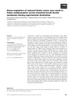
Fig. 1. Proposed pathway for desferrioxamine biosynthesis indicating the conversions catalyzed by the enzymes encoded by desA, desB,
desC and desD (A) and organization of the S. coelicolor des cluster and the upstream SCO2780 and SCO2781 genes (B). The iron box
located upstream of desA is indicated by an open box. RBS, Ribosome-binding site. The hairpin structure corresponds to a stem and loop
structure (putative transcriptional terminator) found downstream of desD. Solid bars indicate the DNA fragments amplified by RT-PCR in the
gene expression studies (see Fig. 4).
Regulation of the desferrioxamine gene cluster S. Tunca et al.
1112 FEBS Journal 274 (2007) 1110–1122 ª 2007 The Authors Journal compilation ª 2007 FEBS
not in the DdesA mutant, as expected (Fig. 3A). In this
conjugant, the Southern blot hybridization pattern
agreed with the integration of the intact wild-type desA
gene (Fig. 3B,C). When the desA fragment (1372 bp)
was used as probe, a band of 3700 bp was found only
in the wild-type and complemented mutant (Fig. 3B).
A 1520-bp positive band was obtained only in the com-
plemented strain when a kan (kanamycin resistance)
fragment (1519 bp) was used as probe, as expected
(Fig. 3C). Complementation of the desA deletion in the
mutant strain with the wild-type gene failed to restore
desferrioxamine production under iron-deficient condi-
tions (data not shown). However, functional comple-
mentation of the DdesA mutant was achieved with
cosmid Stc105, which includes the entire siderophore
biosynthetic gene cluster (Fig. 2C).
The failure of the 4204-bp fragment containing a
wild-type copy of the desA gene to complement the
mutation in trans suggests that the DdesA mutation
affects expression of the downstream genes desBCD in
the des cluster and that the presence of wild-type desA
gene product was not sufficient to restore the ability to
produce these siderophores. The complementation with
cosmid Stc105 indicates that the four genes in the des
cluster are probably transcribed as one polycistronic
mRNA (see below), allowing complementation of the
desA mutant, even if expression of the endogenous des-
BCD genes is disturbed in the DdesA mutant.
Fig. 2. Lack of desferrioxamine production in the S. coelicolor
DdesA mutant and restoration by complementation with the des
cluster. HPLC analysis of siderophore production in S. coelicolor
A3(2) parental and the DdesA mutant strain before and after com-
plementation with the Stc105 cosmid. Desferrioxamine E (retention
time 15.3 min) is the major desferrioxamine produced by S. coeli-
color. Desferrioxamine B showed a retention time of 13.6 min.
AB C
Fig. 3. Verification of the complementation of desA deletion by PCR using primers for the desA gene (A) and by Southern blot hybridization
(B, C). (A) 1-kb Plus DNA ladder (Invitrogen) (lane 1); S. coelicolor A3(2) wild-type strain (lane 2); DdesA mutant (lane 3); complemented strain
(lane 4). (B, C) Southern blot analysis of PvuII-digested genomic DNA probed with a 1372-bp desA fragment (B) and of BamHI + SacI-digested
genomic DNA probed with a kan (kanamycin resistance) fragment (1519 bp) (C). Size markers (kDNA-Hin dIII-digested) (lane 1); S. coelicolor
A3(2) wild-type strain (lane 2); desA-deleted strain (lane 3); complemented strain (lane 4).
S. Tunca et al. Regulation of the desferrioxamine gene cluster
FEBS Journal 274 (2007) 1110–1122 ª 2007 The Authors Journal compilation ª 2007 FEBS 1113
Production of desferrioxamines B and E
is regulated by iron
The desferrioxamines Tris–hydroxamate–Fe
3+
com-
plexes were determined in the supernatants of cultures
grown in (a) iron-limited minimal medium (ILMM),
(b) ILMM with 2,2¢-dipyridyl, and (c) ILMM supple-
mented with 35 lm iron. S. coelicolor A3(2) grown in
iron-deficient medium produces desferrioxamine B and
E. Addition of iron to the culture medium completely
suppressed desferrioxamine production, indicating that
the biosynthesis of these siderophores is strictly regula-
ted by iron (not shown).
Expression of the desABCD cluster is
coordinately derepressed after iron deprivation
Four different genes (desA to desD) have been reported
to be involved in the biosynthesis of desferrioxamine
[14]. Upstream of the desA gene, two other genes enco-
ding siderophore-related proteins are located (Fig. 1B).
The first is a siderophore-interacting protein (viuB
gene), whereas the second encodes a putative secreted
protein (SCO2780) annotated as a hypothetical sidero-
phore-binding lipoprotein [13]. To elucidate if these
two genes are expressed and to study their possible
involvement in desferrioxamine biosynthesis, the tran-
scriptional pattern of the entire region was analyzed
by RT-PCR under iron-deprivation conditions.
Because of the lack of growth after iron deprivation,
the cultures were initially grown in complex medium
[yeast extract and malt extract culture medium
(YEME)] for 36 h and then starved of iron (see
Experimental procedures). After iron deprivation, five
samples (taken at 2, 6, 8, 24 and 48 h) were analyzed,
and the RNA from one nonstarved culture was used
as control. A small increase in dry weight until 6 h
was observed, but no further growth occurred there-
after.
The RT-PCR analysis revealed induction of the tes-
ted desA and desD genes (located at the beginning and
end of the cluster) under iron-limiting conditions, indi-
cating a coordinated transcription (Fig. 4). This result
supports the existence of the desABCD operon sugges-
ted by Barona-Gomez and coworkers [14] that is tran-
scribed as a polycistronic mRNA and confirms the
need of the entire des cluster (as in cosmid Stc105) to
complement the DdesA mutant described above.
Maximum induction of desA and desD was found
6–8 h after iron limitation, and a significant decrease
in expression was observed after 48 h of iron depriva-
tion, indicating that the culture was unable to main-
tain expression of the cluster for prolonged periods,
probably because of the lack of iron-dependent respir-
atory metabolism after extended iron deprivation.
An upstream gene encoding a putative
siderophore-binding protein is also derepressed
after iron deprivation
The transcription pattern of the genes located
upstream of the desA gene was also analyzed. The viuB
gene did not show RT-PCR amplification, suggesting
that it is not expressed, or very poorly so, under the
experimental conditions used. On the other hand, a
transcription pattern similar to that of the desABCD
operon was found for the SCO2780 gene located
upstream of viuB (Fig. 4) encoding a putative sidero-
phore-binding lipoprotein (see Discussion). Our results
confirm the regulation by iron of the expression of this
gene. In contrast with the desABCD operon, the gene
encoding this putative siderophore-binding lipoprotein
(SCO2780) does not show an obvious consensus iron
box in its promoter region, suggesting that SCO2780 is
controlled by indirect iron regulation, probably medi-
ated by a cascade mechanism.
The desA promoter of S. pilosus showed higher
expression ability than the same promoter from
S. coelicolor
Streptomyces pilosus is used industrially for desferriox-
amine production and it produces higher levels of
those siderophores than S. coelicolor [16,17]. To com-
pare the efficiency of expression of the des cluster from
Fig. 4. Expression of the desA–D genes and the upstream genes
at 2, 6, 8, 24 and 48 h after iron starvation (t ¼ 0). Controls without
RNA (lane –) and with DNA instead of RNA (lane +) were per-
formed simultaneously. The hrdB gene was used as control of RNA
amounts.
Regulation of the desferrioxamine gene cluster S. Tunca et al.
1114 FEBS Journal 274 (2007) 1110–1122 ª 2007 The Authors Journal compilation ª 2007 FEBS
these two Streptomyces species, the desA promoter
region (511-bp PCR product) of both S. coelicolor and
S. pilosus were cloned in BamHI–EcoRI-digested
pIJ4083 (7.6 kb) carrying the promoterless xylE repor-
ter gene encoding catechol dioxygenase (constructions
named pCoedesAP and pPildesAP, respectively). The
511-bp fragment of either S. coelicolor or S. pilosus
showed iron-regulated promoter activity when intro-
duced in both S. coelicolor and Streptomyces lividans
(Fig. 5). Catechol oxygenase activity was observed
only under iron-limited conditions in both strains. The
S. pilosus desA promoter clearly showed higher expres-
sion ability than the equivalent S. coelicolor promoter
region when introduced in either S. lividans or S. coeli-
color, suggesting that the S. pilosus promoter is recog-
nized more efficiently by the transcribing RNA
polymerase complex.
Transcription start point: the )10 region overlaps
the iron box
Primer extension experiments with increasing S. coeli-
color RNA concentrations (50–150 lg RNA) using a
fluorescein-labelled 17-bp oligonucleotide [18] as
primer (O6, Table 2) allowed clear identification of the
desA transcription start point at a thymine located 62
nucleotides upstream of the ATG translation initiation
codon of desA. This transcription start point is located
immediately downstream of the iron box (boxed in
Fig. 6) and allowed us to identify the )10 Pribnow
box as TAGGCT in agreement with the proposed con-
sensus sequence for Streptomyces promoters TAgPu-
PuT [19]. It is interesting that the )10 sequence is
located inside the iron box of desA (nucleotides 7–12
of the iron box), explaining the regulation of desA
expression by binding of DmdR1 to the iron box. The
same overlapping was found in S. pilosus.
DmdR1 binds to the promoter region of desABCD
in both S. coelicolor and S. pilosus
Binding of purified DmdR1 to the desA promoter
region of S. coelicolor was studied using a 511-bp PCR
fragment of this region in the DNA-protein binding
reaction. DmdR1 showed a high affinity for the desA
promoter region of both S. coelicolor and S. pilosus,
resulting in retardation of the digoxigenin-labelled
fragment which was prevented by competition with
excess unlabelled probe (Fig. 7). The mobility shift was
clearly higher at increasing protein concentrations,
giving two DNA–protein complexes of different size.
This is in agreement with our previous finding on the
binding of two (or four) DmdR1 molecules to the
iron boxes of either Corynebacterium glutamicum
or
S. pilosus [12].
Discussion
Several desferrioxamines are produced by different
Streptomyces species [5,6]. Desferrioxamine B is used
clinically for the treatment of iron overload during
metabolic alterations in humans. Initial work on the
biosynthesis of desferrioxamine B in S. pilosus indica-
ted that the first step in the desferrioxamine biosynthe-
sis is the decarboxylation of lysine by a lysine
decarboxylase encoded by the desA gene [8,9]. Lysine
decarboxylases occur in different Streptomyces species
and are involved in the utilization of l-lysine as a
nitrogen source [20], but the DesA decarboxylase
might be specific for desferrioxamine biosynthesis.
Unfortunately, the complete desferrioxamine gene clus-
ter in S. pilosus is not known. On the basis of the
sequence of the S. coelicolor genome, Barona-Go
´
mez
et al. [14] proposed a biosynthetic pathway in which
cadaverine formed by lysine decarboxylation is
subsequently hydroxylated to N-hydroxycadaverine by
the protein encoded by desB (Fig. 1) which is later
Fig. 5. S. coelicolor A3(2) and S. pilosus desA promoter activity in
S. lividans and in S. coelicolor A3(2) in ILMM cultures as measured
by determining the catechol oxygenase of the coupled reporter
gene.
S. Tunca et al. Regulation of the desferrioxamine gene cluster
FEBS Journal 274 (2007) 1110–1122 ª 2007 The Authors Journal compilation ª 2007 FEBS 1115
acylated with succinyl-CoA (or alternative acyl-CoA
esters to form succinyl-N-hydroxycadaverine), which is
finally oligomerized by the action of DesD [15].
A separate putative lysine decarboxylase (SCO2017)
showing 38% end to end identity (53% functionally
conserved residues) with DesA occurs in the S. coeli-
color genome. We have shown in this study that the
desA gene is essential for desferrioxamine biosynthesis
but not for growth on lysine as the only carbon and
nitrogen source, indicating that the encoded lysine
decarboxylase is a dedicated enzyme committing
l-lysine to the desferrioxamine pathway, as occurs with
p-aminobenzoic acid synthase in the biosynthesis of
candicidin [21,22] and a few other examples of ‘com-
mitting’ enzymes for secondary metabolites that have
evolved as variants of enzymes involved in primary
metabolism [23].
All the evidence from this work indicates that the
desABCD cluster is expressed as a polycistronic
transcript. Expression of the four genes is coordinately
regulated by iron limitation, as shown by the RT-PCR
analysis, and there is overlapping of the desB transla-
tion termination triplet with the ATG of desC and also
of desC and desD (so-called translational coupling);
moreover, there are no intergenic regions between any
of the four genes. Downstream of desD, we have
located a putative transcriptional terminator [calcula-
ted DG )30.7 kcalÆmol
)1
(128.5 kJÆmol
)1
)] (Fig. 1B). In
the four genes, there is strong overexpression, which is
maximal 8 h after iron deprivation and decreases at
24 h. The coordinated regulation by iron of the expres-
sion of the entire cluster ensures simple and efficient
up-regulation of desferrioxamine biosynthesis after
iron limitation.
The two upstream genes (ORFs SCO2780 and
SCO2781) have been annotated to encode proteins
related to siderophore uptake and metabolism [13]
(SCO database, ), but there
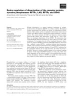
Fig. 6. Primer extension analysis of the transcription start (TS) point of the S. coelicolor desA promoter. Comparison of the reaction
sequences of the promoter region (T, G, C, A) with that of the primer extension reaction product (inclined arrow) using 50, 100 and 150 lg
RNA. The )10 region is shown, and the transcriptional start point is indicated as +1 (bent arrow). The19-bp palindromic region, which con-
tains the repressor-binding site, is boxed, and the ribosome-binding site is underlined. The ATG is shown in bold letters. The reported tran-
scription start site of the S. pilosus desA promoter is indicated by three asterisks. Note the strict conservation of the 19-nucleotide iron box
and the )10 sequence in both Streptomyces species.
Regulation of the desferrioxamine gene cluster S. Tunca et al.
1116 FEBS Journal 274 (2007) 1110–1122 ª 2007 The Authors Journal compilation ª 2007 FEBS
is very little evidence for or against this claim. SCO2780
encoding a putative secreted siderophore-binding
lipoprotein with a conserved Fhu (Fe
2+
siderophore
binding) domain showed an iron-limitation response
similar to that of the desABCD cluster; it was clearly
induced at 2 h and reached maximal expression at 6–8 h
after iron deprivation. There is no consensus iron box
in the upstream region of SCO2780, and its regulation
is probably mediated by a cascade mechanism, rather
than by direct interaction of DmdR1.
On the other hand, the viuB gene (SCO2781) was
not transcribed under the conditions tested (Fig. 4),
and its role in iron metabolism remains obscure. This
putative siderophore-interacting protein is similar to
the Vibrio fischeri ViuB vibriobactin utilization protein
[24].
Primer extension analysis of the promoter region of
the desABCD cluster identified the transcription start
point, which allowed us to deduce the )10 Pribnow
box as TAGGCT, in good agreement with the consen-
sus (TAgPuPuT) )10 sequence of Streptomyces species
[19]. It is very interesting that this )10 region is
located inside the 19-nucleotide iron box identified pre-
viously [12]. Therefore, binding of the iron regulator
DmdR1 will interfere with RNA polymerase interac-
tion and expression of the desferrioxamine cluster.
Indeed, binding of the pure DmdR1 protein to the
S. coelicolor desA promoter region was shown for the
first time in this work. As described previously, bind-
ing of DmdR1 to the iron box requires a bivalent
metal (Fe
2+
,Mn
2+
, or other bivalent metals) [12], and
therefore when iron is depleted, DmdR1 is unable to
bind to the cognate iron box, and transcription is
enhanced leading to siderophore biosynthesis.
It is interesting that the promoter of S. pilosus desA
showed higher transcription ability than the S. coelicol-
or homologous promoter (both of 511 nucleotides,
amplified with the same primers) when coupled to the
reporter xylE gene in either S. coelicolor or S. lividans.
Although the )10 region of desA in both S. coelicolor
and S. pilosus [16,17] was almost identical and in both
species it is located within the iron box palindrome
(Fig. 6), the )35 and upstream regions are different.
These regions were found to be relevant for optimal
expression from the desA promoter, as short promoter
regions gave very poor expression of the reporter xylE
gene.
In summary, we provide evidence that the desA gene
encoding a l-lysine decarboxylase is essential for des-
ferrioxamine biosynthesis in S. coelicolor and appears
to be a desferrioxamine-dedicated enzyme, in contrast
with another putative lysine decarboxylase (SCO2017)
that might be involved in lysine utilization [20].
Expression of the desABCD cluster is coordinately
regulated by iron concentrations in the culture med-
ium, and this regulation is mediated by binding of the
regulatory protein DmdR1 to the iron box located in
the promoter region of the desABCD cluster. Elec-
trophoretic mobility-shift assays of the desA promoters
of both S. coelicolor and S. pilosus revealed that two
different complexes of different size are formed in each
case, supporting earlier suggestions that binding takes
place in the form of dimers or tetramers [12].
Experimental procedures
Bacterial strains, plasmids and culture conditions
Bacterial strains and plasmids used in this work are listed
in Table 1. Streptomyces species were routinely grown in
YEME medium [25,26] at 30 °C. For siderophore produc-
tion and promoter activity experiments Minimal Medium
[3] was used. Escherichia coli strains were grown in Luria–
Bertani broth or Luria–Bertani broth supplemented with
20 mm glucose at 30 °Cor37°C. E. coli BW25113 [27] was
used to propagate the recombination plasmid pIJ790 and
S. coelicolor cosmid Stc105 [28]. E. coli DH5a (Stratagene,
La Jolla, CA, USA) was used as a host for plasmid con-
structions. E. coli ET12567 ⁄ pUZ8002 [29] was used as the
nonmethylating plasmid donor for intergeneric conjugation
with S. coelicolor A3(2). Ampicillin (100 lgÆmL
)1
), apramy-
cin (50 lgÆmL
)1
), chloramphenicol (25 lgÆmL
)1
), and kana-
mycin (50 lgÆmL
)1
) were added to growth media when
AB
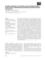
Fig. 7. Binding of the DmdR1 protein to the desA promoter region
of either S. pilosus (left) or S. coelicolor (right) at increasing protein
concentrations. The electrophoretic mobility-shift assays were per-
formed as indicated in Experimental procedures. Lanes 1 and 7,
control probes without protein (dashed arrow). Lanes 2–5 and 8–11
contain 1, 2, 4 or 8 l
M DmdR1 in the binding reactions, respect-
ively. Note the formation of two DNA–protein complexes (arrows)
at high protein concentrations. Lanes 6 and 12 contain 8 l
M
DmdR1 with an excess of cold probe as control. The DmdR1 pro-
tein was purified as described by Flores et al. [12].
S. Tunca et al. Regulation of the desferrioxamine gene cluster
FEBS Journal 274 (2007) 1110–1122 ª 2007 The Authors Journal compilation ª 2007 FEBS 1117
necessary. S. li vidans 1326 was u sed as a host for Strepto-
myces plasmid constructions.
DNA methods
Isolation of plasmid and bacterial chromosomal DNA,
restriction enzyme digestions, agarose gel electrophoresis and
Southern blot analysis were performed according to standard
molecular biology techniques [30]. Plasmids were trans-
formed into E. coli strains by standard chemical methods
or by electroporation. Electroporation-competent cells (50
or 100 lL; 10
9
colony-forming unitsÆmL
)1
) were mixed with
1–5 lL DNA solution in an ice-cold microcentrifuge tube
and electroporated at 2.5 kV with 25 lF and a resistance of
200 ohms or at 2.5 kV with 10 lF and resistance of
500 ohms. DNA fragments used as probes were labelled with
digoxigenin using a random priming kit (DIG DNA labelling
Mix; Roche Diagnostics GmbH, Penzberg, Germany).
Isolation of a S. coelicolor DdesA mutant
Deletion of the desA gene of S. coelicolor A3(2) was per-
formed by replacing the wild-type gene with a cassette con-
taining the apramycin resistance gene as selectable marker
using a PCR-based system [31]. The plasmid pIJ773 which
has a disruption cassette containing the apramycin resist-
ance gene [aac(3)IV] and oriT was used as template. The
mutant was constructed using the oligonucleotides 5¢-
acccc
tctcggaccgtccccaccggaggacccccccatgATTCCGGGGATC
CGTCGACC-3¢ and 5¢-aggccgatgcccacgaagtcgtacggggcgctggctt
caTGTAGGCTGGAGCTGCTTC-3¢ as the forward and
reverse primers, respectively (the sequence identical with the
upstream region of the desA gene is underlined and in low-
ercase; the sequence identical with the downstream region
of the desA gene is shown in italics and in lowercase).
These two long PCR primers (59 and 58 nucleotides) were
designed to produce a deletion of desA just after its start
codon, leaving only its stop codon behind. The 3¢ sequence
of each primer matches the right or left end of the disrup-
tion cassette (the sequence is shown uppercase in both
primers). The extended apramycin resistance cassette was
amplified by PCR, and E. coli BW25113 ⁄ pIJ790 bearing
cosmid Stc105 was electro-transformed with this cassette.
The isolated mutant cosmid was introduced into nonmethy-
lating E. coli ET12567 containing the RP4 derivative
pUZ8002; then the mutant cosmid was transferred to
S. coelicolor by intergeneric conjugation [32,33]. Double
cross-over exconjugants were screened for their kanamycin
sensitivity and apramycin resistance.
S. coelicolor DdesA mutant complementation
A 4204-bp ScaI fragment containing the desA coding region
was cloned into the pBluescript SK EcoRV site. As the
DdesA mutant is apramycin resistant, the kanamycin
resistance marker was cloned into an XbaI site of the new
construct (pSKdesA). A 5723-bp XhoI+NotI fragment
containing the desA gene and kanamycin gene (from
Table 1. Strains and plasmids used in this study.
Strain ⁄ plasmid Relevant genotype ⁄ comments Source ⁄ reference
Plasmids
pIJ790 k-RED (gam, bet, exo), cat, araC, rep101
ts
Gust et al. [31]
pUZ8002 tra, neo, RP4 Paget et al. [29]
Stc105 Cosmid containing the des cluster Redenbach et al. [28]
pBluescript SK E. coli vector Ap
R
lacZ arif1 Stratagene
pRA Integrative and conjugative vector derived from pSET152
pSKdesA Contains desA gene cloned into pBluescript SK This work
pSKdesAKn Contains kan gene cloned into pSKdesA This work
pRAdesAKn Contains desA and neo genes cloned into pRA This work
pIJ4083 High copy number promoter-probe vector carrying the promoterless xylE
gene as reporter
Kieser et al. [25]
pCoedesAP S. coelicolor desA promoter cloned into pIJ4083 This work
pPildesAP S. pilosus desA promoter cloned into pIJ4083 This work
E. coli strains
DH5a F
–
recA1, endA1, gyrA96, thi-1, hsdR17 (r
K
–
,m
K
+
), sup44, relA1k-,
(r80 dLacZAM15), D(lacZYA-argF)U169
Hanahan [39]
BW25113 K12 derivative: DaraBAD, DrhaBAD Datsenko & Wanner [27]
ET12567 dam, dcm, hsdS, cat, tet MacNeil et al. [40]
Streptomyces strains
S. coelicolor A3(2) Prototrophic wild-type Hopwood et al. [32]
S. lividans 1326 Prototrophic wild-type Hopwood et al. [32]
S. pilosus ATCC19797 Prototrophic wild-type Gunter et al. [16]
S. coelicolor M145 SCP1
–
, SCP2
–
Bentley et al. [13]
Regulation of the desferrioxamine gene cluster S. Tunca et al.
1118 FEBS Journal 274 (2007) 1110–1122 ª 2007 The Authors Journal compilation ª 2007 FEBS
pSKdesAKn) was cloned into the EcoRV site of pRA
(5769 bp). The new construct (pRAdesAKn) having a
size of 11492 bp was used to transform E. coli ET12567 ⁄
pUZ8002. After conjugation with the S. coelicolor desA
mutant, Kn
R
colonies were selected.
As complementation of the DdesA mutant was not
obtained with the above construct (see Results), the mutation
was complemented using the cosmid Stc105. A nonessential
gene (SCO2788) downstream of the desferrioxamine gene
cluster was replaced by the cassette from pIJ773 to allow
RP4 oriT-assisted conjugation by the method described
above to obtain the desA mutant. After intergeneric conjuga-
tion between E. coli ET12567 ⁄ pUZ8002 bearing the cosmid
with oriT and the S. coelicolor DdesA mutant, single cross-
over exconjugants were screened for kanamycin resistance.
Kanamycin-resistant colonies were isolated and analysed by
HPLC for their ability to produce desferrioxamines.
Siderophore plate bioassays
Siderophore production assays by colonies were carried out
on chrome azurol S blue plates prepared by the protocol of
Schwyn & Neilands [34].
HPLC analysis of desferrioxamines
Bacteria were grown in ILMM [3] and distributed into 500-
mL flasks (washed with 10% nitric acid and autoclaved).
CaCl
2
Æ2H
2
O (0.01% final concentration) and glucose
(2.5 gÆL
)1
final concentration), autoclaved separately, and fil-
ter-sterilized yeast extract (0.05 gÆL
)1
final concentration)
were then added to the culture flasks. The same medium sup-
plemented with FeSO
4
Æ7H
2
O (35 lm final concentration) was
used as control. Cultures were grown on a rotary shaker
(250 r.p.m.) at 30 ° C. Biomass was removed by filtration,
and 50 mL culture supernatant was freeze-dried. The solid
residue was redissolved in 1 ml distilled water, and, after
removal of the insoluble particles by centrifugation, 10 lL
1 m FeCl
3
was added to form Tris–hydroxamate–Fe
3+
com-
plexes. Insoluble particles were removed by centrifugation,
and the solution was filtered through a Vivaspin concentrator
before HPLC analysis.
HPLC separation of desferrioxamines was performed on a
reverse-phase column (Nucleosil C18, 5 lm, 4.6 by 150 mm)
with 150 lL injection volume and 1 mLÆmin
)1
flow rate for
25 min. A solution of 0.1% aqueous formic acid ⁄ methanol
was used as the solvent system. The Tris–hydroxamate–Fe
3+
complexes were detected at a wavelength of 435 nm.
Determination of desA promoter activity
The desA promoter fragments of S. coelicolor A3(2)
and S. pilosus were amplified by PCR using primers Pf
(5¢-GGAATTCCGCGCGCGGGTCTGGCTTCA-3¢) and
Pr (5¢-CGGGATCCCGGTACTGCTCCGCGGTGGTGT
CGTT-3¢) containing cleavage sites for EcoRI and BamHI
at their ends (in bold). The PCR products were extracted
from gels and digested with EcoRI and BamHI. The pro-
moter fragments were introduced upstream of the xylE
gene (catechol dioxygenase reporter) in the pIJ4083 vector
to generate pCoedesAP and pPildesA P. The correct orienta-
tion was confirmed by sequence analysis. S. coelicolor and
S. lividans cells harbouring pCoedesAP and pPildesAP were
grown in ILMM [3] containing 50 lgÆmL
)1
thiostrepton.
Then 1 mL of the cells was withdrawn at 24, 48, 60 and
72 h and, after being washed with physiological saline, they
were frozen and kept at )20 ° C. Crude extracts of the cells
were obtained by disruption using an ultrasonicator at
4 °C. Cells were sonicated (4 · 15 s with 20 s intervals) in
sample buffer [100 mm phosphate buffer (pH 7.5), 20 mm
EDTA (pH 8.0), 10% (v ⁄ v) acetone]. Triton X-100 was
added to a final concentration of 0.1%, and the mixture
was incubated for 15 min on ice. After clearing of the mix-
ture by centrifugation (10 000 g, Eppendorf 5415R centri-
fuge; Eppendorf, Hamburg, Germany) at 4 °C, the clear
supernatant was assayed for catechol 2,3-dioxygenase activ-
ity as described by Hopwood et al. [32].
Primer extension analysis
RNA was isolated from a 48-h culture of S. coelicolor har-
bouring pCoedesAP plasmid under conditions of iron limi-
tation by the procedure of Kieser et al. [25]. Primer
extension analysis was performed as described by Patek
et al. [35] and Barreiro et al. [36]. The fluorescein-labelled
primer was hybridized to RNA in a solution containing
0.4 m NaCl, 40 mm Pipes (pH 6.4), 1 mm EDTA (pH 8.0),
and 80% formamide at 45 °C for 12 h. The precipitated
RNA was dissolved in 20 lL reaction mixture: 4 lL Super-
Script buffer (Invitrogen, Carlsbad, CA, USA), 5 lL dNTP
(2 mm), 2 lL dithiothreitol (0.1 m), 2 lL actinomycin D
(500 lgÆmL
)1
), 1 lL RNase inhibitor (40 U) and 5 lL
H
2
O. After the addition of SuperScript II RT (Invitrogen),
the reaction was run at 42 °C for 1 h and stopped by heat
inactivation of the enzyme. RNA was removed by RNase
treatment and the protected RNA–DNA sample was preci-
pitated with ethanol. Then, the sample was dissolved in
6 lL TE buffer (10 mm Tris/HCl, 1 m m EDTA, pH 8.0)
and 6 lL stop buffer (Thermo Sequenase Primer Cycle
Sequencing Kit, Amersham Biosciences, Piscataway, NJ,
USA). After heat denaturation, the sample was run in the
ALFexpress DNA sequencer to identify the end of the pro-
tected fragment.
Transcriptional analysis
Culture conditions under iron limitation were as follows.
S. coelicolor inoculum cultures were grown for 36 h in
YEME broth [25]. The cell pellet was harvested by
centrifugation and washed twice with distilled water. Equal
S. Tunca et al. Regulation of the desferrioxamine gene cluster
FEBS Journal 274 (2007) 1110–1122 ª 2007 The Authors Journal compilation ª 2007 FEBS 1119
volumes of cells were inoculated in 100 mL minimal med-
ium [3] supplemented with an iron chelator (2,2¢-dipyridyl,
250 lm final concentration). All the needed material was
washed with 10% of nitric acid and distilled water to
remove iron traces.
For RNA extraction, 300 lL culture was added to 600 lL
RNA Protect Bacteria Reagent (Qiagen, Hilden, Germany),
mixed by vortex (30 s) and maintained for 5 min at room
temperature. The cell pellet was harvested by centrifugation
(5 min, 10 000 g, Eppendorf 5415R centrifuge). Samples
were frozen directly in liquid nitrogen. Total RNA was
extracted as described previously [37] except that the cell pel-
lets were resuspended in 900 lL lysis solution [400 lL acid
phenol, 100 lL chlorophorm:isoamyl alcohol (24 : 1),
400 lL RLT buffer (RNeasy mini kit; Qiagen)] and disrupted
with a Ribolyser instrument by using the lysing matrix B
(BIO 101). Subsequently, total RNA was isolated using an
RNeasy mini kit. DNA was removed in solution by using de-
oxyribonuclease I (Sigma, Haverhill, UK) and in a column
using RNase-Free DNase (Qiagen). RNA concentration was
calculated spectrophotometrically by determining the
absorbance at 260 nm using Nanodrop apparatus (Nano-
drop Technologies, Wilmington, DE, USA).
The transcription patterns were analyzed by the Super-
Script one-step reverse transcriptase PCR (RT-PCR) system
with Platinum Taq DNA polymerase (Invitrogen), using
100 ng total RNA as the template. Conditions were as fol-
lows: first-strand cDNA synthesis, incubation at 50–55 °C
for 40 min followed by 94 °C for 2 min; amplification, 30
to 40 cycles of 96 °C for 30 s, 55 °Cto67°C (depending
on the set of primers used) for 30 s, and 72 °C for 30 s to
1.5 min. Primers (19–24-mers) (Table 2) were designed to
generate PCR products between 879 and 1225 bp. Negative
controls were carried out for every set of primers by using
Taq DNA polymerase (Promega, Madison, WI, USA) to
confirm the absence of contaminating DNA in the RNA
preparations. Besides, specific primers for hrdB were used
as controls of RNA loading amount. Primer specificity was
tested by comparing each sequence against the complete
genome of S. coelicolor by using the web site http://insilico.
ehu.es [38]. The identity of each amplified product was cor-
roborated by direct sequencing with one of the primers
used for each amplification.
DmdR1 protein purification
The dmdRI gene was overexpressed in E. coli using the
pGEX-2T expression system (Amersham Biosciences). Purifi-
cation of the GST hybrid protein in glutathione–Sepharose
columns was performed according to the manufacturer’s
instructions. After elution of glutathione S-transferase
(GST)–DmdR1 fusion protein, DmdR1 protein was separ-
ated from GST by thrombin digestion and filtration through
the GSTrap column.
Electrophoretic mobility-shift assay
The desA promoter fragments were amplified by PCR
using the specific primers Pf and Pr (Table 2) and purified
with the PCR purification kit (GE Healthcare, Chalfont
St Giles, UK). The promoter fragments were then 3¢ end-
labelled with digoxigenin by using terminal transferase
(Roche) according to the manufacturer’s instructions.
Binding reactions were carried out in a 20-lL reaction
mixture containing 20 mm Tris ⁄ HCl (pH 7.5), 5 mm
MgCl
2
,40mm KCl, 100 mm MnCl
2
,2mm dithiothreitol,
10% (v ⁄ v) glycerol, 6.25 lg BSA, 1 lg poly(dI-dC) (GE
Healthcare), purified DmdR1 protein (at concentrations
ranging from 0 to 8 lm) and the labelled desA probe. The
DNA-binding reactions were initiated by the addition of
DmdR1 and incubated at 30 °C for 30 min. Samples were
immediately loaded and resolved on a prerun nondenatur-
ing 5% polyacrylamide gel for 4 h at 80 V in 0.5 · TBE
buffer (45 mm Tris/HCl, 1 mm EDTA, 45 mm boric acid,
pH 8.0) [12] and then electroblotted on to a nylon mem-
Table 2. Oligonucleotides used in this study.
Oligonucleotide Sequence (5¢ ) 3¢) Use
SecPr-5 GCGGCGACGGCGACGGCAAGAG RT-PCR
SecPr-3 CGGGGGAGCGGGCGATGACCT RT-PCR
viuB-5 GCAGATGCGCGTGCCAGACC RT-PCR
viuB-3 CGGCGCCAGTAGCCGACGAAG RT-PCR
desA-5 CGGGTGGCCGCCAAACTCG RT-PCR
desA-3 AGGAAGCGCGGTCAAGGGAGTCTC RT-PCR
desD-5 CGCAAGGCGCTGGCCGAGTTCA RT-PCR
desD-3 TGTGCAGCAGCGGGACGTAGTAGG RT-PCR
Pf GGAATTCCGCGCGCGGGTCTGGCTTCA PCR cloning of the
desA promoter
Pr CGGGATCCCGGTACTGCTCCGCGGTGGTGTCGTT
O6 GCGATCGCTGCCACTGC Primer extension
hrdB-5 GCCGCCGCGCCAAGAACCA RT-PCR
hrdB-3 CCAGCGGCGTGTGCAGCGAGAT RT-PCR
Regulation of the desferrioxamine gene cluster S. Tunca et al.
1120 FEBS Journal 274 (2007) 1110–1122 ª 2007 The Authors Journal compilation ª 2007 FEBS
brane. The digoxigenin-labelled probe was detected by
using anti-(digoxigenin–alkaline phosphatase conjugate)
and the luminogenic substrate CDPstar (Boehringer Mann-
heim, Mannheim, Germany). The signal was captured by
exposure to X-ray film.
Acknowledgements
This work was supported by grants from the Funda-
cio
´
n Ramo
´
n Areces (03 ⁄ 2000–02 ⁄ 2003), Madrid, Spain
and the CICYT (BIO2003-01489) to JFM. We thank
F. Flores for help with preparation of the materials,
F. Barona-Go
´
mez and G. Challis for samples of pure
desferrioxamine B and E, and B. Martin and J. Merino
for excellent technical support.
References
1 Crosa JH (1987) Signal transduction and transcriptional
and post-transcriptional control or iron-regulated genes
in bacteria. Microbiol Mol Biol Rev 61, 319–336.
2 Neilands JB (1995) Siderophores: structure and function
of microbial iron-transport compounds. J Biol Chem
270, 26723–26726.
3 Muller G & Raymond KN (1984) Specificity and
mechanism of ferrioxamine-mediated iron transport in
Streptomyces pilosus. J Bacteriol 160, 304–312.
4 Bunet R, Brock A, Rexer UH & Takano A (2006) Iden-
tification of genes involved in siderophore transport in
Streptomyces coelicolor A3 (2). FEMS Microbiol Lett
262, 57–64.
5 Imbert M, Be
´
chet M & Blondeau R (1995) Comparison
of the main siderophores produced by some species of
Streptomyces. Curr Microbiol 31, 129–133.
6 Yamanaka K, Oikawa H, Ogawa HO, Hosono K,
Shinmachi F, Takano H, Sakuda S, Beppu T & Ueda
K (2005) Desferrioxamine E produced by Streptomyces
griseus stimulates growth and development of Strepto-
myces tanashiensis. Microbiology 151, 2899–2905.
7 Schupp T, Waldmeier U & Divers M (1987) Biosyn-
thesis of desferrioxamine B in Streptomyces pilosus:
evidence for the involvement of lysine decarboxylase.
FEMS Microbiol Lett 42, 135–139.
8 Schupp T, Toupet C & Divers M (1988) Cloning and
expression of two genes of Streptomyces pilosus involved
in the biosynthesis of siderophore desferroxamine
B. Gene 64, 179–188.
9 Tao X & Murphy JR (1992) Binding of the metalloregu-
latory protein DtxR to the diphteria tox operator
requires a divalent heavy metal ion and protects the
palindromic sequence from DNase I digestion. J Biol
Chem 267, 21761–21764.
10 Oguiza JA, Marcos AT, Malumbres M & Martı
´
nJF
(1996) The galE gene encoding the UDP-galactose
4-epimerase of Brevibacterium lactofermentum is coupled
transcriptionally to the dmdR gene. Gene 177,
103–107.
11 Flores FJ, Barreiro C, Coque JJR & Martı
´
n JF (2005)
Functional analysis of two divalent metal-dependent
regulatory genes dmdR1 and dmdR2. Streptomyces
coelicolor and proteome changes in deletion mutants.
FEBS J 272, 725–735.
12 Flores FJ & Martı
´
n JF (2004) Iron-regulatory proteins
DmdR1 and DmdR2 of Streptomyces coelicolor form
two different DNA–protein complexes with iron boxes.
Biochem J 379
, 1–7.
13 Bentley SD, Chater KF, Cerdeno-Tarraga AM, Challis
GL, Thomson NR, James KD, Harris DE, Quail MA,
Kieser H, Harper D et al. (2002) Complete genome
sequence of the model actinomycete Streptomyces coeli-
color A3 (2). Nature 417, 141–147.
14 Barona-Go
´
mez F, Wong U, Giannakopulos AE, Der-
rick PJ & Challis GL (2004) Identification of a cluster
of genes that directs desferrioxamine biosynthesis in
Streptomyces coelicolor M145. J Am Chem Soc 126,
16282–16283.
15 Challis G (2005) A widely distributed bacterial path-
way for siderophore biosynthesis independent of non-
ribosomal peptide synthetases. Chembiochem 6,
601–611.
16 Gu
¨
nter K, Toupet C & Schupp T (1993) Characteriza-
tion of an iron-regulated promoter involved in
desferrioxamine B synthesis in Streptomyces pilosus:
repressor-binding site and homology to the diphtheria
toxin gene promoter. J Bacteriol 175, 3295–3302.
17 Flores FJ, Rinco
´
n J & Martı
´
n JF (2003) Characteriza-
tion of the iron-regulated desA promoter of Strepto-
myces pilosus as a system for controlled gene expression
in actinomycetes. Microb Cell Fact 2,5.
18 Sola-Landa A, Rodrı
´
guez-Garcı
´
a A, Franco-Domı
´
nguez
E & Martı
´
n JF (2005) Binding of PhoP to promoters of
phosphate regulated genes in Streptomyces coelicolor:
identification of PHO boxes. Mol Microbiol 56,
1373–1385.
19 Strohl WR (1992) Compilation and analysis of DNA
sequences associated with apparent streptomycete pro-
moters. Nucleic Acids Res 20, 961–974.
20 Madduri K, Stuttard C & Vining LC (1989) Lysine cat-
abolism in Streptomyces spp. is primarily through cada-
verine: beta-lactam producers also make alpha-
aminoadipate. J Bacteriol 171, 299–302.
21 Gil JA & Hopwood DA (1983) Cloning and expres-
sion of a p-aminobenzoic acid synthetase gene of the
candicidin producer, Streptomyces griseus. Gene 25,
119–132.
22 Martı
´
n JF & Gil JA (1984) Cloning and expression of
antibiotic production genes. Bio ⁄
Technology 2, 63–72.
S. Tunca et al. Regulation of the desferrioxamine gene cluster
FEBS Journal 274 (2007) 1110–1122 ª 2007 The Authors Journal compilation ª 2007 FEBS 1121
23 Martı
´
n JF, Gutie
´
rrez S & Aparicio JF (2000) Secondary
metabolites. In Encyclopedia of Microbiology, Vol. 4,
2nd edn (Lederberg, J, ed.), pp. 213–236. Academic
Press, San Diego, CA.
24 Butterton JR & Calderwood SB (1994) Identification,
cloning, and sequencing of a gene required for ferric
vibriobactin utilization by Vibrio cholerae. J Bacteriol
176, 5631–5638.
25 Kieser T, Bibb MJ, Buttner MJ, Chater KF & Hop-
wood DA (2000) Practical Streptomyces Genetics. John
Innes Foundation, Norwich.
26 Recio E, Aparicio JF, Rumbero A & Martı
´
n JF (2006)
Glycerol, ethylene glycol and propanediol elicit pimari-
cin biosynthesis in the PI-factor-defective strain Strepto-
myces natalensis npi287 and increase polyene production
in several wild-type actinomycetes. Microbiology 152,
3147–3156.
27 Datsenko KA & Wanner BL (2000) One-step inactiva-
tion of chromosomal genes in Escherichia coli K-12
using PCR products. Proc Natl Acad Sci USA 97,
6640–6645.
28 Redenbach M, Kieser HM, Denapaite D, Eichner A,
Cullum J, Kinashi H & Hopwood DA (1996) A set of
ordered cosmids and a detailed genetic and physical
map for the 8 Mb Streptomyces coelicolor A3 (2) chro-
mosome. Mol Microbiol 21 , 77–96.
29 Paget MS, Chamberlin L, Atrih A, Foster SJ & Buttner
MJ (1999) Evidence that the extracytoplasmic function
sigma factor sigmaE is required for normal cell wall
structure in Streptomyces coelicolor A3 (2). J Bacteriol
181, 204–211.
30 Sambrook J & Russell DW (2001) Molecular Cloning: A
Laboratory Manual. Cold Spring Harbor Laboratory
Press, Cold Spring Harbor, NY.
31 Gust B, Challis GL, Fowler K, Kieser T & Chater KF
(2003) PCR-targeted Streptomyces gene replacement
identifies a protein domain needed for biosynthesis of
the sesquiterpene soil odor geosmin. Proc Natl Acad Sci
USA 100, 1541–1546.
32 Hopwood DA, Bibb MJ, Chater KF, Kieser T, Bruton
CJ, Kieser HM, Lydiate DJ, Smith DP, Ward JM &
Schrempf H (1985) Genetic Manipulation of Strepto-
myces: A Laboratory Manual. John Innes Foundation,
Norwich.
33 Enrı
´
quez L, Mendes MV, Anto
´
n N, Tunca S, Guerra
SM, Martı
´
n JF & Aparicio JF (2006) An efficient gene
transfer system for the pimaricin producer Streptomyces
natalensis. FEMS Microbiol Lett 257, 312–318.
34 Schwyn B & Neilands JB (1987) Universal chemical
assay for the detection and determination of sidero-
phores. Anal Biochem 160, 47–56.
35 Patek M, Eikmanns BJ, Patek J & Sahm H (1996) Pro-
moters from Corynebacterium glutamicum: cloning,
molecular analysis and search for a consensus motif.
Microbiology 142, 1297–1309.
36 Barreiro C, Gonza
´
lez-Lavado E, Brand S, Tauch A &
Martı
´
n JF (2005) Heat shock proteome analysis of
wild-type Corynebacterium glutamicum ATCC 13032 and
a spontaneous mutant lacking GroEL1, a dispensable
chaperone. J Bacteriol 187, 884–889.
37 Hu
¨
ser AT, Chassagnole C, Lindley ND, Merkamm M,
Guyonvarch A, Elisakova V, Patek M, Kalinowski J,
Brune I, Puhler A & Tauch A (2005) Rational design of
a Corynebacterium glutamicum pantothenate production
strain and its characterization by metabolic flux analysis
and genome-wide transcriptional profiling. Appl Environ
Microbiol 71, 3255–3268.
38 Bikandi J, San Milla
´
n R, Rementeria A & Garaizar J
(2004) In silico analysis of complete bacterial genomes:
PCR, AFLP-PCR and endonuclease restriction. Bioin-
formatics 20, 798–799.
39 Hanahan D (1983) Studies on transformation of
Escherichia coli with plasmids. J Mol Biol 166, 557–580.
40 MacNeil DJ, Gewain KM, Ruby CL, Dezeny G,
Gibbons PH & MacNeil T (1992) Analysis of Strepto-
myces avermitilis genes required for avermectin bio-
synthesis utilizing a novel integration vector. Gene 111,
61–68.
Regulation of the desferrioxamine gene cluster S. Tunca et al.
1122 FEBS Journal 274 (2007) 1110–1122 ª 2007 The Authors Journal compilation ª 2007 FEBS