Báo cáo khoa học: Cellular response to unfolded proteins in the endoplasmic reticulum of plants pptx
Bạn đang xem bản rút gọn của tài liệu. Xem và tải ngay bản đầy đủ của tài liệu tại đây (305.78 KB, 20 trang )
REVIEW ARTICLE
Cellular response to unfolded proteins in the endoplasmic
reticulum of plants
Reiko Urade
Division of Food Science and Biotechnology, Graduate School of Agriculture, Kyoto University, Japan
Introduction
The unfolded protein response (UPR) is a fundamental
system common to unicellular organisms, plants, ani-
mals, and humans, and is conserved in all eukaryotic
cells. However, there are differences in the molecular
mechanisms underlying the UPR between organisms.
In yeast, the UPR increases the folding and degrada-
tion capacities of unfolded proteins by inducing the
expression of genes related to those capacities [1]. Inos-
itol-requiring enzyme-1 (IRE1), an endoplasmic reticu-
lum (ER)-transmembrane protein that is activated by
ER stress, splices basic leucine zipper (bZIP) transcrip-
tion factor HAC1 mRNA in a nonconventional man-
ner [2,3]. HAC1 is translated from the spliced mRNA
[4–6] and subsequently activates the transcription of a
group of genes possessing UPR cis-activating regula-
tory elements in their promoter regions [7–9]. This
pathway was the first example of a protein signal that
is transduced from the ER to the nucleus, and this
finding opened the door to investigation of the details
of UPR signaling events.
In comparison with that of yeast, the UPR of mam-
malian cells is a much more complicated event, in
which general attenuation of translation, apoptosis,
and folding or degrading of unfolded proteins occurs
[10–12]. The mammalian UPR is triggered by at least
three ER stress sensors, including the mammalian
Keywords
endoplasmic reticulum; ER-associated
degradation; molecular chaperones; protein
folding; quality control of proteins; unfolded
protein response
Correspondence
R. Urade, Division of Food Science and
Biotechnology, Graduate School of
Agriculture, Kyoto University, Gokasho, Uji,
Kyoto 611-0011, Japan
Fax: +81 774 38 3757
Tel. +81 774 38 3758
E-mail:
(Received 23 November 2006, accepted 22
December 2006)
doi:10.1111/j.1742-4658.2007.05664.x
Secretory and transmembrane proteins are synthesized in the endoplasmic
reticulum (ER) in eukaryotic cells. Nascent polypeptide chains, which are
translated on the rough ER, are translocated to the ER lumen and folded
into their native conformation. When protein folding is inhibited because
of mutations or unbalanced ratios of subunits of hetero-oligomeric pro-
teins, unfolded or misfolded proteins accumulate in the ER in an event
called ER stress. As ER stress often disturbs normal cellular functions, sig-
nal-transduction pathways are activated in an attempt to maintain the
homeostasis of the ER. These pathways are collectively referred to as the
unfolded protein response (UPR). There have been great advances in our
understanding of the molecular mechanisms underlying the UPR in yeast
and mammals over the past two decades. In plants, a UPR analogous to
those in yeast and mammals has been recognized and has recently attracted
considerable attention. This review will summarize recent advances in the
plant UPR and highlight the remaining questions that have yet to be
addressed.
Abbreviations
ATF, activating transcription factor; BiP, binding protein; bZIP, basic leucine zipper; eIF2a, initiation factor-2a; ER, endoplasmic reticulum;
ERAD, ER-associated degradation; ERSE, ER stress response element; fl-2, floury-2; GFP, green fluorescent protein; GLS, Golgi body
localization sequence; GPT, UDP-N-acetylglucosamine–dolichol phosphate N-acetylglucosamine-1-phosphate transferase; IRE1, inositol-
requiring enzyme-1; PCD, programmed cell death; PDI, protein disulfide isomerase; PERK, interferon-induced dsRNA-activated protein
kinase-related protein; S1P, site-1 protease; S2P, site-2 protease; UGGT, UDP-glucose–glycoprotein glucosyltransferase; UPR, unfolded
protein response; UPS, ubiquitin-proteasome system; XBP-1, X-box binding protein 1.
1152 FEBS Journal 274 (2007) 1152–1171 ª 2007 The Author Journal compilation ª 2007 FEBS
ortholog of yeast IRE1 [13,14], activating transcription
factor (ATF) 6 [15], and interferon-induced dsRNA-
activated protein kinase-related protein (PERK) [16].
IRE1 is activated during ER stress and splices invalid
mRNA, similar to yeast IRE1, into the mature X-box
binding protein 1 (XBP-1) mRNA, a bZIP-like tran-
scription factor [17–20]. XBP-1 is translated from the
spliced mRNA and is translocated to the nucleus to
regulate transcription of target genes. In addition,
IRE1 independently mediates the rapid degradation of
a specific subset of mRNAs due to their localization
on the ER membrane and to the amino-acid sequence
they encode [21]. This response could selectively halt
production of proteins that challenge the ER and
could make available the translocation and folding
machinery for the subsequent remodeling process. In
addition, IRE1 forms a trimeric complex with phos-
phorylated tumor necrosis factor receptor-associated
factor 2, apoptosis signal regulating kinase 1 and the
c-Jun N-terminal kinase and subsequently causes cell
death [11,22,23]. ATF6 is an ER transmembrane pro-
tein that senses ER stress through its luminal domain,
and then moves to Golgi bodies to be cleaved. The
ATF6 cytosolic domain produced as a result of this
cleavage event is released from the Golgi membrane
into the nucleus, where it induces the expression of tar-
get genes [24–28]. PERK is an ER transmembrane pro-
tein that senses ER stress through its luminal domain
and phosphorylates a specific serine residue of transla-
tion initiation factor-2a (eIF2a), resulting in general
inhibition of translation [16,29]. Phosphorylation of
eIF2a also stimulates translation of ATF4 [30], a
bZIP-like transcription factor that induces the tran-
scription of many amino-acid synthetic enzymes,
amino-acid transporters, and antioxidation enzymes.
ATF6 and ATF4 also stimulate the transcription of
CHOP, a gene important for apoptotic cell death [31].
It has recently been shown that UPR signaling not
only maintains the homeostasis of the ER, but also
plays an important role in nutritional and differentia-
tion programs in healthy and unstressed yeast and
mammalian cells [11,32,33]. Furthermore, organ-specific
UPR signaling pathways have been identified in mam-
malian cells [34–37]. These findings suggest that the
UPR functions during normal processes as well as
during emergency situations. The UPR pathways act
cooperatively such that the fate of the cell depends on
the balance between the individual UPR pathways.
Therefore, disturbance of these functions causes mal-
function of the ER transport machinery and defective
UPR signaling, resulting in diseases such as neurode-
generative disorders, diabetes, and endocrine defects
[11].
The UPR in plants is an important and constantly
expanding topic. However, study of the plant UPR is
a relatively new field, and its molecular details are only
now becoming clear. Recent developments in this field
will be explored in this review.
Transcriptional regulation of UPR
genes
The most prominent phenomenon induced by ER
stress is transcriptional regulation of UPR genes. The
induction of genes assumed to be related to the UPR
in plant cells has been reported. Binding protein (BiP)
is a representative UPR gene. BiP is induced in the
presence of drugs that cause ER stress, such as tunica-
mycin [38–45]. Tunicamycin inhibits UDP-N-acetyl-
glucosamine– dolichol phosphate N-acetylglucosamine-1-
phosphate transferase (GPT), such that the initial step
of the biosynthesis of dolichol-linked oligosaccharides
is blocked [46]. Treatment with tunicamycin results in
the inability of asparagine (N)-linked glycoproteins
synthesized in the ER to be glycosylated. Transgenic
Arabidopsis thaliana plants with a 10-fold higher level
of GPT activity were resistant to tunicamycin at a con-
centration that was lethal to control plants [44]. Like-
wise, transgenic plants grown in the presence of
tunicamycin have N-glycosylated proteins, and expres-
sion levels of BiP mRNA was lower than in control
plants. These findings suggest that treatment with tu-
nicamycin results in the generation of misfolded or
unfolded proteins by inhibiting N-glycosylation and
activation of the UPR. Transcription of BiP mRNA
is activated by other drugs such as the proline analog
azetidine-2-carboxylase, which is incorporated into
nascent polypeptides and prevents their folding [47],
and dithiothreitol, which inhibits formation of disulfide
bonds on nascent polypeptides and prevents their fold-
ing [39].
Two comprehensive analyses of the transcriptome of
A. thaliana during drug-induced ER stress have been
performed using two kinds of DNA microarray meth-
ods. Martı
`
nez & Chrispeels [48] performed experiments
using an Affymetrix GeneChip with a 8297 probe set
(7372 independent genes of the 27 000 protein-coding
genes of A. thaliana). The UPR was induced by treat-
ing Arabidopsis plants with tunicamycin or dithiothrei-
tol. Fifty-three genes were identified as up-regulated
genes under ER stress, whereas 31 genes were identi-
fied as down-regulated genes. Kamauchi et al. [49]
analyzed the transcriptome of Arabidopsis UPR genes
by fluid microarray analysis of tunicamycin-treated
plantlets. Using this method, target genes were cloned
from selected fluid microarray beads [50], and 215
R. Urade Response to unfolded proteins in ER of plants
FEBS Journal 274 (2007) 1152–1171 ª 2007 The Author Journal compilation ª 2007 FEBS 1153
up-regulated genes and 17 down-regulated genes were
identified. These genes were reanalyzed with functional
DNA microarrays using DNA clones from the fluid
microarray analysis. Together, 36 up-regulated genes
and two down-regulated genes in all samples treated
with the three drugs, tunicamycin, dithiothreitol or
azetidine-2-carboxylase were recognized as UPR genes.
The up-regulated UPR genes identified by the two
research groups are shown in Table 1, and include ER
chaperones, glycosylation ⁄ modification-related pro-
teins, translocon subunits, vesicle transport proteins,
and ER-associated degradation (ERAD) proteins.
Most of these proteins are orthologs of the genes iden-
tified as being related to the UPR in yeast and mam-
malian cells [1,30,51–54]. In addition, genes related to
the regulation of translation (P58
IPK
) [55] and apop-
tosis (BAX inhibitor 1) [56,57] were also identified as
being up-regulated during the UPR in plants [49,58].
Phospholipid biosynthetic enzymes increase in expres-
sion in the maize (Zea mays) floury-2 (fl-2) mutant
(described below) and soybean (Glycine max) suspen-
sion cultures when treated with tunicamycin [45], and,
in yeast, a number of lipid metabolism-related genes
are up-regulated by ER stress [1]. On the other hand,
neither of the DNA microarray analyses of the
Arabidopsis transcriptome described above detected
any up-regulation of lipid metabolism-related genes,
suggesting that additional experiments are needed to
assess if phospholipid metabolism-related genes are
related to the UPR in plant cells.
Signal-transduction-related proteins such as protein
kinases and transcription factors are also up-regulated
during the plant UPR. WRKY33 and ATAF2 were
identified as repressors of the signal-transduction path-
way activated in response to pathogens [59,60]. Zat12
enhances the expression of oxidative-stress and light
stress-response transcripts and plays a central role in
reactive oxygen and abiotic stress signaling [61], imply-
ing that the UPR signal-transduction pathway con-
nects other stress signaling pathways. Genes regulated
by other transduction pathways connected with UPR
signal transduction may eventually be identified as
being either up-regulated or down-regulated after
treatment with drugs that induce ER stress. The role
of these genes under these circumstances remains to be
elucidated in plants.
There are discrepancies in the identification and ana-
lysis of genes down-regulated during ER stress
obtained from the two DNA microarray assays des-
cribed above. Thirty-one down-regulated genes were
identified using the Affymetrix GeneChip, and among
them, 29 genes were predicted to encode proteins con-
taining signal peptides. Lowering the threshold of
detection from 2.5-fold to 2-fold inhibition increases
this amount to 129 independent genes. Among these
genes, 82% of the encoded proteins have signal pep-
tides. On the other hand, only two down-regulated
genes, vegetative storage proteins Vsp1 and Vsp2, were
identified by the fluid microarray method. Both of
these proteins also have a signal peptide. In mamma-
lian cells, expression of abundant genes is repressed
during ER stress depending on IRE1 but not on XBP-1.
Repression of these genes is fast compared with
expression changes mediated by XBP-1. Furthermore,
functional signal sequences of proteins encoded by
down-regulated genes are required for this repression
event to occur. Taken together, it is possible that
IRE1-mediated mRNA degradation occurs during co-
translational translocation [21]. The fact that more
than 80% of the encoded proteins in Arabidopsis with
down-regulated expression during ER stress have sig-
nal peptides raises the possibility that similar systems
may function in plant cells.
In both DNA microarray analyses, only the genes
that complied with certain restrictive criteria were
designated UPR genes, implying that some UPR genes
were missed during the analysis as a result of these cri-
teria. Thus, genes expressed at very low levels might
have been unintentionally eliminated from the analysis
because of difficulty in assessing differences in their
expression levels. For example, AtbZIP60, which was
not designated a UPR gene by DNA microarray ana-
lysis, is induced in response to ER stress as detected
by Northern blot and RT-PCR analyses [62]. It is
expected that genes identified by the DNA microarray
analyses will eventually be confirmed by other methods
such as mRNA quantification and promoter analysis.
A pivotal role of the UPR is to maintain ER home-
ostasis. Therefore, the presence of mutated proteins
that are unable to fold into their native conformation
in the ER induces the UPR in an effort to restabilize
the ER environment. Many examples of this phenom-
enon have been described in yeast and mammalian
cells, and few examples have been found in plants. For
example, maize high-lysine starchy endosperm (opaque)
mutants are characterized by a decrease in the accumu-
lation of storage proteins in the ER and by alterations
in protein body morphology in their endosperm. The
opaque mutants fl-2 and defective endosperm B30 have
a defective signal peptide in the 24-kDa a-zein and the
19-kDa a-zein endosperm storage proteins, respect-
ively. These mutant proteins are translocated into the
lumen of the ER, but remain anchored to the mem-
branes through the noncleaved signal peptide [63,64].
A decrease in the expression of a-zein is accompanied
by an increase in the level of b-70, a water-soluble
Response to unfolded proteins in ER of plants R. Urade
1154 FEBS Journal 274 (2007) 1152–1171 ª 2007 The Author Journal compilation ª 2007 FEBS
Table 1. Genes up-regulated during ER stress. Data from [48,49] are combined. NEM, N-Ethylmaleimide; GST, glutathione S-transferase.
AGI gene Description cis-Acting regulatory element
a
References
Protein folding
At1g09080 BiP-like ERSE like (2), XBP1-BS-like 48, 49
At5g28540 BiP P-UPRE 48, 49
At5g42420 BiP XBP1-BS-like, P-UPRE 49
At4g21180 Similar to ERdj3 48
At5g61790 Calnexin 1 ERSE like, XBP1-BS-like 48, 49
At5g07340 Calnexin 2 P-UPRE 48, 49
At1g56340 Calreticulin 1 ERSE like 49
At1g09210 Calreticulin 2 ERSE like, XBP1-BS-like 48, 49
At4g24190 AtHsp90–7 ERSE like, XBP1-BS-like 48, 49
At2g47470 Similar to PDI ERSE like, XBP1-BS-like 48, 49
At1g77510 Similar to PDI ERSE like 49
At2g32920 Similar to PDI 48, 49
At1g04980 Similar to PDI ERSE like, XBP1-BS-like 49
At5g58710 AtCYP20-1 (cyclophilin ROC7) ERSE like, XBP1-BS-like 49
Glycosylation ⁄ modification
At2g02810 UDP-glucose ⁄ UDP-galactose transporter ERSE like 48, 49
At2g41490 UDP-GlcNAc:dolichol phosphate
N-acetylglucosamine-1-phosphate transferase
ERSE like 48, 49
At2g47180 Putative galactinol synthase XBP1-BS-like 48
At2g41490 GPT ERSE like, XBP1-BS-like 48
At4g15550 UDP-glucose indole-3-acetate
b-
D-glucosyltransferase
48
Translocation
At5g50460 SEC61 c subunit XBP1-BS-like 49
At1g29310 Similar to SEC61 a subunit ERSE like 49
At2g34250 Similar to SEC61 a subunit 49
At2g45070 Similar to SEC61 b subunit XBP1-BS-like 48, 49
At4g24920 Similar to SEC61 c subunit XBP1-BS-like 48, 49
At1g27330 Similar to SERP1 ⁄ RAMP4 ERSE like 49
At1g27350 Similar to SERP1 ⁄ RAMP4 ERSE like 48, 49
At3g51980 Similar to ER chaperone SIL 1 ERSE like, XBP1-BS-like 49
At5g03160 P58
IPK
ERSE like (2) 49
At2g18190 Putative AAA-type ATPase 48
At2g03120 Similar to signal peptide peptidase 48
Protein degradation
At1g65040 Similar to HRD1 ERSE like 49
At4g21810 Similar to DER1 48, 49
At1g18260 Similar to HRD3 ⁄ SEL1 ERSE like 49
At5g35080 Similar to OS-9 48
At2g46500 Similar to ubiquitin 48
Vacuolar
At3g52190 Similar to SP12p 48
At1g78920 Similar to H
+
-pyrophosphatase 48
Translation
At5g03160 P58
IPK
ERSE like (2) 49
Vesicle trafficking
At3g07680 Similar to Emp24p ERSE like, XBP1-BS-like (2) 49
At4g21730 Similar to NEM-sensitive fusion protein 49
At1g11890 Similar to vesicle trafficking protein XBP1-BS-like 48, 49
At1g62020 Similar to coatomer a subunit 49
At1g09180 Similar to SAR1B ERSE like 48
At4g01810 Similar to SEC23p XBP1-BS-like 48
PCD
At5g47120 BAX inhibitor 1 ERSE like 49
R. Urade Response to unfolded proteins in ER of plants
FEBS Journal 274 (2007) 1152–1171 ª 2007 The Author Journal compilation ª 2007 FEBS 1155
maize BiP ortholog associated with both the ER and
protein bodies [64–70]. The increase in maize BiP
mRNA and corresponding protein concentrations
in mutants compared with those of wild-type maize
was endosperm-specific and inversely proportional to
changes in mutant zein synthesis [66]. The pattern of
gene expression in normal and the seven opaque
mutants o1, o2, o5, o9, o11, Mc and fl-2, protein syn-
thesis of which is the molecular basis of the mutation,
was assayed by profiling endosperm mRNA transcripts
with an Affymetrix GeneChip containing more than
1400 selected maize gene sequences [71]. Compared
with normal maize, alterations in the gene expression
patterns of the opaque mutants were pleiotropic, where
the expressions of BiP, protein disulfide isomerase
(PDI), calreticulin, GRP94 and cyclophilin, and other
physiological stress-related genes were increased in the
opaque mutants. The transcriptional response in fl-2
may be induced by the UPR, as the change in the
pattern of gene expression was restricted to the endo-
sperm in which the mutant a-zein was synthesized. The
expression pattern of o2 and fl-2 depends on the
molecular basis of the mutation. It remains necessary
to evaluate the relationship between the expression
patterns and the molecular basis of each mutation in
the other mutants before a complete understanding of
how these mutants affect ER homeostasis in plants will
be obtained.
Signal transduction during the UPR
Transcription of genes related to the UPR is controlled
by the specific transcription factor that binds to the
cis-acting regulatory element on the promoter of a
UPR gene. Many experiments have revealed the details
of the signal-transduction mechanism by which yeast
and mammalian cells adapt to ER stress [10,11,72,73].
In yeast, a 22-bp segment in the promoter of KAR2
(yeast BiP) was identified as the first regulatory ele-
ment responding to ER stress [7–9], and the sequence
CAGCGTG within this 22-bp segment was identified
as the minimal regulatory element and named UPRE
(UPR cis-acting regulatory element). HAC1 produced
from mRNA spliced by IRE1 binds to the UPRE and
induces the transcription of UPR genes [4,5]. In mam-
malian cells, bZIP-like transcription factors XBP1 [17–
20], ATF6 [15], ATF4 [30], ATF3 [74], CHOP [75],
nuclear factor-erythroid 2-related factor 2 [76], OASIS
[35], CREB-H [36] and Tisp40 [37] function under ER
stress. These transcription factors bind to one or more
cis-acting regulatory elements and activate or repress
the transcription of target genes. More than 10 types
Table 1. (Continued ).
AGI gene Description cis-Acting regulatory element
a
References
Kinase
At1g08650 Putative calcium-dependent protein kinase ERSE like 48
Transcription factor
At3g24050 GATA-1 48
At1g56170 Hap5b 48
At2g38470 WRKY-33 48
At5g08790 ATAF2 ERSE like 48
At5g59820 Zat12 48
Stress protein
At5g16660 HSP-like (D2T2) ERSE like 48
At1g67360 Putative stress-related protein XBP1-BS-like 48
Unclassified
At2g25110 Similar to stromal cell derived factor-2 48, 49
At5g09410 Similar to anther ethylene-up-regulated
calmodulin-binding protein ER1
ERSE like, XBP1-BS-like 49
At4g12720 Similar to growth factor protein with
mutT domain
48
At4g19880 GST ERSE like 48
At2g16060 Similar to AHB1 48
At4g26400 Putative ring zinc finger protein 48
At4g14430 Carnitine racemase-like protein ERSE like 48
At1g07670 ER-type calcium transporter ATPase 4 ERSE like, XBP1-BS-like 48
At5g39580 Peroxidase ATP24a 48
At4g10040 Cytochrome c ERSE like 48
a
Numbers in parentheses show the number of elements on the promoter.
Response to unfolded proteins in ER of plants R. Urade
1156 FEBS Journal 274 (2007) 1152–1171 ª 2007 The Author Journal compilation ª 2007 FEBS
of cis-acting regulatory elements that respond to ER
stress are known in mammals [11]. Among them, ER
stress response element (ERSE) and ERSE-II are tar-
gets for both ATF6 and XBP1 [15,77–79]. ATF6 is
constitutively synthesized as a type II transmembrane
protein in the ER [24]. When the ER-membrane-bound
precursors of ATF6 are cleaved by the serine protease
site-1 protease (S1P) and the metalloprotease site-2
protease (S2P) in response to ER stress, the N-terminal
halves become soluble transcription factors. These sol-
uble factors are translocated into the nucleus and bind
to ERSE and ERSE-II [24–28]. ERSE controls the
expression of ER-localized molecular chaperones
[80,81]. Transcription from another cis-acting regula-
tory element, XBP1-BS, is entirely controlled by
XBP1, and induces expression of components of the
ERAD system [80,81]. In plants, cis-acting regulatory
elements that respond to ER stress have also been dis-
covered. The soybean BiP paralog genes gsBIP6 and
gsBIP9 have domains similar to ERSE and ERSE-II
in their 5¢ flanking sequences that are responsive to
treatment with tunicamycin [82]. Similarly, a 24-bp
sequence in the 5¢ flanking sequences of Arabidopsis
BiP is crucial for gene induction by tunicamycin [83].
This 24-bp sequence is called P-UPRE and contains
two overlapping elements similar to mammalian
ERSE-II and XBP-BS. Putative cis-acting regulatory
sequences similar to ERSE, XBP1-BS, and P-UPRE
are found at high frequencies (> 65%) in the 5¢ flank-
ing sequences of the Arabidopsis UPR genes identified
by the DNA microarray analyses (Table 1).
Novel transcription factor AtbZIP60 has been identi-
fied as a member of the plant UPR signal-transduction
pathway. To date, every transcription factor related to
the UPR in mammals and yeast is bZIP-like. Hence,
Iwata & Koizumi [84] analyzed transcripts of 75 puta-
tive bZIP transcription factors in the Arabidopsis
genome. Among them, only AtbZIP60, a factor that is
induced by treatment with tunicamycin, dithiothreitol
and azetidine-2-carboxylase, activates transcription
from P-UPRE and ERSE elements. The AtbZIP60
gene encodes a predicted type II transmembrane pro-
tein of 295 amino acids with an N-terminal bZIP
DNA-binding domain, a putative transmembrane
domain, and a 56-amino-acid small C-terminal domain
(Fig. 1A). A truncated form of AtbZIP60 lacking the
transmembrane domain (AtbZIP60 DC) fused with
green fluorescent protein (GFP) localized to the nuc-
leus. In other experiments, AtbZIP60 DC clearly acti-
vated both P-UPRE and ERSE-like sequences in a
dual luciferase assay using protoplasts of cultured
tobacco (Nicotiana tabacum) cells. Therefore, Atb-
ZIP60 is considered to be a transcription factor
responding to ER stress, where AtbZIP60 DC induces
the expression of AtbZIP60 through ERSE-like
sequences present in the promoter of AtbZIP60. In
contrast, wild-type AtbZIP60 is unable to activate
ERSE-like sequences and P-UPRE, probably because
it is anchored to the membrane. This suggests that
native AtbZIP60 may be released from the membrane
into the cytosol during ER stress to act as a transcrip-
tion factor in the nucleus (Fig. 2). In the Arabidopsis
genome, the At4g20310 gene encodes a membrane pro-
tein analogous to S2P, but it remains to be confirmed
whether AtbZIP60 is cleaved and released from the
membrane during ER stress. In addition, no conserved
sequence necessary for cleavage by S1P and S2P has
been identified near the putative transmembrane
domain of AtbZIP60, suggesting that it is possible that
AtbZIP60 is released by an unknown intramembrane
proteolysis event unique to plant cells.
It is not known how AtbZIP60 senses ER stress.
Two Golgi body localization sequences (GLS1 and
GLS2) were identified in the ER-luminal domain of
ATF6 [85]. ATF6 localizes to the ER through interac-
tion between GLS1 and BiP. In the absence of BiP,
ATF6 is constitutively transported to the Golgi bodies.
Thus, when unfolded proteins sequester BiP from
GLS1 under ER stress, ATF6 is transported into the
Golgi body to become a substrate for S1P and S2P.
A
B
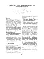
Fig. 1. Comparison of the primary structure
of ATF6 and Arabidopsis bZIP60 (A) and of
yeast IRE1, Arabidopsis IRE1-1 (AtIre1-1)
and Arabidopsis IRE1-2 (AtIre1-2) (B). The
black bar represents the region required for
oligomerization. The dotted bars represent
regions that interact with BiP. TAD, Tran-
scriptional activation domain; TM, trans-
membrane domain; SP, signal peptide.
Arrows indicate the positions cut by S1P
and S2P.
R. Urade Response to unfolded proteins in ER of plants
FEBS Journal 274 (2007) 1152–1171 ª 2007 The Author Journal compilation ª 2007 FEBS 1157
However, because the luminal domain of AtbZIP60 is
much smaller than that of ATF6 (Fig. 1A), it remains
unclear whether it functions as a sensor for ER stress
in a manner similar to ATF6. Investigation into the
cellular localization of AtbZIP60 will probably clarify
these issues.
Orthologs of IRE1 have been identified in Arabidop-
sis (AtIre1-1 and AtIre1-2) and rice (Oryza sativa)
(OsIre1) [86–88]. Fusion proteins of AtIre1-1, AtIre1-2
or OsIre1 with GFP expressed in tobacco By2 cells
localize to the perinuclear ER. The expression patterns
of AtIre1-1 and AtIre1-2 have been examined with
fusion genes of their promoter and a reporter gene.
The expression of AtIre1-1 is restricted to certain tis-
sues at specific developmental stages such as the apical
meristem, the leaf margins where vascular bundles end,
the anthers before pollen is formed, the ovules at an
early stage of development, and the cotyledons imme-
diately after germination. AtIre1-2 is generally
expressed in plants. The C-terminal cytosolic domain
of IRE1ps is conserved among a variety of organisms
(Fig. 1B). The C-terminal halves of recombinant
AtIre1-2 and OsIre1 have autophosphorylation activ-
ity. When Lys442 of AtIre1-2 was mutated to Ala, this
activity was lost. The N-terminal luminal domains of
AtIre1-1, AtIre1-2 and OsIre1 function as ER stress
sensors in yeast cells, although the amino-acid
sequences of these N-terminal domains are dissimilar
from that of yeast IRE1. Thus, when chimeric genes
were created by fusing the N-terminal domains of
AtIre1-1, AtIre1-2 and OsIre1 with the C-terminal
domain of yeast IRE1, and were introduced into a
yeast DIre1 mutant, treatment with tunicamycin no
longer inhibited growth, and treatments with tunica-
mycin or dithiothreitol induced the UPR [86,88].
Yeast and mammalian IRE1 function as a sensor to
ER stress through a process involving homodimeriza-
tion and autophosphorylation. The luminal domain
has a BiP-binding site in a region neighboring the
transmembrane domain, and dissociation and associ-
ation of BiP with this domain regulates the activation
of IRE1 [89–91]. Thus, IRE1 is inactive when its lumi-
nal domain is bound by BiP. Upon accumulation of
unfolded proteins in the ER, BiP is competitively titra-
ted from the luminal domain of IRE1 by the abundant
unfolded proteins in the ER lumen, and IRE1 is acti-
vated. Structural studies of the luminal domains of
yeast and human IRE1 show that dimerization of lu-
minal domain monomers creates a major histocompati-
bility complex-like groove at the interface [92,93].
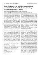
Fig. 2. Model of ER-stress signaling path-
ways in plants. Question marks indicate
incompletely understood relationships.
Response to unfolded proteins in ER of plants R. Urade
1158 FEBS Journal 274 (2007) 1152–1171 ª 2007 The Author Journal compilation ª 2007 FEBS
However, it remains unknown if plant IRE1 orthologs
function as regulators of transcription during ER
stress, but it is possible that BiP plays an important
role in sensing unfolded proteins in the ER, as overex-
pression of BiP in tobacco cells results in a decrease in
the UPR induced by tunicamycin [94].
Plant ER is different from animal ER, in that it is
continuous throughout the entire plant by way of the
plasmodesmata network [95]. Certain stress signals,
such as an attack by a pathogen, are transmitted
throughout the plant, giving rise to systematic induc-
tion of specific genes through this continuity of the
ER. However, the UPR is restricted to the cells where
the stress was initiated and cannot induce a systemic
response in plants, as transcription of BiP mRNA was
found to be restricted to leaves treated with tunica-
mycin [96].
Enhancing cellular quality control
systems by the UPR
Folding
Folding of nascent polypeptides in cells is not as effi-
cient as was once thought. More than 30% of the nas-
cent polypeptides are assumed to be degraded as junk
products before being folded into their proper confor-
mation in the cytosol of animal cells [97]. Nascent
polypeptides produced in the ER are presumed to
undergo a similar fate. However, folding of polypep-
tides translocated into the ER lumen may fail more
often than that of the polypeptides in the cytosol
because these folding events require more complicated
steps such as glycosylation and ⁄ or formation of disul-
fide bonds. Therefore, the UPR is considered to be
weakly but constitutively activated and maintains the
homeostasis of the ER even in apparently unstressed
cells. In particular, developmental events associated
with high secretory activity are predicted to induce the
UPR [98,99]. The quality control of proteins includes
the folding of nascent polypeptide chains into their
native conformation, post-translational modifications
important for proper folding, and the degradation of
misfolded proteins and nonassociated subunit proteins.
Enhancement of folding is accompanied by induction
of ER-localized molecular chaperones and foldases
(PDI-related proteins). In Arabidopsis,mRNAofBiP,
the SIL1 homolog, cyclophilin, GRP94 and PDI-related
proteins are up-regulated by the UPR as described
above. BiP is best characterized by its role in protein
folding and assembly [100,101]. In addition, BiP plays
an essential role in maintaining the permeability bar-
rier of the ER translocon during early stages of protein
translocation [102], targeting misfolded proteins for
proteasomal degradation [103,104], sensing ER stress
[85,89], and contributing to the ER calcium stores
[105]. Most of these functions require its ATPase activ-
ity, where in the ATP-bound state, BiP is in an ‘open’
form that binds and releases unfolded substrates rap-
idly. Hydrolysis of ATP drives it to the ADP-bound or
‘closed’ state, thus stabilizing its association with
unfolded proteins. The release of ADP and the rebind-
ing of ATP reopens the substrate-binding domain to
release and fold the nascent protein. SIL1 is a cochap-
erone of BiP and regulates its ATPase cycle by stimu-
lating ATP hydrolysis and accelerating the ADP–ATP
exchange [106].
Proline can exist in either the cis or trans form in
a polypeptide chain, and its orientation dramati-
cally influences the secondary structure of the protein.
Peptidyl-prolyl-cis-trans isomerases (cyclophilin) survey
the status of the proline residues and rearrange them
from the cis to the trans form to ensure proper folding
of the nascent polypeptide chains. Twenty-nine genes
encoding cyclophilin family members are present in the
Arabidopsis genome, and five gene products are
assumed to be targeted to the ER lumen with N-ter-
minal signal peptides [107]. Among them, ATCYP20-1
is up-regulated during ER stress, and contains a
domain essential for peptidyl-prolyl-cis-trans isomerase
activity.
Four PDI-related genes are up-regulated during ER
stress. PDI catalyzes the formation and rearrangement
of disulfide bonds between correct pairs of Cys resi-
dues in nascent polypeptide chains in the ER [108].
PDI and related proteins are characterized by thiore-
doxin motifs within their primary structure [109,110];
Arabidopsis PDI-related proteins, the expression of
which is induced during ER stress, have two of these
motifs. A comprehensive search of the Arabidopsis gen-
ome identified 22 orthologs of known PDI-like pro-
teins [111]. PDI purified from plants or recombinant
PDI-related proteins expressed in Escherichia coli have
protein disulfide oxidoreductase activity [38,112–116],
and their importance in protein folding has been dem-
onstrated in rice endosperm [117]. In endosperm of rice
esp2 mutants lacking PDI, a precursor of the storage
protein proglutelin forms aggregates with other storage
proteins via interchain disulfide bonds within the
ER lumen, whereas in wild-type rice, proglutelins are
processed normally into acidic and basic subunits and
accumulate in protein storage vacuoles. In soybean
cotyledon, PDI-related proteins GmPDIS-1 (an ortho-
log of At2g47470) [116] associates with a precursor
of the storage protein glycinin in the ER, suggesting
that the PDI-related protein participates in glycinin
R. Urade Response to unfolded proteins in ER of plants
FEBS Journal 274 (2007) 1152–1171 ª 2007 The Author Journal compilation ª 2007 FEBS 1159
folding. Yeast and mammalian PDI are activated
by the FAD-dependent oxidases ERO1 and Erv2p
[118–121]. Similarly, the Arabidopsis genome encodes
an ERO1 homolog, At2g38960, and an Erv2p homo-
log, At1g15020 or At2g01270, but so far the plant
varieties have not been characterized. Mammalian PDI
not only folds polypeptides, but it also aggregates
unfolded proteins via disulfide bonds for retention in
the ER lumen [122], and reduces aggregated proteins
before retro-translocation into the cytosol for degrada-
tion [123]. No evidence for the function of PDI pro-
teins in plants has been reported.
The high-capacity calcium-binding proteins, calnexin
(an ER transmembrane protein) [124,125] and calreticu-
lin (an ER luminal protein) [126,127], are molecular
chaperones in mammalian cells specific for unfolded
N-glycosylated proteins [128]. The first step in the
N-glycosylation of a protein is the transfer of a core
glycan Glc
3
Man
9
GlucNac
2
from a membrane-bound
dolichol phosphate anchor to consensus Asn-X-Ser ⁄
Thr residues in the polypeptide chain. The glucose resi-
dues on the transferred core glycan are sequentially
trimmed to Glc
1
Man
9
GlucNac
2
by b-glucosidase I and
b-glucosidase II. The monoglucosylated glycan on the
polypeptide chain is trapped by calnexin or calreticulin
to protect it from degradation, resulting in retention of
the polypeptide in the ER for folding [129,130].
The monoglucosylated form of the unfolded protein
shuttles through cycles of deglucosylation by b-glucosi-
dase II and reglucosylation by UDP-glucose–glycopro-
tein glucosyltransferase (UGGT), which preferentially
recognizes unfolded glucosylated glycoproteins [131]. This
process is called the calnexin ⁄ calreticulin cycle, and is
one arm of the quality control machinery in the mam-
malian ER. It is possible that interaction between
monoglucosylated N-glycan with calnexin ⁄ calreticulin
functions for the quality control of N-glycosylated pro-
teins in plants, although the calnexin ⁄ calreticulin cycle
remains to be elucidated in plants. However, circum-
stantial evidence supports the idea that the calnexin ⁄
calreticulin cycle is present in plant cells [132]. For
example, it has been shown in in vitro translation sys-
tems with wheat germ extract and bean microsomes that
the rate of phaseolin assembly is accelerated when a glu-
cosidase inhibitor is included to stop glucose trimming
of the N-glycan [133]. In this system, phaseolin with par-
tially trimmed glycans was unable to assemble into trim-
ers, probably because of being trapped by calnexin or
calreticulin. In kaiware radish (Raphanus sativus), the
glucosidase inhibitors castanospermine and deoxynojiri-
mycin suppressed the growth of seedlings by inhibiting
glucose trimming of the N-glycan [134,135], and, in
Arabidopsis, homozygous deletion of b-glucosidase I by
T-DNA tagging is lethal [136]. In potato, curled leaves
and low yields have been reported when expression of
the b-glucosidase II gene MAL1 was knocked-down by
antisense RNA [137]. Furthermore, the knock-down of
MAL1 caused an increase in the expression of BiP, sug-
gesting the presence of ER stress. In Arabidopsis rsw3, a
temperature-sensitive mutant of the b-glucosidase II
b-subunit, some morphological abnormalities and
growth impairments were observed [138]. As trimming
glucose residues of N-glycan by b-glucosidase I and
b-glucosidase II is a prerequisite for modification of the
ER-type glycan to the complex glycan in Golgi bodies,
it is possible that the impairment of this process is
responsible for the adverse effects on plant morphology.
However, this explanation may be unlikely, as neither
growth inhibition nor reproduction defects have been
observed in Arabidopsis mutants defective in GlcNAc-
transferase I, which catalyzes the first modification reac-
tion to the complex-type glycan [139].
UDP-glucose, the substrate for re-glucosylation of
N-glycan by UGGT, is synthesized in the cytosol, indi-
cating that a UDP-glucose transporter would be
required for the calnexin ⁄ calreticulin cycle. AtUTr1
from Arabidopsis is an ER-localized membrane pro-
tein, the expression of which is induced by treatment
with dithiothreitol [140], and is recognized as a UDP
galactose ⁄ glucose transporter [141]. In addition, up-
regulation of the ER chaperones, BiP and calnexin,
has been observed in an AtUTr1 insertional mutant,
suggesting that these plants may constitutively activate
the UPR. Taken together, it is possible that the calnex-
in ⁄ calreticulin cycle discriminates between folded and
unfolded glycoproteins in plant cells. In mammalian
cells, the recognition of the unfolded glycoproteins by
calnexin ⁄ calreticulin is coupled with the formation of
disulfide bonds, where the PDI-related thiol-oxidore-
ductase, ER-60 ⁄ ERp57, interacts with the P domain of
calnexin or calreticulin to fold N-glycosylated proteins
[142–144]. The amino-acid sequence of the P domain
of plant calnexin and calreticulin is highly conserved
compared with that of its mammalian counterparts
[145,146]. However, it is not known whether plant
calnexin or calreticulin cooperates with any plant PDI-
related oxidoreductase to form disulfide bonds in
N-glycosylated proteins.
Degradation of unfolded proteins
Unfolded proteins generated in the rough ER are
predominantly degraded by ERAD in yeast and
mammalian cells [147], requiring that the unfolded
polypeptides be transported across the ER membrane
into the cytosol via a translocon located on the ER
Response to unfolded proteins in ER of plants R. Urade
1160 FEBS Journal 274 (2007) 1152–1171 ª 2007 The Author Journal compilation ª 2007 FEBS
membrane [148] to be degraded by the cytoplasmic
ubiquitin-proteasome system (UPS) [149].
In plants, misfolded storage proteins generated in
the ER are degraded by an unidentified system
[150,151]. However, it has been proposed that both
ERAD and a vacuolar system may degrade the unfol-
ded proteins generated in the rough ER, although the
details of this mechanism have not been established.
In plants, UPS-dependent and UPS-independent
ERAD-like degradation have been observed. Ricin is a
heterodimeric ribosome-inactivating protein that accu-
mulates in castor beans (Ricinus communis). The
mature ricin comprises a catalytic A chain and a B
chain linked by a single disulfide bond. The ER-tar-
geted A chain is degraded by a pathway that closely
resembles ERAD when expressed in tobacco proto-
plasts in the absence of a B chain [152]. The degrada-
tion of ricin A chain is brefeldin A-insensitive and is
inhibited by the proteasome inhibitor clasto-lactacystin
b-lactone, resulting in the accumulation of ricin
A chains. These stabilized ricin A chains are partly
deglycosylated by a peptide–N-glycanase-like activity.
Taken together, these results indicate that the ricin A
chain behaves as a substrate of the ERAD where it is
exported into the cytosol, deglycosylated, and degra-
ded by the proteasome [153,154]. A mutant of barley
(Hordeum vulgare) mildew resistance O protein-1 is
also degraded by UPS-dependent ERAD in plants
[155]. Individual mutant mildew resistance O protein-1
proteins with single amino-acid substitutions in its
seven-transmembrane domain exhibit markedly
reduced half-lives, are polyubiquitinated, and can be
stabilized through inhibition of proteasome activity.
When the mutant mildew resistance O protein-1 is
transfected into Arabidopsis plants previously transfected
with dominant negative mutants of the putative AAA
ATPase AtCDC48A ⁄ p97 (a component of the ERAD
machinery) [156,157], the degradation of the mutant
mildew resistance O protein-1 is impaired. This
strongly suggests that mildew resistance O protein-1 is
an endogenous substrate of a UPS-dependent ERAD-
related quality control mechanism in plants.
In plants, several misfolded proteins are translocated
across the ER membrane to the cytosol and degraded
by an unknown UPS-independent system. The C-ter-
minal extension mutant of phaseolin transfected into
tobacco protoplasts is degraded very rapidly in a bre-
feldin A- and proteasome inhibitor-insensitive manner
[158], suggesting that it is performed in a pre-Golgi
compartment, probably in the cytosol. Likewise, when
both endogenous and recombinant cell wall invertases
are synthesized without their N-glycans in BY2
tobacco cells, they both degrade very rapidly [159].
This degradation does not occur in an acidic com-
partment and is also insensitive to brefeldin A and
proteasome inhibitor. Furthermore, a fusion protein
consisting of misfolded N-terminally truncated calreti-
culin with GFP is also retrotranslocated from the ER
lumen to the cytosol and is subsequently degraded
[160,161]. The dislocated fusion proteins accumulate in
the nucleoplasm in a microtubule-dependent manner
and are degraded very slowly by an unknown UPS-
independent system. These UPS-independent ERAD-
like degradations are unique in plants. However, any
underlying molecular mechanism of the system remains
unknown.
Some genes relevant to the translocation of misfolded
proteins across the ER membrane into the cytosol are
induced during ER stress in Arabidopsis (Table 1).
SEC61 subunits form the specific translocon required
for retro-translocation of misfolded polypeptides [162].
Stress-associated ER protein 1 (SERP1) ⁄ Ribosome-
associated membrane protein 4 (RAMP4) orthologs are
also up-regulated during ER stress. SERP1 ⁄ RAMP4
interacts with the SEC61 a-subunit, the SEC61 b-sub-
unit, and calnexin [163,164]. This complex stabilizes
membrane proteins in the ER membrane through a
translocational pausing mechanism [165]. P58
IPK
was
previously implicated in translational control (described
below). Recently, the novel role of mammalian P58
IPK
in the control of the translocation of newly synthesized
polypeptides to the ER lumen was reported by
Oyadomari et al. [166]. P58
IPK
associates with SEC61,
recruits HSP70 chaperones to the cytosolic face of
SEC61 and associates with translocating polypeptides
during ER stress. In P58
IPK
-knockout mice, cells with a
high secretory burden are markedly compromised in
their ability to cope with ER stress. On the basis of
these results, P58
IPK
is thought to be a key mediator
of cotranslocational ER protein degradation, and
probably contributes to ER homeostasis in stressed
cells.
Genes that stimulate vesicle transport from the ER
to the cis-Golgi are induced during ER stress in Ara-
bidopsis (Table 1). Among them, EMP24, SAR1B and
SEC23 are shown to make a complex with subunits of
the COPII coat, which are key molecules for export of
proteins from the ER, and promote transport of newly
synthesized proteins from the ER into ER subdomains
or Golgi in yeast [167–170]. Newly synthesized proteins
that do not fold correctly in the ER are targeted for
ERAD through distinct sorting mechanisms; soluble
luminal ERAD substrates require ER–Golgi transport
and retrieval for degradation, whereas transmembrane
ERAD substrates are retained in the ER [169].
Retained transmembrane proteins are often seques-
R. Urade Response to unfolded proteins in ER of plants
FEBS Journal 274 (2007) 1152–1171 ª 2007 The Author Journal compilation ª 2007 FEBS 1161
tered into ER subdomains containing BiP. Sequestra-
tion and degradation of membrane proteins is disrupted
in a mutant yeast strain lacking guanine-nucleotide
exchange factor SAR1, SEC23 or SEC13 [170]. There-
fore, it has been proposed that SAR1 ⁄ COPII-mediated
sorting of membrane proteins into ER subdomains is
essential for its entry into the proteasomal degradation
pathway. In plants, a similar sorting system for mem-
brane proteins is presumed to function.
In the plant UPS-dependent ERAD system, HRD1
complex-like machinery may play an important role in
the elimination of misfolded proteins. Putative ortho-
logs of the constituents of a yeast ERAD system,
HRD1, HRD3 ⁄ SEL-1 L, DER1 and YOS9, are
induced during ER stress in Arabidopsis (Table 1).
These components constitute the HRD1 complex,
which functions in recognition and ubiquitination of
proteins with misfolded ER-luminal domains (ERAD-L)
and proteins with misfolded intramembrane domains
(ERAD-M) in yeast [171–173]. HRD1 is an E3 ubiqu-
itin ligase, specialized for ERAD-L and ERAD-M,
which catalyzes the final reaction of ubiquitination of
misfolded proteins. HRD1 is stabilized by forming a
complex with HRD3 ⁄ SEL-1 L [174]. HRD3 ⁄ SEL-1 L
is a type I transmembrane protein equipped with a
large luminal domain that recognizes proteins that
deviate from their native conformation [173]. DER1 is
a small, membrane-bound protein, the function of
which remains unclear, but its deletion abolishes
degradation of misfolded proteins in yeast [175].
Remarkably, maize DER1-like gene (Zm Derlins)is
capable of functionally complementing a yeast DER1
deletion mutant [176]. YOS9 is a member of the OS-9
protein family and shows similarity to mannose-6-
phosphate receptors. It is an essential component for
degradation of misfolded ER-luminal glycoproteins
[177], and specifically associates with misfolded ERAD
substrates [171].
ERAD is considered to be the primary disposal
route for unfolded and misfolded proteins, but grow-
ing evidence suggests a vacuolar role in protein quality
control. Even in plants, the vacuolar system is involved
in the degradation of misfolded proteins generated in
the ER. Pimpl et al. [178] demonstrated that BiP is
constitutively transported to the vacuole in a wortmannin-
sensitive manner in tobacco, and that it could play an
active role in this second disposal route for misfolded
proteins. ER export of BiP to the Golgi apparatus is
dependent on COPII. BiP is transported to the lytic
vacuole via multivesicular bodies, which represent the
plant prevacuolar compartment. When the plant is
treated with tunicamycin, a subset of BiP-unfolded
protein complexes is transported to the vacuole and
degraded. As this degradation process is very rapid,
the transported BiP–ligand complexes in the vacuole
are not detected under normal circumstances. When
the route from the Golgi apparatus to vacuoles is
blocked in the presence of wortmannin, BiP–ligand
complexes are secreted into the medium and are subse-
quently detected. In tobacco seeds, a misfolded phase-
olin mutant is degraded in vacuole-derived organelles,
protein storage vacuoles [179]. Vacuolar disposal may
function with ERAD to maximize the quality control
of proteins in the secretory pathway. It is not known
whether the vacuolar function is enhanced by the UPR
in plants.
Other UPR in plants
The UPR is composed of three steps in mammalian
cells: enhancement of the folding and degradation of
unfolded proteins (described above), attenuation of
translation, and apoptosis. ER stress causes transla-
tional arrest through phosphorylation of eIF2a (Ser51)
by PERK, which senses ER stress through its luminal
domain and leads to the degradation of ER-localized
mRNAs by IRE1 [16,21,29]. In plants, however, a
PERK ortholog has yet to be described, and an
increase in phosphorylation of eIF2a (Ser51) and
attenuation of translation has not been confirmed dur-
ing ER stress [49]. Mammalian P58
IPK
is an inhibitor
of PERK [180] which is induced at a later phase of
ER stress by the XBP-1 signal transduction pathway
[58]. Because deletion of P58
IPK
increases the amount
of phosphorylated eIF2a in the cell [58], it is thought
to function as a feedback regulator of translation in
the later phase of ER stress. In Arabidopsis, the P58
IPK
gene is up-regulated and the phosphorylation of eIF2a
(Ser51) is partially inhibited by ER stress [49], but
translation as a whole is not affected. Induction of
Arabidopsis P58
IPK
and a subsequent decrease in phos-
phorylation of eIF2a (Ser51) may increase the transla-
tional efficiency of unidentified gene(s). Alternatively,
induction of P58
IPK
could be required for the cotrans-
locational degradation of ER proteins in an effort to
maintain the homeostasis of the ER as described
above.
The idea that programmed cell death (PCD) func-
tions during the UPR in plants is supported by several
lines of indirect evidence. van Doorn & Woltering
[181] categorized plant PCD into three morphological
types, including apoptotic-like PCD, autophagy, and
nonlysosomal PCD. In cultured sycamore (Acer pseudo-
platanus L) cells, treatment with tunicamycin induced
apoptotic PCD, as indicated by nuclear morphology
and DNA fragmentation [182,183]. In cultured soy-
Response to unfolded proteins in ER of plants R. Urade
1162 FEBS Journal 274 (2007) 1152–1171 ª 2007 The Author Journal compilation ª 2007 FEBS
bean cells, inhibition of ER-type IIA Ca
2+
-pumps by
cyclopiazonic acid induced ER stress and PCD [184].
However, the regulatory mechanism that underlies
apoptotic-like PCD induced during ER stress remains
unclear. Two apoptotic-like PCD-related genes, BAX
inhibitor 1 [49] and Hsr203J [185], have been identified
as UPR genes. BAX inhibitor 1 is a conserved integral
membrane protein localized in the ER that is a pro-
apoptotic member of the multidomain Bcl2 family
[56,57]. In mammalian cells, BAX inhibitor 1 affords
protection from apoptosis induced by ER stress by
inhibiting the activation of BAX and its translocation
to the mitochondria, by preserving the mitochondrial
membrane potential, and by suppressing caspase acti-
vation [186]. Plant BAX inhibitor 1 is induced by stres-
sors such as wounding and infection with pathogens
[187]. It also suppresses fungal elicitor-induced apop-
totic PCD in rice and barley [188,189]. Therefore,
BAX inhibitor 1 is thought to be one of the key fac-
tors required for regulation of plant apoptotic PCD.
However, BAX, Bcl2 and their relatives have not been
found in plants, and the underlying mechanism of
BAX inhibitor 1 remains unknown. The ERSE-like
cis-acting regulatory element is found in the promoter
region of Arabidopsis BAX inhibitor 1 gene (Table 1),
suggesting that BAX inhibitor 1 may be induced by
the AtZIP60 signal-transduction system during ER
stress.
Hsr203J is a PCD-related serine hydrolase that is
induced by ER stress and is traditionally used as a
marker for PCD [190,191]. Accumulation of Hsr203J
mRNA begins at 10 h and plateaus at 24 h after
treatment with tunicamycin, whereas accumulation of
BiP and PDI mRNA begins 2 h after treatment with
tunicamycin [185]. This suggests that transcription of
Hsr203J mRNA is induced by a signal-transduction
system different from the UPR governing the induc-
tion of molecular chaperones during ER stress. Taken
together, these data suggest that apoptotic PCD is
induced in plants when ER homeostasis is not restored
after stress.
Future perspectives
Plant ER is an extremely flexible and adaptable organ-
elle, which differentiates into various types of organelle
to cope with internal and external stresses and to con-
tain the enormous number of proteins that are actively
synthesized there [192–194]. Therefore, the UPR that is
unique to plants is expected to function widely,
although the molecular mechanisms underlying the
UPR system in plants, animals, and yeast share com-
mon components. This is supported by the fact that a
number of plant-specific genes are induced by ER
stress, but the functional significance of their induction
has not yet been established. Recent studies in yeast
and mammals have highlighted the importance of the
UPR in nutrient sensing and control of differentiation
[11,32,33]. In diploid yeast, nitrogen starvation inhibits
HAC1 splicing and induces pseudohyphal growth. As
this phenomenon is repressed in strains defective in the
UPR, the latter is thought to have an important
underlying role in differentiation depending on nutri-
tional conditions. Many data also support a role for
the UPR in the control of nutritional and differenti-
ation programs in the mammalian system. Under con-
ditions of low glucose concentration, translation of
proinsulin in pancreatic b-cells is repressed by activa-
tion of PERK, and the UPR controls the terminal dif-
ferentiation of B-cells into antibody-secreting plasma
cells. In plants, abundant unfolded storage proteins are
loaded into the ER during seed development, where
the UPR is presumed to enhance the ability of the ER
to fold these proteins [195]. However, there is currently
no experimental confirmation of this, and the role of
the UPR in seed development remains to be explored
in greater detail.
The ER stress-regulated genes identified by the
DNA microarray analyses described in this review are
valuable for understanding the plant UPR. However,
these analyses may have identified either genes primar-
ily regulated under the UPR or genes regulated by
other signal-transduction systems cross-talking with
the UPR. Isolation of mutants deficient in sensor pro-
teins and transcription factors that function in UPR
signal transduction will provide valuable tools for fur-
ther study of the plant UPR.
Acknowledgements
The author thanks Dr Makoto Kito, Emeritus Profes-
sor of Kyoto University, for critical reading of the
manuscript, valuable advice, and warm encourage-
ment.
References
1 Travers KJ, Patil CK, Wodicka L, Lockhart DJ,
Weissman JS & Walter P (2000) Functional and geno-
mic analyses reveal essential coordination between the
unfolded protein response and ER-associated degrada-
tion. Cell 101, 249–258.
2 Cox JS, Shamu CE & Walter P (1993) Transcriptional
induction of genes encoding endoplasmic reticulum
resident-proteins requires a transmembrane protein
kinase. Cell 73, 1197–1206.
R. Urade Response to unfolded proteins in ER of plants
FEBS Journal 274 (2007) 1152–1171 ª 2007 The Author Journal compilation ª 2007 FEBS 1163
3 Mori K, Ma W, Gething M-J & Sambrook JF (1993)
A transmembrane protein with cdc2+ ⁄ CDC28-related
kinase activity is required for signaling from the ER to
the nucleus. Cell 74, 743–756.
4 Cox JS & Walter P (1996) A novel mechanism for
regulating the activity of a transcription factor that
controls the unfolded protein response. Cell 87, 391–
404.
5 Mori K, Kawahara T, Yoshida H, Yanagi H & Yura T
(1996) Signalling from the endoplasmic reticulum to the
nucleus: transcription factor with a basic-leucine zipper
motif is required for the unfolded protein-response path-
way. Genes Cells 1, 803–817.
6 Nikawa J, Akiyoshi M, Hirata S & Fukuda T (1996)
Saccharomyces cerevisiae IRE2 ⁄ HAC1 is involved in
IRE1-mediated KAR2 expression. Nucleic Acids Res
24, 4222–4226.
7 Kohno K, Normington K, Sambrook J, Gething M-J
& Mori K (1993) The promoter region of the yeast
KAR2 (BiP) gene contains a regulatory domain that
responds to the presence of unfolded proteins in the
endoplasmic reticulum. Mol Cell Biol 13, 877–890.
8 Mori K, Ogawa N, Kawahara T, Yanagi H & Yura T
(1998) Palindrome with a spacer of one nucleotide is
characteristic of the cis-acting unfolded protein
response element in Saccharomyces cerevisiae. J Biol
Chem 273, 9912–9920.
9 Mori K, Sant A, Kohno K, Normington K, Gething
MJ & Sambrook JF (1992) A 22 bp cis-acting element
is necessary and sufficient for the induction of the
yeast KAR2 (BiP) gene by unfolded proteins. EMBO J
11, 2583–2593.
10 Harding PH, Calfon M, Urano F, Novoa I & Ron D
(2002) Transcriptional and translational control in the
mammalian unfolded protein response. Annu Rev Cell
Dev Biol 18, 575–599.
11 Schro
¨
der M & Kaufman RJ (2005) The mammalian
unfolded response. Annu Rev Biochem 74, 739–789.
12 Brewer JW & Diehl JA (2000) PERK mediates cell-
cycle exit during the mammalian unfolded protein
response. Proc Natl Acad Sci USA 97, 12625–12630.
13 Tirasophon W, Welihinda AA & Kaufman RJ (1998)
A stress response pathway from the endoplasmic reti-
culum to the nucleus requires a novel bifunctional pro-
tein kinase ⁄ endoribonuclease (Ire1p) in mammalian
cells. Genes Dev 12, 1812–1824.
14 Bertolotti A & Ron D (2001) Alterations in an IRE1-
RNA complex in the mammalian unfolded protein
response. J Cell Sci 114, 3207–3212.
15 Yoshida H, Haze K, Yanagi H, Yura T & Mori K
(1998) Identification of the cis-acting endoplasmic
reticulum stress response element responsible for trans-
criptional induction of mammalian glucose-regulated
proteins. Involvement of basic leucine zipper transcrip-
tion factors. J Biol Chem 273, 33741–33749.
16 Harding HP, Zhang Y & Ron D (1999) Protein trans-
lation and folding are coupled by an endoplasmic-
reticulum-resident kinase. Nature 397, 271–274.
17 Wang XZ, Harding HP, Zhang Y, Jolicoeur EM,
Kuroda M & Ron D (1998) Cloning of mammalian
Ire1 reveals diversity in the ER stress responses.
EMBO J 17, 5708–5717.
18 Calfon M, Zeng H, Urano F, Till JH, Hubbard SR,
Harding HP, Clark SG & Ron D (2002) IRE1 couples
endoplasmic reticulum load to secretory capacity by
processing the XBP-1 mRNA. Nature 415, 92–96.
19 Yoshida H, Matsui T, Yamamoto A, Okada T &
Mori K (2001) XBP1 mRNA is induced by ATF6 and
spliced by IRE1 in response to ER stress to produce a
highly active transcription factor. Cell 107, 881–891.
20 Lee AH, Iwakoshi NN & Glimcher LH (2003) XBP-1
regulates a subset of endoplasmic reticulum-resident
chaperone genes in the unfolded protein response. Mol
Cell Biol 23, 7448–7459.
21 Hollien J & Weissman JS (2006) Decay of endoplasmic
reticulum-localized mRNAs during the unfolded pro-
tein response. Science 313, 104–107.
22 Yoneda T, Imaizumi K, Oono K, Yui D, Gomi F,
Katayama T & Tohyama M (2001) Activation of cas-
pase-12, an endoplastic reticulum (ER) resident cas-
pase, through tumor necrosis factor receptor-associated
factor 2-dependent mechanism in response to the ER
stress. J Biol Chem 276 , 13935–13940.
23 Nishitoh H, Matsuzawa A, Tobiume K, Saegusa K,
Takeda K, Inoue K, Hori S, Kakizuka A & Ichijo H
(2002) ASK1 is essential for endoplasmic reticulum
stress-induced neuronal cell death triggered by expanded
polyglutamine repeats. Genes Dev 16, 1345–1355.
24 Haze K, Yoshida H, Yanagi H, Yura T & Mori K
(1999) Mammalian transcription factor ATF6 is
synthesized as a transmembrane protein and activated
by proteolysis in response to endoplasmic reticulum
stress. Mol Biol Cell 10, 3787–3799.
25 Okada T, Haze K, Nadanaka S, Yoshida H, Seidah
NG, Hirano Y, Sato R, Negishi M & Mori K (2003)
A serine protease inhibitor prevents endoplasmic reti-
culum stress-induced cleavage but not transport of the
membrane-bound transcription factor ATF6. J Biol
Chem 278, 31024–31032.
26 YeJ, Rawson RB, Komuro R, Chen X, Dave
´
UP,
Prywes R, Brown MS & Goldstein JL (2000) ER stress
induces cleavage of membrane-bound ATF6 by the
same proteases that process SREBPs. Mol Cell 6,
1355–1364.
27 Haze K, Okada T, Yoshida H, Yanagi H, Yura T,
Negishi M & Mori K (2001) Identification of the G13
(cAMP-response-element-binding protein-related pro-
tein) gene product related to activating transcription
factor 6 as a transcriptional activator of the mamma-
lian unfolded protein response. Biochem J 355, 19–28.
Response to unfolded proteins in ER of plants R. Urade
1164 FEBS Journal 274 (2007) 1152–1171 ª 2007 The Author Journal compilation ª 2007 FEBS
28 Yoshida H, Okada T, Haze K, Yanagi H, Yura T,
Negishi M & Mori K (2000) ATF6 activated by proteo-
lysis binds in the presence of NF-Y (CBF) directly to the
cis-acting element responsible for the mammalian
unfolded protein response. Mol Cell Biol 20, 6755–6767.
29 Harding HP, Zhang Y, Bertolotti A, Zeng H & Ron D
(2000) Perk is essential for translational regulation and
cell survival during the unfolded protein response. Mol
Cell 5, 897–904.
30 Harding HP, Zhang Y, Zeng H, Novoa I, Lu PD,
Calfon M, Sadri N, Yun C, Popko B, Paules R et al.
(2003) An integrated stress response regulates amino
acid metabolism and resistance to oxidative stress. Mol
Cell 11, 619–633.
31 Ma Y, Brewer JW, Diehl JA & Hendershot LM (2000)
Two distinct stress signaling pathways converge upon
the CHOP promoter during the mammalian unfolded
protein response. J Mol Biol 318, 1351–1365.
32 Schro
¨
der M, Chang JS & Kaufman RJ (2000) The
unfolded protein response represses nitrogen-starvation
induced developmental differentiation in yeast. Genes
Dev 14, 2962–2975.
33 Kaufman RJ, Scheuner D, Schroder M, Shen X, Lee
K, Liu CY & Arnold SM (2002) The unfolded protein
response in nutrient sensing and differentiation. Nat
Rev Mol Cell Biol 3, 411–421.
34 Gass JN, Gifford NM & Brewer JW (2002) Activation
of an unfolded protein response during differentiation
of antibody-secreting B cells. J Biol Chem 277, 49047–
49054.
35 Kondo S, Murakami T, Tatsumi K, Ogata M, Kanemo-
to S, Otori K, Iseki K, Wanaka A & Imaizumi K (2005)
OASIS, a CREB ⁄ ATF-family member, modulates UPR
signalling in astrocytes. Nat Cell Biol 7, 186–194.
36 Chin KT, Zhou HJ, Wong CM, Lee JM, Chan CP,
Qiang BQ, Yuan JG, Ng IO & Jin DY (2005) The
liver-enriched transcription factor CREB-H is a growth
suppressor protein underexpressed in hepatocellular
carcinoma. Nucleic Acids Res 33, 1859–1873.
37 Nagamori I, Yabuta N, Fujii T, Tanaka H, Yomogida
K, Nishimune Y & Nojima H (2005) Tisp40, a sperma-
tid specific bZip transcription factor, functions by
binding to the unfolded protein response element via
the Rip pathway. Genes Cells 10, 575–594.
38 Shorrosh BS & Dixon RA (1991) Molecular cloning of
a putative plant endomembrane protein resembling
vertebrate protein disulfide-isomerase and a phosphati-
dylinositol-specific phospholipase C. Proc Natl Acad
Sci USA 88, 10941–10945.
39 D’Amico L, Valsasina B, Daminati MG, Fabbrini MS,
Nitti G, Bollini R, Ceriotti A & Vitale A (1992) Bean
homologs of the mammalian glucose-regulated pro-
teins: induction by tunicamycin and interaction with
newly synthesized seed storage proteins in the endo-
plasmic reticulum. Plant J 2, 443–455.
40 Wrobel RL, O’Brian GR & Boston RS (1997) Com-
parative analysis of BiP gene expression in maize endo-
sperm. Gene 204, 105–113.
41 Oliver SC, Venis MA, Freedman RB & Napier RM
(1995) Regulation of synthesis and turnover of maize
auxin-binding protein and observations on its passage
to the plasma membrane: comparisons to maize immu-
noglobulin-binding protein cognate. Planta 197, 465–
474.
42 Denecke J, Carlsson LE, Vidal S, Hs
ˇ
glund A, Ek B,
van Zeijl MJZ, Sinjorgo KMC & Palva ET (1995) The
tabacco homolog of mammalian calreticulin is present
in protein complexes in vivo. Plant Cell 7, 391–406.
43 Cascardo JCM, Almeida RS, Buzeli RAA, Carolino
SMB, Otoni WC & Fontes EPB (2000) The phosphry-
lation state and expression of soybean BiP isoforms
are differentially regulated following abiotic stresses.
J Biol Chem 275, 14494–14500.
44 Koizumi N, Ujino T, Sano H & Chrispeels MJ (1999)
Overexpression of a gene that encodes the first enzyme
in the biosynthesis of asparagine-linked glycans makes
plants resistant to tunicamycin and obviates the tunica-
mycin-induced unfolded response. Plant Physiol 121,
353–361.
45 Shank KJ, Su P, Brglez I, Boss WF, Dewey RE &
Boston RS (2001) Induction of lipid metabolic enzymes
during the endoplasmic reticulum stress response in
plants. Plant Physiol 126, 267–277.
46 Lehrman MA (1991) Biosynthesis of N-acetylglucosa-
mine-P-P-dolicol, the committed step of asparagine-
linked oligosaccharide assembly. Glycobiology 1, 553–
562.
47 Lee AS (1987) Coordinated regulation of a set of genes
by glucose and calcium ionophores in mammalian
cells. Trends Biochem Sci 12, 20–23.
48 Martı
`
nez IM & Chrispeels MJ (2003) Genomic analysis
of the unfolded protein response in Arabidopsis shows
its connection to important cellular processes. Plant
Cell 15, 561–576.
49 Kamauchi S, Nakatani H, Nakano C & Urade R
(2005) Gene expression in response to endoplasmic
reticulum stress in Arabidopsis thaliana. FEBS J 272,
3461–3476.
50 Brenner S, Williams SR, Vermaas EH, Storck T,
Moon K, McCollum C, Mao J-I, Luo S, Kirchner JJ,
Eletr S et al. (2000) In vitro cloning of complex mix-
tures of DNA on microbeads: physical separation of
differentially expressed cDNAs. Proc Natl Acad Sci
USA 97, 1665–1670.
51 Ng DTW, Spear ED & Walter P (2000) The unfolded
protein response regulates multiple aspects of secretory
and membrane protein biogenesis and endoplasmic
quality control. J Cell Biol 150, 77–88.
52 Scheuner D, Song B, McEwen E, Liu C, Laybutt R,
Gillespie P, Saunders T, Bonner-Weir S & Kaufman RJ
R. Urade Response to unfolded proteins in ER of plants
FEBS Journal 274 (2007) 1152–1171 ª 2007 The Author Journal compilation ª 2007 FEBS 1165
(2001) Translational control is required for the unfolded
protein response and in vivo glucose homeostasis. Mol
Cell 7, 1165–1176.
53 Okada T, Yoshida H, Akazawa R, Negishi M &
Mori K (2002) Distinct roles of activating transcription
factor 6 (ATF6) and double-stranded RNA-activated
protein kinase-like endoplasmic reticulum kinase
(PERK) in transcription during the mammalian
unfolded protein response. Biochem J 366, 585–594.
54 Lee AH, Iwakoshi NN & Glimcher LH (2003) XBP-1
regulates a subset of endoplasmic reticulum resident
chaperone genes in the unfolded protein response. Mol
Cell Biol 23, 7448–7459.
55 Bilgin DD, Liu Y, Schiff M & Dinesh-Kumar SP
(2003) P58
IPK
, a plant ortholog of double-stranded
RNA-dependent protein kinase PKR inhibitor, func-
tions in viral pathogenesis. Dev Cell 4, 651–661.
56 Bolduc N, Ouellet M, Pitre F & Brisson LF (2003)
Molecular characterization of two plant BI-1 homolo-
gues which suppress Bax-induced apoptosis in human
293 cells. Planta 216, 377–386.
57 Kawai-Yamada M, Jin L, Yoshinaga K, Hirata A &
Uchimiya H (2001) Mammalian Bax-induced plant
cell death can be down-regulated by overexpression of
Arabidopsis Bax Inhibitor-1 (AtBI-1). Proc Natl Acad
Sci USA 98, 12295–12300.
58 van Huizen R, Martindale JL, Gorospe M &
Holbrook NJ (2003) P58
IPK
, a novel endoplasmic
reticulum stress-inducible protein and potential nega-
tive regulator of eIF2a signaling. J Biol Chem 278,
15558–15564.
59 Andreasson E, Jenkins T, Brodersen P, Thorgrimsen S,
Petersen NH, Zhu S, Qiu JL, Micheelsen P, Rocher A,
Petersen M et al. (2005) The MAP kinase substrate
MKS1 is a regulator of plant defense responses.
EMBO J 24, 2579–2589.
60 Delessert C, Kazan K, Wilson IW, Van Der Straeten
D, Manners J, Dennis ES & Dolferus R (2005) The
transcription factor ATAF2 represses the expression of
pathogenesis-related genes in Arabidopsis. Plant J 43,
745–757.
61 Davletova S, Schlauch K, Coutu J & Mittler R (2005)
The zinc-finger protein Zat12 plays a central role in
reactive oxygen and abiotic stress signaling in Arabi-
dopsis. Plant Physiol 139, 847–856.
62 Iwata Y & Koizumi N (2005) An Arabidopsis tran-
scription factor, AtbZIP60 regulates the endoplasmic
reticulum stress response in a manner unique to plants.
Proc Natl Acad Sci USA 102, 5280–5285.
63 Gillikin JW, Zhang F, Coleman CE, Bass HW,
Larkins BA & Boston RS (1997) A defective signal
peptide tethers the floury-2 zein to the endoplasmic
reticulum membrane. Plant Physiol 114 , 345–352.
64 Kim CS, Hunter BG, Kraft J, Boston RS, Yans S,
Jung R & Larkins BA (2004) A defective signal peptide
in a 19-kD alpha-zein protein causes the unfolded pro-
tein response and an opaque endosperm phenotype in
the maize De*-B30 mutant. Plant Physiol 134, 380–
387.
65 Galante E, Vitale A, Manzocchi L, Soave C &
Salamini F (1983) Genetic control of a membrane
component and zein deposition in maize endosperm.
Mol Gen Genet 192, 316–321.
66 Boston RS, Fontes EB, Shank BB & Wrobel RL
(1991) Increased expression of the maize immuno-
globulin binding protein homolog b-70 in three zein
regulatory mutants. Plant Cell 3, 497–505.
67 Fontes EB, Shank BB, Wrobel RL, Moose SPO, Brian
GR, Wurtzel ET & Boston RS (1991) Characterization
of an immunoglobulin binding protein homolog in the
maize floury-2 endosperm mutant. Plant Cell 3, 483–496.
68 Marocco A, Santucci A, Cerioli S, Motto M, Di
Fonzo N, Thompson R & Salamini F (1991) Three
high-lysine mutations control the level of ATP-binding
HSP70-like proteins in the maize endosperm. Plant
Cell 3, 507–515.
69 Zhang F & Boston RS (1992) Increases in binding
protein (BiP) accompany changes in protein body
morphology in three high-lysine mutants of maize.
Protoplasma 171, 142–152.
70 Li CP & Larkins BA (1996) Expression of protein
disulfide isomerase is elevated in the endosperm of the
maize floury-2 mutant. Plant Mol Biol 30, 873–882.
71 Hunter BG, Beatty MK, Singletary GW, Hamaker
BR, Dilkes BP, Larkins BA & Jung R (2002) Maize
opaque endosperm mutations create extensive changes
in patterns of gene expression. Plant Cell 14, 2591–
2612.
72 Mori K (2000) Tripartite management of unfolded pro-
teins in the endoplasmic reticulum. Cell 101, 451–454.
73 Patil C & Water P (2001) Intracellular signaling from
the endoplasmic reticulum to the nucleus: the unfolded
protein response in yeast and mammals. Curr Opin Cell
Biol 13
, 349–355.
74 Zhang C, Kawauchi J, Adachi MT, Hashimoto Y,
Oshiro S, Aso T & Kitajima S (2001) Activation of
JNK and transcriptional repressor ATF3 ⁄ LRF1
through the IRE1 ⁄ TRAF2 pathway is implicated in
human vascular endothelial cell death by homocyste-
ine. Biochem Biophys Res Commun 289, 718–724.
75 Wang XZ, Kuroda M, Sok J, Batchvarova N, Kimmel
R, Chung P, Zinszner H & Ron D (1998) Identifica-
tion of novel stress-induced genes downstream of
CHOP. EMBO J 17, 3619–3630.
76 Cullinan SB, Zhang D, Hannink M, Arvisais E,
Kaufman RJ & Diehl JA (2003) Nrf2 is a direct PERK
substrate and effector of PERK-dependent cell survi-
val. Mol Cell Biol 23, 7198–7209.
77 Roy B & Lee AS (1999) The mammalian endoplas-
mic reticulum stress response element consists of an
Response to unfolded proteins in ER of plants R. Urade
1166 FEBS Journal 274 (2007) 1152–1171 ª 2007 The Author Journal compilation ª 2007 FEBS
evolutionarily conserved tripartite structure and inter-
acts with a novel stress-inducible complex. Nucleic
Acids Res 27, 1437–1443.
78 Wang Y, Shen J, Arenzana N, Tirasophon W, Kauf-
man RJ & Prywes R (2000) Activation of ATF6 and
an ATF6 DNA binding site by the endoplasmic reticu-
lum stress response. J Biol Chem 275, 27013–27020.
79 Kokame K, Kato H & Miyata T (2001) Identification
of ERSE-II, a new cis-acting element responsible for
the ATF-6-dependent mammalian unfolded protein
response. J Biol Chem 276, 9199–9205.
80 Yoshida H, Matsui T, Hosokawa N, Kaufman RJ,
Nagata K & Mori K (2003) A time-dependent phase
shift in the mammalian unfolded protein response. Dev
Cell 4, 265–271.
81 Yamamoto K, Yoshida H, Kokame K, Kaufman RJ
& Mori K (2004) Differential contributions of ATF6
and XBP1 to the activation of endoplasmic reticulum
stress-responsive cis-acting elements ERSE, UPRE and
ERSE-II. J Biochem 136, 343–350.
82 Buzeli RAA, Cascardo JCM, Rodrigues LAZ, And-
rade MO, Almeida RS, Loureiro ME, Otoni WC &
Fontes EPB (2002) Tissue-specific regulation of BiP
genes: a cis-acting regulatory domain is required for
BiP promoter activity in plant meristems. Plant Mol
Biol 50, 757–771.
83 Oh DH, Kwon CS, Sano H, Chung WI & Koizumi N
(2003) Conservation between animals and plants of the
cis-acting element involved in the unfolded protein
response. Biochem Biophys Res Commun 301, 225–230.
84 Iwata Y & Koizumi N (2005) An Arabidopsis tran-
scription factor, AtbZIP60, regulates the endoplasmic
reticulum stress response in a manner unique to plants.
Proc Natl Acad Sci USA 102, 5280–5285.
85 Shen J, Chen X, Hendershot L & Prywes R (2002) ER
stress regulation of ATF6 localization by dissociation
of BiP ⁄ GRP78 binding and unmasking of Golgi local-
ization signals. Dev Cell 3, 99–111.
86 Koizumi N, Martinez IM, Kimata Y, Kohno K, Sano H
& Chrispeels MJ (2001) Molecular characterization of
two Arabidopsis Ire1 homologs, endoplasmic reticulum-
located transmembrane protein kinases. Plant Physiol
127, 949–962.
87 Noh SJ, Kwon CS & Chung WI (2002) Characteriza-
tion of two homologs of Ire1p, a kinase ⁄ endoribonuc-
lease in yeast. Arabidopsis thaliana . Biochim Biophys
Acta 1575, 130–134.
88 Okushima Y, Koizumi N, Yamaguchi Y, Kimata Y,
Kohno K & Sano H (2002) Isolation and characteriza-
tion of a putative transducer of endoplasmic reticulum
stress in Oryza sativa. Plant Cell Physiol 43, 532–539.
89 Bertolotti A, Zhang Y, Hendershot LM, Harding HP
& Ron D (2000) Dynamic interaction of BiP and ER
stress transducers in the unfolded-protein response.
Nat Cell Biol 2
, 326–332.
90 Liu CY, Xu Z & Kaufman RJ (2003) Structure and
intermolecular interactions of the luminal dimerization
domain of human IRE1alpha. J Biol Chem 278 ,
17680–17687.
91 Kimata Y, Oikawa D, Shimizu Y, Ishiwata-Kimata Y
& Kohno K (2004) A role for BiP as an adjustor for
the endoplasmic reticulum stress-sensing protein Ire1.
J Cell Biol 167, 445–456.
92 Credle JJ, Finer-Moore JS, Papa FR, Stroud RM &
Walter P (2005) On the mechanism of sensing unfolded
protein in the endoplasmic reticulum. Proc Natl Acad
Sci USA 102, 18773–18784.
93 Zhou J, Liu CY, Back SH, Clark RL, Peisach D,
Xu Z & Kaufman RJ (2006) The crystal structure of
human IRE1 luminal domain reveals a conserved
dimerization interface required for activation of the
unfolded protein response. Proc Natl Acad Sci USA
103, 14343–14348.
94 Leborgne-Castel N, Jelitto-Van Dooren EP, Crofts AJ
& Denecke J (1999) Overexpression of BiP in tobacco
alleviates endoplasmic reticulum stress. Plant Cell 11,
459–470.
95 Ghoshroy S, Lartey R, Sheng JS & Citovsky V (1997)
Transport of proteins and nucleic acids through
plasmodesmata. Annu Rev Plant Physiol Plant Mol
Biol 48, 25–48.
96 Jelitto-Van Dooren EPWM, Vidal S & Denecke J
(1999) Anticipating endoplasmic reticulum stress: a
novel early response before pathgenesis-related gene
induction. Plant Cell 11, 1935–1943.
97 Schubert U, Anton LC, Gibbs J, Norbury CC,
Yewdell JW & Bennink JR (2000) Rapid degradation
of a large fraction of newly synthesized proteins by
proteasomes. Nature 404, 770–774.
98 Boston RS, Viitanen PV & Vierling E (1996) Molecu-
lar chaperones and protein folding in plants. Plant Mol
Biol 32, 191–222.
99 Galili G, Sengupta-Gopalan C & Ceriotti A (1998)
The endoplasmic reticulum of plant cells and its role in
protein maturation and biogenesis of oil bodies. Plant
Mol Biol 38, 1–29.
100 Simons JF, Ferro-Novick S, Rose MD & Helenius A
(1995) BiP ⁄ Kar2p serves as a molecular chaperone
during carboxypeptidase Y folding in yeast. J Cell Biol
130, 41–49.
101 Hendershot L, Wei J, Gaut J, Melnick J, Aviel S &
Argon Y (1996) Inhibition of immunoglobulin folding
and secretion by dominant negative BiP ATPase
mutants. Proc Natl Acad Sci USA 93, 5269–5274.
102 Hamman BD, Hendershot LM & Johnson AE (1998)
BiP maintains the permeability barrier of the ER mem-
brane by sealing the luminal end of the translocon pore
before and early in translocation. Cell 92, 747–758.
103 Skowronek MH, Hendershot LM & Haas IG (1998)
The variable domain of nonassembled Ig light chains
R. Urade Response to unfolded proteins in ER of plants
FEBS Journal 274 (2007) 1152–1171 ª 2007 The Author Journal compilation ª 2007 FEBS 1167
determines both their half-life and binding to the cha-
perone BiP. Proc Natl Acad Sci USA 95 , 1574–1578.
104 Brodsky JL, Werner ED, Dubas ME, Goeckeler JL,
Kruse KB & McCracken AA (1999) The requirement
for molecular chaperones during endoplasmic reticu-
lum-associated protein degradation demonstrates that
protein export and import are mechanistically distinct.
J Biol Chem 274, 3453–3460.
105 Lievremont JP, Rizzuto R, Hendershot L & Meldolesi
J (1997) BiP, a major chaperone protein of the endo-
plasmic reticulum lumen, plays a direct and important
role in the storage of the rapidly exchanging pool of
Ca
2+
. J Biol Chem 272, 30873–30879.
106 Tyson JR & Stirling CJ (2000) LHS1 and SIL1 provide
a lumenal function that is essential for protein translo-
cation into the endoplasmic reticulum. EMBO J 19,
6440–6452.
107 Romano PG, Horton P & Gray JE (2004) The Arabi-
dopsis cyclophilin gene family. Plant Physiol 134, 1268–
1282.
108 Gilbert HF (1998) Protein disulfide isomerase. Methods
Enzymol 290, 26–50.
109 Freedman RB, Hirst TR & Tuite MF (1994) Protein
disulphide isomerase: building bridges in protein fold-
ing. Trends Biochem Sci 19, 331–336.
110 Creighton TE, Zapun A & Darby NJ (1995) Mechan-
isms and catalysts of disulfide bond formation in pro-
teins. Trends Biotechnol 13, 18–23.
111 Houston NL, Fan C, Xiang QY, Schulze JM, Jung R
& Boston RS (2005) Phylogenetic analyses identify 10
classes of the protein disulfide isomerase family in
plants, including single-domain protein disulfide iso-
merase-related proteins. Plant Physiol 137, 762–778.
112 Shorrosh BS, Subramaniam J, Schubert KR & Dixon
RA (1993) Expression and localization of plant protein
disulfide isomerase. Plant Physiol 103, 719–726.
113 Shimoni Y, Zhu X, Levanony H, Segal G & Galili G
(1995) Purification, characterization, and intracellular
localization of glycosylated protein disulfide isomerase
from wheat grains. Plant Physiol 108, 327–335.
114 Kainuma K, Ookura T & Kawamura Y (1995) Purifi-
cation and characterization of protein disulfide isomer-
ase from soybean. J Biochem 117, 208–215.
115 Coughlan SJ, Hastings C & Winfrey RJ Jr (1996)
Molecular characterization of plant endoplasmic reti-
culum. Identification of protein-disulfide isomerase as
the major reticuloplasmin. Eur J Biochem 235, 215–
224.
116 Wadahama H, Kamauchi S, Ishimoto M, Kawada T
& Urade R (2007) Protein disulfide isomerase family
proteins involved in soybean protein biogenesis.
FEBSJ 274, 687–703.
117 Takemoto Y, Coughlan SJ, Okita TW, Satoh H,
Ogawa M & Kumamaru T (2002) The rice mutant
esp2 greatly accumulates the glutelin precursor and
deletes the protein disulfide isomerase. Plant Physiol
128, 1212–1222.
118 Tu BP & Weissman JS (2004) Oxidative protein fold-
ing in eukaryotes: mechanisms and consequences.
J Cell Biol 164, 341–346.
119 Coppock DL & Thorpe C (2006) Multidomain flavin-
dependent sulfhydryl oxidases. Antioxid Redox Signal
8, 300–311.
120 Fassio A & Sitia R (2002) Formation, isomerisation
and reduction of disulphide bonds during protein qual-
ity control in the endoplasmic reticulum. Histochem
Cell Biol 117, 151–157.
121 Kaiser CA (2002) Formation and transfer of disulphide
bonds in living cells. Nat Rev Mol Cell Biol 3, 836–847.
122 Puig A & Gilbert HF (1994) Protein disulfide isomer-
ase exhibits chaperone and anti-chaperone activity in
the oxidative refolding of lysozyme. J Biol Chem 269,
7764–7771.
123 Forster ML, Sivick K, Park YN, Arvan P, Lencer WI
& Tsai B (2006) Protein disulfide isomerase-like pro-
teins play opposing roles during retrotranslocation.
J Cell Biol 173, 853–859.
124 Wada I, Rindress D, Cameron PH, Ou WJ, Doherty
JJ, 2nd Louvard D, Bell AW, Dignard D, Thomas DY
& Bergeron JJM (1991) SSR alpha and associated
calnexin are major calcium binding proteins of the
endoplasmic reticulum membrane. J Biol Chem 266,
19599–19610.
125 Tjoelker LW, Seyfried CE, Eddy RL Jr, Byers MG,
Shows TB, Calderon J, Schreiber RB & Gray PW
(1994) Human, mouse, and rat calnexin cDNA clon-
ing: identification of potential calcium binding motifs
and gene localization to human chromosome 5.
Biochemistry 33 , 3229–3236.
126 Michalak M, Milner RE, Burns K & Opas M (1992)
Calreticulin. Biochem J 285, 681–692.
127 Baksh S & Michalak M (1991) Expression of calreticu-
lin in Escherichia coli and identification of its Ca
2+
binding domains. J Biol Chem 266, 21458–21465.
128 Helenius A & Aebi M (2004) Roles of N-linked gly-
cans in the endoplasmic reticulum. Annu Rev Biochem
73, 1019–1049.
129 Hammond C & Helenius A (1994) Folding of VSV G
protein: sequential interaction with BiP and calnexin.
Science 266, 456–458.
130 Nauseef WM, McCormick SJ & Clark RA (1995)
Calreticulin functions as a molecular chaperone in the
biosynthesis of myeloperoxidase. J Biol Chem 270,
4741–4747.
131 Sousa MC, Ferrero-Garcia MA & Parodi AJ (1992)
Recognition of the oligosaccharide and protein moi-
eties of glycoproteins by the UDP-Glc:glycoprotein
glucosyltransferase. Biochemistry 31, 97–105.
132 Vitale A (2001) Uncovering secretory secrets: inhibition
of endoplasmic reticulum (ER) glucosidases suggests a
Response to unfolded proteins in ER of plants R. Urade
1168 FEBS Journal 274 (2007) 1152–1171 ª 2007 The Author Journal compilation ª 2007 FEBS
critical role for ER quality control in plant growth and
development Plant. Cell 13, 1260–1262.
133 Lupattelli F, Pedrazzini E, Bollini R, Vitale A &
Ceriotti A (1997) The rate of phaseolin assembly is
controlled by the glucosylation state of its N-linked
oligosaccharide chains. Plant Cell 9, 597–609.
134 Mega T (2004) Conversion of the carbohydrate struc-
tures of glycoproteins in roots of Raphanus sativus
using several glycosidase inhibitors. J Biochem
(Tokyo) 136, 525–531.
135 Mega T (2005) Glucose trimming of N-glycan in endo-
plasmic reticulum is indispensable for the growth of
Raphanus sativus seedling (kaiware radish). Biosci
Biotechnol Biochem 69, 1353–1364.
136 Boisson M, Gomord V, Audran C, Berger N,
Dubreucq B, Granier F, Lerouge P, Faye L, Caboche
M & Lepiniec L (2001) Arabidopsis glucosidase I
mutants reveal a critical role of N-glycan trimming in
seed development. EMBO J 20, 1010–1019.
137 Taylor MA, Ross HA, McRae D, Stewart D, Roberts
I, Duncan G, Wright F, Millam S & Davies HV (2000)
A potato alpha-glucosidase gene encodes a glycopro-
tein-processing alpha-glucosidase II-like activity.
Demonstration of enzyme activity and effects of down-
regulation in transgenic plants. Plant J 24, 305–316.
138 Burn JE, Hurley UA, Birch RJ, Arioli T, Cork A &
Williamson RE (2002) The cellulose-deficient Arabidop-
sis mutant rsw3 is defective in a gene encoding a puta-
tive glucosidase II, an enzyme processing N-glycans
during ER quality control. Plant J 32, 949–960.
139 von Schaewen A, Sturm A, O’Neill J & Chrispeels MJ
(1993) Isolation of a mutant Arabidopsis plant that
lacks N-acetyl glucosaminyl transferase I and is unable
to synthesize Golgi-modified complex N-linked glycans.
Plant Physiol 102, 1109–1118.
140 Reyes F, Marchant L, Norambuena L, Nilo R, Silva
H & Orellana A (2006) AtUTr1, a UDP-glucose ⁄
UDP-galactose transporter from Arabidopsis thaliana,
is located in the endoplasmic reticulum and up-regula-
ted by the unfolded protein response. J Biol Chem 281,
9145–9151.
141 Norambuena L, Marchant L, Berninsone P, Hirsch-
berg CB, Silva H & Orellana A (2002) Transport of
UDP-galactose in plants. Identification and functional
characterization of AtUTr1, an Arabidopsis thaliana
UDP-galactos ⁄ UDP-glucose transporter. J Biol Chem
277, 32923–32929.
142 Oliver JD, van der Wal FJ, Bulleid NJ & High S
(1997) Interaction of the thiol-dependent reductase
ERp57 with nascent glycoproteins. Science 275, 86–88.
143 Urade R, Okudo H, Kato H, Moriyama T & Arakaki Y
(2004) ER-60 domains responsible for interaction with
calnexin and calreticulin. Biochemistry 43, 8858–8868.
144 Zapun A, Darby NJ, Tessier DC, Michalak M,
Bergeron JJ & Thomas DY (1998) Enhanced catalysis of
ribonuclease B folding by the interaction of calnexin or
calreticulin with ERp57. J Biol Chem 273, 6009–6012.
145 Hayes PM, Mulrooney DM & Pan A (1994) Identifica-
tion and characterization of cDNA clones encoding
plant calreticulin in barley. Plant Cell 6, 835–843.
146 Huang L, Franklin AE & Hoffman NE (1993) Primary
structure and characterization of an Arabidopsis thali-
ana calnexin-like protein. J Biol Chem 268, 6560–6566.
147 Meusser B, Hirsch C, Jarosch E & Sommer T (2005)
ERAD: the long road to destruction. Nat Cell Biol 7,
766–772.
148 Tsai B, YeY & Rapoport TA (2002) Retro-transloca-
tion of proteins from the endoplasmic reticulum into
the cytosol. Nat Rev Mol Cell Biol 3, 246–255.
149 Sommer T & Jentsch S (1993) A protein translocation
defect linked to ubiquitin conjugation at the endoplas-
mic reticulum. Nature 365, 176–179.
150 Hoffman LM, Donaldson DD & Herman EM (1988)
A midified storage protein is synthesized, processed,
and degraded in the seeds of transgenic plants. Plant
Mol Biol 11, 717–729.
151 Pedrazzini E, Giovinazzo G, Bielli A, de Virgilio M,
Frigerio L, Pesca M, Faoro F, Bollini R, Ceriotti A &
Vitale A (1997) Protein quality control along the route
to the plant vacuole. Plant Cell 9, 1869–1880.
152 Frigerio L, Vitale A, Lord JM, Ceriotti A & Roberts
LM (1998) Free ricin A chain, proricin, and native
toxin have different cellular fates when expressed in
tobacco protoplasts. J Biol Chem 273, 14194–14199.
153 Di Cola A, Frigerio L, Lord JM, Ceriotti A & Roberts
LM (2001) Ricin A chain without its partner B chain
is degraded after retrotranslocation from the endoplas-
mic reticulum to the cytosol in plant cells. Proc Natl
Acad Sci USA 98, 14726–14731.
154 Di Cola A, Frigerio L, Lord JM, Roberts LM &
Ceriotti A (2005) Endoplasmic reticulum-associated
degradation of ricin A chain has unique and plant-
specific features. Plant Physiol 137 , 287–296.
155 Mu
¨
ller J, Piffanelli P, Devoto A, Miklis M, Elliott C,
Ortmann B, Schulze-Lefert P & Panstruga R (2005)
Conserved ERAD-like quality control of a plant poly-
topic membrane protein. Plant Cell 17, 149–163.
156 Feiler HS, Desprez T, Santoni V, Kronenberger J,
Caboche M & Traas J (1995) The higher plant Arabi-
dopsis thaliana encodes a functional CDC48 homolo-
gue which is highly expressed in dividing and
expanding cells. EMBO J 14, 5626–5637.
157 Rancour DM, Dickey CE, Park S & Bednarek SY
(2002) Characterization of AtCDC48. Evidence for
multiple membrane fusion mechanisms at the plant of
cell division in Plants. Plant Physiol 130, 1241–1253.
158 Nuttall J, Vitale A & Frigerio L (2003) C-terminal
extension of phaseolin with a short methionine-rich
sequence can inhibit trimerisation and result in high
instability. Plant Mol Biol 51, 885–894.
R. Urade Response to unfolded proteins in ER of plants
FEBS Journal 274 (2007) 1152–1171 ª 2007 The Author Journal compilation ª 2007 FEBS 1169
159 Pagny S, Denmat-Ouisse LA, Gomord V & Faye L
(2003) Fusion with HDEL protects cell wall invertase
from early degradation when N-glycosylation is inhib-
ited. Plant Cell Physiol 44, 173–182.
160 Brandizzi F, Hanton S, DaSilva LL, Boevink P, Evans
D, Oparka K, Denecke J & Hawes C (2003) ER qual-
ity control can lead to retrograde transport from the
ER lumen to the cytosol and the nucleoplasm in
plants. Plant J 34, 269–281.
161 Pedrazzini E, Giovinazzo G, Boilini R, Ceriotti A &
Vitale A (1994) Binding of BiP to an assembly-defec-
tive protein in plant cells. Plant J 5, 103–110.
162 Wiertz EJ, Tortorella D, Bogyo MYuJ, Mothes W,
Jones TR, Rapoport TA & Ploegh HL (1996) Sec61-
mediated transfer of a membrane protein from the
endoplasmic reticulum to the proteasome for destruc-
tion. Nature 384, 432–438.
163 Go
¨
lich D & Rapoport TA (1993) Protein translocation
into proteoliposomes reconstituted from purified com-
ponents of the endoplasmic reticulum membrane. Cell
75, 615–630.
164 Schro
¨
der K, Martoglio B, Hofmann M, Holscher C,
Hartmann E, Prehn S, Rapoport TA & Dobberstein B
(1999) Control of glycosylation of MHC class II-asso-
ciated invariant chain by translocon-associated
RAMP4. EMBO J 18 , 4804–4815.
165 Yamaguchi A, Hori O, Stern DM, Hartmann E,
Ogawa S & Tohyama M (1999) Stress-associated
endoplasmic reticulum protein 1 (SERP1) ⁄ Ribosome-
associated membrane protein 4 (RAMP4) stabilizes
membrane proteins during stress and facilitates subse-
quent glycosylation. J Cell Biol 147, 1195–1204.
166 Oyadomari S, Yun C, Fisher EA, Kreglinger N,
Kreibich G, Oyadomari M, Harding HP, Goodman
AG, Harant H, Garrison JL, et al. (2006) Cotransloca-
tional degradation protects the stressed endoplasmic
reticulum from protein overload. Cell 126, 727–739.
167 Belden WJ & Barlowe C (2001) Distinct roles for the
cytoplasmic tail sequences of Emp24p and Erv25p in
transport between the endoplasmic reticulum and
Golgi complex. J Biol Chem 276, 43040–43048.
168 Shoulders CC, Stephens DJ & Jones B (2004) The
intracellular transport of chylomicrons requires the
small GTPase, Sar1b. Curr Opin Lipidol 15, 191–197.
169 Vashist S, Kim W, Belden WJ, Spear ED, Barlowe C &
Ng DT (2001) Distinct retrieval and retention mechan-
isms are required for the quality control of endoplasmic
reticulum protein folding. J Cell Biol 155, 355–368.
170 Fu L & Sztul E (2003) Traffic-independent function of
the Sar1p ⁄ COPII machinery in proteasomal sorting of
the cystic fibrosis transmembrane conductance regula-
tor. J Cell Biol 160, 157–163.
171 Denic V, Quan EM & Weissman JS (2006) A luminal
surveillance complex that selects misfolded glycopro-
teins for ER-associated degradation. Cell 126, 349–359.
172 Carvalho P, Goder V & Rapoport TA (2006) Distinct
ubiquitin-ligase complexes define convergent pathways
for the degradation of ER proteins. Cell 126, 361–373.
173 Gauss R, Jarosch E, Sommer T & Hirsch C (2006) A
complex of Yos9p and the HRD ligase integrates
endoplasmic reticulum quality control into the degra-
dation machinery. Nat Cell Biol 8, 849–854.
174 Gardner RG, Swarbrick GM, Bays NW, Cronin SR,
Wilhovsky S, Seelig L, Kim C & Hampton RY (2000)
Endoplasmic reticulum degradation requires lumen to
cytosol signaling: Transmembrane control of Hrd1p by
Hrd3p. J Cell Biol 151, 69–82.
175 Knop M, Finger A, Braun T, Hellmuth K & Wolf DH
(1996) Der1, a novel protein specifically required for
endoplasmic reticulum degradation in yeast. EMBO J
15, 753–763.
176 Kirst ME, Meyer DJ, Gibbon BC, Jung R & Boston
RS (2005) Identification and characterization of endo-
plasmic reticulum-associated degradation proteins dif-
ferentially affected by endoplasmic reticulum stress.
Plant Physiol 138, 218–231.
177 Cormier JH, Pearse BR & Hebert DN (2005) Yos9p: a
sweet-toothed bouncer of the secretory pathway. Mol
Cell 19, 717–719.
178 Pimpl P, Taylor JP, Snowden C, Hillmer S, Robinson
DG & Denecke J (2006) Golgi-mediated vacuolar sort-
ing of the endoplasmic reticulum chaperone BiP may
play an active role in quality control within the secre-
tory pathway. Plant Cell 18, 198–211.
179 Pueyo JJ, Chrispeels MJ & Herman EM (1995) Degra-
dation of transport-competent destabilized phaseolin
with a signal for retention in the endoplasmic reticu-
lum occurs in the vacuole. Planta 196, 586–596.
180 Lee TG, Tang N, Thompson S, Miller J & Katze MG
(1994) The 58,000-dalton cellular inhibitor of the inter-
feron-induced double-stranded RNA-activated protein
kinase (PKR) is a member of the tetratricopeptide
repeat family of proteins. Mol Cell Biol 14, 2331–2342.
181 van Doorn WG & Woltering EJ (2005) Many ways to
exit? Cell death categories in plants. Trends Plant Sci
3, 117–122.
182 Crosti P, Malerba M & Bianchetti R (2001) Tunicamy-
cin and brefeldin A induce in plant cells a programmed
cell death showing apoptosis features. Protoplasma
216, 31–38.
183 Malerba M, Cerana R & Crosti P (2004) Comparison
between the effects of fusicoccin, tunicamycin, and bre-
feldin A on programmed cell death of cultured syca-
more (Acer pseudoplatanus L.) cells. Protoplasma 224,
61–70.
184 Zuppini A, Navazio L & Mariani P (2004) Endoplas-
mic reticulum stress-induced programmed cell death in
soybean cells. J Cell Sci 117, 2591–2598.
185 Iwata Y & Koizumi N (2005) Unfolded protein
response followed by induction of cell death in
Response to unfolded proteins in ER of plants R. Urade
1170 FEBS Journal 274 (2007) 1152–1171 ª 2007 The Author Journal compilation ª 2007 FEBS
cultured tobacco cells treated with tunicamycin. Planta
220, 804–807.
186 Chae H-J, Kim H-R, Xu C, Bailly-Maitre B,
Krajewska M, Krajewski S, Banares S, Cui J,
Digicaylioglu M, Ke N et al. (2004) BI-1 regulates an
apoptosis pathway linked to endoplasmic reticulum
stress. Mol Cell 15, 355–366.
187 Sanchez P, de Torres Zabala M & Grant M (2000)
AtBI-1, a plant homologue of Bax inhibitor-1, sup-
presses Bax-induced cell death in yeast and is rapidly
upregulated during wounding and pathogen challenge.
Plant J 21, 393–399.
188 Hu
¨
ckelhoven R, Dechert C & Kogel K-H (2003) Over-
expression of barley BAX inhibitor 1 induces breakdown
of mlo-mediated penetration resistance to Blumeria gra-
minis. Proc Natl Acad Sci USA 100, 5555–5560.
189 Matsumura H, Nirasawa S, Kiba A, Urasaki N, Saitoh
H, Ito M, Kawai-Yamada M, Uchimiya H & Terauchi
R (2003) Overexpression of Bax inhibitor suppresses
the fungal elicitor-induced cell death in rice (Oryza
sativa L) cells. Plant J 33, 425–434.
190 Baudouin E, Charpenteau M, Roby D, Marco Y,
Ranjeva R & Ranty B (1997) Functional expression of
a tobacco gene related to the serine hydrolase family.
Esterase activity towards short chain dinitrophenyl
acylesters. Eur J Biochem 248, 700–706.
191 Pontier D, Tronchet M, Rogowsky P, Lam E & Roby
D (1998) Activation of hsr203, a plant gene expressed
during incompatible plant–pathogen interactions, is
correlated with programmed cell death. Mol Plant
Microbe Interact 11, 544–554.
192 Staehelin LA (1997) The plant ER: a dynamic organ-
elle composed of a large number of discrete functional
domains. Plant J 11, 1151–1116.
193 Chrispeels MJ & Herman EM (2000) Endoplasmic reti-
culum-derived compartments function in storage and
as mediators of vacuolar remodeling via a new type of
organelle, precursor protease vesicles. Plant Physiol
123, 1227–1233.
194 Hara-Nishimura I, Matsushima R, Shimada T &
Nishimura M (2004) Diversity and formation of
endoplasmic reticulum-derived compartments in plants.
Are these compartments specific to plant cells? Plant
Physiol 136, 3435–3439.
195 Vitale A & Ceriotti A (2004) Protein quality control
mechanisms and protein storage in the endoplasmic
reticulum. A conflict of interests? Plant Physiol 136,
3420–3426.
R. Urade Response to unfolded proteins in ER of plants
FEBS Journal 274 (2007) 1152–1171 ª 2007 The Author Journal compilation ª 2007 FEBS 1171