Báo cáo khoa học: Inactivation of tyrosine phenol-lyase by Pictet–Spengler reaction and alleviation by T15A mutation on intertwined N-terminal arm docx
Bạn đang xem bản rút gọn của tài liệu. Xem và tải ngay bản đầy đủ của tài liệu tại đây (363.21 KB, 10 trang )
Inactivation of tyrosine phenol-lyase by Pictet–Spengler
reaction and alleviation by T15A mutation on intertwined
N-terminal arm
Seung-Goo Lee
1
, Seung-Pyo Hong
2
, Do Young Kim
1
, Jae Jun Song
1
, Hyeon-Su Ro
3
and
Moon-Hee Sung
2,4
1 Systems Microbiology Research Center, KRIBB, Daejeon, Korea
2 Bioleaders Corporation, Daejeon, Korea
3 Department of Microbiology and Research Institute of Life Science, KyeongSang National University, Chinju, Korea
4 Department of Bio- and Nanochemistry, Kookmin University, Seoul, Korea
Tyrosine phenol-lyase (TPL; EC 4.1.99.2) is a carbon-
carbon lyase that catalyzes the a,b-elimination and
b-replacement of l-tyrosine and its related amino
acids, with pyridoxal-5¢-phosphate (PLP) as the cofac-
tor [1]. Meanwhile, at high concentrations of ammo-
nium pyruvate, the enzyme catalyzes the synthesis of
aromatic amino acids from phenolic substrates
through the reverse reaction of a,b-elimination [2,3]
(Scheme 1). Application of the enzyme for the synthe-
sis of 3,4-dihydroxyphenyl-l-alanine (l-DOPA) from
catechol has also attracted particular attention [4–6],
because l-DOPA is used as a general medicine for
the treatment of Parkinson’s disease [7].
Investigations on the metabolic fate of l-DOPA in
biological fluids have discovered the formation of con-
densation adducts with endogenous aldehydes, like PLP,
Keywords
cofactor affinity;
L-DOPA; N-terminal arm;
Pictet–Spengler condensation; tyrosine
phenol-lyase
Correspondence
M H. Sung, Department of Bio- and
Nanochemistry, Kookmin University,
Seoul 136-702, Korea
Fax: +82 2 910 4415
Tel: +82 2 910 4808 ⁄ 5098
E-mail:
(Received 27 August 2006, revised 16
October 2006, accepted 18 October 2006)
doi:10.1111/j.1742-4658.2006.05546.x
Citrobacter freundii l-tyrosine phenol-lyase (TPL) was inactivated by a
Pictet–Spengler reaction between the cofactor and a substrate, 3,4-dihyd-
roxyphenyl-l-alanine (l-dopa), in proportion to an increase in the reaction
temperature. Random mutagenesis of the tpl gene resulted in the genera-
tion of a Thr15 to Ala mutant (T15A), which exhibited a two-fold
improved activity towards l-DOPA as the substrate. The Thr15 residue
was located on the intertwined N-terminal arm of the TPL structure, and
comprised an H-bond network in proximity to the hydrophobic core
between the catalytic dimers. The maximum activity of the mutant and
native enzymes with l-DOPA was detected at 45 and 40 °C, respectively,
which was 15 °C lower than when using l-tyrosine as the substrate. The
half-lives at 45 °C were about 16.8 and 6.4 min for the mutant and native
enzymes, respectively, in 10 mml-DOPA. On treatment with excess pyrid-
oxal-5¢-phosphate (PLP), the l-DOPA-inactivated enzymes recovered over
80% of their original activities, thereby attributing the inactivation to a
loss of the cofactor through Pictet–Spengler condensation with l-DOPA.
Consistent with the extended half-life, the apparent Michaelis constant of
the T15A enzyme for PLP (K
m,PLP
) increased slowly when increasing the
temperature, while that of the native enzyme showed a sharp increase at
temperatures higher than 50 °C, implying that the loss of the cofactor with
the Pictet–Spengler reaction was prevented by the tighter binding and smal-
ler release of the cofactor in the mutant enzyme.
Abbreviations
AspAT, aspartate aminotransferase; IPTG, isopropyl thio-b-
D-galactoside; LDH, lactate dehydrogenase; L-DOPA, 3,4-dihydroxyphenyl-L-alanine;
PLP, pyridoxal-5¢-phosphate; TNA, tryptophan indole-lyase; TPL, tyrosine phenol-lyase.
5564 FEBS Journal 273 (2006) 5564–5573 ª 2006 The Authors Journal compilation ª 2006 FEBS
excreting tetrahydroisoquinolines in the urine of patients
after the oral administration of l-DOPA [8,9]. The for-
mation of l-DOPA-PLP cyclic adducts has also been
detected in the inactivation of l-DOPA decarboxylase
by a substrate [10,11], eventually leading to the dissoci-
ation of the cofactor. However, despite extensive studies
on TPL as a biocatalyst [2–6,12,13], the inhibitory effect
of l-DOPA-PLP adduct formation on the enzymatic
synthesis of l-DOPA has not yet been addressed.
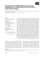
Structural studies on the enzymes from Citrobacter
freundii (PDB entries: 1TPL, 2TPL) and Erwinia
herbicola (1C7G) have found them to be composed of
four identical subunits, each with one molecule of PLP
[14,15]. Each subunit of C. freundii TPL is comprised of
an N-terminal arm (residues 1–19), small domain, and
PLP-binding large domain. The active site is located in
a cleft surrounded by one subunit and the large domain
of the adjacent subunit, constituting a catalytic dimer.
The two dimers are then tightly combined through a
hydrophobic cluster at the center of the tetramer and
intertwined N-terminal arms (Fig. 1).
The above mentioned architecture is conserved in
many a-family PLP-enzymes including tryptophan
indole-lyase (TNA; PDB entry: 1AX4) and aspartate
aminotransferases (AspAT; PDB entry: 1ARI) [16–18].
In porcine cytosolic aspartate aminotransferase
(AspAT), the N-terminal arm protruding toward the
large domain of the other subunit is essential for both
the catalytic activity and thermal stability of the enzyme
[19–21]. Similarly, the AspAT of Bacillus circulans
shows a weakened cofactor affinity at the truncation of
the N-terminal arm, resulting in a monomeric nonfunc-
tional conformation [22]. Meanwhile, structural studies
of Proteus vulgaris TNA have revealed an intimate
correlation between cofactor binding and the interfacial
H-bonds formed on the subunit interface [17].
In this study, a random mutagenesis approach to
evolve a robust TPL for l-DOPA synthesis resulted in
an effective mutation, T15A, located on the N-terminal
arm of C. freundii TPL. Biochemical characterization
of the native and mutant enzyme proved the mutation
on the interface increased the stability of the catalytic
capability of the enzyme by preventing cyclic conden-
sation between l-DOPA and PLP (Fig. 1).
Results
Random mutagenesis and structural
identification of T15A mutant
An error-prone PCR of C. freundii TPL and subse-
quent cloning into Escerichia coli XL1-Blue resulted in
a mutant library containing 1–5 mutations that were
evenly distributed over the entire TPL sequence. About
10 000 colonies from the library were subjected to
rapid screening on microtiter plates with l-DOPA
as the substrate. To select a highly active mutant from
the library, the activity with l-DOPA was divided by
the corresponding activity when using l-tyrosine as the
substrate, thereby compensating for a variation in the
expression levels. When comparing the normalized
activities, mutant #44 was identified as the most active,
with a two-fold increased activity with l-DOPA. A
sequence analysis of #44 exhibited an amino acid
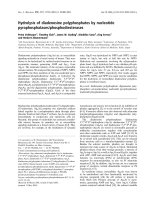
change from Thr15 to Ala, while a structural analysis
of C. freundii TPL (1TPL, 2TPL) revealed that Thr15
was located on the intertwined N-terminal arm, com-
prising an H-bond network between the catalytic
dimers within a proximal distance of the hydrophobic
core (Fig. 2A). The hydroxyl group of Thr15 was
H-bonded to the sidechain of Lys59, and connected to
the sidechain of Asp58 via a water molecule, which
was also linked to the backbone nitrogen of Thr15
(Fig. 2B). In addition, the sidechain of Thr15 was
involved in nonbonded interactions with the Lys59
and Glu308 sidechains from the other catalytic dimer.
Fig. 1. Schematic view of Pictet–Spengler reaction and cofactor
release from holo-TPL enzymes. The adductive reaction between
L-DOPA and pyridoxal-5¢-phosphate (PLP) leads to the depletion of
the cofactor in the reaction solution, inactivating the enzyme
depending on the cofactor binding affinity.
Scheme 1. Synthesis reaction by TPL.
S G. Lee et al. T15A mutation effect on C. freundii TPL cofactor stability
FEBS Journal 273 (2006) 5564–5573 ª 2006 The Authors Journal compilation ª 2006 FEBS 5565
In summary, the proximate interaction of Thr15 with
the other subunits suggested that the effect of T15A
on the catalytic capability was related to changes in
the interdomain architectures of the catalytic dimers.
Purification, kinetic parameters, and catalytic
stability with
L-DOPA as substrate
The E. coli XL-1 Blue cells bearing the plasmid
pHR1001 or pDA44 revealed a thick protein band
with a molecular mass of 52 kDa in an SDS ⁄ PAGE
analysis after induction with 1 mm isopropyl thio-b-
d-galactoside (IPTG). Based on ammonium sulfate
precipitation between 50 and 70% saturation, followed
by ion exchange and hydrophobic chromatography,
the native TPL and T15A mutant were purified to
homogeneity with a recovery yield of 45% and 39%,
respectively. The purified proteins were preserved in
a refrigerator after being reprecipitated in 70%
(NH
4
)
2
SO
4
, then desalted just before use to recover
their original specific activities of around 1.2 and 0.64
unitsÆmg
)1
, respectively, with l-tyrosine as the sub-
strate.
The kinetic parameters were determined in triplicate
experiments at 30 °C, with 0.05–1 mml-tyrosine or
0.5–12 mml-DOPA as the substrate. The catalytic rate
constants (k
cat
) for the native and mutant enzymes
with l-DOPA were 0.31 s
)1
and 0.68 s
)1
, respectively
(Table 1), while the Michaelis constants with l-tyrosine
were determined as 0.24 and 0.22 mm, respectively,
indicating a conserved geometry at the binding site,
and with l-DOPA were determined to be 3.2 and
4.6 mm, respectively, yet with larger error limits. Inves-
tigations of the substrate range of the TPLs revealed
that 3-chloro-l-tyrosine, dl-serine, and dl-cysteine
also served as substrates to a lesser extent, whereas
d-tyrosine, d-DOPA, dl-tryptophane, dl-phenylalan-
ine, and dl-alanine were all inert towards the enzymes.
The native and mutant enzymes were then investi-
gated for their stability and activity at temperatures
between 15 and 80 ° C. When heated for 20 min in the
standard buffer, both enzymes remained stable up to
55 °C in a 0.1 m potassium phosphate buffer (pH 8.0)
(Fig. 3A). The half-inactivation temperatures for the
native and mutant enzymes were calculated to be 62.2
and 65.2 °C, respectively, with a four-parameter sig-
moid equation using sigmaplot (Systat Software Inc.,
Richmond, CA, USA). Plus, the inclusion of two sub-
strates for the synthesis of l-DOPA (20 mm catechol
and 1.0 m ammonium acetate) increased the half-inac-
tivation temperatures to 72.1 and 73.9 °C, respectively,
similar to the stabilization of porcine cytosolic AspAT
by a-ketoglutarate [20]. The maximum activity for the
a,b-elimination of l-tyrosine was observed at 55 and
60 °C for the native and mutant enzymes, respectively
(Fig. 3B). However, when l-DOPA was applied as the
substrate, the temperatures producing the maximum
activity were down-shifted by 15 °C for both the native
and mutant enzymes to 40 and 45 °C, respectively
(Fig. 3C).
A B
Fig. 2. Interfacial architectures of catalytic
dimers of C. freundii TPL. (A) Overall struc-
ture, extracted from prediction of oligomeric
states server at EBI ( />Yellow colored molecules represent 3-(4¢-
hydroxyphenyl)propionic acid adopted from
2TPL PDB file. (B) Magnified view of red-
lined box in overall structure. Green lines
represent intimate molecular interactions
including hydrogen bond networks in vicinity
of Thr15 on intertwined N-terminal arm.
H-bond information was extracted from
entry code 1TPL of Protein Data Bank.
Table 1. Kinetic constants for C. freundii TPL and T15A mutant.
Enzymes
L-Tyrosine L-DOPA PLP
K
m
(mM) k
cat
(sec
-1
) k
cat
⁄ K
m
K
m
(mM) k
cat
(sec
-1
) k
cat
⁄ K
m
(K
m,PLP
, lM)
Native 0.24 ± 0.1 1.8 ± 0.2 7.5 3.2 ± 0.8 0.31 ± 0.04 0.10 2.0
T15A 0.22 ± 0.03 1.2 ± 0.1 5.5 4.6 ± 1.8 0.68 ± 0.15 0.15 2.5
T15A mutation effect on C. freundii TPL cofactor stability S G. Lee et al.
5566 FEBS Journal 273 (2006) 5564–5573 ª 2006 The Authors Journal compilation ª 2006 FEBS
Inactivation of C. freundii TPL by Pictet–Spengler
reaction
Cyclic adducts of l-DOPA with endogenous aldehydes
have been detected in biological solutions for decades.
As such, when the C. freundii TPL (30 lm) was incu-
bated with 10 mml-DOPA in a 0.1 m potassium
phosphate buffer (pH 7.5) at 30 °C, a time-dependant
decrease in the absorbance at 400 nm was detected,
resulting in a new absorption peak at 330 nm
(Fig. 4A), corresponding to previous literature on the
inactivation of l-DOPA decarboxylases when using
l-DOPA as the substrate [5,6,23]. When a pseudo
first-order kinetic (low initial concentration of the
enzyme) was applied for the decolorization rate of
C. freundii TPL with l-DOPA, the rate constant
(k
1
) was calculated as 0.012 min
)1
using a kinetic
equation, log
A
t
A
0
¼Àk
1
t , where A
0
and A
t
are the
Fig. 3. Effect of temperature on stability and activity of C. freundii
TPL and T15A mutant. (A) Stability, (B) activity with
L-tyrosine, and
(C) activity with
L-DOPA as substrates. The stability was evaluated
as the remaining activity after the enzymes were incubated in a
100 m
M potassium phosphate buffer (pH 8.0) at the indicated tem-
peratures for 20 min. The activity with
L-tyrosine and L-DOPA as
the substrates was measured in the standard reaction mixture for
20 min at different temperatures and the amount of pyruvate pro-
duced determined by the salicylaldehyde method. Closed symbols
represent native enzyme (d,r) and open symbols represent T15A
mutant (s,e). Diamond symbols indicate improved stability in the
presence of 20 m
M catechol and 1.0 M ammonium acetate.
Fig. 4. Pictet–Spengler adduct formation from C. freundii TPL in
presence of
L-DOPA. (A) Spectral analyses of enzyme solution
(30 l
M) treated with 10 mML-DOPA in 0.1 M potassium phosphate
buffer (pH 7.5) at 30 °C. Inset: Time-dependent decrease in absorb-
ance at 400 nm. (B) HPLC analyses of enzyme mixture. After the
spectral change was completed, the reaction solution was subjec-
ted to centrifugal ultrafiltration (molecular cutoff: 10 000), the fil-
trate loaded on a DOWEX 50 W column (pH 3.0), and the eluted
solution precipitated with three volumes of isopropanol on ice. The
precipitates were dissolved in water, then analyzed by HPLC. The
standard was a 2 : 1 mixture of pyridoxal-5¢-phosphate and
L-DOPA-PLP adduct synthesized in the authors’ laboratory.
S G. Lee et al. T15A mutation effect on C. freundii TPL cofactor stability
FEBS Journal 273 (2006) 5564–5573 ª 2006 The Authors Journal compilation ª 2006 FEBS 5567
absorbance at times 0 and t, respectively (inset in
Fig. 4A). After the spectral change was completed, the
reaction solution was treated with a cation exchange
resin (DOWEX 50 W), analyzed by HPLC, and found
to include an l-DOPA-PLP adduct with the same
retention time and molecular mass (426 Da) as the
Pictet–Spengler type adduct (Fig. 4B) synthesized as
described below. Meanwhile, the rate constant (k
1
)of
free PLP was 0.12 ± 0.02 min
)1
under the pseudo
first-order reaction conditions. Consequently, the free
cofactor was estimated to be 10-fold more susceptible
to adduct formation than the enzyme-bound PLP.
The rate constants also increased with the pH and
temperature, as previously reported for the reaction
between l-DOPA and d -glucose [7]. In particular, the
k
1
values increased up to 0.5 ± 0.1 min
)1
when 1.0 m
ammonium chloride (pH 8.2) was included in the reac-
tion solution.
As seen in Fig. 3C, the maximum activity of the
mutant and native enzymes with l-DOPA as the sub-
strate was 15 °C lower than when using l-tyrosine as
the substrate, plus both enzymes were similarly stable
up to 55 °C. Consequently, because the spectral and
kinetic studies on the decolorization of TPL suggested
that the compromised activity was closely related to
the loss of the coenzyme via adduct formation, an
experiment on the stability of the enzyme-bound cofac-
tors was performed in a 1.0 m ammonium chloride
solution (pH 8.2) with 10 mml-DOPA. During incu-
bation at 45 °C, the enzyme mixtures were withdrawn
intermittently, 100-fold diluted in an assay solution,
and examined for their remaining activity using 1 mm
l-tyrosine as the substrate. The remaining activity of
the mutant and native enzymes decreased according to
the incubation time, down to 30% and 6% of the ini-
tial activity (dotted lines in Fig. 5) with a half-life of
16.8 and 6.4 min, respectively.
However, when the same samples were assayed in
the presence of excess PLP (200 lm), both enzymes
recovered over 80% of their original activity (solid
lines in Fig. 5), indicating that the inactivation could
be attributed to the loss of the cofactor through a con-
densation reaction with l-DOPA.
Stabilization of cofactor binding by T15A
mutation
The extended lifetime of T15A in 10 mml-DOPA sug-
gested that the intertwined N-terminal architecture,
where Thr15 is located, was closely related with the
cofactor binding affinity of C. freundii TPL. To verify
the effect of the T15A mutation on the cofactor affin-
ity, the apparent Michaelis constants for PLP (K
m,PLP
)
with the native and mutant enzymes were investigated
at temperatures ranging from 30 to 60 °C.
As shown in Fig. 6A, the K
m,PLP
for C. freundii TPL
increased slowly below 45 °C, accompanied by an
increase in the catalytic rate constant (k
cat
). However,
above 50 °C, the binding constants increased very
sharply, while the k
cat
remained at a similar level
(Fig. 6B). An increase in the K
m,PLP
was also detected
with the T15A mutant, yet significantly slower than
that with the native enzyme (Fig. 6A). As such, the
cofactor release from the active site was increased rel-
ative to the temperature, likely accelerating the adduct
formation with l-DOPA, yet this was significantly
relieved by the T15A mutation located on the inter-
twined N-terminal arm.
Finally, the effect of the T15A mutation on l-DOPA
synthesis was investigated in a reaction solution (10
mL) including 0.65 m ammonium chloride (pH 8.5),
50 mm sodium pyruvate, 50 mm catechol, 0.1 mm
PLP, 0.1% sodium sulfite, and 15 units of the enzyme.
In addition, because alcoholic additives have been
shown to be beneficial for the synthesis of l-DOPA by
C. freundii TPL [3], 10% ethanol was also included in
the reaction solution. When the synthesis reaction was
carried out for 2.5 h at 45 °C, the concentration of
l-DOPA increased rapidly up to the maximum level
within an hour, then slightly decreased (Fig. 7), prob-
ably because of the adduct formation between
l-DOPA and remaining pyruvate [2,5]. However, the
upward curve flattened much earlier with C. freundii
TPL, consequently the l-DOPA productivity of T15A
was at least two-fold higher than that with C. freundii
Fig. 5. Inactivation of TPL enzymes by L-DOPA and its reactivation
by PLP. Timecourse profiles of inactivation and activity recovery in
presence of excess pyridoxal 5¢-phosphate. An enzyme mixture
containing 0.01 unitsÆml
)1
of TPL, 0.1 mM PLP, and 10 mML-DOPA
in a 100 m
M potassium phosphate buffer (pH 8.0) was incubated at
45 °C for different times, and the remaining activity determined in
the presence of excess PLP (d,s) or without PLP (r,e). Closed
symbols represent native enzyme and open symbols represent
T15A mutant.
T15A mutation effect on C. freundii TPL cofactor stability S G. Lee et al.
5568 FEBS Journal 273 (2006) 5564–5573 ª 2006 The Authors Journal compilation ª 2006 FEBS
TPL, consistent with the robustness of T15A at eleva-
ted temperatures (Figs 5 and 6C). No oxidation of
l-DOPA was detected while the solutions were flushed
with nitrogen gas.
Discussion
The enzymatic synthesis of l-DOPA using E. herbicola
TPL is more successful at a low temperature range
from 15 to 24 °C [5,24]. Likewise, with C. freundii
TPL, the synthesis was facilitated at 18 °C [2],
although the enzyme activity was about 20% of the
maximal activity (Fig. 3C). The compromised produc-
tivity at high temperatures has been attributed to the
formation of byproducts and the oxidative deterior-
ation of catechol or pyruvate during the reaction, all
of which are accelerated by the temperature [2,5].
In this study, it was postulated that the lowered pro-
ductivity of C. freundii TPL at elevated temperatures
partly resulted from a decolorization reaction in the
enzyme mixture, which eventually led to the depletion
of the cofactor PLP, accompanied by the inactivation
of the enzyme. HPLC and
1
H NMR analyses of the
purified adduct revealed that the inactivation resulted
from Pictet–Spengler type condensation between
l-DOPA and PLP. Notwithstanding previous reports
on the inactivation of PLP enzymes, aromatic decarb-
oxylases, by l-DOPA [10,23], this is the first time the
rapid inactivation of TPL has been explained based on
a Pictet–Spengler reaction.
Consistent with the observation that a Pictet–Spen-
gler reaction is accelerated relative to the reaction
temperature [8], the optimal temperature for enzyme
activity in the presence of l-DOPA was 15 °C lower
than that with l-tyrosine as the substrate (Fig. 3B,C).
The inactivation profile of the enzyme with 10 mm
l-DOPA (Fig. 5) also agreed with the optimal tem-
perature results. Meanwhile, the incubation of TPL
with d-DOPA, a stereoisomer of l-DOPA that does
Fig. 6. Effect of temperature on kinetic constants for C. freundii
TPL and T15A mutant. (A) Apparent binding constant (K
m,PLP
) for
PLP, (B) catalytic rate constant (k
cat
), and (C) ratio of k
cat
⁄ K
m
,
PLP
.
The kinetic constants were determined from double reciprocal plots
of the reaction rate versus the PLP concentration at different tem-
peratures. Closed symbols represent K
m
,
PLP
values for C. freundii
TPL, while open symbols represent K
m
,
PLP
values for T15A mutant.
Fig. 7. Synthesis of L-DOPA by C. freundii TPL and T15A mutant.
The synthetic reaction was carried out using partially purified
enzymes in a solution (10 mL) containing 0.65
M ammonium chlor-
ide (pH 8.5), 50 m
M sodium pyruvate, 50 mM catechol, 0.1 mM
PLP, 0.1% sodium sulfite, 10% ethanol and 15 units of enzyme.
The reaction bottle was flushed with nitrogen gas, tightly sealed
with a rubber stopper, and incubated at 45 °C. Samples were with-
drawn using a syringe in a stream of nitrogen gas to prevent oxida-
tion of the ingredients. Closed symbols represent native enzyme
and open symbols represent T15A mutant.
S G. Lee et al. T15A mutation effect on C. freundii TPL cofactor stability
FEBS Journal 273 (2006) 5564–5573 ª 2006 The Authors Journal compilation ª 2006 FEBS 5569
not serve as a substrate for the enzyme reaction, pro-
duced a similar effect to l-DOPA, indicating that
the adduct-forming reaction was independent of the
enzyme reaction and a chemical reaction between
l-DOPA and the free PLP released from the active
site. The release of PLP from the enzyme was acceler-
ated at an elevated temperature, as shown by the
K
m,PLP
versus temperature profile of the native enzyme
(Fig. 6A). The enzyme-bound PLP reacted with the
l-DOPA in the reaction mixture to form an l-DOPA-
PLP adduct at a rate of 0.012 min
)1
, as shown by the
inset in Fig. 4A, which was 10 times slower than
the rate with the free PLP and l-DOPA (Fig. 1). The
removal of PLP by release and the subsequent Pictet–
Spengler reaction may have been responsible for the
rapid decrease in the k
cat
⁄ K
m,PLP
value of the native
enzyme at temperatures above 45 °C (Fig. 6C). Note
that the k
cat
⁄ K
m,PLP
value was the catalytic rate in the
presence of a limited concentration of PLP.
In contrast, the K
m,PLP
-value for T15A was less
sensitive to the temperature (Fig. 6A), suggesting a
tight binding of the cofactor at the enzyme active
site. Therefore, the mutation on the intertwined
N-terminal arm stabilized the cofactor binding affin-
ity, thereby improving the catalytic properties at
elevated temperatures (Fig. 7), as indicated by the
higher stability of the k
cat
⁄ K
m,PLP
value for the T15A
enzyme (Fig. 6C).
Citrobacter freundii TPL has a 50% sequence iden-
tity with the tryptophanase from P. vulgaris, which
degrades tryptophan to indole, ammonia, and pyruvate
[14,25]. The secondary, tertiary, and quaternary struc-
tures are also highly conserved, plus a hydrophobic
cluster and intertwined N-terminal arms are formed on
the intersubunit interface, contributing to its stability.
The network of hydrogen bonds and salt bridges
formed upon the binding of PLP is known to influence
the quaternary structure of tryptophanases [17]. There-
fore, when considering the common structural features
of a-family PLP enzymes [26,27], the T15A mutation
on the N-terminal arm may have increased the rigidity
of the cofactor binding architecture of C. freundii TPL
through adjusting the quaternary interfaces. One poss-
ible communication between the N-terminal arm and
the active site is through Tyr71, which belongs to the
adjacent subunit of the catalytic dimer. Tyr71 is
known to be essential for activity, as a general acid
catalyst for the elimination of the leaving group from
a quinonoid intermediate, and also for PLP binding
[28]. The PLP binding constant for the Y71F mutant
of C. freundii TPL was estimated to be 1 mm, while
the wildtype TPL showed a binding constant of 0.6 lm
based on spectrophotometric titration. Consistently,
the equivalent Tyr70 in aspartate aminotransferase
also has a PLP binding function [29].
Thus, this study demonstrated that the deterioration
of the cofactor through a Pictet–Spengler reaction with
l-DOPA appeared to be a significant interference with
the biotechnological production of l-DOPA when
using C. freundii TPL. The T15A mutation improved
the cofactor binding affinity at high temperatures,
along with the apparent turnover rate when using
l-DOPA as the substrate, through an interfacial inter-
action between the N-terminal arm and the cleft active
site. However, l-DOPA synthesis at a high temperature
also increases the adduct formation between l-DOPA
and a substrate pyruvate [2,5], eventually decreasing
the l-DOPA concentration during a prolonged reaction
at a high temperature, as observed in Fig. 7. Thus,
despite the increased catalytic efficiency and stability of
the T15A mutant, l-DOPA synthesis at a high
temperature should be further scrutinized to minimize
the adduct formation between l-DOPA and pyruvate.
For example, a continuous limited supply of pyruvate
into the reaction solution could be used to maintain the
pyruvate concentration at a minimal level, thereby
decreasing the adduct formation rate. In addition,
based on the effect of alcohols [3], the reaction ingre-
dients could also be optimized to increase the l-DOPA
synthesis and relieve the adduct formation.
Consequently, with its enhanced l-DOPA synthesis
activity and stability, the T15A enzyme of this work
could be used for the development of a new bioconver-
sion strategy for the efficient production of l-DOPA
at high temperatures, where it can catalyze the reaction
more actively.
Experimental procedures
Materials
The PLP was purchased from Sigma (St Louis, MO, USA)
and the l-DOPA purchased from Boehringer Mannheim
(Mannheim, Germany). The restriction endonucleases and
T4 DNA ligase were purchased from New England Biolabs
(Beverly, MA, USA) and the Taq DNA polymerase from
PerkinElmer (Branchburg, NJ, USA). The oligonucleotides
were synthesized at Bioneer Co. (Daejeon, Korea) and the
DNA sequencing performed at Solgent Co. (Daejon, Korea).
The l-DOPA-PLP adduct was synthesized by mixing
l-DOPA (0.32 g) and PLP (0.2 g) in a 50 mm sodium
phosphate buffer (80 mL, pH 8.0) at 45 °C for 30 min.
The reaction product was purified on a DOWEX 50 W
column (pH 3.0, Sigma) and the eluted solution precipitated
with isopropanol (240 mL) on ice for 2 h. The precipitates
were then washed on a sintered glass filter with acetone and
T15A mutation effect on C. freundii TPL cofactor stability S G. Lee et al.
5570 FEBS Journal 273 (2006) 5564–5573 ª 2006 The Authors Journal compilation ª 2006 FEBS
stored in a deep freezer after vacuum-drying. The molecular
mass of the adduct was 426 Da on a ESI-MS spectrometer,
and the chemical shift values in D
2
O determined by
300 MHz
1
H NMR experiments were as follows: d 2.36
(3 H, s, H-2¢), 3.2 (2 H, m, H-b), 4.0 (1 H, m, H-a), 4.91
(2 H, d, H-5¢), 5.77 (1 H, s, H-4¢), 6.20 (1H, s, H-5¢¢), 6.69
(1 H, s, H-2¢¢), and 7.74 (1 H, s, H-6). The chemical struc-
ture of the adduct was identified as shown in Fig. 1. All
other chemicals used were chemical reagent grade.
Random mutagenesis and screening
on microtiter plates
The plasmid pHR1001 harboring the C. freundii tpl gene
(gene bank accession no. DQ907529) [3] was used as the
template for an error-prone PCR with the following prim-
ers: 5¢-AATTATCCGGCAGAACCCTT-3¢ (forward) and
5¢-GATC
AAGCTTTTAGATATAGTCAAAGCGTGC-3¢
(reverse, underlined HindIII). The thermal cycling was per-
formed using a DNA Thermal Cycler (PerkinElmer): 5 min
at 95 °C, a subsequent 25 cycles of 1 min at 95 °C, 2 min at
50 °C, 3 min at 72 °C, and a final extension of 7 min at
72 °C. The amplified PCR products were digested with Hin-
dIII to yield a 1.37 kb DNA fragment. The plasmid
pTrc99A was then digested with NcoI, blunt-ended by
Klenow treatment, and digested with HindIII. The resulting
plasmid was ligated with the HindIII-treated PCR product
by blunt-cohesive ligation at 16 °C with a T4 DNA ligase.
E. coli XL1 Blue cells were then transformed with the ligate
by electroporation and spread on LB-ampicillin plates. After
being incubated overnight at 37 °C, the evolved colonies
were transferred by toothpick to fresh LB-ampicillin plates.
The mutant library was inoculated into an LB-ampicillin-
IPTG medium (500 lL) contained in a deep 96-well plate,
and cultivated in a wellplate culture system, Megagrow
TM
(Bioneer Co.). The cultivated cells were centrifuged at
5000 g for 20 min with a wellplate centrifuge Union
5KR
TM
, rotor type TM96-65 (Hanil Sci. Ind., Inchon,
Korea), washed in a 50 mm Tris ⁄ HCl buffer (pH 8.0), and
treated with 200 lL Cellytic
TM
B (Sigma) for 1 h at 37 °C.
The cell lysate (100 lL) was then transferred into 96-well
PCR plates and mixed with the same amount of substrate
solutions, including 10 mml-DOPA or 1 mml-tyrosine,
and 20 lm PLP in a 50 mm potassium phosphate buffer
(pH 8.0). After being incubated at 37 °C for 20 min, the
reaction solutions were heated for 3 min at 95 °C, centri-
fuged at 5000 g for 20 min with Union 5KR
TM
centrifuge to
remove any insoluble aggregates, and analyzed for pyruvate
formation using the salicylaldehyde method [25] to compare
the enzyme activities towards l-DOPA and l-tyrosine.
Expression and purification
Escerichia coli XL-1 Blue cells harboring pHR1001 or
pDA44 were cultivated at 37 °C for 16 h in 1 litre of an LB
medium containing 100 lgÆmL
)1
ampicillin. Protein expres-
sion was induced by the addition of 1 mm IPTG when the
absorbance at 600 nm reached 0.5. The harvested cells were
then disrupted by sonification in a standard buffer, inclu-
ding 0.01% 2-mercaptoethanol, 0.05 mm PLP, and 50 mm
Tris ⁄ HCl (pH 8.0). The centrifugation supernatant was col-
lected, and subjected to ammonium sulfate fractionation
between 50% and 70% saturation. The enzyme dissolved in
the standard buffer was then loaded on to a Resource Q
ion exchange (Pharmacia, Uppsala, Sweden), washed with
the standard buffer, and eluted using a KCl gradient from
0 to 0.5 m. Most of the active fractions were then pooled,
adjusted to include 1.7 m (NH
4
)
2
SO
4
, and loaded on to a
Phenyl Superose (Pharmacia). The elution from the hydro-
phobic column was performed using a reverse gradient of
(NH
4
)
2
SO
4
from 1.7 m to 0 m, then the active fractions
were dialyzed against a 100 mm Tris ⁄ HCl buffer (pH 8.0)
containing 0.2 m KCl, reprecipitated in 70% saturated
(NH
4
)
2
SO
4
, and stored in a refrigerator. All the column
procedures were carried out using an AKTA system (Amer-
sham Bioscience, Uppsala, Sweden) at room temperature.
Determination of kinetic parameters and cofactor
binding affinity
The kinetic constants for l-DOPA and l-tyrosine as sub-
strates were determined using a lactate dehydrogenase
(LDH)-coupled assay of the pyruvate formation rate. The
reaction was started by the addition of 0.05–1.0 mml-tyro-
sine or 0.5–12 mml-DOPA as the substrate, and the
decrease in A
340
monitored at 30 °C using a spectrophoto-
meter, Ultrospec3000 (Pharmacia Biotech, Uppsala,
Sweden), equipped with a Peltier cuvette-heating system.
The pyruvate formation rate was calculated using the
extinction coefficient of NADH (6200 m
)1
Æcm
)1
) from the
slope between 0.5 and 5.0 min, after the early perturbation
of the absorbance was settled.
The apparent binding constants of PLP to the enzymes
were presumed as the concentration of PLP for half the
maximal activity of the enzyme. The assay mixture with dif-
ferent PLP concentrations (0.5–200 lm) and 2.5 mml-tyro-
sine was equilibrated to different temperatures for 5 min in
a thermo-controlled spectrophotometer, and the enzyme
activity measured using an LDH coupling assay, as des-
cribed above. The apparent binding constants for PLP
(K
m,PLP
) were calculated from a double reciprocal plot
of the reaction rate (v) versus the PLP concentration:
m
V
max
¼
½PLP
K
m;PLP
þ½PLP
, where V
max
is the maximum reaction
rate at saturating PLP concentrations. All the kinetic
experiments were performed in triplicate.
Enzyme assay and analysis
The a,b-elimination activity of TPL was calculated from
the pyruvate formation rate determined by a coupling assay
S G. Lee et al. T15A mutation effect on C. freundii TPL cofactor stability
FEBS Journal 273 (2006) 5564–5573 ª 2006 The Authors Journal compilation ª 2006 FEBS 5571
with LDH (Roche Diagnostics, Bazel, Switzerland) or using
the salicylaldehyde method [25]. The standard reaction mix-
ture contained 10 mml-DOPA or 1 mml-tyrosine as the
substrate, 50 l m PLP, 0.2 mm NADH, 10 lgÆmL
)1
LDH,
and TPL in a 0.1 m potassium phosphate buffer (pH 8.0).
One unit of enzyme was defined as the activity to catalyze
the formation of 1 lmol of pyruvate per min at 30 °C. The
protein concentration was determined using a Bradford rea-
gent (Bio-Rad, Hercules, CA, USA) with bovine serum
albumin as the standard.
The analysis of the l-DOPA-PLP adduct was performed
on a HPLC system (Young-in Co., Seoul, Korea) equipped
with an ODS18 column (Shimazu, Kyoto, Japan) and UV-
detector (295 nm). The elution was carried out using a co-
solvent consisting of a 50 mm potassium phosphate buffer
with 2 mm sodium dodecylsulfate (pH 3.0), methanol, and
acetonitrile (volumetric ratio ¼55 : 40 : 5) at a flow rate of
0.6 mLÆmin
)1
.
Acknowledgements
This project was supported by a grant from the Clea-
ner Production Program 10007946 of NCPC, the
KRIBB Research Initiative Program, and the 2006
research fund of Kookmin University, Korea.
References
1 Kumagai H, Yamada H, Matsui H, Ohkishi H &
Ogata K (1970) Tyrosine phenol lyase. I. purification,
crystallization, and properties. J Biol Chem 245,
1767–1772.
2 Yamada H & Kumagai H (1975) Synthesis of 1-tyro-
sine-related amino acids by beta-tyrosinase. Adv Appl
Microbiol 19, 249–288.
3 Lee SG, Ro HS, Hong SP, Lee KJ, Wang JW, Tae DN,
Uhm KN, Bang SG, Kim YJ & Sung MH (1996) Pro-
duction of 3,4-dihydroxyphenyl-l-alanine by using the
b-tyrosinase of Citrobacter freundii overexpressed in
recombinant Escherichia coli. Kor J Appl Microbiol
Biotechnol 24, 44–49.
4 Ajinomoto Co. Inc. (1993) Method for producing l-3,4-
dihydroxyphenylalanine by Erwinia having tyrosinase
activity. US Patent 05338672.
5 Enei H, Nakazawa H, Okumura S & Yamada H (1973)
Synthesis of 1-tyrosine and 3,4-dihydroxyphenyl-l-ala-
nine from pyruvic acid, ammonia, and phenol or pyro-
catechol. Agr Biol Chem 37, 725–735.
6 Foor F, Morin N & Bostian KA (1993) Production of
1-dihydroxyphenylalanine in Escherichia coli with the
tyrosine phenol-lyase gene cloned from Erwinia herbi-
cola. Appl Environ Microbiol 59, 3070–3075.
7 Dougan D, Wade D & Mearric PK (1975) Effects of
l-DOPA metabolites at a dopamine receptor suggest a
basis for ‘on-off’ effect in Parkinson’s disease. Nature
254, 70.
8 Manini P, Ischia M & Prota G (2001) Pictet-Spengler
condensation of the antiparkinsonian drug l-DOPA with
d-glyceraldehyde. Opposite kinetic effects of Fe
3+
and
Cu
2+
ions and possible implications for the origin of
therapeutic side effects. Bioorg Med Chem 9, 923–929.
9 Sandler M, Carter SB, Hunter KR & Stern GM (1973)
Tetrahydroisoquinoline alkaloids: in vivo metabolites of
l-DOPA in man. Nature 241, 439–443.
10 Bertoldi M & Voltattorn CBI (2000) Reaction of dopa
decarboxylase with 1-aromatic amino acids under aero-
bic and anaerobic conditions. Biochem J 352, 533–538.
11 Nishino J, Hayashi H, Ishii S & Kagamiyama H (1997)
An anomalous side reaction of the Lys303 mutant aro-
matic 1-amino acid decarboxylase unravels the role of
the residue in catalysis. J Biochem 121, 604–611.
12 Faleev NG, Ruvinov SB, Demidkina TV, Myagkikh I,
Gololobov M, Bakhmutov VI & Belikov VM (1988)
Tyrosine phenol-lyase from Citrobacter intermedius: fac-
tors controlling substrate specificity. Eur J Biochem 177,
395–401.
13 Faleev NG, Spirina SN, Ivoilov VS, Demidkina TV &
Phillips RS (1996) The catalytic mechanism of tyrosine
phenol-lyase from Erwinia herbicola: the effect of sub-
strate structure on pH-dependence of kinetic parameters
in the reactions with ring-substituted tyrosines. Z Natur-
forsch [C] 51, 363–370.
14 Antson AA, Demidkina TV, Gollnick P, Dauter Z, von
Tersch RL, Long J, Berezhnoy SN, Phillips RS, Har-
utyunyan EH & Wilson KS (1993) Three-dimensional
structure of tyrosine phenol-lyase. Biochemistry 32,
4195–4206.
15 Sundararaju B, Antson AA, Phillips RS, Demidkina
TV, Barbolina MV, Gollnick P, Dodson GG & Wilson
KS (1997) The crystal structure of Citrobacter freundii
tyrosine phenol-lyase complexed with 3-(4-hydroxyphe-
nyl) propionic acid, together with site-directed mutagen-
esis and kinetic analysis, demonstrates that arginine 381
is required for substrate specificity. Biochemistry 36,
6502–6510.
16 Alexander FW, Sandmeier E, Mehta PK & Christen P
(1994) Evolutionary relationships among pyridoxal-5¢-
phosphate-dependent enzymes. regio-specific a, b, and !
families. Eur J Biochem 219, 953–960.
17 Isupov MN, Antson AA, Dodson EJ, Dodson GG,
Dementieva IS, Zakomirdina LN, Wilson KS, Dauter
Z, Lebedev AA & Harutyunyan EH (1998) Crystal
structure of tryptophanase. J Mol Biol 276, 603–623.
18 Iwamori S, Yoshino S, Ishiwata K & Makiguchi N
(1991) Structure of tyrosine phenol-lyase genes from
Citrobacter freundii and structural comparison with
tryptophanase from Escherichia coli. J Ferment Bioeng
72, 147–151.
T15A mutation effect on C. freundii TPL cofactor stability S G. Lee et al.
5572 FEBS Journal 273 (2006) 5564–5573 ª 2006 The Authors Journal compilation ª 2006 FEBS
19 Ford GC, Eichele G & Jansonius JN (1980) Three-
dimensional structure of a pyridoxal-phosphate-depen-
dent enzyme, mitochondrial aspartate aminotransferase.
Proc Natl Acad Sci USA 77, 2559–2563.
20 Fukumoto Y, Tanase S, Nagashima F, Ueda
S, Ikegami K & Morino Y (1991) Structural and
functional role of the amino-terminal region of
porcine cytosolic aspartate aminotransferase. J Biol
Chem 266, 4187–4193.
21 Sandmeier E & Christen P (1980) Mitochondrial aspar-
tate aminotransferase 27 ⁄ 32-410. Partially active enzyme
derivative produced by limited proteolytic cleavage of
native enzyme. J Biol Chem 255, 10284–10289.
22 Kravchuk Z, Tsybovsky Y, Koivulehto M, Vlasov
A, Chumanevich A, Battchikova N, Martsev S &
Korpela T (2001) Truncated aspartate aminotransfer-
ase from alkalophilic Bacillus circulans with deletion
of N-terminal 32 amino acids is a non-functional
monomer in a partially structured state. Protein Eng
14, 279–285.
23 Bertoldi M, Frigeri P, Paci M & Voltattorni CB (1999)
Reaction specificity of native and nicked 3,4-dihydroxy-
phenylalanine decarboxylase. J Biol Chem 274, 5514–
5521.
24 Koyanagi T, Katayama T, Suzuki H, Nakazawa H,
Yokozeki K & Kumagai H (2005) Effective production
of 3,4-dihydroxyphenyl-l-alanine (l-DOPA) with Erwi-
nia herbicola cells carrying a mutant transcriptional
regulator TyrR. J Biotechnol 115, 303–306.
25 Berntsson S (1955) Spectrophotometric determination
of pyruvic acid by the salicylaldehyde method. Anal
Biochem 27, 1659–1660.
26 Phillips RS, Demidkina TV & Faleev NG (2003) Struc-
ture and mechanism of tryptophan indole-lyase and tyro-
sine phenol-lyase. Biochim Biophys Acta 1647, 167–172.
27 Phillips RS, Chen HY, Shim D, Lima S, Khadijeh K &
Sundararaju B (2004) Role of lysine-256 in Citrobacter
freundii tyrosine phenol-lyase in monovalent cation acti-
vation. Biochemistry 43, 14412–14419.
28 Chen HY, Demidkina TV & Phillips RS (1995) Site-
directed mutagenesis of tyrosine-71 to phenylalanine in
Citrobacter freundii tyrosine phenol-lyase: evidence for
dual roles of tyrosine-71 as a general acid catalyst in the
reaction mechanism and in cofactor binding. Biochemis-
try 34, 12276–12283.
29 Toney MD & Kirsch JF (1991) Tyrosine 70 fine-tunes
the catalytic efficiency of aspartate aminotransferase.
Biochemistry 30, 7456–7461.
S G. Lee et al. T15A mutation effect on C. freundii TPL cofactor stability
FEBS Journal 273 (2006) 5564–5573 ª 2006 The Authors Journal compilation ª 2006 FEBS 5573