Báo cáo khoa học: Aberrant interchain disulfide bridge of tissue-nonspecific alkaline phosphatase with an Arg433 fi Cys substitution associated with severe hypophosphatasia pdf
Bạn đang xem bản rút gọn của tài liệu. Xem và tải ngay bản đầy đủ của tài liệu tại đây (446.03 KB, 13 trang )
Aberrant interchain disulfide bridge of tissue-nonspecific
alkaline phosphatase with an Arg433
fi
Cys substitution
associated with severe hypophosphatasia
Makiko Nasu
1
, Masahiro Ito
2
, Yoko Ishida
2
, Natsuko Numa
3
, Keiichi Komaru
4
, Shuichi Nomura
1
and Kimimitsu Oda
2,5
1 Division of Oral Health in Aging and Fixed Prosthodontics, Niigata University Graduate School of Medical and Dental Sciences,
Japan
2 Division of Oral Biochemistry, Niigata University Graduate School of Medical and Dental Sciences, Japan
3 Division of Pediatric Dentistry, Niigata University Graduate School of Medical and Dental Sciences, Japan
4 Kitasato Junior College of Health and Hygienic Sciences, Yamatomachi, Minami-Uonuma-shi, Niigata, Japan
5 Center for Transdisciplinary Research, Niigata University, Japan
Keywords
alkaline phosphatase; bone; disulfide bridge;
hypophosphatasia; loss of function
Correspondence
K. Oda, Division of Oral Biochemistry,
Niigata University Graduate School of
Medical and Dental Sciences, 2-5274,
Gakkocho-dori Niigata 951-8514, Japan
Fax: +81 25 227 0803
Tel: +81 25 227 2827
E-mail:
(Received 24 September 2006, accepted
23 October 2006)
doi:10.1111/j.1742-4658.2006.05550.x
Various mutations in the tissue-nonspecific alkaline phosphatase (TNSALP)
gene are responsible for hypophosphatasia characterized by defective bone
and tooth mineralization; however, the underlying molecular mechanisms
remain largely to be elucidated. Substitution of an arginine at position 433
with a histidine [TNSALP(R433H)] or a cysteine [TNSALP(R433C)] was
reported in patients diagnosed with the mild or severe form of hypo-
phosphatasia, respectively. To define the molecular phenotype of the two
TNSALP mutants, we sought to examine them in transient (COS-1) and
conditional (CHO-K1 Tet-On) heterologous expression systems. In contrast
to an 80 kDa mature form of the wild-type and TNSALP(R433H), a unique
disulfide-bonded 160 kDa molecular species appeared on the cell surface
of the cells expressing TNSALP(R433C). Sucrose density gradient centri-
fugation demonstrated that TNSALP(R433C) forms a disulfide-bonded
dimer, instead of being noncovalently assembled like the wild-type. Of the
five cysteine residues per subunit of the wild-type, only Cys102 is thought to
be present in a free form. Replacement of Cys102 with serine did not affect
the dimerization state of TNSALP(R433C), implying that TNSALP(R433C)
forms a disulfide bridge between the cysteine residues at position 433 on
each subunit. Although the cross-linking did not significantly interfere with
the intracellular transport and cell surface expression of TNSALP(R433C),
it strongly inhibited its alkaline phosphatase activity. This is in contrast to
TNSALP(R433H), which shows enzyme activity comparable to that of the
wild-type. Importantly, addition of dithiothreitol to the culture medium was
found to partially reduce the amount of the cross-linked form in the cells
expressing TNSALP(R433C), concomitantly with a significant increase in
enzyme activity, suggesting that the cross-link between two subunits distorts
the overall structure of the enzyme such that it no longer efficiently carries
out its catalytic function. Increased susceptibility to proteases confirmed a
Abbreviations
Endo H, endo-b-N-acetylglucosaminidase H; ER, endoplasmic reticulum; GPI, glycosylphosphatidylinositol, PI-PLC, phosphatidylinositol-
specific phospholipase C; TNSALP, tissue-nonspecific alkaline phosphatase; TNSALP(R433C), tissue-nonspecific alkaline phosphatase with
an arginine to cysteine substitution at position 433; TNSALP(R433H), tissue-nonspecific alkaline phosphatase with an arginine to histidine
substitution at position 433.
5612 FEBS Journal 273 (2006) 5612–5624 ª 2006 The Authors Journal compilation ª 2006 FEBS
Hypophosphatasia is characterized by defective osteo-
genesis with various degree of failure in mineralization
of hard tissues such as bone and tooth [1–3]. Various
mutations in the human tissue-nonspecific alkaline
phosphatase (TNSALP, EC 3.1.3.1) gene are thought
to be responsible for hypophosphatasia [1–5]. Hypo-
phosphatasia is customarily divided into: (a) perinatal
hypophosphatasia; (b) infantile hypophosphatasia; (c)
childhood hypophosphatasia; (d) adult hypophospha-
tasia; and (e) odonto-type hypophosphatasia. The bio-
chemical hallmark of the disease is reduction in serum
alkaline phosphatase activity. Variation in clinical
expression is known to correlate well with variable
residual enzymatic activities in hypophosphatasia
patients (6,7). In general, the lower the activity, the
more severe the symptoms. As of 24 July 2006, 184
mutations had been reported in the TNSALP gene
worldwide, and about 80% of them are missense muta-
tions [7] (. ⁄ Database.html).
Recently, using a computer-assisted, three-dimensional
model of TNSALP, Mornet et al. have proposed the
categorization of missense mutations into different
functional domains, such as the active site, the
homodimer interface and the crown domain [8]. It is
now easier to predict, estimate and probably under-
stand the effects of some of the missense mutations on
the TNSALP molecule. However, the structural
evidence in itself may not be sufficient to assess the
effects of other mutations on TNSALP, especially if a
particular amino acid plays an essential role in the
adoption of the native structure other than its role in
maintaining the structure and function of the fully
folded enzyme. In this respect, we previously reported
that several TNSALP mutant proteins, which were
reported in severe hypophosphatasia patients, tend to
form a high molecular mass aggregate in the endoplas-
mic reticulum (ER), resulting in decreased cell surface
appearance of the TNSALP mutants, suggesting
impairment of the folding and assembly process for
TNSALP [9–13]. Furthermore, some mutant proteins
undergo proteasomal degradation [11–13]. Obviously,
an ER exit defect could be an important factor in
the etiology of severe forms of hypophosphatasia,
irrespective of whether mutant enzymes exhibit vari-
able residual enzyme activity [3]. TNSALP is an
ectoenzyme anchored to the plasma membrane via
glycosylphosphatidylinositol (GPI), and is believed to
regulate biomineralization by hydrolyzing inorganic
pyrophosphate, the extracellular matrix mineralization
inhibitor, on the surface of osteoblasts, chondrocytes
and matrix vesicles derived from them [3,14].
TNSALP(R433H arginine to histidine substitution)
was found in a compound heterozygote (R433H ⁄
D389G) diagnosed with odontohypophosphatasia [15],
whereas TNSALP(R433C arginine to cysteine substitu-
tion) was found in two independent homozygous
patients with infantile hypophosphatasia [16]. The
three-dimensional structure of human TNSALP predicts
that an arginine residue at position 433 is unique to
TNSALP and is located at the entrance of the active site
pocket, raising the possibility of its involvement in sub-
strate positioning [8]. Because of its conservative nature,
the replacement of arginine with histidine was assumed
to affect the catalytic function of TNSALP less severely
than replacement with cysteine. Here, we report that
both TNSALP(R433H) and TNSALP(R433C) are
anchored to the plasma membrane via GPI, like the
wild-type. Nonetheless, in contrast to the wild-type and
TNSALP(R433H), TNSALP(R433C) forms a covalent-
ly cross-linked dimer with low catalytic efficiency, pre-
sumably explaining the severity of the disease when this
particular mutation is present in a homozygous state.
Results
Transient expression of TNSALP mutants
in COS-1 cells
Human TNSALP folds and assembles as a noncova-
lently associated homodimer in the ER and then pro-
ceeds through the secretory pathway to the plasma
membrane, where it is anchored via GPI [9,10]. Of five
potential N-glycosylation sites of TNSALP, three sites
are attached by oligosaccharide chains when the pro-
tein is expressed in COS-1 cells [9]. TNSALP is syn-
thesized as a 66 kDa endo-b-N-glucosaminidase H
(Endo H)-sensitive form, is processed to a mature
80 kDa Endo H-resistant form, and finally appears on
the cell surface. To examine whether the two missense
mutations at position 433 of TNSALP affect the
biosynthesis of TNSALP, we transfected COS-1
cells with a plasmid encoding TNSALP(R433C) or
gross conformational change of TNSALP(R433C) compared with the wild-
type. Thus, loss of function resulting from the interchain disulfide bridge is
the molecular basis for the lethal hypophosphatasia associated with TNS-
ALP(R433C).
M. Nasu et al. Aberrant interchain disulfide bridge
FEBS Journal 273 (2006) 5612–5624 ª 2006 The Authors Journal compilation ª 2006 FEBS 5613
TNSALP(R433H). The cells were metabolically labeled
with [
35
S]methionine ⁄ cysteine for 3 h and subjected to
immunoprecipitation using anti-TNSALP serum, fol-
lowed by SDS ⁄ PAGE⁄ fluorography as shown in
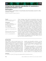
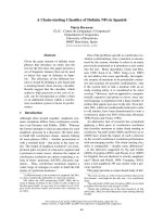
Fig. 1. Under reducing conditions, the wild-type and
the two TNSALP mutants gave a similar electropho-
retic pattern, consisting of the 66 kDa and 80 kDa
forms. However, strikingly, a distinct pattern was
obtained under nonreducing conditions. In addition to
the two molecular forms, a 160 kDa and a 130 kDa
form were found only in the cells expressing TNS-
ALP(R433C) (Fig. 1, lanes 2 and 6), indicating that a
considerable portion of newly synthesized TNS-
ALP(R433C) is covalently cross-linked via a disulfide
bond. As reported previously [13], TNSALP(D289V) is
not processed to the 80 kDa form, as this mutant is
transport-incompetent (Fig. 1, lanes 4 and 8). Instead
of being conveyed to the Golgi apparatus, it accumu-
lates in the ER, and is eventually degraded in the
ubiquitin–proteasome pathway [13]. We consistently
observed a high molecular mass aggregate even in
the cells expressing the wild-type under nonreducing
conditions (see the top of the gel, Fig. 1, lanes 5–8).
Previously, we reported that a proportion of the newly
synthesized TNSALP fails to be modified with GPI,
and resultant GPI-anchorless TNSALP molecules form
the aggregate in transfected cells [17]. This probably
reflects a shortage of a GPI precursor pool in the ER
of COS-1 cells where TNSALP is overexpressed ectopi-
cally.
The two TNSALP mutants appear
on the cell surface
Next, we investigated whether the TNSALP mutants
gain access to the cell surface like the wild-type. The
cells that expressed each TNSALP mutant were meta-
bolically labeled and further incubated with phosphati-
dylinositol-specific phospholipase C (PI-PLC). Upon
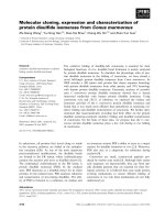
digestion, the 80 kDa form was the only form in the
culture media of the cells that expressed the wild-type
or TNSALP(R433H) (Fig. 2, lanes 4 and 12). However,
the 160 kDa form as well as the 80 kDa form were
released into the medium from the cells expressing
TNSALP(R433C) (Fig. 2, lane 8), indicating that the
dimerization via a disulfide bridge does not severely
affect the cell surface appearance of TNSALP(R433C).
As a negative control, no TNSALP(D289V) was
released into the medium by digestion with PI-PLC,
because this mutant fails to exit from the ER. Immuno-
fluorescence studies also confirmed the cell surface
appearance of the wild-type, TNSALP(R433C) and
TNSALP(R433H), but not TNSALP(D289V) (data not
shown).
Catalytic activity of TNSALP mutants
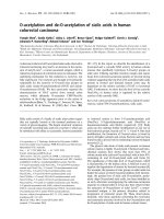
An immunoblotting method showed essentially the
same result for steady-state expression of the TNSALP
mutants as the biosynthetic experiments (Fig. 3A, lanes
5 and 6), confirming that TNSALP(R433C) tends to
become a disulfide-bonded form that is clearly differ-
ent from the noncovalently associated forms of the
wild-type, which migrates on the SDS gel as the
66 kDa or 80 kDa form.
To address the question of whether the replacement
of arginine at position 433 affects the catalytic function
of TNSALP, the cells expressing the two mutants were
assayed for alkaline phosphatase activity using p-nitro-
phenylphosphate as a substrate (Fig. 3B). Conservative
replacement of arginine with histidine was expected
not to greatly change the catalytic function of
TNSALP(R433H), although we consistently detected
higher specific enzyme activity in the cells expres-
sing TNSALP(R433H) than in those expressing the
a
Dk061
a
Dk031
a
Dk08
a
Dk66
87654321
dernonder
Fig. 1. Biosynthesis of TNSALP mutants in COS-1 cells. COS-1
cells, which had been transfected for 24 h with a plasmid enco-
ding the wild-type (lanes 1 and 5), TNSALP(R433C) (lanes 2 and 6),
TNSALP(R433H) (lanes 3 and 7) or TNSALP(D289V) (lanes 4 and 8),
were labeled with [
35
S]methionine ⁄ cysteine for 3 h. The cell lysates
were immunoprecipitated with anti-TNSALP, and the immune com-
plexes were then analyzed by SDS ⁄ PAGE ⁄ fluorography under redu-
cing (lanes 1–4) or nonreducing (lanes 5–8) conditions. Double and
single arrowheads indicate the tops of the stacking and resolving
gels, respectively. Left lane:
14
C-methylated protein markers of
200, 97.4, 66, 46 and 30 kDa, from the top of the gel.
Aberrant interchain disulfide bridge M. Nasu et al.
5614 FEBS Journal 273 (2006) 5612–5624 ª 2006 The Authors Journal compilation ª 2006 FEBS
wild-type. In contrast to this, the cell homogenate of
the cells that expressed TNSALP(R433C) showed a
much reduced level of activity as compared with the
wild-type. As a negative control, TNSALP(D289V) did
not exhibit any enzyme activity, in agreement with a
previous report [13]. K
m
(V
max
) values for the wild-type,
TNSALP(R433C) and TNSALP(R433H), which were
determined using Lineweaver–Burk plots, were
0.23 mm (2.57 lmolÆmin
)1
), 0.50 mm (1.05 lmolÆmin
)1
)
and 0.34 mm (3.69 lmolÆmin
)1
), respectively. As the
expression level of TNSALP(R433H) was higher than
that of the wild-type in the COS-1 cells, based on the
immunoblotting results (Fig. 3A, lanes 1 and 3), it
seems reasonable to assume that replacement of argin-
ine with histidine at position 433 does not have much
affect on the catalytic function of TNSALP, although a
definite conclusion awaits its purification. In the case of
TNSALP(R433C), however, we were uncertain whether
the decrease in specific enzyme activity could be attrib-
uted to disulfide bond formation, as a significant
amount of the noncross-linked molecular species was
also present in the cell homogenate (Fig. 3A, lane 6).
Expression of TNSALP(R433C) in CHO-K1 Tet-On
cells
As it was difficult to separate the noncross-linked and
the cross-linked form of TNSALP(R433C) from each
other in the native state by means of biochemical
methods such as gel filtration and electrophoresis, we
turned to another strategy. We reasoned that if expres-
sion levels of TNSALP(R433C) are kept at a relatively
low level compared with transient expression, most of
the newly synthesized TNSALP(R433C) molecules
might be oxidized to become disulfide-bonded in the
CLP-I
P
++ ++ ++
+
+
MCMCMCMCMCMCMCMC
C334Rd-typeliWV982DH334R
aDk061
aDk031
aDk08
aDk66
16
15141312
11
1098
7
6
5
43
21
Fig. 2. Cell surface appearance of TNSALP
mutants in COS-1 cells. COS-1 cells, which
had been transfected with a plasmid enco-
ding the wild-type, TNSALP(R433C), TNS-
ALP(R433H) or TNSALP(D289V) for 24 h,
were labeled with [
35
S]methionine ⁄ cysteine
for 3 h and chased for 2 h. The cells were
then further incubated in the absence or
presence of PI-PLC. The cell lysates (C) and
media (M) were immunoprecipitated with
anti-TNSALP, and the immune complexes
were analysed by SDS ⁄ PAGE (nonreduc-
ing) ⁄ fluorography. The single arrowhead
indicates the top of the resolving gels. Left
lane:
14
C-methylated protein markers as in
Fig. 1.
0
00
0
1
0002
00
0
3
0004
0
00
5
V982DH334RC334RTW
Enzyme activity (U/mg protein)
B
4321
8765
dernonder
a
Dk061
aDk031
aDk08
a
D
k6
6
A
Fig. 3. Steady-state expression of TNSALP mutants in COS-1 cells.
(A) COS-1 cells, which had been transfected with a plasmid enco-
ding the wild-type (lanes 1 and 5), TNSALP(R433C) (lanes 2 and 6),
TNSALP(R433H) (lanes 3 and 7) or TNSALP(D289V) (lanes 4 and 8)
for 24 h, were homogenized, and 10 lg of each homogenate was
directly separated by SDS ⁄ PAGE under reducing (lanes 1–4) or non-
reducing (lanes 5–8) conditions and subjected to immunoblotting
using anti-TNSALP. Double and single arrowheads indicate the tops
of the stacking and resolving gels, respectively. (B) The same
homogenates as described in (A) were assayed for alkaline phos-
phatase activity and protein. Values are means of two independent
experiments.
M. Nasu et al. Aberrant interchain disulfide bridge
FEBS Journal 273 (2006) 5612–5624 ª 2006 The Authors Journal compilation ª 2006 FEBS 5615
ER. This was the case. We succeeded in establishing a
CHO-K1 Tet-On (Tet-On) cell line that expresses
TNSALP(R433C) only in response to the addition of
doxycycline (a tetracycline analog). In marked contrast
to transient expression (Fig. 3), the 160 kDa disulfide-
bonded form was the predominant molecular species
in the Tet-On cells, with a trace amount of the 80 kDa
noncross-linked form, over a wide range of expression
conditions (Fig. 4). Induction of TNSALP(R433C)
was found to be regulated tightly, as no band was
observed in the absence of doxycycline (Fig. 4). Con-
sistent with this, the alkaline phosphatase activity of
Tet-On cells was negligible in the absence of the indu-
cer (data not shown). When its synthesis was induced,
TNSALP(R433C) was localized on the cell surface of
the Tet-On cells, as judged by immunofluorescence
(Fig. 5A) and PI-PLC digestion (Fig. 5B). Next, the
detergent extracts of cells expressing the wild-type or
TNSALP(R433C) were fractionated by sucrose density
gradient centrifugation, and the distribution of
TNSALP was analyzed by immunoprecipitation
(Fig. 6). Both the wild-type and TNSALP(R433C)
appeared at exactly the same position across the
gradient, demonstrating that the disulfide-bonded
0.5
0.20DOX 1.00.50.201.0
nonredred
a
Dk061
a
Dk031
aD
k
0
8
a
D
k66
Fig. 4. Steady-state expression of TNS-
ALP(R433C) in Tet-On cells.The established
Tet-On cells harboring a plasmid encoding
TNSALP(R433C) were cultured with differ-
ent concentrations of doxycycline for 24 h.
The cells were homogenized, and 5 lgof
each homogenate was separated by
SDS ⁄ PAGE under reducing (red) or non-
reducing (nonred) conditions; this was fol-
lowed by immunoblotting with anti-TNSALP.
DOX, doxycycline.
PCLP-I ++
MCMC
aDk061
aDk031
a
Dk08
a
Dk66
AB
Fig. 5. Cell surface appearance of TNSALP(R433C) in Tet-On cells. (A) Established Tet-On cells harboring a plasmid encoding TNS-
ALP(R433C) were cultured with 0.5 lgÆmL
)1
doxycycline for 24 h. After fixation, the cells were reacted with anti-TNSALP and then with
anti-(rabbit IgG)–rhodamine. (B) The established Tet-On cells, which had been cultured with 1.0 lgÆmL
)1
doxycycline for 14 h, were labeled
with [
35
S]methionine ⁄ cysteine for 0.5 h and chased for 1 h. The cells were further incubated in the absence or presence of PI-PLC. The cell
lysates (C) and media (M) were immunoprecipitated with anti-TNSALP, and the immune complexes were analysed by SDS ⁄ PAGE (nonreduc-
ing) ⁄ fluorography. Left lane:
14
C-methylated protein markers as in Fig. 1.
Aberrant interchain disulfide bridge M. Nasu et al.
5616 FEBS Journal 273 (2006) 5612–5624 ª 2006 The Authors Journal compilation ª 2006 FEBS
TNSALP(R433C) forms a dimer like the wild-type.
The pulse-chase experiments demonstrated that the
wild-type 66 kDa form was efficiently processed to the
mature 80 kDa form, and this mature form was the
only form found in the cell at 2 h chase time (Fig. 7A).
Similarly, the majority of TNSALP(R433C) was
121110987654321
Wild-type
C
334R
aDk
08
a
Dk061
a
D
k
0
8
c
ab
Fig. 6. Sucrose density gradient analysis of TNSALP(R433C). The established Tet-On cells harboring a plasmid encoding the wild-type or
TNSALP(R433C) were cultured with 1.0 lgÆmL
)1
doxycycline for 12 h. The cells were labeled with [
35
S]methionine ⁄ cysteine for 1 h and fur-
ther chased for 3 h. The cells were lysed, loaded on the top of the gradient [5–35% (w ⁄ w) sucrose], and centrifuged for 18 h at 4 °C. Each
400 lL fraction was collected from the top (fraction 1) of the gradient and immunoprecipitated. The immune complexes were separated by
SDS ⁄ PAGE (nonreducing), followed by fluorography. The arrowhead indicates an unknown band. BSA (b, 68 kDa), alcohol dehydrogenase (a,
141 kDa) and catalase (c, 250 kDa) were applied on a separate gradient as size markers. Left lane:
14
C-methylated protein markers of 200,
97.4 and 66 kDa from the top of the gel.
aDk08
aDk66
aDk
0
61
aDk0
31
a
Dk66
0)h(esahC
AB
215.0
epyt-
d
liW
C33
4
R
a
Dk031
aDk66
+-HodnE
Fig. 7. Biosynthesis of TNSALP(R433C) in Tet-On cells. (A) The established Tet-On cells harboring a plasmid encoding the wild-type or
TNSALP(R433C) were cultured with 1.0 lgÆmL
)1
doxycycline for 14 h, labeled with [
35
S]methionine ⁄ cysteine for 0.5 h, and chased for up to
2 h. The cells were lysed and immunoprecipitated with anti-TNSALP, and the immune complexes were separated by SDS ⁄ PAGE (non-
reducing), followed by fluorography. Left lane:
14
C-methylated protein markers of 200, 97.4 and 66 kDa from the top of the gel. (B) The
established Tet-On cells harboring a plasmid encoding TNSALP(R433C) were cultured with 1.0 lgÆmL
)1
doxycycline for 14 h. The cells were
pulse-labeled with [
35
S]methionine ⁄ cysteine for 0.5 h and immunoprecipitated for Endo H digestion. The immunoprecipitates were analyzed
by SDS ⁄ PAGE (nonreducing) ⁄ fluorography.
M. Nasu et al. Aberrant interchain disulfide bridge
FEBS Journal 273 (2006) 5612–5624 ª 2006 The Authors Journal compilation ª 2006 FEBS 5617
efficiently converted to the 160 kDa form, although a
small proportion of it remained unprocessed even after
2 h of chase. Thus we cannot exclude the possibility
that this missense mutation also affects acquisition of
the transport competence of TNSALP. The dimeri-
zation of TNSALP(R433C) must occur at the ER, as
the 130 kDa form appeared immediately after the
pulse period (Fig. 7A). Furthermore, the 130 kDa
disulfide-bonded TNSALP(R433C) was sensitive to
Endo H digestion (Fig. 7B).
The disulfide bridge suppresses the catalytic
function of TNSALP(R433C)
The predominance of the dimer form of TNS-
ALP(R433C) in the Tet-On cells in response to doxy-
cycline allowed us to unambiguously evaluate the
enzyme activity of this disulfide-bonded TNSALP
(R433C) (Fig. 8). The specific enzyme activity of the
cells expressing TNSALP(R433C) was only one-twen-
tieth of those expressing the wild-type enzyme. K
m
(and
V
max
) values obtained by kinetic studies are: 0.45 m m
(6.75 lmolÆmin
)1
) for the wild-type and 0.66 mm
(0.34 lmolÆmin
)1
) for the disulfide-bonded TNSALP
(R433C). As the wild-type and TNSALP(R433C) in
Tet-On cells were comparable in their expression levels
as estimated by immunoblotting (Fig. 9, lanes 1 and 2),
it is likely that the disulfide bond formation substan-
tially suppresses the catalytic efficiency of TNS-
ALP(R433C) without much affecting its substrate
binding.
Figure 9 shows the effect of dithiothreitol on the
biosynthesis of TNSALP(R433C). Dithiothreitol is a
membrane-permeable reducing agent and is known to
render the lumen of the ER unfavorable for oxida-
tion of sulfhydryl groups on cysteine residues [18].
The cells were incubated with doxycycline in the
absence or presence of dithiothreitol for 12 h or
24 h. A small but significant amount of the 80 kDa
form of TNSALP(R433C) was found to appear in
the cells only with dithiothreitol (Fig. 9A, lanes 3
and 7). A concentration of 1 mm of dithiothreitol
was optimal, and higher concentrations of dithiothrei-
tol tended to inhibit the synthesis of TNSALP
(R433C) induced by doxycycline. Importantly, we
detected an increase in the enzyme activity of the
cells concomitantly with the appearance of the
80 kDa form (Fig. 9B), suggesting that TNSALP
(R433C) is capable of exhibiting its catalytic activity
unless it is oxidized to form an interchain disulfide
bond. However, we failed to increase the enzyme
activity of the cell homogenate prepared from Tet-On
cells expressing TNSALP(R433C) by incubating them
with dithiothreitol or 2-mercaptoethanol under var-
ious conditions.
0
0002
0004
0
00
6
0008
00001
00021
00041
C334RTW
Enzyme activity (U/mg protein)
Fig. 8. Alkaline phosphatase activity in the Tet-On cells expressing
TNSALP(R433C). After the established Tet-On cells harboring a
plasmid encoding the wild-type or TNSALP(R433C) had been cul-
tured with 1 lgÆmL
)1
doxycycline for 24 h, the cells were homo-
genized and assayed for alkaline phosphatase and protein. The
homogenates (5 lg each) were also used for immunoblotting
(Fig. 9, lanes 1 and 2). Values are means of two independent
experiments.
Fig. 9. Effects of dithiothreitol on the expression of TNS-
ALP(R433C). After the established Tet-On cells harboring a plasmid
encoding the wild-type (lane 1) or TNSALP (R433C) (lanes 2–9) had
been cultured with 1 lgÆmL
)1
doxycycline for 12 h or 24 h in the
presence of different concentrations of dithiothreitol (A), the cell
homogenates (5 lg each) were used for immunoblotting (nonreduc-
ing). (B) The same cell homogenates as described in (A) were
investigated for alkaline phosphatase activity. The open bar and
closed bar represent 24 h or 12 h of incubation with dithiothreitol,
respectively. Values are means of two experiments.
Aberrant interchain disulfide bridge M. Nasu et al.
5618 FEBS Journal 273 (2006) 5612–5624 ª 2006 The Authors Journal compilation ª 2006 FEBS
Next, we compared protease susceptibility between
the wild-type and TNSALP(R433C). As shown in
Fig. 10, the wild-type enzyme was largely resistant to
trypsin digestion at concentrations up to 50 lgÆmL
)1
,
whereas the mutant protein was found to be degraded
at higher concentrations of trypsin. The same holds
true for proteinase K digestion. The mutant protein
completely disappeared even at 0.5 lgÆmL
)1
(lane 7),
but not the wild-type (lane 2). These results therefore
suggest that the interchain disulfide bond markedly
changes the tertiary structure of TNSALP such that
TNSALP(R433C) becomes more susceptible to the
proteases.
An interchain disulfide bridge forms between
two cysteines at position 433
Human TNSALPs have five cysteine residues (C102,
C122, C184, C472 and C480) per subunit, and their
positions are well conserved among four isoenzymes
[3,19]. C122 and C472 are thought to bond to C184
and C480 in the same subunit, respectively, whereas
C102 is in a free state, raising the possibility that
C102 is involved in the interchain disulfide bridge of
TNSALP(R433C). To address this question, we
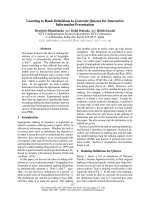
replaced C102 with serine and expressed TNSALP
(C102S) in the COS-1 cells as shown in Fig. 11.
TNSALP(C102S) consists of the 66 kDa immature and
80 kDa mature forms, and showed a similar specific
enzyme activity to that of the wild-type. Also, a TNS-
ALP double mutant (C102S ⁄ R433C) was found to be
indistinguishable from TNSALP(R433C) as assessed
by immunoblotting, as shown in Fig. 11A (lanes 7 and
8), suggesting that a disulfide bond forms between
C433 residues on two subunits of TNSALP(R433C).
Discussion
Hypophosphatasia and TNSALP mutants
Inorganic pyrophosphate is believed to play a pivotal
role in bone matrix mineralization [20,21]. At lower
concentrations (0.01–0.1 mm), pyrophosphate enhances
mineralization, whereas it inhibits the formation of
hydroxyapatite at concentrations higher than 1 mm.
TNSALP is thought to promote mineralization by
hydrolyzing pyrophosphate into phosphate. Fine regu-
lation of pyrophosphate levels at the site of mineral-
ization also requires at least two other proteins:
nucleoside triphosphate pyrophosphatase phospho-
diesterase (or PC-1), which generates pyrophosphate
from nucleoside triphosphate, and a channel protein
ANK (ankylosis), which mediates transport of pyro-
phosphate across the plasma membrane of osteoblasts
[3,22,23].
Various mutations in the TNSALP gene cause a her-
editary disease known as hypophosphatasia, which is
characterized by defective osteogenesis, unequivocally
pointing to the physiologic relevance of the enzyme in
biomineralization [4,5,7]. In support of this, relevant
knock-out mice develop rickets and osteomalacia, thus
recapitulating infantile hypophosphatasia [24–26]. In
TNSALP-deficient mice, the initiation of mineral
crystallization occurs within matrix vesicles; however,
2101
9
87654321 019876543
C334Rd-typeliwC334Rd-typeliw
nispyrtKesanietorp
aDk08
aDk061
Fig. 10. Protease sensitivity. After the established Tet-On cells harboring a plasmid encoding the wild-type or TNSALP(R433C) had been cul-
tured with 1 lgÆmL
)1
doxycycline for 24 h, the cells were homogenized in 10 mM Tris ⁄ HCl (pH 8.0), using a sonicator. The homogenates
were incubated with trypsin or proteinase K in an ice ⁄ water bath for 30 min at the indicated concentrations. For trypsin, the final concentra-
tions were (lg ⁄ mL): lanes 1 and 6, 0; lanes 2 and 7, 5; lanes 3 and 8, 10; lanes 4 and 9, 20; lanes 5 and 10, 50. For proteinase K, the final
concentrations were (lg ⁄ mL): lanes 1 and 6, 0; lanes 2 and 7, 0.5; lanes 3 and 8, 1.0; lanes 4 and 9, 5.0; lanes 5 and 10, 10. Lanes 1–5 and
lanes 6–10 were analyzed by SDS ⁄ PAGE in the presence or absence of 2-mercaptoethanol, respectively.
M. Nasu et al. Aberrant interchain disulfide bridge
FEBS Journal 273 (2006) 5612–5624 ª 2006 The Authors Journal compilation ª 2006 FEBS 5619
the subsequent proliferation and growth stage of min-
eralization is severely impaired, leading to an increase
in noncalcified bone matrix (osteoid) [27], consistent
with what is seen in hypophosphatasia patients [28].
Hypophosphatasia patients show wide-ranging clinical
manifestations, from stillbirth with an almost unminer-
alized skeleton to premature loss of deciduous teeth in
childhood and pseudofracture first presenting in adult
life [1,2]. The symptoms of hypophosphatasia are well
known to correlate with the residual enzyme activities
of affected patients [1,2,6,7]. During the course of our
studies on the biosynthesis of several TNSALP
mutants, we found that the missense mutations associ-
ated with severe hypophosphatasia variously affect the
efficiency with which TNSALP properly folds and cor-
rectly assembles, depending upon the position of a
missense mutation and the nature of a substituted
amino acid. For example, TNSALP(A162T), found in
a homozygous patient diagnosed with a lethal infantile
form of hypophosphatasia [4], mainly formed a high
molecular mass aggregate in the ER, and only a small
proportion of newly synthesized TNSALP(A162T)
reached its site of action, the cell surface [9,10]. Conse-
quently, the cell surface expression of this TNSALP
mutant is much reduced compared with that of the
wild-type. More TNSALP(D277A) was found to gain
access to the cell surface than TNSALP(A162T),
albeit with a significant population in the ER [10].
Alternatively, TNSALP(R54C), TNSALP(N153D),
TNSALP(E218G), TNSALP(D289V) and TNSALP
(G317D) never appeared on the plasma membrane
[9–13]. Interestingly, a recent study has shown that
alkaline phosphatase acquires Zn
2+
, which is indis-
pensable for its catalytic activity, in the Golgi
apparatus on its way to the plasma membrane [29].
This leads to the speculation that TNSALP mutants,
which are retained in the ER due to a folding defect,
not only fail to appear on the cell surface, but also
are not able to acquire Zn
2+
. Consistent with this,
the TNSALP mutants with defective ER-to-Golgi
transport did not show measurable alkaline phospha-
tase activity when being expressed in COS-1 cells
[10–13].
TNSALP(R433C) becomes cross-linked via
a disulfide bridge
Mammalian alkaline phosphatases have five cysteine
residues per subunit, and their positions are well con-
served [3,19,30]. C102 is believed to be present only
in a free state, whereas C122 and C472 bind to C184
and C480, respectively, in the same subunit. Both
TNSALP(C184Y) and TNSALP(C472S) have been
reported in perinatal hypophosphatasia patients
[15,31], implying that the two interchain disulfide
bonds are necessary for the correct folding and
assembly of TNSALP. TNSALP(R433C) was repor-
ted in homozygous patients diagnosed with lethal
infantile hypophosphatasia [16,32]. In contrast to the
TNSALP mutants showing various degrees of folding
defect, TNSALP(R433C) did not form a high molecu-
lar mass aggregate. Instead, it formed a covalently
cross-linked homodimer, as evidenced by sucrose den-
sity gradient centrifugation (Fig. 6). As replacement
reducing
AB
nonreducing
123 4 5678
6000
5000
4000
160 kDa
80 kDa
Enzyme activity (U/mg protein)
3000
2000
1000
0
WT C102S R433C C102S/R433C
Fig. 11. Expression of a TNSALP double mutant (C102S ⁄ R433C) in COS-1 cells. COS-1 cells expressing wild-type enzyme (lanes 1 and 5),
TNSALP(C102S) (lanes 2 and 6), TNSALP(R433C) (lanes 3 and 7) or TNSALP(C102S ⁄ R433C) (lanes 4 and 8) were homogenized. The homo-
genates were analyzed by SDS ⁄ PAGE under reducing or nonreducing conditions, and this was followed by immunoblotting with anti-
TNSALP (A) or assayed for alkaline phosphatase (B).
Aberrant interchain disulfide bridge M. Nasu et al.
5620 FEBS Journal 273 (2006) 5612–5624 ª 2006 The Authors Journal compilation ª 2006 FEBS
of C102 with serine did not affect the cross-linking of
TNSALP(R433C) (Fig. 11), this result strongly indi-
cates that a sulfhydryl group on the cysteine residue
at position 433 of one subunit is oxidized to bond to
the counterpart of the other subunit. This covalent
cross-linkage of TNSALP(R433C) occurs in an early
stage of the secretory pathway, as the cross-linked
molecular species appeared in the cell immediately
after a pulse-labeling period, and besides this, the
130 kDa form was sensitive to Endo H digestion.
Also, the results of the pulse-chase experiments
suggest that most newly synthesized TNSALP(R433C)
migrated from the ER to the Golgi apparatus at a
similar rate to the wild-type enzyme (Fig. 7). The cell
surface appearance of TNSALP(R433C) was shown
by immunofluorescence microscopy and PI-PLC
digestion (Fig. 5), indicating that TNSALP(R433C)
resides on the cell surface as a GPI-anchored ecto-
enzyme, like the wild-type. Thus, it is likely that the
cross-linkage between the subunits did not greatly
affect the biosynthesis and intracellular transport of
this mutant protein. However, the intersubunit cross-
linkage did severely affect the catalytic activity of
TNSALP(R433C). This is based on the findings in
Tet-On cells, which predominantly express the cross-
linked form of TNSALP(R433C) in response to doxy-
cycline. Considering that the expression levels of
TNSALP(R433C) and the wild-type in each Tet-On
cell line are very similar (Fig. 9A), comparison of K
m
and V
max
values suggests that the catalytic efficiency
of the mutant protein is dramatically reduced com-
pared with that of the wild-type. Increased suscepti-
bility of TNSALP(R433C) to proteases supports the
notion that the disulfide bridge has a profound effect
on the structure of TNSALP (Fig. 10). The effects of
substitution of R433 either with alanine or aspartate
on the catalytic properties of TNSALP were reported
by Kozlenkov et al. [33]. Both TNSALP(R433A) and
TNSALP(R433D) showed a noticeable decrease in
k
cat
with a moderate increase in K
m
. One might argue
that the substitution of arginine with cysteine itself,
but not the disulfide bridge, decreases the catalytic
activity of TNSALP(R433C). However, this is unli-
kely, for the following reasons: first, the COS-1 cells,
which express both noncross-linked and cross-linked
TNSALP(R433C), showed considerable enzyme acti-
vity (Fig. 3). Second, when the Tet-On (R433C) cells
were cultured in the presence of doxycycline and di-
thiothreitol, a significant amount of TNSALP(R433C)
failed to become cross-linked, and concomitantly we
detected an increase in enzyme activity in the cell
homogenates. Taken together, these facts suggest that
the diminished catalytic function of TNSALP(R433C)
due to its disulfide-bonded linkage is a likely cause
for the lethal hypophosphatasia resulting from the
homozygous presence of this mutation. To our know-
ledge, this is the first TNSALP missense mutation
associated with severe hypophosphatasia that abro-
gates the catalytic activity of TNSALP without signi-
ficantly affecting its cell surface expression.
TNSALP(R433H) was reported in a compound
heterozygote (R433H ⁄ D389G) diagnosed with a mild
form of hypophosphatasia [15]. Therefore, it is reason-
able to assume that TNSALP(R433H) does not have a
severe effect, unlike TNSALP(R433C). Also, as the
substitution of arginine with histidine is a conservative
replacement, it was expected that this mutation would
not much affect TNSALP activity. When expressed in
COS-1 cells, TNSALP(R433H) showed enzyme activity
comparable to that of the wild-type. Also, its biosyn-
thesis and cell surface appearance were not measurably
disturbed (Figs 1–3), further highlighting the clinical
importance of the substitution of arginine at position
433 with cysteine.
Experimental procedures
Materials
Express
35
S
35
S protein labeling mix (> 1000 CiÆmmol
)1
)
was obtained from Dupont-New England Nuclear
(Boston, MA), and
14
C-methylated proteins and enhanced
chemiluminescence western blotting detection reagent, per-
oxidase-conjugated donkey anti-(rabbit IgG) and protein
A–Sepharose CL-4B were obtained from Amersham Phar-
macia Biotech (Arlington Heights, IL); the pALTER-MAX,
Altered sites II mammalian mutagenesis system was
obtained from Promega (Madison, WI); the QuikChange II
Site-Directed Mutagenesis kit was obtained from Stratagene
(La Jolla, CA); G418 and pansorbin were obtained from
Calbiochem (La Jolla CA); Lipofectamine Plus Reagent was
obtained from Invitrogen (Carlsbad, CA); PI-PLC was
obtained from BIOMOL International, L.P. (Plymouth
Meeting, PA); aprotinin, doxycycline and saponin (Quillaja
Bark) and l-1-tosylamide-2-phenylethyl-chloromethyl
ketone-treated bovine pancreas trypsin were obtained from
Sigma Chemical Co. (St Louis, MO); proteinase K was
obtained from Roche Diagnotics (London, UK); antipain,
chymostatin, elastatinal, leupeptin and pepstatin A were
obtained from the Protein Research Foundation (Osaka,
Japan); hygromycin B and p-amidinophenylmethanesulfonyl
fluoride were obtained from Wako Pure Chemicals (Tokyo,
Japan); and serum against recombinant human TNSALP
was raised in rabbits as described previously [34]. pTRE2
and the BD CHO-K1 Tet-On cell line and Tet system
approved fetal bovine serum were obtaied from BD
Biosciences Clontech (Palo Alto, CA).
M. Nasu et al. Aberrant interchain disulfide bridge
FEBS Journal 273 (2006) 5612–5624 ª 2006 The Authors Journal compilation ª 2006 FEBS 5621
Plasmids and transfection
The pALTER-Max encoding the wild-type TNSALP was
constructed as described previously [12]. Mutations were
introduced at specific sites using the Altered sites II mamma-
lian mutagenesis system as described previously [12,13].
The oligonucleotides used were: R433H, 5¢-CGTGGGT
CTCATGATGCAGGGGCAC-3¢; and R433C, 5¢-CGT
GGGTCTCATGACACAGGGGCAC-3¢. For the conver-
sion of the cysteine residue at position 102, the QuikChange
II Site-Directed Mutagenesis kit was used with two
primers: 5¢-ACCGCCTACCTGAGTGGGGTGAAGGCC
AAT-3¢ and 5¢-ATTGGCCTTCACCCCACTCAGGTA
GGCGGT-3¢. The DNA sequence of the mutation sites was
verified by DNA sequence analyses. The cDNA encoding
TNSALP(R433C) was further subcloned into pTRE2 to
establish stable cell lines. Transfection and screening of stable
cell lines were performed essentially according to the manu-
facturer’s protocol. Tet-On cells, which successfully produced
the mutant TNSALP in the presence of doxycycline, but not
in its absence, were identified using immunofluorescence. The
establishment and characterization of Tet-On cells expressing
the wild-type TNSALP will be published elsewhere. Estab-
lished Tet-On cells were cultured and passaged in the absence
of doxycycline until they were used for experiments. For
immunoblotting or immunofluorescence studies, the cells
were cultured in the presence of 0.5–1 lgÆ mL
)1
doxycycline
for 12 h or 24 h before being used. Alternatively, cells were
cultured in the presence of 0.2–1 lgÆmL
)1
doxycycline for
14 h before biosynthesis experiments. For transient expres-
sion, COS-1 cells (1.0–1.3 · 10
5
cells per 35 mm dish) were
transfected with 0.5–0.8 lg of each plasmid using Lipofecta-
mine Plus, according to the manufacturer’s protocol as
described previously [12,13], and the transfected cells were
incubated for 24 h in a 5% CO
2
⁄ 95% air incubator before
use. COS-1 cells were cultured in DMEM supplemented with
10% fetal bovine serum [9].
Metabolic labeling and immunoprecipitation
For pulse-chase experiments, cells were preincubated for
0.5–1 h in the methionine ⁄ cysteine-free DMEM and labeled
with 50–100 lCi of [
35
S]methionine ⁄ cysteine for 0.5 h in
fresh methionine ⁄ cysteine-free MEM. After a pulse period,
cells were washed and chased in DMEM as described previ-
ously [12,13]. After metabolic labeling, the medium was
removed, and the cells were lysed in 0.5 mL of lysis buffer
[1% (w ⁄ v) Triton X-100, 0.5% (w ⁄ v) sodium deoxycholate
and 0.05% (w ⁄ v) SDS in NaCl ⁄ P
i
]. A protease inhibitor
cocktail (antipain, aprotinin, chymostatin, elastatinal, leu-
peptin, pepstatin A) was added to cell lysates and media
(10 lgÆmL
)1
). The lysates were incubated for 20 min at
37 °C to extract TNSALP. The lysates and media were sub-
jected to immunoisolation as described previously [9,10].
The immune complexes ⁄ Protein A beads were boiled in the
absence or presence of 1% (v ⁄ v) 2-mercaptoethanol, and
then analyzed by SDS ⁄ PAGE [9% (w ⁄ v) gels], followed by
fluorography [9].
Endo H digestion
As the cross-linked form of TNSALP(R433C) was found to
be resistant to conventional Endo H digestion [9,10], the
immune complex ⁄ protein A beads were boiled in 1% (w ⁄ v)
SDS (in 5 mm Tris ⁄ HCl, pH 7.4) and centrifuged at
20 000 g (Sigma model 3615 centrifuge with 12024H rotor).
The supernatant was then adjusted to final concentrations
of 50 mm acetate buffer (pH 5.5), 1% (w ⁄ v) Triton X-100,
and 0.1% SDS. Digestion with Endo H (final concentration
0.2 U ⁄ mL) was carried out in the presence of the protease
inhibitor cocktail for 16 h at 37 °C. TNSALP was precipi-
tated with cold acetone containing 0.1 m HCl as previously
described [35].
Protease digestion
Tet-On cells harboring the wild-type or TNSALP(R433C)
cDNA were cultured in the presence of 1 lgÆmL
)1
doxycy-
cline for 1 day. Cells were homogenized in 10 mm Tris ⁄ HCl
(pH 8.0) using a sonicator. Each 5 lg of homogenate was
incubated with increasing concentrations of trypsin or pro-
teinase K at pH 8.0 in a total volume of 20 lL in an ice ⁄
water bath, essentially according to Akiyama and Ito
[36]. After 30 min, 1 lL of 100 mm p-amidinophenyl
methanesulfonyl fluoride was added to the reaction mix-
tures to stop the reaction. Each sample was mixed with
3 · SDS sample buffer, and this was followed by
SDS ⁄ PAGE ⁄ immunoblotting.
Miscellaneous procedures
Immunofluorescence determination of alkaline phosphatase
was performed as described previously [12,13]. Sucrose den-
sity gradient centrifugation was performed as described pre-
viously [10,12]. Electrical transfer of proteins and
subsequent procedures were as described previously [12,13].
Proteins on membranes were detected with enhanced chemi-
luminescence western blotting detection reagents. Protein
and alkaline phosphatase assays were performed as des-
cribed previously [9,10]. One unit of alkaline phosphatase
activity is defined as nmoles of p-nitrophenylphosphate
hydrolyzed per min at 37 °C.
Acknowledgements
We thank Dr Yoshio Misumi, Dr Miwa Sohda and Dr
Tsuneo Imanaka for their advice on establishing Tet-
On cells expressing a TNSALP mutant. We also thank
anonymous reviewers for bringing Cys102 to our
Aberrant interchain disulfide bridge M. Nasu et al.
5622 FEBS Journal 273 (2006) 5612–5624 ª 2006 The Authors Journal compilation ª 2006 FEBS
attention and suggesting the protease sensitivity assay.
This work was supported in part by a Grant-in-Aid
for Scientific Research from the Ministry of Education,
Culture, Sports and Technology of Japan (to KO) and
by a grant for the Promotion of Niigata University
Research Project (to KO).
References
1 Harris H (1989) The human alkaline phosphatases: what
we know and what we don’t know. Clin Chim Acta 186,
133–150.
2 Whyte MP (2001) Hypophosphatasia. In The Metabolic
and Molecular Basis of Inherited Disease (Scriver CR,
Beaudet AL, Sly WS, Valle D, Childs B, Kinzler KW &
Vogelstein B, eds), 8th edn, Vol. 4, pp. 5313–5329.
McGraw-Hill, New York, NY.
3 Milla
´
n JL (2006) Mammalian Alkaline Phosphatases:
from Biology to Applications in Medicine and Biotechno-
logy. Wiley-VCH-Verlag GmbH KGaA, Weinheim.
4 Weiss MJ, Cole DE, Ray K, Whyte MP, Lafferty MA,
Mulivor RA & Harris H (1988) A missense mutation in
the human liver ⁄ bone⁄ kidney alkaline phosphatase gene
causing a lethal form of hypophosphatasia. Proc Natl
Acad Sci USA 85, 7666–7669.
5 Mumm S, Jones J, Finnegan P & Whyte MP (2001)
Hypophosphatasia: molecular diagnosis of Rathbun’s
original case. J Bone Miner Res 16, 1724–1727.
6 Zurutuza L, Muller F, Gibrat JF, Taillandier A, Simon-
Bouy B, Serre JL & Mornet E (1999) Correlations of
genotype and phenotype in hypophosphatasia. Hum
Mol Genet 8 , 1039–1046.
7 Mornet E (2000) Hypophosphatasia: the mutations in
the tissue-nonspecific alkaline phosphatase gene. Hum
Mutat 15, 309–315.
8 Mornet E, Stura E, Lia-Baldin AS, Stigbrand T, Menez
A & Le Du MH (2001) Structural evidence for a func-
tional role of human tissue nonspecific alkaline phos-
phatase in bone mineralization. J Biol Chem 276,
31171–31178.
9 Shibata H, Fukushi M, Igarashi A, Misumi Y, Ikehara
Y, Ohashi Y & Oda K (1998) Defective intracellular
transport of tissue-nonspecific alkaline phosphatase with
an Ala
162
fi Thr mutation associated with lethal
hypophosphatasia. J Biochem (Tokyo) 123, 968–977.
10 Fukushi-Irie M, Ito M, Amaya Y, Amizuka N, Ozawa
H, Omura S, Ikehara Y & Oda K (2000) Possible inter-
ference between tissue-non-specific alkaline phosphatase
with an Arg
54
fi Cys substitution and a counterpart
with an Asp
277
fi Ala substitution found in a com-
pound heterozygote associated with severe hypopho-
sphatasia. Biochem J 348, 633–642.
11 Fukushi M, Amizuka N, Hoshi K, Ozawa H, Kumagai
H, Omura S, Misumi Y, Ikehara Y & Oda K (1998)
Intracellular retention and degradation of tissue-nonspe-
cific alkaline phosphatase with a Gly
317
fi Asp substitu-
tion associated with lethal hypophosphatasia. Biochem
Biophys Res Commun 246, 613–618.
12 Ito M, Amizuka N, Ozawa H & Oda K (2002) Reten-
tion at the cis-Golgi and delayed degradation of tissue-
non-specific alkaline phosphatase with an Asn
153
fi Asp
substitution, a cause of perinatal hypophosphatasia.
Biochem J 361, 473–480.
13 Ishida Y, Komaru K, Ito M, Amaya Y, Kohno S &
Oda K (2003) Tissue-nonspecific alkaline phosphatase
with an Asp
289
fi Val mutation fails to reach the cell
surface and undergoes proteasome-mediated degrada-
tion. J Biochem (Tokyo) 134, 63–70.
14 Hessle L, Johnson KA, Anderson HC, Narisawa S, Sali
A, Goding JW, Terkeltaub R & &Millan JL (2002)
Tissue-nonspecific alkaline phosphatase and plasma cell
membrane glycoprotein-1 are central antagonistic regu-
lators of bone mineralization. Proc Natl Acad Sci USA
99, 9445–9449.
15 Taillandier A, Cozien E, Muller F, Merrien Y, Bonnin
E, Fribourg C, Simon-Bouy B, Serre JL, Bieth E, Bren-
ner R et al. (1999) Fifteen new mutations (-195C>T,
L-12X,298–2A>G, T117N, A159T, R229S, 997+2T>
A, E274X, A331T, H364R, D389G,1256delC, R433H,
N461I, C472S) in the tissue-nonspecific alkaline phos-
phatase (TNSALP) gene in patients with hypophospha-
tasia. Hum Mutat 15, 293.
16 Mornet E, Taillandier A, Peyramaure S, Kaper F,
Muller F, Brenner R, Bussiere P, Friesinger P, Godard J,
Le Merrer M et al. (1998) Identification of fifteen novel
mutations in the tissue-nonspecific alkaline phosphatase
(TNSALP) gene in European patients with severe hypo-
phosphatasia. Eur J Hum Genet 6, 308–314.
17 Komaru K, Ishida Y, Amaya Y, Goseki-Sone M, Ori-
mo H & Oda K (2005) Novel aggregate formation of a
frame-shift mutant protein of tissue-nonspecific alkaline
phosphatase is ascribed to three cysteine residues in the
C-terminal extension. Retarded secretion and proteaso-
mal degradation. FEBS J 272, 1704–1717.
18 Lodish HF & Kong N (1993) The secretory pathway is
normal in dithiothreitol-treated cells, but disulfide-bonded
proteins are reduced and reversibly retained in the endo-
plasmic reticulum. J Biol Chem 268, 20598–20605.
19 Le Du MH, Stigbrand T, Taussig MJ, Menez A & Stura
EA (2001) Crystal structure of alkaline phosphatase from
human placenta at 1.8 A
˚
resolution. Implication for a
substrate specificity. J Biol Chem 276, 9158–9165.
20 Anderson HC, Garimella R & Tague SE (2005) The
role of matrix vesicles in growth plate development and
biomineralization. Front Biosci 10, 822–837.
21 Murshed M, Harmey D, Milla
´
n JL, MaKee MD &
Karsenty G (2005) Unique coexpression in osteoblasts
of broadly expressed genes accounts for the spatial
M. Nasu et al. Aberrant interchain disulfide bridge
FEBS Journal 273 (2006) 5612–5624 ª 2006 The Authors Journal compilation ª 2006 FEBS 5623
restrictionof ECM mineralization to bone. Genes Dev
19, 1093–1104.
22 Harmey D, Hessle L, Narisawa S, Johnson KA, Terkel-
taub R & Millan JL (2004). Concerted regulation of
inorganic pyrophosphate and osteopontin by akp2,
enpp1 and a; an integrated model of the pathogenesis of
mineralization disorders. Am J Pathol 164, 1199–2019.
23 Anderson HC, Harmey D, Camacho NP, Garimella R,
Sipe JB, Tague S, Bi X, Johnson K, Terkeltaub R &
Millan JL (2005) Sustained osteomalacia of long bones
despite major improvement in other hypophosphatasia-
related mineral deficits in tissue nonspecific alkaline
phosphatase ⁄ nucleotide pyrophosphatase phosphodiest-
erase 1 double-deficient mice. Am J Pathol 166, 1711–
1720.
24 Waymire KG, Mahuren JD, Jaje JM, Guilarte TR,
Coburn SP & MacGregor GR (1995) Mice lacking
tissue non-specific alkaline phosphatase die from
seizures due to defective metabolism of vitamin B-6.
Nat Genet 11, 45–51.
25 Narisawa S, Frohlander N & Milla
´
n JL (1997) Inactiva-
tion of two mouse alkaline phosphatase genes and
establishment of a model of infantile hypophosphatasia.
Devdyn 208, 432–446.
26 Fedde KN, Blair L, Silverstein J, Coburn SP, Ryan
LM, Weinstein RS, Waymire K, Narisawa S, Milla
´
n JL,
MacGregor GR et al. (1999) Alkaline phosphatase
knock-out mice recapitulate the metabolic and skeletal
defects of infantile hypophosphatasia. J Bone Mineral
Res 14, 2015–2026.
27 Anderson HC, Sipe JB, Hessle L, Dhanyamraju R, Atti
E, Camacho NP & Milla
´
n JL (2004) Impaired calcifica-
tion around matrix vesicles of growth plate and bone in
alkaline phosphatase-deficient mice. Am J Pathol 164,
841–847.
28 Anderson HC, Hsu HH, Morris DC, Fedde KN &
Whyte MP (1997) Matrix vesicles in osteomalacic hypo-
phosphatasia bone contain apatite-like mineral crystals.
Am J Pathol 151, 1555–1561.
29 Suzuki T, Ishihara K, Migaki H, Matsuura W, Kohda
A, Okumura K, Nagao M, Yamaguchi-Iwai Y &
Kambe T (2005) Zinc transporters, ZnT5 and ZnT7, are
required for the activation of alkaline phosphatases,
zinc-requiring enzymes that are glycosylphosphatidyl-
inositol-anchored to the cytoplasmic membrane. J Biol
Chem 280, 637–643.
30 Kozlenkov A, Manes T, Hoylaerts MF & Millan JL
(2002) Function assignment to conserved residues in
mammalian alkaline phosphatases. J Biol Chem 277,
22992–22999.
31 Taillandier A, Zurutuza L, Muller F, Simon-Bouy B,
Serre JL, Bird L, Brenner R, Boute O, Cousin J, Gail-
lard D et al. (1999) Characterization of eleven novel
mutations (M45L, R119H, 544delG, G145V, H154Y,
C184Y, D289V, 862+5A, 1172delC, R411X, E459K) in
the tissue-nonspecific alkaline phosphatase (TNSALP)
gene in patients with severe hypophosphatasia. Hum
Mutat 13, 171–172.
32 Whyte MP, Essmyer K, Geimer M & Mumm S (2006)
Homozygosity for TNSALP mutation 1348C>T
(Arg433Cys) causes infantile hypophosphatasia mani-
festing transient disease correction and variably lethal
outcome in a kindred of black ancestry. J Pediatr 148,
753–758.
33 Kozlenkov A, Le Du MH, Cuniasse P, Ny T, Hoylaerts
MF & Milla
´
n JL (2004) Residues determining the bind-
ing specificity of uncompetitive inhibitors to tissue-non-
specific alkaline phosphatase. J Bone Miner Res 19,
1862–1872.
34 Oda K, Amaya Y, Fukushi-Irie M, Kinameri Y, Ohsuye
K, Kubota I, Fujimura S & Kobayashi J (1999) A gen-
eral method for rapid purification of soluble versions of
glycosylphosphatidylinositol-anchored proteins
expressed in insect cells: an application for human tis-
sue-nonspecific alkaline phosphatase. J Biochem
(Tokyo)
126, 694–699.
35 Oda K, Koriyama Y, Yamada E & Ikehara Y (1986)
Effects of weakly basic amines on proteolytic proces-
sing and terminal glycosylation of secretory proteins
in cultured rat hepatocytes. Biochem J 240, 739–
745.
36 Akiyama Y & Ito K (1989) Export of Escherichia coli
alkaline phosphatase attached to an integral membrane
protein, Sec Y. J Biol Chem 264, 437–442.
Aberrant interchain disulfide bridge M. Nasu et al.
5624 FEBS Journal 273 (2006) 5612–5624 ª 2006 The Authors Journal compilation ª 2006 FEBS