Báo cáo khoa học: Separation and characterization of caveolae subclasses in the plasma membrane of primary adipocytes; segregation of specific proteins and functions docx
Bạn đang xem bản rút gọn của tài liệu. Xem và tải ngay bản đầy đủ của tài liệu tại đây (429.62 KB, 12 trang )
Separation and characterization of caveolae subclasses in
the plasma membrane of primary adipocytes; segregation
of specific proteins and functions
Unn O
¨
rtegren
1
, Lan Yin
1
, Anita O
¨
st
1
, Helen Karlsson
2
, Fredrik H. Nystrom
3
and Peter Stra
˚
lfors
1
1 Department of Cell Biology and Diabetes Research Centre, University of Linko
¨
ping, Sweden
2 Department of Molecular and Clinical Medicine, University of Linko
¨
ping, Sweden
3 Department of Medicine and Care and Diabetes Research Centre, University of Linko
¨
ping, Sweden
Caveolae are defined by electron microscopy as un-
coated, omega- or flask-shaped invaginations of the
plasma membrane and also by the presence of the
structural protein caveolin (isoforms 1–3) [1]. Caveo-
lin-1 and caveolin-2 are found in most cell types, inclu-
ding adipocytes. The caveolae membrane is rich in
cholesterol and sphingomyelin [2–4]. Adipocytes have
a very high number of caveolae in their plasma
Keywords
cholesterol; FATP; GLUT4; insulin
receptorSR-BI
Correspondence
P. Stralfors, Department of Cell Biology,
Faculty of Health Sciences, SE58185
Linko
¨
ping, Sweden
Fax: +46 13 224314
Tel: +46 13 224315
E-mail:
(Received 7 April 2006, revised 27 April
2006, accepted 26 May 2006)
doi:10.1111/j.1742-4658.2006.05345.x
Caveolae are nearly ubiquitous plasma membrane domains that in adipo-
cytes vary in size between 25 and 150 nm. They constitute sites of entry
into the cell as well as platforms for cell signalling. We have previously
reported that plasma membrane-associated caveolae that lack cell surface
access can be identified by electron microscopy. We now report the identifi-
cation, after density gradient ultracentrifugation, of a subclass of very
high-density apparently closed caveolae that were not labelled by cell sur-
face protein labelling of intact cells. These caveolae contained caveolin-1
and caveolin-2. Another class of high-density caveolae contained
caveolin-1, caveolin-2 and specifically fatty acid transport protein-1, fatty
acid transport protein-4, fatty acyl-CoA synthetase, hormone-sensitive
lipase, perilipin, and insulin-regulated glucose transporter-4. This class of
caveolae was specialized in fatty acid uptake and conversion to triacylglyc-
erol. A third class of low-density caveolae contained the insulin receptor,
class B scavenger receptor-1, and insulin-regulated glucose transporter-4.
Small amounts of these proteins were also detected in the high-density
caveolae. In response to insulin, the insulin receptor autophosphorylation
and the amount of insulin-regulated glucose transporter-4 increased in
these caveolae. The molar ratio of cholesterol to phospholipid in the three
caveolae classes varied considerably, from 0.4 in very high-density caveolae
to 0.9 in low-density caveolae. There was no correlation between the caveo-
lar contents of caveolin and cholesterol. The low-density caveolae, with the
highest cholesterol concentration, were particularly enriched with the cho-
lesterol-rich lipoprotein receptor class B scavenger receptor-1, which medi-
ated cholesteryl ester uptake from high-density lipoprotein and generation
of free cholesterol in these caveolae, suggesting a specific role in cholesterol
uptake ⁄ metabolism. These findings demonstrate a segregation of functions
in caveolae subclasses.
Abbreviations
FATP, fatty acid transport protein; GLUT4, insulin-regulated glucose transporter; HD-caveolae, high-density caveolae; HDL, high-density
lipoprotein; LD-caveolae, low-density caveolae; LDL, low-density lipoprotein; SR-B1, class B scavenger receptor-1; sulfo-NHS-biotin, sulfo-N-
hydroxysuccinimidyl-biotin; VHD-caveolae, very high-density caveolae; VLDL, very low-density lipoprotein.
FEBS Journal 273 (2006) 3381–3392 ª 2006 The Authors Journal compilation ª 2006 FEBS 3381
membranes. About one-third of the plasma membrane
of these cells constitutes caveolae membrane and the
caveolae vary considerably in size, from about 25 to
over 100 nm in diameter [5].
In adipocytes the insulin receptor resides in caveolae
[6,7], and insulin-stimulated uptake of glucose takes
place in caveolae after insulin-regulated glucose trans-
porter-4 (GLUT4) translocation to caveolae in the
plasma membrane [8,9]. We recently reported that a
distinct class of caveolae in adipocytes harbour the
enzymatic machinery for synthesis of triacylglycerol
and represents a location for uptake of exogenous
fatty acids and their conversion to triacylglycerol [10].
We have earlier also reported that almost half of the
roughly 10
6
caveolae in rat adipocytes are smaller than
about 50 nm in diameter [5]. By labelling intact cells
with ruthenium red, which is electron dense and does
not permeate the plasma membrane, and examination
by electron microscopy we have demonstrated that
these caveolae are not open to the cell surface and the
surrounding medium [5]. In contrast, the caveolae that
are larger than about 50 nm are labelled by ruthenium
red and are thus open cell surface caveolae [5]. This
indicates the presence of additional specialized sub-
groups or classes of caveolin-containing membranes in
the plasma membrane.
We now report the separation and biochemical char-
acterization of three classes of caveolae, which demon-
strate a segregation of functions in caveolae subclasses.
Results
Primary rat adipocytes were homogenized and plasma
membranes isolated by differential and density gradi-
ent ultracentrifugation (Fig. 1). The purified plasma
membrane fraction contained less than 1% of the cell’s
mitochondrial cytochrome oxidase and less than 7%
of the cell Golgi TGN38 protein, indicating a very lim-
ited contamination by other membrane fractions of the
purified plasma membrane [2].
Purified plasma membranes were disrupted by soni-
cation to release caveolae [2,11,12] and subjected to lin-
ear sucrose gradient ultracentrifugation. Membranes in
collected fractions were pelleted and analysed for cave-
olin content by SDS ⁄ PAGE and immunoblotting with
anticaveolin-1 antibodies. A broad peak with a shoul-
der of caveolin suggested heterogeneity and the partial
separation of caveolin-containing membranes (Fig. 2B).
Analysis of the specific protein and lipid composition
reported below demonstrated the presence of at least
three subclasses of caveolae, each with its specific
composition: a very high-density class of caveolae at
about 31% sucrose (VHD-caveolae; corresponding to
density ¼ 1.11 gÆmL
)1
), and two caveolae classes of
lower density, a high-density class of caveolae at about
25% sucrose (HD-caveolae; 1.09 gÆmL
)1
) and a low-
density class of caveolae at about 18% sucrose (LD-
caveolae; 1.06 gÆmL
)1
), respectively (Fig. 2B). This
should be compared to intact plasma membranes with
a density of 1.14 gÆmL
)1
[13]. It was not possible to
attain complete separation of all three caveolae classes
in a sucrose gradient. Utilizing a step sucrose gradient,
we demonstrated that the HD-caveolae and LD-caveo-
lae can be separated and thus represent separate entities
(Fig. 3), although at the cost of the resolution from the
caveolae at 31% sucrose.
The distribution of caveolin-2 coincided with that
of caveolin-1 over all three caveolae (Fig. 2C). Insulin
did not affect the distribution between or amount of
Fig. 1. Outline of the procedure used for isolation of caveolae sub-
classes. Also shown is the step at which other cellular fractions are
removed.
Identification of caveolae subclasses U. O
¨
rtegren et al.
3382 FEBS Journal 273 (2006) 3381–3392 ª 2006 The Authors Journal compilation ª 2006 FEBS
A
B
C
D
E
F
G
H
I
J
K
L
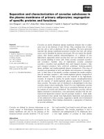
Fig. 2. Linear density gradient ultracentrifugal separation of closed and open invaginated plasma membrane caveolae. Purified adipocyte
plasma membranes were disrupted in alkaline carbonate buffer and subjected to sucrose density gradient ultracentrifugation as detailed in
Experimental procedures. Equal volumes of fractions were collected from the bottom of the tube, and membranes were collected by centrif-
ugation and analysed for the distribution of indicated protein or EZ-Link-sulfo-NHS-LC-biotin labelling by SDS ⁄ PAGE and immunoblotting or
avidin-labelled HRP, respectively. The amounts of indicated proteins are expressed in arbitrary densitometric units and normalized to percent-
age of maximum. (A) Protein concentration (mgÆmL
)1
). (B) Caveolin-1. (C) Caveolin-2. (D) Cell surface biotin labelling of proteins. (E) TGN38.
(F) Insulin receptor. (G) GLUT4. (H) Hormone-sensitive lipase (HSL). (I) FATP1. (J) FATP4. (K) Perilipin. (L) SR-BI.
U. O
¨
rtegren et al. Identification of caveolae subclasses
FEBS Journal 273 (2006) 3381–3392 ª 2006 The Authors Journal compilation ª 2006 FEBS 3383
caveolin-1 in the different caveolae (Fig. 4A). The pro-
tein profile mirrored the caveolin profile (Figs 2A and
4A), indicating that the caveolae fractions are not con-
taminated by noncaveolar membranes [2]. Noncaveolar
membrane protein was found at the bottom of the
sucrose gradient (Fig. 2A). By immunogold labelling
and transmission electron microscopy, membrane vesi-
cles in all three classes of caveolae were found to con-
tain caveolin-1 (Fig. 5). In all three classes of caveolae,
the same fraction of membrane vesicles were labelled
against caveolin: VHD-caveolae 58 ± 3% (n ¼ 13);
HD-caveolae 58 ± 5% (n ¼ 12); LD-caveolae
59 ± 5% (n ¼ 8) (mean ± SE, n ¼ number of grids
examined from two separate preparations).
We labelled the cell surface proteins of intact
adipocytes with sulfo-N-hydroxysuccinimidyl-biotin
(sulfo-NHS-biotin), which cannot penetrate the plasma
membrane, before isolation of the caveolin-containing
membranes. After SDS ⁄ PAGE, blotting, and detection
with horseradish peroxidase-labelled avidin, biotinylated
Fig. 3. Separation of HD-caveolae and LD-caveolae by two-step
density gradient ultracentrifugation. Purified adipocyte plasma
membranes were disrupted in alkaline carbonate buffer and subjec-
ted to step sucrose, density gradient ultracentrifugation: a sample
suspended in 45% sucrose was overlayered with 35%, 26% and
5% sucrose. Equal volumes of fractions were collected from the
bottom of the tube, membranes were collected by centrifugation,
and analysed by SDS ⁄ PAGE and immunoblotting for the distribution
of caveolin (——), perilipin ( ), and SR-BI (– — –). The HD-
caveolae were collected at the 26 ⁄ 35% sucrose interphase and the
LD-caveolae at the 5 ⁄ 26% sucrose interphase.
A
B
C
Fig. 4. Effects of insulin on caveolin-1, insulin receptor autophosph-
orylation, and GLUT4 translocation. Isolated adipocytes were incu-
bated with (filled bars; insert lanes 4–6) or without (open bars;
insert lanes 1–3) 5 n
M insulin for 20 min, after which caveolae were
prepared and separated by density gradient ultracentrifugation as in
Fig. 2. Fractions corresponding to the three caveolae peaks were
analysed by SDS ⁄ PAGE and immunoblotting by loading equal
amounts of protein (lanes 1 and 4, VHD-caveolae; lanes 2 and 5,
HD-caveolae; lanes 3 and 6, LD-caveolae). The amount of the
analysed proteins is expressed as arbitrary densitometric units and
normalized to percentage of maximum. (A) Caveolin-1. (B) Tyrosine-
phosphorylated insulin receptor b-subunit. (C) GLUT4.
Identification of caveolae subclasses U. O
¨
rtegren et al.
3384 FEBS Journal 273 (2006) 3381–3392 ª 2006 The Authors Journal compilation ª 2006 FEBS
proteins were detected in the HD-caveolae and
LD-caveolae, particularly in the LD-caveolae, but there
was no labelling coinciding with the VHD-caveolae
(Fig. 2D). As the sulfo-NHS-biotin reagent is negatively
charged and this may interfere with access to caveolae
and caveolar proteins, we repeated the experiment
with the noncharged TFP-PEO-biotin protein-labelling
reagent. This produced protein labelling that was
again limited to the HD-caveolae and LD-caveolae
and especially the LD-caveolae (not shown). To
examine whether the biotin labelling was restricted to
the LD-caveolae, we separately subjected the higher-
and lower-density half of the sulfo-NHS-biotin-labelled
peak to a second density gradient ultracentrifugal separ-
ation. Each biotin-labelled density peak remained as a
discrete entity, indicating that both HD-caveolae and
LD-caveolae were subject to cell surface labelling
(Fig. 6).
SDS ⁄ PAGE and silver staining of the three caveo-
lin-containing membrane fractions showed that all
three caveolae share a set of major proteins (Fig. 7A).
The major protein pattern also revealed that the indi-
cated proteins with molecular masses of 75 kDa and
100 kDa were nearly absent from the VHD-caveolae
and concentrated in the LD-caveolae. Caveolin was
enriched in all three caveolae fractions compared to
the purified plasma membrane fraction (Fig. 7B).
TGN38, which is a trans-Golgi protein involved in
trafficking to the plasma membrane, was present in the
VHD-caveolae fractions (Fig. 2E).
The insulin receptor was present in both HD-caveo-
lae and LD-caveolae fractions, but was mainly concen-
trated in the LD-caveolae (Fig. 2F). The distribution
of the insulin receptor was not affected by insulin (not
shown), although the receptor was tyrosine phosphor-
ylated in response to insulin, predominantly in the
LD-caveolae (Fig. 4B).
The insulin-stimulated glucose transporter GLUT4
is present in low amounts in the plasma membrane,
where it increases by translocation from intracellular
locations in response to insulin stimulation of adipo-
cytes [14,15]. GLUT4 of the plasma membrane has
been shown to mainly reside in caveolae under basal
noninsulin-stimulated conditions and to translocate
from intracellular stores to caveolae for glucose uptake
in response to insulin [8,9]. Under basal noninsulin-
stimulated conditions, GLUT4 was present mainly in
the HD-caveolae (Fig. 2G), but increased in response
to insulin in both the LD-caveolae and HD-caveolae,
with the increase being particularly pronounced in the
LD-caveolae (Fig. 4C).
The presence of the triacylglycerol-hydrolysing
enzyme hormone-sensitive lipase was identified specific-
ally in the HD-caveolae (Fig. 2H). The two fatty acid
transport proteins FATP1 and FATP4 were largely
confined to the HD-caveolae (Fig. 2I,J). The triacyl-
glycerol-synthesizing fatty acyl-CoA synthetase (not
shown) [10] and the lipid-droplet protein perilipin
(Fig. 2K) were likewise confined to the HD-caveolae.
FATP1 has been found to translocate to the plasma
A
C
B
Fig. 5. Immunogold labelling and electron microscopic visualization
of caveolin-1 in caveolae fractions. Caveolae fractions from density
gradient ultracentrifugation were immunogold labelled against cave-
olin-1 and examined by transmission electron microscopy. (A) VHD-
caveolae. (B) HD-caveolae. (C) LD-caveolae.
U. O
¨
rtegren et al. Identification of caveolae subclasses
FEBS Journal 273 (2006) 3381–3392 ª 2006 The Authors Journal compilation ª 2006 FEBS 3385
membrane in response to insulin stimulation of 3T3-L1
adipocytes, but much less so in primary rat adipocytes
[16]. We found, however, no significant effect of
insulin on the amount of FATP1 in the plasma mem-
brane or in caveolae (not shown).
Cholesterol, which is a critical and major component
of caveolae membranes [2,4], was, as expected, present
in all caveolae fractions. The molar ratio of cholesterol
to phospholipid varied widely between the caveolae
classes, however, from about 0.9 for the LD-caveolae
to about 0.4 for the VHD-caveolae (Table 1). Like-
wise, the ratio of caveolin to cholesterol varied widely,
being about six times lower in the LD-caveolae than in
the closed VHD-caveolae (Table 1).
Interestingly, the class B type 1 scavenger receptor
(SR-BI), which mediates uptake and efflux of choles-
terol, was mainly found in the caveolae fraction with
the highest concentration of cholesterol (Fig. 2L). We
confirmed the presence of SR-BI using different anti-
bodies against the protein (see Experimental proce-
dures), with the same results. A functional role for the
LD-caveolae in cholesterol metabolism was suggested
by a time-dependent uptake and hydrolysis of radiola-
belled cholesteryl ester from high-density lipoprotein
(HDL) in the LD-caveolae membrane (Fig. 8). Uptake
was apparently mediated by SR-BI, as it was inhibited
by the SR-BI inhibitor BLT-1 by 45% (not shown),
which is similar to what has been reported for BLT-1
inhibition of SR-BI [17].
Discussion
Herein we have identified three caveolin-containing
membrane fractions by their composition and cell sur-
face accessibility, thus demonstrating the segregation
of proteins and functions in different classes of caveo-
lae. We refer to all three as caveolae, because we have
earlier used immunogold labelling and electron micro-
Fig. 6. Separate reseparation of cell surface biotin-labelled HD-caveolae and LD-caveolae by density gradient centrifugation. Fractions con-
taining HD-caveolae or LD-caveolae from the linear density gradient ultracentrifugation (corresponding to Fig. 2 fractions 5 and 7, respect-
ively) were separately subjected to a second round of linear density gradient ultracentrifugation, and analysed by SDS ⁄ PAGE and
immunoblotting. (A) HD-caveolae. (B) LD-caveolae. Caveolin-1 (——), cell surface biotin-labelled proteins (– – –).
A
B
Fig. 7. Protein pattern and caveolin enrichment in caveolae frac-
tions. (A) VHD-caveolae from density gradient ultracentrifugation
(lane a); HD-caveolae (lanes b1 and b2); LD-caveolae (lane c). a, b
1
and b
2
, and c were separately subjected to SDS ⁄ PAGE (9% acryla-
mide) and silver staining. Indicated are apparent molecular masses
of reference proteins. (B) SDS ⁄ PAGE and immunoblotting against
caveolin-1 of VHD-caveolae (lane 1), HD-caveolae (lane 2),
LD-caveolae (lane 3), and purified plasma membrane fraction (lane
4). Equal amounts of protein were subjected to analysis.
Identification of caveolae subclasses U. O
¨
rtegren et al.
3386 FEBS Journal 273 (2006) 3381–3392 ª 2006 The Authors Journal compilation ª 2006 FEBS
scopy to demonstrate that the localization of the cave-
olin in the plasma membrane of primary rat adipocytes
is found in caveolar structures, with negligible amounts
of caveolin in noninvaginated, nonvesicular structures
[5]. Moreover, we herein used immunogold labelling
and electron microscopy to verify that membranes in
the sucrose density gradient, corresponding to all three
classes of caveolae, contained caveolin-1.
The kinship between the three caveolae classes iden-
tified herein, which further justifies referring to all of
them as caveolae, was demonstrated by their content
of both caveolin-1 and caveolin-2. The coexistence of
caveolin-1 and caveolin-2 in all three caveolae sub-
classes is in line with earlier findings that caveolin-1
expression is a prerequisite for proper expression of
caveolin-2 [18]. That at least three discrete classes of
caveolae were indeed identified was demonstrated by
both distinct subsets of specific proteins and substan-
tially different concentrations of cholesterol in them.
In Table 1 we have summarized the composition of the
three subclasses of caveolae.
The three classes of caveolae were isolated from
purified plasma membranes. None of the three caveo-
lae classes, which each represent about one-third of
the caveolin, can for quantitative reasons therefore
be explained as contamination by other membrane
fractions containing caveolin, such as Golgi mem-
branes or lipid bodies that contain minor fractions of
total adipocyte caveolin. It needs to be kept in mind
that all cellular membrane compartments communicate
through continuous membranes or by vesicular traf-
ficking, and therefore biochemically isolated mem-
branes represent fractions of a continuum. This is
especially true for fractions of the plasma membrane
where any fractionation is artificial and a complete
separation based on density is unlikely. Indeed, the
classes of caveolae with specific functions overlap in
terms of their constituents as well as in the sucrose
gradient. It was not possible to completely separate the
caveolae subclasses, but their distinct identities were
ascertained by using a step sucrose gradient that
clearly demonstrated the presence of HD-caveolae and
LD-caveolae (Fig. 3). Overlapping densities were indi-
cated by a small amount of the LD-caveolae protein
SR-B1 (Fig. 2L) collecting at the higher sucrose den-
sity step together with the HD-caveolae and, likewise,
by the small amount of HD-caveolae protein perilipin
(Fig. 2K) collecting at the low-density step together
Table 1. Constituent proteins and lipids of three subclasses of
caveolae. A compilation of components and properties.
Property
VHD-
caveolae
HD-
caveolae
LD-
caveolae
Approximate density
(gÆmL
)1
)
1.11 1.09 1.06
Caveolin-1 + + +
Caveolin-2 + + +
Biotin cell surface labelling – + + +
Insulin receptor – + + +
TGN38 + – –
Perilipin – + + –
Fatty acyl-CoA synthetase – + + –
Enzymes of triacylglycerol
synthesis from fatty acids
–++–
FATP1 – + + –
FATP4 – + + –
Hormone-sensitive lipase – + + –
GLUT4 – + +
SR-B1 – + + +
Cholesterol
(lmolÆmg protein
)1
)
0.2 ± 0.01 0.4 ± 0.03 1.1 ± 0.1
Mean ± SE
(n ¼ 4 preparations)
a
Phospholipids
(lmolÆmg protein
)1
)
0.4 ± 0.1 0.8 ± 0.1 1.2 ± 0.1
Mean ± SE
(n ¼ 3 preparations)
a
Cholesterol ⁄ phospholipid
ratio (mol ⁄ mol)
0.43 0.46 0.91
Caveolin ⁄ cholesterol
relative ratio
1 ⁄ 11⁄ 2.3 1 ⁄ 6
a
Measured in the indicated caveolae subclasses, represented by
fractions 3–4, 5–6, and 7, respectively, in Fig. 2.
Fig. 8. HDL cholesteryl ester uptake and conversion to free choles-
terol. Purified HDL with oleoyl-[
3
H]cholesterol were prepared as
in Experimental procedures and incubated with isolated adipocytes
for 2 min (open bars) or 10 min (closed bars), after which cells
were homogenized and caveolae classes fractionated as in Fig. 2.
Fractions corresponding to the HD-caveolae or LD-caveolae were
analysed for their content of free [
3
H]cholesterol or oleoyl-[
3
H]cho-
lesterol by TLC.
U. O
¨
rtegren et al. Identification of caveolae subclasses
FEBS Journal 273 (2006) 3381–3392 ª 2006 The Authors Journal compilation ª 2006 FEBS 3387
with the LD-caveolae (Fig. 3). It cannot be excluded
that additional classes of caveolae are hidden in the
partly separated peaks of caveolae defined herein. Our
findings nevertheless clearly demonstrate the segrega-
tion of different functions in different caveolae in the
adipocyte plasma membrane. This identification of
caveolae subclasses can be compared with the initial
isolation and separation of subclasses of lipoproteins
from blood [e.g. chylomicrons, very low-density lipo-
protein (VLDL), low-density lipoprotein (LDL)],
which were partially separated by gradient ultracentrif-
ugation as a crucial step in their identification and
definition. This has been fundamental for understand-
ing cardiovascular disease.
The concentration of cholesterol varied widely in the
three identified caveolae classes. Also, the ratio of
caveolin to cholesterol varied widely, being about six
times lower in the LD-caveolae than in the closed
VHD-caveolae, demonstrating that factors other than
caveolin control the concentration of cholesterol in the
plasma membrane and its domains.
Others have noticed two bands after sucrose gradi-
ent ultracentrifugation of the detergent-resistant resi-
due of whole 3T3-L1 adipocytes, but only one was
found to represent caveolae [19]. We have earlier
shown that detergent extraction is a poor method for
isolating caveolae from fat cells and that, for example,
the insulin receptor [6] and lipids are extracted with
the detergent [2], even at 0 °C. Two bands have also
been found after discontinuous sucrose gradient cen-
trifugation of a carbonate extract of rat adipocyte
plasma membranes [20].
VHD-caveolae lack cell surface access
The biotin labelling of cell surface proteins indicates
that the VHD-caveolae in particular, but to some
extent also the HD-caveolae, are not accessible to the
labelling reagents (negatively charged and uncharged).
This is fully supported by our previous electron micro-
scopic identification [5] of two morphologically distinct
classes of caveolae at the plasma membrane ) canon-
ical caveolae that are open to the extracellular space
and caveolae lacking access from the cell surface.
Closed caveolae have restricted access through caveo-
lae openings, and the presence of a ‘diaphragm’ over
some caveolae openings has indeed been demonstrated
by thin-section electron microscopy [21]. However, we
cannot rule out the possibility that the lack of labelling
was due to these caveolae being void of cell surface
proteins.
In addition to the high buoyant density, low choles-
terol concentration, and low cholesterol to caveolin
ratio of the VHD-caveolae, these differed from the
open caveolae in that they contained no or very little
insulin receptor, SR-BI or GLUT4 ) membrane pro-
teins that interact with extracellular ligands or sub-
strates ) or FATP1 and FATP4, which, presumably,
bind fatty acids taken up in those caveolae [10]. This
again supports the interpretation that the VHD-caveo-
lae are without cell surface access.
We cannot exclude the possibility that these VHD-
caveolae membranes originate intracellularly. TGN38
was found in the fractions corresponding to the closed
VHD-caveolae, and this protein is found in highest
amount in Golgi and is involved in vesicle transport
between the plasma membrane and the trans-Golgi
network [22,23]. Golgi ⁄ microsomal contamination of
the plasma membrane fraction may be the source of
the small amounts of TGN38, but it is unlikely that
the closed VHD-caveolae fraction is Golgi ⁄ microsomal
membrane, for the following reasons: (a) the ratio of
TGN38 to caveolin content was 15 times higher in the
microsomal fraction than in the VHD-caveolae frac-
tion (not shown); (b) it has been reported that Golgi
predominantly contains caveolin-2 [24–26], which was
present in the VHD-caveolae with the same relation to
caveolin-1 as in the HD-caveolae and LD-caveolae; (c)
the VHD-caveolae contained almost a third of the
plasma membrane caveolin, and the majority of cellu-
lar caveolin is found in the plasma membrane of
adipocytes; (d) the pattern of major proteins revealed
by SDS ⁄ PAGE demonstrated a relation between the
VHD-caveolae and HD-caveolae and LD-caveolae;
and (e) closed caveolae without cell surface access have
been demonstrated in the plasma membrane by elec-
tron microscopy [5].
We do not know the function of this class of closed
caveolae, but obvious possibilities are vesicular trans-
port between the Golgi and plasma membrane, and a
readily available pool of membrane and caveolin for
replenishment and formation of open caveolae, or
potocytosis [27]. A related possibility is the recently
described caveolae that cycle between fused and free
forms, which remain close to the plasma membrane in
a volume limited by microfilaments [28].
HD-caveolae as sites of fatty acid uptake and
triacylglycerol synthesis
We have previously used biochemical analysis, fluores-
cence confocal microscopy and electron microscopy to
identify the HD-caveolae as specific sites of fatty acid
uptake and conversion to triacylglycerol, including the
unique presence of fatty acyl-CoA synthetase and
perilipin in these caveolae [10]. These findings are here
Identification of caveolae subclasses U. O
¨
rtegren et al.
3388 FEBS Journal 273 (2006) 3381–3392 ª 2006 The Authors Journal compilation ª 2006 FEBS
corroborated by the identification of the fatty acid-
binding membrane proteins FATP1 and FATP4 in this
specific class of caveolae. FATP1 and FATP4 are the
only FATP proteins expressed in adipocytes [16],
although very low levels of FATP2 mRNA have been
detected [16]. Taken together, these findings strongly
implicate the HD-caveolae as specific sites of fatty acid
entry into the fat cells. As previously pointed out [10],
the role of caveolae as gateways for fatty acid entry is
particularly relevant, since fatty acids are potent deter-
gents that dissolve cell membranes and lyse cells [29].
Owing to their relatively high detergent resistance,
caveolae are adapted to cope with the detergent prop-
erties of fatty acids.
LD-caveolae as sites of cholesteryl ester uptake
The very high concentration of cholesterol in the
LD-caveolae, also when compared to the concentration
of caveolin, indicates a specific function in cholesterol
metabolism. Such a notion is supported by the abun-
dance in this caveolae subclass of SR-BI, which medi-
ates binding of LDL and selective uptake of HDL
cholesteryl esters and the efflux of cholesterol [30,31].
SR-BI has previously been found in a caveolin-
enriched fraction of a cell line stably transfected with
the protein [32]. As SR-BI has been difficult to identify
in adipocytes, despite very high levels of the corres-
ponding mRNA [33,34], we confirmed its presence in
the LD-caveolae peak with antibodies from two differ-
ent sources. We also demonstrated that these caveolae
functioned in the uptake and hydrolysis of cholesteryl
ester from HDL. The involvement of SR-B1 in this
uptake was indicated by the inhibition of the uptake
by the SR-BI inhibitor BLT-1.
Conclusions
In conclusion, we demonstrate three subclasses of
caveolae with compositions that suggest their involve-
ment in specific processes at the plasma membrane.
The HD-caveolae are apparently specialized in fatty
acid uptake and triacylglycerol synthesis [10], and the
LD-caveolae appear to be specifically involved in
cholesterol metabolism, while both classes have a role
in insulin-stimulated glucose uptake. Further investiga-
tions are needed to determine the genesis and func-
tion(s) of the apparently closed VHD-caveolae vesicles.
Obvious possibilities are Golgi–plasma membrane
vesicular transport, potocytosis [27], or a readily avail-
able pool of membrane and caveolin for formation of
open caveolae. It will be important to elucidate the
functional and dynamic relationships between closed
and open caveolae, as well as how targeting and
sequestration of the different proteins and lipids are
regulated and maintained.
Experimental procedures
Materials
Harlan Sprague Dawley rats (130–160 g) were obtained
from B & K Universal (Sollentuna, Sweden). The animals
were treated in accordance with Swedish animal care regu-
lations. Antibodies against insulin receptor b-subunit (rab-
bit polyclonal) and caveolin-2 (mouse monoclonal) were
from Santa Cruz Biotech. (Santa Cruz, CA, USA), those
against caveolin-1 (mouse monoclonal) were from Trans-
duction Laboratories (Lexington, KY, USA), those against
TGN38 (mouse monoclonal) were from Affinity Biorea-
gents. Inc. (Golden, CO, USA), those against GLUT4 (rab-
bit polyclonal) were from Biogenesis (Poole, UK) and those
against SR-BI (rabbit polyclonal) were from Novus Biologi-
cals (Littleton, CO, USA). Antibodies against FATP1 and
FATP4 were a generous gift from A Stahl (Stanford Uni-
versity School of Medicine), those against SR-BI were from
M Krieger (Massachusetts Institute of Technology), those
against perilipin were from C Londos (National Institutes
of Health), those against hormone-sensitive lipase were
from C Holm (Lund University), and those against fatty
acyl-CoA synthetase were from JE Schaffer (Washington
School of Medicine). EZ-link sulfo-NHS-LC-biotin and
EZ-Link TFP-PEO-biotin were from Perbio ⁄ Pierce (Tatten-
hall, UK), and HRP-linked streptavidin was from Amer-
sham Bisosciences (Amersham, UK). Other chemicals were
from Sigma-Aldrich (St Louis, MO, USA) or Boehringer
Mannheim (Mannheim, Germany) or as indicated.
Isolation and incubation of adipocytes
Adipocytes were isolated by collagenase digestion [35].
Cells were kept in Krebs–Ringer solution (0.12 m NaCl,
4.7 mm KCl, 2.5 mm CaCl
2
, 1.2 mm MgSO
4
, 1.2 mm
KH
2
PO
4
) containing 20 mm Hepes, pH 7.40, 1% (w ⁄ v)
fatty acid-free BSA, 100 nm phenylisopropyladenosine,
0.5 UÆmL
)1
adenosine deaminase and 2 mm glucose, at
37 °C on a shaking water bath. When indicated, cells
were incubated with 5 nm insulin for 20 min before
homogenization.
To label plasma membrane proteins with biotin, adipo-
cytes were incubated for 20 min at 13 °C in the Krebs–
Ringer solution with 0.1% (w ⁄ v) fatty acid-free BSA and
0.5 mgÆmL
)1
EZ-Link Sulfo-NHS-LC-biotin or EZ-Link
TFP-PEO-biotin, as indicated. Labelling was terminated by
incubation for 10 min with 20 mm glycine. Cells were
washed three times with Krebs–Ringer solution, 1% (w ⁄ v)
fatty acid-free BSA and 20 mm glycine.
U. O
¨
rtegren et al. Identification of caveolae subclasses
FEBS Journal 273 (2006) 3381–3392 ª 2006 The Authors Journal compilation ª 2006 FEBS 3389
Preparation of subfractions of caveolae
To prepare caveolae fractions without detergent [2], adipo-
cytes were homogenized in 10 mm Tris ⁄ HCl, pH 7.4, 1 mm
EDTA, 0.5 mm EGTA, 0.25 m sucrose, 25 mm NaF, 1 mm
Na
2
-pyrophosphate, with protease inhibitors, 10 lm leupep-
tin, 1 lm pepstatin, 1 lm aprotinin, 4 mm iodoacetate, and
50 lm phenylmethylsulfonyl fluoride using a motor-driven
Teflon ⁄ glass homogenizer at room temperature. Subsequent
procedures were carried out at 0–4 °C. Cell debris and nuclei
were removed by centrifugation (JA21, Beckman Instru-
ments, Fullerton, CA, USA) at 1000 g for 10 min. A plasma
membrane-containing pellet, obtained by centrifugation
(JA21, Beckman) at 16 000 g for 20 min, was resuspended in
10 mm Tris ⁄ HCl, pH 7.4, 1 mm EDTA and protease inhibi-
tors. Purified plasma membranes, obtained by sucrose den-
sity gradient centrifugation, were pelleted and resuspended
in 0.5 m Na
2
CO
3
, pH 11, and sonicated with a probe-type
sonifier (Soniprep 150, MSE, Crawley, UK) for 3 · 20 s.
The homogenate was then adjusted to 40% (w ⁄ v) sucrose in
12 mm Mes, pH 6.5, 75 mm NaCl, 0.25 m Na
2
CO
3
, made
into a linear sucrose gradient with 10% (w ⁄ v) sucrose in
12 mm Mes, pH 6.5, 75 mm NaCl, 0.25 m Na
2
CO
3
, and
centrifuged at 200 000 g for 16–20 h in an SW41 rotor
(Beckman). Fractions of 1 mL were collected from the bot-
tom of the tube. Caveolae purity has been documented
[2,12]. A microsomal fraction was obtained as described [2].
SDS ⁄ PAGE and immunoblotting
Membranes were pelleted by centrifugation, and after
SDS ⁄ PAGE, separated proteins were electrophoretically
transferred to a polyvinylidene difluoride blotting mem-
brane (Immobilone-P, Millipore, Bedford, MA, USA) and
incubated with indicated antibodies. When membranes were
reprobed after stripping bound antibodies, stripping com-
pleteness was ascertained by incubation with secondary
antibodies. Bound antibodies were detected using ECL-plus
with HRP-conjugated anti-IgG as secondary antibodies
(Amersham Biosciences). Blots were quantitated by chemi-
luminiscence imaging (Las 1000, Fuji, Tokyo Japan).
Electron microscopy of immunogold-labelled
caveolae membranes
Membranes were collected on carbon formvar-covered nickel
grids by spotting a drop of indicated caveolae-containing
fractions from the ultracentrifugation sucrose gradient on
the grids. After washing and fixation in 3% paraformalde-
hyde for 15 min, grids were air-dried. Membranes on grids
were rehydrated and blocked in 5% (w ⁄ v) BSA (BSA-c,
Aurion, The Netherlands), 1% (v ⁄ v) normal goat serum, and
0.1% (w ⁄ v) gelatine for 1 h at 37 °C. The grids were incuba-
ted with rabbit caveolin-1 polyclonal antibody overnight
at 4 °C and then with secondary 15 nm colloidal gold-
conjugated goat antirabbit antibodies (Aurion) for 1 h at
37 °C. After washing, membranes on the grids were further
fixed in 2% glutaraldehyde for 10 min and visualized with 1%
(w ⁄ v) uranylacetate. Transmission electron microscopy was
performed with a Jeol EX1200 TEM-SCAN (Tokyo, Japan).
Cholesteryl ester uptake from HDL and analysis
of cholesteryl ester hydrolysis
HDL
3
was isolated from human blood as previously des-
cribed [36,37]. [1a,2a(n)-
3
H]Cholesteryl oleate (0.4 mCi) was
dried on 20 mg of celite and incubated with HDL
3
(3 mL,
about 1 mg of protein) overnight at 37 °C under N
2
, and
then the mixture was filtered (pore size 0.22 lm). The
[1a,2a(n)-
3
H]cholesteryl oleate-HDL was added to the cells
and incubated at 37 °C. Lipids were extracted from caveolae
fractions using CHCl
3
⁄ CH
3
OH ⁄ sample (1 : 1 : 0.9, v ⁄ v).
Cholesterol and cholesteryl oleate were quantitated after
separation by TLC developed in CHCl
3
⁄ CH
3
OH ⁄ H
2
O
(65 : 35 : 2.5, v ⁄ v) for 2 cm and, after drying, in hexane ⁄ di-
ethylether ⁄ CH
3
COOH (70 : 30 : 1, v ⁄ v) to the top of the
plate. Silica acid was scraped off plates, suspended in 0.2 mL
of methanol, and analysed for radioactivity by liquid scintil-
lation. To inhibit cholesteryl ester uptake, cells were incuba-
ted with 100 lm BLT-1 (#5234221, Chembridge, San Diego,
CA, USA) for 30 min at 37 ° C prior to addition of HDL
3
,
when radioactivity taken up by the cells was determined.
Cholesterol determination
For determination of cholesterol content, membranes were
pelleted by centrifugation and lipids extracted with 2-
propanol. Cholesterol was then quantitated spectrofluoro-
metrically by measuring the amount of H
2
O
2
produced
by cholesterol oxidase [38].
Phospholipid determination
For determination of phospholipid content, membranes
were pelleted by centrifugation and lipids extracted with
CHCl
3
⁄ CH
3
OH ⁄ H
2
O (1 : 1 : 0.9, v ⁄ v). Phospholipids were
then determined as phosphate molybdate complexes after
charring in perchloric acid, according to Fiske and Subar-
row [39] and modified as in Svennerholm and Vanier [40].
Protein determination
Protein was quantitated using the protein quantitation kit
Micro BCA from Pierce, with BSA as reference.
Acknowledgements
We thank Drs A. Stahl, M. Krieger, C. Londos,
C. Holm, and J. E. Shaffer for generously sharing their
Identification of caveolae subclasses U. O
¨
rtegren et al.
3390 FEBS Journal 273 (2006) 3381–3392 ª 2006 The Authors Journal compilation ª 2006 FEBS
antibodies. The authors declare that they have no
competing financial interests. Financial support was
obtained from Lions Foundation, O
¨
stergo
¨
tland
County Council, the Swedish Foundation for Strategic
Research (Glycoconjugates in Biological Systems), the
Novo Nordisk Foundation, the Swedish Diabetes
Association, and the Swedish Research Council.
References
1 Williams TM & Lisanti MP (2004) The caveolin pro-
teins. Genome Biol 5, 214.1–214.8.
2O
¨
rtegren U, Karlsson M, Blazic N, Blomqvist M,
Nystrom FH, Gustavsson J, Fredman P & Stra
˚
lfors P
(2004) Lipids and glycosphingolipids in caveolae and
surrounding plasma membrane of primary rat adipo-
cytes. Eur J Biochem 271, 2028–2036.
3 Razani B, Woodman SE & Lisanti MP (2002) Caveolae:
from cell biology to animal physiology. Pharmacol Rev
54, 431–467.
4 Pike LJ, Han X, Chung K-N & Gross RW (2002) Lipid
rafts are enriched in arachidonic acid and plasmenyl-
ethanolamine and their composition is independent of
caveolin-1 expression: a quantitative electrospray ioniza-
tion ⁄ mass spectrometric analysis. Biochemistry 41,
2075–2088.
5 Thorn H, Stenkula KG, Karlsson M, O
¨
rtegren U,
Nystrom FH, Gustavsson J & Stra
˚
lfors P (2003) Cell
surface orifices of caveolae and localization of caveolin
to the necks of caveolae in adipocytes. Mol Biol Cell 14,
3967–3976.
6 Gustavsson J, Parpal S, Karlsson M, Ramsing C, Thorn
H, Borg M, Lindroth M, Peterson KH, Magnusson
K-E & Stra
˚
lfors P (1999) Localisation of the insulin
receptor in caveolae of adipocyte plasma membrane.
FASEB J 13, 1961–1971.
7 Parpal S, Karlsson M, Thorn H & Stra
˚
lfors P (2001)
Cholesterol depletion disrupts caveolae and insulin
receptor signaling for metabolic control via IRS-1, but
not for MAP-kinase control. J Biol Chem 276, 9670–
9678.
8 Gustavsson J, Parpal S & Stra
˚
lfors P (1996) Insulin-
stimulated glucose uptake involves the transition of
glucose transporters to a caveolae-rich fraction within
the plasma membrane: implications for type II diabetes.
Mol Med 2, 367–372.
9 Karlsson M, Thorn H, Parpal S, Stra
˚
lfors P &
Gustavsson J (2001) Insulin induces translocation of
glucose transporter GLUT4 to plasma membrane caveo-
lae in adipocytes. FASEB J 16, 249–251.
10 O
¨
st A, O
¨
rtegren U, Gustavsson J, Nystrom FH &
Stra
˚
lfors P (2005) Triacylglycerol is synthesized in a
specific subclass of caveolae in primary adipocytes.
J Biol Chem 280, 5–8.
11 Song KS, Li S, Okamoto T, Quilliam LA, Sargiacomo M
& Lisanti MP (1996) Co-purification and direct interac-
tion of ras with caveolin, an integral membrane protein
of caveolae microdomains. Detergent-free purification of
caveolae membranes. J Biol Chem 271, 9690–9697.
12 Aboulaich N, Vainonen J, Stra
˚
lfors P & Vener AV
(2004) Vectorial proteomics reveal targeting, phosphory-
lation and specific fragmentation of polymerase I and
transcript release factor (PTRF) at the surface of caveo-
lae in human adipocytes. Biochem J 383, 237–248.
13 McKeel DW & Jarett L (1970) Preparation and charac-
terization of a plasma membrane fraction from isolated
fat cells. J Cell Biol 44, 417–432.
14 Suzuki K & Kono T (1980) Evidence that insulin causes
translocation of glucose transport activity to the plasma
membrane from an intracellular storage site. Proc Natl
Acad Sci USA 77, 2542–2545.
15 Cushman SW & Wardzala LJ (1980) Potential mechan-
ism of insulin action on glucose transport in the isolated
rat adipose cell. J Biol Chem 255, 4758–4762.
16 Stahl A, Evans JG, Pattel S, Hirsch D & Lodish HF
(2002) Insulin causes fatty acid transport protein trans-
location and enhanced fatty acid uptake in adipocytes.
Dev Cell 2, 477–488.
17 Nieland TJ, Penman M, Dori L, Krieger M & Kirch-
hausen T (2002) Discovery of chemical inhibitors of the
selective transfer of lipids mediated by the HDL recep-
tor SR-BI. Proc Natl Acad Sci USA 99, 15422–15427.
18 Razani B, Engelman JA, Wang XB, Schubert W, Zhang
XL, Marks CB, Macaluso F, Russel RG, Li M, Pestell
RG et al. (2001) Caveolin-1 null mice are viable but
show evidence of hyperproliferative and vascular abnor-
malities. J Biol Chem 276, 38121–38138.
19 Corley Mastick C, Brady MJ & Saltiel AR (1995) Insu-
lin stimulates the tyrosine phosphorylation of caveolin.
J Cell Biol 129, 1523–1531.
20 Muller G, Hanekop N, Wied S & Frick W (2002) Cho-
lesterol depletion blocks redistribution of lipid raft com-
ponents and insulin-mimetic signaling by glimperiride
and phosphoinositolglycans in rat adipocytes. Mol Med
8, 120–136.
21 Simionescu N, Simionescu M & Palade GE (1981) Dif-
ferentiated microdomains on the luminal surface of the
capillary endothelium. I. Preferential distribution of
anionic sites. J Cell Biol 90, 605–613.
22 Jones SM, Crosby JR, Salamero J & Howell KE (1993)
A cytosolic complex of p62 and rab6 associates with
TGN38 ⁄ 41 and is involved in budding of exocytotic ves-
icles from the trans-Golgi network. J Cell Biol 122,
775–788.
23 Reaves B, Horn M & Banting G (1993) TGN38 ⁄ 41
recycles between the cell surface and the TGN: brefeldin
A affects its rate of return to the TGN. Mol Biol Cell 4,
93–105.
U. O
¨
rtegren et al. Identification of caveolae subclasses
FEBS Journal 273 (2006) 3381–3392 ª 2006 The Authors Journal compilation ª 2006 FEBS 3391
24 Breuza L, Corby S, Arsanto JP, Delgrossi MH,
Scheiffele P & LeBivic A (2002) The scaffolding domain
of caveolin 2 is responsible for its Golgi localization in
Caco-2 cells. J Cell Sci 115, 4457–4467.
25 Gargalovic P & Dory L (2001) Caveolin-1 and caveolin-
2 expression in mouse macrophages. High density lipo-
protein 3-stimulated secretion and a lack of significant
subcellular co-localization. J Biol Chem 276, 26164–
26170.
26 Mora R, Bonilha VL, Marmorstein A, Scherer PE,
Brown D, Lisanti MP & Rodriguez-Boulan E (1999)
Caveolin-2 localizes to the golgi complex but redistri-
butes to plasma membrane, caveolae, and rafts when
co-expressed with caveolin-1. J Biol Chem 274, 25708–
25717.
27 Anderson RGW, Kamen BA, Rothberg KG & Lacey
SW (1992) Potocytosis: sequestration and transport of
small molecules by caveolae. Science 255, 410–411.
28 Tagawa A, Mezzacasa, a. Hayer A, Longatti A,
Pelkmans L & Helenius A (2005) Assembly and traffick-
ing of caveolar domains in the cell: caveolae as stable,
cargo-triggered vesicular transporters. J Cell Biol 170 ,
769–779.
29 Stra
˚
lfors P (1990) Autolysis of isolated adipocytes by
endogenously produced fatty acids. FEBS Lett 263,
153–154.
30 Acton SL, Scherer PE, Lodish HF & Krieger M (1994)
Expression cloning of SR-BI, a CD36-related class B
scavenger receptor. J Biol Chem 269, 21003–21009.
31 Connelly MA & Williams DL (2004) Scavenger receptor
B1: a scavenger receptor with a mission to transport
high density lipoprotein lipids. Curr Opin Lipidol 15,
287–295.
32 Babitt J, Trigatti B, Rigotti A, Smart EJ, Anderson
RGW, Xu S & Krieger M (1997) Murine SR-B1, a high
density lipoprotein receptor that mediates selective lipid
uptake, is N-glycosylated and fatty acylated and coloca-
lizes with plasma membrane caveolae. J Biol Chem 272,
13242–13249.
33 Acton S, Rigotti A, Landschulz KT, Xu S, Hobbs HH
& Krieger M (1996) Identification of scavenger receptor
SR-B1 as a high density lipoprotein receptor. Science
271, 518–520.
34 Landschulz KT, Pathak RK, Rigotti A, Krieger M &
Hobbs HH (1996) Regulation of scavenger receptor,
class B, type 1, a high density lipoprotein receptor, in
liver and steroidogenic tissues of the rat. J Clin Invest
98, 984–995.
35 Stra
˚
lfors P & Honnor RC (1989) Insulin-induced depho-
sphorylation of hormone-sensitive lipase. Correlation
with lipolysis and cAMP-dependent protein kinase
activity. Eur J Biochem 182, 379–385.
36 Sattler W, Mohr D & Stocker R (1994) Rapid isolation
of lipoproteins and assessment of their peroxidation by
high-performance liquid chromatography postcolumn
chemiluminescence. Methods Enzymol 233, 469–489.
37 Karlsson H, Leanderson P, Tagesson C & Lindahl M
(2005) Lipoproteomics II: mapping of proteins in high-
density lipoprotein using two-dimensional gel electro-
phoresis and mass spectrometry. Proteomics 5, 1431–
1445.
38 Heider JG & Boyett RL (1978) The picomole determi-
nation of free and total cholesterol in cells in culture.
J Lipid Res 19, 515–518.
39 Fiske CH & Subbarow YJ (1925) The colorimetric
determination of phosphorus. J Biol Chem 66 , 375–400.
40 Svennerholm L & Vanier MT (1972) The distribution of
lipids in the human nervous system. II. Lipid composi-
tion of human fetal and infant brain. Brain Res 47,
457–468.
Identification of caveolae subclasses U. O
¨
rtegren et al.
3392 FEBS Journal 273 (2006) 3381–3392 ª 2006 The Authors Journal compilation ª 2006 FEBS