Báo cáo khoa học: Enhanced stereoselective hydrolysis of toxic organophosphates by directly evolved variants of mammalian serum paraoxonase ppt
Bạn đang xem bản rút gọn của tài liệu. Xem và tải ngay bản đầy đủ của tài liệu tại đây (656.4 KB, 14 trang )
Enhanced stereoselective hydrolysis of toxic
organophosphates by directly evolved variants of
mammalian serum paraoxonase
Gabriel Amitai
1
, Leonid Gaidukov
2
, Rellie Adani
1
, Shelly Yishay
1
, Guy Yacov
1
, Moshe Kushnir
1
,
Shai Teitlboim
1
, Michal Lindenbaum
1
, Peter Bel
1
, Olga Khersonsky
2
, Dan S. Tawfik
2
and Haim Meshulam
1
1 Division of Medicinal Chemistry, Israel Institute for Biological Research, Ness Ziona, Israel
2 Department of Biological Chemistry, Weizmann Institute of Science, Rehovot, Israel
Keywords
acetylcholinesterase; detoxification;
organophosphates; paraoxanase;
stereoselective degradation
Correspondence
G. Amitai, Department of Pharmacology,
IIBR, PO Box 19, Ness Ziona 74100, Israel
Fax: +972 8 938 1559
Tel: +972 8 938 1591
E-mail:
(Received 4 September 2005, revised 16
February 2006, accepted 23 February 2006)
doi:10.1111/j.1742-4658.2006.05198.x
We addressed the ability of various organophosphorus (OP) hydrolases to
catalytically scavenge toxic OP nerve agents. Mammalian paraoxonase
(PON1) was found to be more active than Pseudomonas diminuta OP
hydrolase (OPH) and squid O,O-di-isopropyl fluorophosphatase (DFPase)
in detoxifying cyclosarin (O-cyclohexyl methylphosphonofluoridate) and
soman (O-pinacolyl methylphosphonofluoridate). Subsequently, nine
directly evolved PON1 variants, selected for increased hydrolytic rates with
a fluorogenic diethylphosphate ester, were tested for detoxification of
cyclosarin, soman, O-isopropyl-O-(p-nitrophenyl) methyl phosphonate
(IMP-pNP), DFP, and chlorpyrifos-oxon (ChPo). Detoxification rates were
determined by temporal acetylcholinesterase inhibition by residual non-
hydrolyzed OP. As stereoisomers of cyclosarin and soman differ signifi-
cantly in their acetylcholinesterase-inhibiting potency, we actually measured
the hydrolysis of the more toxic stereoisomers. Cyclosarin detoxification
was $ 10-fold faster with PON1 mutants V346A and L69V. V346A also
exhibited fourfold and sevenfold faster hydrolysis of DFP and ChPo,
respectively, compared with wild-type, and ninefold higher activity towards
soman. L69V exhibited 100-fold faster hydrolysis of DFP than the wild-
type. The active-site mutant H115W exhibited 270–380-fold enhancement
toward hydrolysis of the P–S bond in parathiol, a phosphorothiolate ana-
log of parathion. This study identifies three key positions in PON1 that
affect OP hydrolysis, Leu69, Val346 and His115, and several amino-acid
replacements that significantly enhance the hydrolysis of toxic OPs.
GC ⁄ pulsed flame photometer detector analysis, compared with assay of
residual acetylcholinesterase inhibition, displayed stereoselective hydrolysis
of cyclosarin, soman, and IMP-pNP, indicating that PON1 is less active
toward the more toxic optical isomers.
Abbreviations
ChPo, chlorpyrifos-oxon [O,O-diethyl O-(3,5,6-trichloro-2-pyridyl)phosphate]; cyclosarin, O-cyclohexyl methylphosphonofluoridate; DEPCyC:
O,O-diethyl phosphate O-(3-cyano-7-coumarinyl); DFP, O,O-di-isopropyl fluorophosphate; IMP-pNP, O-isopropyl O-(p-nitrophenyl)methyl-
phosphonate; OPAA, organophosphorus acid anhydrolase; OP, organophosphate; OPH, organophosphate hydrolase; paraoxon,
O,O-diethyl O-(p-nitrophenyl) phosphate; parathiol, O,O-diethyl S-(p-nitrophenyl) phosphorothiolate; PC, the annotation of PON1 variants
screened by the phospho-coumarin DEP-CyC; PFPD, pulsed flame photometer detector; PON1, mammalian paraoxonase (EC 3.1.8.1);
soman, O-pinacolyl methylphosphonofluoridate; VX, O-ethyl S-(N,N-di-isopropylaminoethyl) methylphosphonothiolate.
1906 FEBS Journal 273 (2006) 1906–1919 ª 2006 Israel Institute for Biological Research
Toxic organophosphates (OPs) that serve as nerve
agents, such as O,O-di-isopropyl fluorophosphate
(DFP), soman and cyclosarin (O-cyclohexyl methyl-
phosphonofluoridate), and various insecticides, such as
chlorpyrifos, parathion and their oxo-metabolites,
chlorpyrifos-oxon (ChPo) and paraoxon [O,O-diethyl
O-(p-nitrophenyl) phosphate] (Scheme 1), exert their
toxicity by irreversible inhibition of acetylcholinesterase
[1]. Inhibition of acetylcholinesterase results in severe
cholinergic toxic signs caused by increased concentra-
tions of acetylcholine at cholinergic nerve–nerve and
nerve–muscle synapses [1]. The treatment of OP poison-
ing is based mainly on therapeutic combination of
anti-cholinergic drugs such as atropine together with
quaternary oxime reactivators of inhibited acetylcholin-
esterase such as 2-pyridinealdoximemethiodide and tox-
ogonin [2–4]. The potential use of acetylcholinesterase
and butyrylcholinesterase for stoichiometric scavenging
of toxic OPs and various OP hydrolases (OPHs) as cata-
lytic scavengers has been studied extensively [5–8].
OPHs could also be used for noncorrosive decontam-
ination of sensitive surfaces including human skin [9].
Four groups of hydrolases have been studied with
regard to OP degradation: (a) bacterial (Pseudomonas
diminuta or Flavobacterium sp.) OPH (also known as
phosphotriesterase) was cloned and exhibited hydrolytic
activity toward various nerve agents [10]; (b) organo-
phosphorus acid anhydrolase (OPAA) from Alteromon-
as sp. JD6.5 [12], a halophilic prolidase that exhibits
marked hydrolytic activity toward soman, DFP and
cyclosarin [13]; (c) recombinant Loligo vulgaris squid
DFPase cloned by Scharff et al. [14] is active toward
DFP and other toxic OP compounds; (d) mammalian
serum paraoxonases (PON1), isolated from human, and
other mammalian sera. PON1 is a group of calcium-
dependent hydrolases capable of catalyzing the hydro-
lysis of various lactones, esters and certain OP
compounds [11]. The human serum paraoxonase ⁄
arylesterase gene (PON1) is a member of a multigene
family [15], the primary function of which appears to be
lactonase [27–29]. The hydrolysis of OPs, including
paraoxon which gave PON1 its name, turned out to be
a promiscuous activity of PON1 [20,27,29]. The rate of
hydrolysis of certain nerve agents such as sarin and
soman by human serum PON1 is comparable to that of
Ps. diminuta OPH, with bimolecular rate constants
(k
cat
⁄ K
m
)of10
5
)10
6
m
)1
Æmin
)1
[16]. The catalytic effi-
ciency of PON1 in the hydrolysis of sarin and soman
and the possibility to re-inject it in humans render
PON1 a possible candidate for medical countermeasure
against nerve agent poisoning [16]. Pertinently, it was
estimated that a 10-fold increase in wild-type PON1 cat-
alytic activity toward toxic OPs would be sufficient to
provide substantial in vivo protection against certain
nerve agents [17]. It was also noted recently that bacter-
ial OPAA and OPH catalyze preferentially the hydro-
lysis of the less toxic optical isomer of cyclosarin [30].
The 3D structure of mammalian PON1 was described at
2.2 A
˚
resolution [18]. It is a six-bladed b-propeller with
a unique active-site lid which seems also to be involved
in high-density lipoprotein binding [18]. Interestingly,
the 3D structures of DFPase and PON1 are similar,
Scheme 1. Chemical structure of toxic OP substrates.
G. Amitai et al. Enhanced stereoselective OP hydrolysis by PON1
FEBS Journal 273 (2006) 1906–1919 ª 2006 Israel Institute for Biological Research 1907
both showing a secondary structure of a six-bladed
b-propeller [14,18]. Using directed evolution, various
variants of PON1 were generated by Aharoni et al. [19].
The first series of PON1 variants were evolved for
heterologous expression in Escherichia coli and exhibit
enzymatic properties that are essentially identical with
the serum-purified PON1 [19]. The recombinant vari-
ants were subjected to further mutation and selection
with the aim of increasing their activity towards various
substrates [18–20]. In particular, a series of PON1 vari-
ants were selected after three generations of enhanced
evolution using the fluorogenic OP ester O,O-diethyl-
phosphate O-(3-cyano-7-coumarinyl) (DEPCyC) which
resembles in its structure the oxo-metabolite of the
insecticide coumaphos. Certain newly evolved variants
selected with DEPCyC exhibit improved rates of OPH
activity toward DEPCyC and paraoxon compared with
wild-type PON1 by factors of up to 155-fold and
10-fold, respectively [19,20]. As noted above, PON1 is a
multifunction enzyme exhibiting lactonase, esterase and
OPH activities [19]. It was noted that different muta-
tions affect differently the lactonase, esterase and OPH
activity of PON1 [18,20]. The amino-acid residues that
affect the OPH activity are primarily Val346, Leu69,
Lys192 and Ser193, but the effect of mutations on these
positions has thus far only been examined with para-
oxon and DEPCyC [19]. It was therefore important to
examine the newly evolved PON1 variants and evaluate
their detoxification activity toward nerve agents and
other toxic OPs. In this report, we demonstrate mark-
edly enhanced catalytic activity of certain newly evolved
mammalian PON1 variants mainly toward ChPo, DFP,
cyclosarin and soman. We identify the residues that
affect the rate of hydrolysis of nerve agents such as
cyclosarin, DFP and soman, and mutations that dra-
matically enhance their degradation. We further des-
cribe the PON1 variant H115W, in which the His115
that catalyzes lactone and ester hydrolysis is mutated to
Trp [21]. This variant was found to display unexpectedly
high activity toward parathiol [O,O -diethyl S-(p-nitro-
phenyl) phosphorothiolate], a P–S bond-containing OP.
The enantioselectivity of OP hydrolysis by PON1 and
some of its variants is also demonstrated here with
cyclosarin, soman and the sarin analog O-isopropyl
O-(p-nitrophenyl)methylphosphonate (IMP-pNP).
Results
Detoxification of cyclosarin and soman by bacterial
OPH, squid DFPase and mammalian PON1
The rate of enzymatic hydrolysis of cyclosarin, soman,
DFP, ChPo, IMP-pNP, paraoxon and parathiol
(Scheme 1) was determined primarily by measuring the
temporal acetylcholinesterase inhibition caused by the
residual nonhydrolyzed OP. This enzymatic hydrolysis
of OPs measured by the acetylcholinesterase inhibition
assay actually reflects detoxification of the more toxic
stereoisomers of chiral OPs. Our attempts to determine
K
m
and k
cat
values for cyclosarin and soman using the
acetylcholinesterase inhibition assay were unsuccessful
because the rate of hydrolytic detoxification did not
increase with increasing substrate concentrations.
Therefore, the time-course of OP detoxification was
analyzed by measuring the initial rates of hydrolysis.
The first-order initial rate constant (k
obs
)
,
was calcula-
ted from the slope of the linear decrease in ln(% resid-
ual OP) with time. Equal concentrations of OPs as
well as OPH, DFPase and PON1 variants were used in
all kinetic studies. These conditions enable the compar-
ison of initial rate constants obtained for OPH,
DFPase or PON1 variant relative to wild-type PON1.
Thus, changes in OPH activity observed for the newly
evolved PON1 variants were evaluated by the ratio
k
obs
(mutant) ⁄ k
obs
(wild-type).
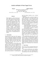
The hydrolytic activity of recombinant PON1
toward cyclosarin was sevenfold and ninefold higher
than that of squid DFPase and Ps. diminuta OPH,
respectively (at 0.03 mgÆmL
)1
enzyme, 10 lm cyclo-
sarin, k
obs
¼ 25.4 · 10
)3
, 3.8 · 10
)3
and 2.7 · 10
)3
min
)1
, respectively, Fig. 1A). Furthermore, PON1 was
more active than DFPase and OPH in detoxifying
soman, with fourfold higher rates (at 0.03 mgÆmL
)1
enzyme, 10 lm soman, k
obs
¼ 7.5 · 10
)3
, 1.8 · 10
)3
and 1.7 · 10
)3
min
)1
, respectively, Fig. 1B). The con-
centration of each enzyme was 0.03 mgÆmL
)1
or
0.75 lm (when the molecular mass of OPH, DFPase
and PON1 is taken as 40 kDa) and OP substrate con-
centration was 10 lm. All kinetic data obtained for
detoxification of cyclosarin and soman using the ace-
tylcholinesterase inhibition assay were fitted to a single
exponential decay function (Figs 2 and 3).
Modified rates of OP detoxification by newly
evolved PON1 variants
The enhanced rate of detoxification of cyclosarin and
soman by wild-type PON1 compared with DFPase and
OPH (Fig. 1) led us to study further PON1 and its vari-
ants as catalytic OP scavengers. New PON1 variants
were evolved by directed evolution using the fluorogen-
ic OP substrate DEPCyC [19,20]. Nine of these variants
were evaluated for their hydrolysis of cyclosarin, so-
man, DFP, paraoxon, parathiol, IMP-pNP and ChPo
(Scheme 1). The most rapid detoxification of cyclosarin
was obtained with the single-site mutants V346A
Enhanced stereoselective OP hydrolysis by PON1 G. Amitai et al.
1908 FEBS Journal 273 (2006) 1906–1919 ª 2006 Israel Institute for Biological Research
(1.2PC) and L69V (1.1PC): k
obs
¼ 270 · 10
)3
and
250 · 10
-3
min
)1
, respectively, versus 25 · 10
)3
min
)1
with wild-type PON1 (Fig. 2). The double mutant
L69V ⁄ S193P (2.1PC) exhibited a fourfold faster detoxi-
fication rate toward cyclosarin (k
obs
¼ 92 · 10
)3
min
)1
;
Figs 2 and 4). The variants L69V ⁄ S138L ⁄ S193P
(3.1PC), L69V ⁄ S138L ⁄ S193P ⁄ N287D (3.2PC) and
L69V ⁄ S138L ⁄ S193P ⁄ N287D ⁄ V346A (3.2PC ⁄ V346A)
displayed 2.5–3.5-fold higher activity than wild-type
PON1 (Figs 2 and 4). The rate of soman hydrolysis by
wild-type PON1 was significantly slower than hydroly-
sis of cyclosarin and DFP (k
obs
¼ 7.5 · 10
)3
compared
with 25 · 10
)3
and 17 · 10
)3
min
)1
, respectively; Figs 2,
3 and 4; time-course for DFP hydrolysis is not shown).
However, the variant V346A (1.2PC) exhibited a nine-
fold enhancement of hydrolysis toward soman com-
pared with wild-type PON1 (k
obs
¼ 65 · 10
)3
and
7.5 · 10
)3
min
)1
, respectively, Figs 3 and 4). In
addition, the five-site mutant L69V ⁄ S138L ⁄ S193P ⁄
N287D ⁄ V346A (3.2PC ⁄ V346A) catalyzed soman
detoxification twofold faster than wild-type PON1
(Figs 3 and 4). All other variants exhibited equal or
slower hydrolytic rates than wild-type PON1 toward
soman (Figs 3 and 4). The kinetic data obtained for en-
zymatic hydrolysis of soman with all tested PON1 vari-
ants indicate the importance of the V346A mutation
for the enhancement of cyclosarin and soman hydro-
lysis. The PON1 variant V346A also exhibited fourfold
and sevenfold faster hydrolysis than wild-type PON1
toward DFP and ChPo (kinetic data not shown; see
k
obs
ratios in Fig. 4). The most active variant toward
DFP was the single-site mutant L69V, with a 100-fold
enhancement over that of wild-type PON1 (k
obs
¼ 1.7
versus 0.017 min
)1
, Fig. 4). All other multiple mutants
(with three to five active-site mutations) yielded faster
rates than wild-type PON1 for DFP, cyclosarin and
parathiol hydrolysis, but to a lower extent than the sin-
gle and double mutants (Fig. 4). These multiple muta-
tion variants also exhibited lower activity than wild-type
PON1 toward soman and ChPo (Figs 3 and 4). Thus,
the most universally active PON1 variant toward DFP,
cyclosarin, soman and ChPo was the single-site mutant
Fig. 1. Time-course of enzymatic detoxification of cyclosarin (A) and soman (B) by Ps. diminuta OPH, squid DFPase and mammalian wild-
type PON1, measured by the acetylcholinesterase inhibition assay. Cyclosarin and soman concentration 10 l
M;20mM Tris ⁄ HCl, pH 7.0;
enzyme concentration 0.03 mgÆmL
)1
(0.75 lM); CaCl
2
1mM;25°C. Initial rates of OP detoxification (k
obs
,min
)1
mean ± SEM, n ¼ 3) were
estimated from the slopes of the linear plot of ln[% OP] versus time. All k
obs
values are summarized in the attached table. ND, not deter-
mined. The linear plot is based on points transformed from the initial part (up to 50% of OP hydrolysis) of the experimental nonlinear curve.
All kinetic experiments were performed in triplicate. The curves were fitted by one-phase exponential decay (r
2
¼ 0.96–0.99). The plots
shown are taken from one representative experiment.
G. Amitai et al. Enhanced stereoselective OP hydrolysis by PON1
FEBS Journal 273 (2006) 1906–1919 ª 2006 Israel Institute for Biological Research 1909
V346A, which exhibited a 4–11-fold enhanced activity
compared with wild-type PON1 (Fig. 4).
It was of particular interest to search for a PON1
variant that could hydrolyze parathiol, a P–S bond-
containing paraoxon congener (Scheme 1) and thereby
to learn about putative residues involved in the hydro-
lysis of the P–S bond in OP insecticides (e.g. Demeton,
malathion) and toxic nerve agents such as O-ethyl
S-(N,N-di-isopropylaminoethyl) methylphosphonothio-
late (VX). Therefore, the activity of PON1 variants
with parathiol, paraoxon and cyclosarin was also
compared at higher OP substrate concentration
(100 lm) (Fig. 5). The rate of parathiol hydrolysis by
wild-type PON1 was 88-fold slower than with para-
oxon (k
obs
¼ 6 · 10
)4
and 0.053 min
)1
, respectively).
However, parathiol was hydrolyzed 380-fold faster by
the H115W variant (k
obs
¼ 0.23 min
)1
, Fig. 5) than by
wild-type PON1.
A complete Michaelis–Menten kinetic analysis was
performed with the chromogenic symmetrical OP sub-
strates paraoxon and parathiol using selected PON1
variants. Table 1 summarizes the kinetic data obtained
for hydrolysis of paraoxon and parathiol by wild-type
PON1 and the following variants: H115W, L69V,
V346A, L69V ⁄ S138L ⁄ S193P ⁄ N287D and L69V ⁄
S138L ⁄ S193P ⁄ N287D ⁄ V346A. Figure 6 shows the kin-
etics of hydrolysis of paraoxon and parathiol by
H115W and wild-type PON1. It was noted that
H115W enhanced the rate of parathiol hydrolysis by
270-fold compared with wild-type PON1 (k
cat
⁄ K
m
¼
1.6 · 10
4
versus 60 m
)1
Æs
)1
; Table 1, Fig. 7). These
results corroborate those obtained for H115W with
parathiol as substrate using the acetylcholinesterase
inhibition assay (Figs 4 and 5). H115W enhanced
paraoxon hydrolysis only 16-fold (k
cat
⁄ K
m
¼ 6.4 · 10
4
versus 4 · 10
3
m
)1
Æs
)1
; Table 2, Fig. 7). All other
PON1 variants exhibited 17–28-fold enhancement of
parathiol hydrolysis compared with that of the wild-
type (Table 2, Fig. 7). Similarly, these variants also
showed a lower increase in activity toward paraoxon,
with a 2–10-fold increase in k
cat
⁄ K
m
values (Table 2,
Fig. 7). These results strongly corroborate the data
Fig. 2. Time-course of enzymatic detoxification of cyclosarin by PON1 variants measured by the acetylcholinesterase inhibition assay. Cyclo-
sarin concentration 10 l
M; PON1 0.03 mgÆmL
)1
(0.75 lM); CaCl
2
1mM;20mM Tris ⁄ HCl, pH 7.0. All experimental kinetics data were fitted
to mono-exponential decay curves drawn on the left (r
2
¼ 0.98–0.99). Initial rate value for each PON1 variant (first-order rate constant k
obs
,
min
)1
, mean ± SEM, n ¼ 3) were calculated from the slopes of the linear plots of ln(% OP) versus time shown in the right panel. Correlation
coefficients (r
2
) for the linear plots were 0.94–0.99. The kinetic plots shown are taken from a single representative experiment out of three
replicates. All k
obs
values are summarized in the attached table. ND, not determined.
Enhanced stereoselective OP hydrolysis by PON1 G. Amitai et al.
1910 FEBS Journal 273 (2006) 1906–1919 ª 2006 Israel Institute for Biological Research
Fig. 3. Time-course of enzymatic degradation of Soman by PON1 variants measured by acetylcholinesterase inhibition assay. Soman concen-
tration 10 l
M; PON1 0.03 mgÆmL
)1
(0.75 lM); CaCl
2
1mM;20mM Tris ⁄ HCl, pH 7.0. All experimental kinetics data were fitted to mono-expo-
nential decay curves drawn on the left (r
2
¼ 0.98–0.99). Initial rate values for each PON1 variant (first-order rate constant k
obs
,min
)1
; mean
± SEM, n ¼ 3) were calculated from the slopes of the linear plots of ln(% OP) versus time shown on the right. Each k
obs
value is based on
triplicate kinetic measurements. The kinetic plots shown are taken from a single representative experiment out of three replicates. All k
obs
values are summarized in the attached table. ND, not determined.
Fig. 4. Changes in hydrolytic activity (k
obs
) toward toxic OP sub-
strates of PON1 variants compared with wild-type PON1 (PON1
0.03 mgÆmL
)1
,OP10lM). Detoxification was followed by residual
acetylcholinesterase inhibition assay. The change in activity of each
PON1 variant versus PON1 wild-type is expressed as the ratio
k
obs
(mutant) ⁄ k
obs
(wild-type) drawn on a logarithmic scale. The value
of this ratio for wild-type PON1 is 1. The asterisk designates a
value of 1.0 obtained for the L69V variant with ChPo.
Fig. 5. Changes in hydrolytic activity (k
obs
) of PON1 variants using
higher concentrations (100 l
M) of cyclosarin, paraoxon and parathiol
by PON1 variants (PON1, 0.3 mgÆmL
)1
). Detoxification was fol-
lowed by acetylcholinesterase inhibition assay. The change in activ-
ity is expressed as the ratio k
obs
(mutant) ⁄ k
obs
(wild-type) drawn on a
logarithmic scale.
G. Amitai et al. Enhanced stereoselective OP hydrolysis by PON1
FEBS Journal 273 (2006) 1906–1919 ª 2006 Israel Institute for Biological Research 1911
obtained for paraoxon and parathiol using the acetyl-
cholinesterase inhibition assay (Figs 4 and 5), confirm-
ing that the results obtained by the acetylcholinesterase
inhibition assay at single substrate concentration
clearly reflect the Michaelis–Menten kinetic analysis of
enzymatic activity.
Stereoselective degradation of cyclosarin, soman
and IMP-pNP by PON1
As indicated previously, cyclosarin is a racemic mix-
ture of its S and R optical isomers configured around
the phosphorus (P) atom [P(–) and P(+) optical iso-
mers]. Soman is a mixture of four stereoisomers con-
sisting of two pairs of diastereoisomers with two
chiral centers: one on the phosphorus atom (P) and a
second on the asymmetric carbon (C) atom of the
pinacolyl group [P(–)C(+), P(–)C(–), P(+)C(+) and
P(+)C(–) stereoisomers]. Benschop et al. [23] have
noted that the pair of soman stereoisomers that are
configured with the (–) isomer on the P atom
[P(–)C(+ ⁄ –)] are 20–150-fold more toxic than the
P(+)C(+ ⁄ –) pair of diastereoisomers. It was previ-
ously noted that Ps. diminuta OPH preferentially
Table 1. Michaelis–Menten analysis for the hydrolysis of paraoxon and parathiol by wild-type PON1 and its evolved variants. Each value rep-
resents the mean of at least two independent experiments. Standard deviations were less then 10% of parameter values. Values in paren-
theses are the x-fold increase in k
cat
⁄ K
m
relative to the wild-type PON1.
Variant Mutations
Paraoxon Parathiol
k
cat
(s
)1
)
K
M
(mM)
k
cat
⁄ K
M
(M
)1
Æs
)1
)
k
cat
(s
)1
)
K
M
(mM)
k
cat
⁄ K
M
(M
)1
Æs
)1
)
Wild-type — 6.9 1.7 4 · 10
3
0.05 0.9 60
H115W H115W 25.5 0.4 6.4 · 10
4
(16) 11 0.7 1.6 · 10
4
(270)
1.1PC L69V 11.4 0.8 1.5 · 10
4
(4) 0.8 0.7 1.1 · 10
3
(18)
1.2PC V346A 12.4 0.3 4.1 · 10
4
(10) 0.8 0.5 1.6 · 10
3
(27)
3.2PC L69V 8.0 0.2 4.0 · 10
4
(10) 0.7 0.7 1.0 · 10
3
(17)
S138L
S193P
N287D
3.2PC ⁄ L69V 16.4 2.3 7.1 · 10
3
(2) 3.4 2.0 1.7 · 10
3
(28)
V346A S138L
S193P
N287D
V346A
Fig. 6. Kinetics of hydrolysis of paraoxon (A) and parathiol (B) by the PON1 variant H115W and wild-type. Hydrolysis of OP substrates was fol-
lowed by measuring the increase in p-nitrophenol A
405
at pH 8 and 25 °C. Enzymatic parameters with paraoxon and parathiol were determined
by Michaelis–Menten analysis of initial rates {v
0
¼ k
cat
[E]
0
[S]
0
⁄ ([S]
0
+K
M
)}. Values in parentheses represent molar concentrations of PON1.
Enhanced stereoselective OP hydrolysis by PON1 G. Amitai et al.
1912 FEBS Journal 273 (2006) 1906–1919 ª 2006 Israel Institute for Biological Research
hydrolyzes the less toxic optical isomers of cyclosarin
[30] and those of p-nitrophenol analogs of sarin and
soman [25]. Therefore, it was of interest to examine
the stereoselectivity of cyclosarin and soman hydro-
lysis exerted by PON1 variants. We compared the
results of GC ⁄ pulsed flame photometer detector
(PFPD) analysis, monitoring the chemical degradation
of all stereoisomers of soman and cyclosarin at speci-
fied time intervals, with those of the residual acetyl-
cholinesterase inhibition assay, measuring its
detoxification rate. Table 2 summarizes values of
chemical degradation of soman and cyclosarin com-
pared with its detoxification level at specified time
intervals. It was noted that soman is 50% hydrolyzed
by V346A within the first minute (based on GC ⁄ PFPD
analysis; Table 2), whereas acetylcholinesterase inhibi-
tion bioassay reveals practically no detoxification at
this short time interval (1 min). Similarly, cyclosarin
was degraded by 50% within the first minute
(GC ⁄ PFPD analysis; Table 2) compared with less
than 5% detoxification measured by acetylcholinest-
erase inhibition at this short time interval (Table 2).
After 100 min incubation of soman or 15 min incuba-
tion of cyclosarin with V346A PON1, each agent was
both degraded and detoxified by 91–98%. Soman and
cyclosarin were 95–98% degraded and detoxified by
wild-type PON1 only after 470 and 100 min, respect-
ively. These data are consistent with faster hydrolysis
of the less toxic optical isomer of cyclosarin [P(+)]
and the two less toxic diastereoisomers of soman
[P(+)C(+ ⁄ –)] by V346A. Stereoselective hydrolysis of
chiral OP esters by PON1 was further demonstrated
by using the sarin analog IMP-pNP as substrate.
IMP-pNP degradation by wild-type PON1 and
V346A was followed using three different analytical
methods: quantitative GC ⁄ PFPD analysis, direct spec-
trophotometric determination of p-nitrophenol
released during hydrolysis, and detoxification kinetics
measured by acetylcholinesterase inhibition assay.
Table 3 summarizes the levels of degradation of
Fig. 7. Changes in bimolecular rate constants (k
2
¼ k
cat
⁄ K
M
)of
paraoxon and parathiol hydrolysis by PON1 variants compared with
wild-type PON1 determined by Michaelis–Menten analysis of the
enzymatic activity. The changes in activity of each variant toward
degradation of paraoxon and parathiol are expressed by the ratio
k
2
(mutant) ⁄ k
2
(wild-type) drawn on a logarithmic scale.
Table 2. Comparison of degradation and detoxification levels of
soman and cyclosarin by wild-type PON1 and V346A PON1 variant
at specified time intervals. % Degradation (Deg) was determined
by GC ⁄ PFPD analysis and percentage detoxification (Detox) was
determined by residual acetylcholinesterase inhibition assay.
Enzyme ⁄ buffer
Soman Cyclosarin
Time
(min)
%
Deg
%
Detox
Time
(min)
%
Deg
%
Detox
Tris, pH 7.0 1–100 < 9 < 5 1 < 9 < 5
PON1 V346A 1 50 < 5 1 50 < 5
Tris, pH 7.0 – – – 15 10 < 5
PON1 V346A 100 > 91 > 95 15 > 98 98
Tris, pH 7.0 470 30 20 100 10 10
PON1 wild-type 470 > 95 > 95 100 > 98 98
Table 3. Stereoselective hydrolysis of IMP-pNP by wild-type and
V346A PON1 measured in parallel by GC ⁄ PFPD, spectrophotomet-
ric and acetylcholinesterase inhibition assays. IMP-pNP concentrat-
ion 10 l
M; PON1 0.03 mgÆmL
)1
;50mM Tris ⁄ HCl, pH 8, 25 °C. GC
analysis: samples of enzymatic degradation solutions were extract-
ed at specified time intervals with equal volumes of methyl t-butyl
ether that were used for quantitative GC analysis. Spectrophoto-
metric analysis was performed by measuring increases in p-nitro-
phenol absorbance. acetylcholinesterase inhibition assay was
measured by 5 min incubation with a 20-fold dilution aliquot of
IMP-pNP sampled from the hydrolysis reaction. Deg, Degradation;
Detox, detoxification.
Enzyme
Time
(min)
% Deg
(GC)
%Deg
(A
400
min
)1
)
% Detox
(acetylcholinesterase
activity)
Tris 1 10 5 0
V346A 1 52 57 2.5
Wild-type 1 40 52 0
Tris 10 0 4 0
V346A 10 46 63 8.0
Wild-type 10 44 53 0
Tris 60 0 5 0
V346A 60 70 82 17.5
Wild-type 60 49 53 0
Tris 180 6 5 0
V346A 180 87 98 54.5
Wild-type 180 45 53 0
Tris 420 14 5 0
V346A 420 98 100 87.4
Wild-type 420 61 53 0
G. Amitai et al. Enhanced stereoselective OP hydrolysis by PON1
FEBS Journal 273 (2006) 1906–1919 ª 2006 Israel Institute for Biological Research 1913
IMP-pNP by wild-type PON1 and its single mutation
variant V346A (1.2PC) at specified time intervals
using both direct spectrophotometric assay and
GC ⁄ PFPD analysis, used for determination of degra-
dation levels of both stereoisomers. These degradation
levels of IMP-pNP were compared with the levels of
detoxification measured by the acetylcholinesterase
inhibition assay (Table 3). Figure 8 shows the time-
course of IMP-pNP detoxification as well as degrada-
tion by wild-type PON1 and its variant V346A using
the acetylcholinesterase inhibition assay and the spec-
trophotometric method, respectively. Detoxification of
IMP-pNP by the V346A PON1 variant measured by
acetylcholinesterase inhibition assay fits well to a sin-
gle exponential decay function (Fig. 8), whereas the
time-course of p-nitrophenol release induced by
V346A is biphasic (Fig. 8). A mono-exponential decay
fit to the experimental detoxification data yields a sin-
gle rate constant k ¼ 0.005 min
)1
(r
2
¼ 0.987). An
excellent nonlinear fit (r
2
¼ 0.999) to the experimental
degradation data measured by p-nitrophenol release
was obtained with the following double exponential
decay function:
%IMP-pNP ¼½A  expðÀk
1
tÞ þ ½B  expðÀk
2
tÞ
This fit provides two rate constants k
1
¼ 0.98 min
)1
and k
2
¼ 0.014 min
)1
with almost equal spans (A ¼ 55
and B ¼ 45) consistent with equal amounts of two
enantiomers in the racemic mixture. The lower rate
constant of the biphasic degradation curve (k
2
) (Fig. 8)
is consistent with the first-order rate constant obtained
from the acetylcholinesterase inhibition assay reflect-
ing IMP-pNP detoxification (k
2
¼ 0.014min
)1
derived
from the double exponential decay fit, shown by the
left ordinate in Fig. 8, and k ¼ 0.005 min
)1
obtained
from detoxification kinetics presented on the right
ordinate in Fig. 8).
As shown by the spectrophotomertic and GC analy-
sis, IMP-pNP was already degraded 40–52% and
52–57% by wild-type and V346A PON1, respectively,
within the first minute (second and third row in the
third and fourth column of Table 3, Fig. 9). In con-
trast, no detoxification was observed with the V346A
variant within 10 min and up to seven hours with
wild-type PON1 as evidenced by the residual acetyl-
cholinesterase inhibition assay (fifth column in Table 3,
Fig. 9). These results are consistent with significantly
faster degradation of the less toxic isomer [P(+)] of
IMP-pNP compared with its more toxic stereoisomer
[P(–)] by wild-type and V346A [23,24]. After 3 h in the
presence of V346A, IMP-pNP was detoxified by 54%
and degraded by 87–98% (third, fourth and fifth col-
umn at the 11th row in Table 3, Fig. 9). Interestingly,
wild-type PON1 degraded IMP-pNP only up to a level
of 50% even after 21 h (Fig. 8), whereas the V346A
variant caused complete degradation within 4 h (Figs 8
and 9, Table 3). This property of wild-type PON1 was
utilized to enzymatically separate the more toxic P(–)
stereoisomer of IMP-pNP. Racemic IMP-pNP
(500 lm) was incubated with wild-type PON1
(0.1 mgÆmL
)1
) for 2 h. The enzymatic reaction was
monitored spectrophotometrically by measuring the
increase in the absorbance of the released p-nitrophe-
nol up to the plateau level obtained at 50% degrada-
tion, as demonstrated in Fig. 8. After hydrolysis by
PON1, the nonhydrolyzed stereoisomer was extracted
with methyl t-butyl ether. IMP-pNP concentration in
methyl t-butyl ether was determined by quantitative
GC analysis. The bimolecular rate constant of human
acetylcholinesterase by the separated stereoisomer of
IMP-pNP was k
i
¼ 6.3 · 10
6
min
)1
Æm
)1
, which is four-
fold higher than that of racemic IMP-pNP (k
i
¼
1.6 · 10
6
min
)1
Æm
)1
). These results are consistent with
a 16-fold difference in the rate of human acetylcholin-
esterase inhibition by the P(–) compared with P(+)
stereoisomer of IMP-pNP.
Fig. 8. Time-course of IMP-pNP degradation and detoxification by
wild-type and V346A PON1. The spectrophotometric method meas-
uring the increase in A
400
of p-nitrophenol (pNP) was used for de-
gradation kinetics, and the acetylcholinesterase inhibition assay
was used for detoxification kinetics (50 m
M Tris ⁄ HCl, pH 8, 25 °C).
The left ordinate presnts the scale for residual percentage IMP-pNP
during its degradation determined spectrophotometrically by p-nitro-
phenol release. The right ordinate represents the percentage of
putative P(–)IMP-pNP during detoxification as determined by the
acetylcholinesterase inhibition assay.
Enhanced stereoselective OP hydrolysis by PON1 G. Amitai et al.
1914 FEBS Journal 273 (2006) 1906–1919 ª 2006 Israel Institute for Biological Research
Discussion
Hydrolysis of all the OPs was measured by the acetyl-
cholinesterase inhibition assay. Acetylcholinesterase
inhibition was measured by diluting (50–1000-fold) the
intact OP remaining in solution at various time
intervals during the enzymatic hydrolysis. The acetyl-
cholinesterase inhibition assay is therefore sensitive to
changes in concentration of the more toxic isomer of
chiral OPs and reflects the rate of detoxification, rather
than degradation, of cyclosarin, IMP-pNP and soman.
In the case of symmetric OPs such as DFP, ChPo,
paraoxon and parathiol, the acetylcholinesterase inhibi-
tion assay reflects the rate of both degradation and
detoxification. The rates of detoxification of soman and
cyclosarin catalyzed by Ps. diminuta OPH, squid
DFPase and PON1 shown in Fig. 1 were determined
by calculating the initial rates of hydrolysis. The initial
rate (k
obs
) is equal to the slope of linear dependence of
ln(% acetylcholinesterase inhibition) [parallel with
ln(% residual OP)] with time. It is pertinent to note a
recent report on the stereoselective hydrolysis of cyclos-
arin by bacterial OPAA and OPH [30]. Hydrolysis was
followed by measuring the fluoride ions released during
hydrolysis. This study demonstrated a 12–24.3-fold fas-
ter rate of hydrolysis by OPH and OPAA for the P(+)
isomer than for the P(–) isomer. As the acetylcholinest-
erase inhibition assay measures exclusively the hydroly-
sis of the more toxic stereoisomer P(–)cyclosarin, the
time-course profile of cyclosarin detoxification fits bet-
ter a single-exponential decay (Fig. 1A) rather than a
double-exponential profile, as demonstrated previously
by the fluoride-release assay [30]. Possible racemization
induced by fluoride ions released during hydrolysis is
unlikely, as the maximal concentration of fluoride
released from 10 lm cyclosarin is not sufficient for the
conversion of cyclosarin enantiomers at the time scale
used in our study (not shown). The slow phase of
P(–)cyclosarin hydrolysis observed by Harvey et al. [30]
is consistent with the slow detoxification rate of cyclos-
arin by bacterial OPH measured in the present report
by the acetylcholinesterase inhibition assay (k
obs
¼
2.7 · 10
)3
min
)1
; Fig. 1).
Comparison of the rate of enzymatic detoxification of
cyclosarin and soman using constant substrate and
enzyme concentrations clearly demonstrates faster
detoxification by wild-type mammalian PON1 than bac-
terial OPH and squid DFPase (Fig. 1). Therefore, it was
of particular interest to develop and study new PON1
variants with enhanced activity. This work describes
several PON1 variants with significantly improved
detoxification rates toward toxic OP substrates. Most
notably, the single mutants V346A and H115W exhib-
ited higher rates (11–380-fold) of hydrolysis of certain
OPs compared with wild-type PON1. The newly evolved
PON1 variants could be segregated into four main
groups: group 1, H115W showing 270–380-fold
enhanced hydrolytic activity toward the P–S bond in pa-
rathiol compared with wild-type PON1 (Figs 4, 5 and
7); group 2, the single mutant L69V showing 10–100-
fold enhanced activity toward P–F-containing OP com-
pounds (i.e. DFP, cyclosarin and soman; Figs 2, 3 and
4); group 3, V346A, L69V ⁄ S193P ⁄ V346A and the
five-site mutant L69V ⁄ S138L ⁄ S193P ⁄ N287D ⁄ V346A
exhibiting a 4–10-fold higher activity toward both P–O-
containing (ChPo) and P–F-containing OP esters
(Fig. 4); group 4, includes the variants S193P,
L69V ⁄ S193P, L69V ⁄ S138L ⁄ S193P, L69V ⁄ S138L ⁄
S193P ⁄ N287D displaying no enhancement or lower
activity than wild-type PON1 toward soman and ChPo
(Fig. 4).
The H115W mutant is an interesting variation.
His115 and His134 have been proposed as the key cat-
alytic residues of PON1 [18]. However, Yeung et al.
[17] have shown that the paraoxonase activity of
H115W PON1 is even higher than that of the wild-
Fig. 9. Time-course of IMP-pNP degradation and detoxification elici-
ted by wild-type and V346A PON1 at specified time intervals pre-
sented in three dimensions. Degradation of IMP-pNP was
measured by GC ⁄ PFPD analysis during hydrolysis (left side of the
cube: black bars, Tris buffer; red, wild-type; blue, V346A). Detoxifi-
cation was monitored by residual acetylcholinesterase inhibition by
IMP-pNP (right side of the cube: green bars, Tris buffer; pink, wild-
type; khaki, V346A). The time axis (minutes) is drawn on a logarith-
mic scale. PON1 wild-type and V346A concentration is
0.03 mgÆmL
)1
;50mM Tris, pH 8; 1 mM CaCl
2
;25°C.
G. Amitai et al. Enhanced stereoselective OP hydrolysis by PON1
FEBS Journal 273 (2006) 1906–1919 ª 2006 Israel Institute for Biological Research 1915
type, although the esterase [20] and lactonase activities
are severely compromised [27]. Although the residue(s)
that mediate the OPH activity of PON1 are still to be
identified, it is clear that this activity, in particular the
P–S-hydrolyzing activity, can be greatly enhanced by
mutating His115 into Trp, and possibly to other aro-
matic residues. Notably, some of the OP substrates
studied (e.g. soman, cyclosarin and IMP-pNP) are
racemic mixtures of optical stereoisomers that differ
significantly in their toxicity [23–25]. As the acetylcho-
linesterase inhibition assay measures primarily hydroly-
sis of the more toxic optical isomer of racemic OPs, it
was pertinent to use an analytical method that would
measure the degradation of all stereoisomers. Thus,
GC analysis was used to monitor the enzymatic hydro-
lysis of all stereoisomers of cyclosarin, soman and
IMP-pNP. The GC analysis demonstrates 50% degra-
dation of either cyclosarin or soman already within the
first minute as compared with practically no detoxifica-
tion (< 5%) during this period (Table 1). Similarly,
the degradation of the sarin analog IMP-pNP is signi-
ficantly faster than its detoxification, as is evident from
both GC and spectrophotometric analysis compared
with acetylcholinesterase inhibition assay (Table 3,
Fig. 8). Our data indicate faster hydrolysis of the less
toxic stereoisomers of cyclosarin, soman and IMP-
pNP (Tables 2 and 3 and Figs 8 and 9). These results
are consistent with those obtained by Li et al. [25] with
wild-type Ps. diminuta OPH using p-nitrophenol ana-
logs of soman and sarin and with biphasic stereoselec-
tive hydrolysis of cyclosarin by Alteromonas sp. JD6.5
and A. haloplanktis OPAA demonstrated by Harvey
et al. [30].
Altogether, our results show that the OP detoxifica-
tion activities of PON1 can be dramatically enhanced,
not only toward fluorogenic symmetric OPs that were
used for screening the PON1 gene libraries, but also
toward other toxic OPs that were never screened for.
The main obstacle for the generation of highly effect-
ive OP-detoxifying enzymes toward nerve agents such
as sarin, soman and VX has been their extremely high
toxicity and the lack of an easy screening method.
These factors effectively excluded the screening of
large libraries (> 10
3
variants) with these substrates.
This study identifies three key positions that have dra-
matic effects on OP hydrolytic rates, Leu69, Val346
and His115, and several amino-acid exchanges that are
favored at this position. It therefore opens the road to
the generation of small libraries (< 100 variants) in
which these three positions are mutated in a combina-
torial manner [26] and their screening is performed
directly with the nerve agent of interest using the ace-
tylcholinesterase inhibition assay. The best variants
could then be further optimized by mutations at the
second-shell residues identified by this study (e.g.
Ser138, Ser193 and Asn287), to obtain a level of cata-
lytic efficiency that is sufficient for decontamination of
these agents. In summary, screening newly evolved
PON1 variants, with symmetric fluorogenic OP ester
DEPCyC, produced PON1 variants that exhibit higher
rates than the wild-type toward toxic chiral nerve
agents with enhanced stereoselective hydrolytic activity
toward the less toxic OP stereoisomers.
Experimental procedures
Chemicals
Acetylthiocholine iodide, 5,5¢-dithiobis-(2-nitrobenzoic
acid), paraoxon and DFP are products of Sigma, St Louis,
MO, USA. All other chemical reagents for buffer prepar-
ation were purchased at their highest available purity.
Parathiol, VX, cyclosarin, soman and ChPo were synthes-
ized at the Department of Organic Chemistry, IIBR,
according to previously published procedures.
Synthesis of (IMP-pNP)
To a stirred solution of methylphosphonic dichloride
(1.37 g, 10.30 mmol) and triethylamine (1.15 g, 11.39
mmol) in dry benzene (40 mL) under nitrogen was added
propan-2-ol (0.685 g, 11.42 mmol). The reaction mixture
was stirred at room temperature for 3 h, then dried 4-nitro-
phenol sodium salt (1.45 g, 9.00 mmol) was added, and the
reaction mixture stirred at room temperature overnight.
The reaction mixture was filtered, and the solid washed
with dry ether. The filtrate and washing were combined and
evaporated at reduced pressure to give the crude product,
which was purified by column chromatography on silica
gel. Elution was performed with an ethyl acetate ⁄ hexane
mixture using a gradual increase in ethyl acetate content
(1 : 3 fi 1 : 1] to provide IMP-pNP as a colorless liquid
(1.25 g, 53.6% yield;
1
H-NMR (CDCl
3
) d 1.28 (d, j ¼ 6.2
Hz (CH
3
)
2
C-); 1.69 (d, j ¼ 17.7Hz, CH
3
P-); 4.84 (m, -CHO-);
7.40 (m, 2H); 8.25 (m, 2H)).
31
P-NMR (CDCl
3
) d 24.0; MS
(CI) m ⁄ z 260 (M +1)
+
, 218.
Enzymes
Purified recombinant human acetylcholinesterase was a gift
from Dr A. Shafferman, IIBR. It was diluted to 5 UÆmL
)1
in 50 mm phosphate ⁄ 0.1% BSA ⁄ 1mm EDTA, pH ¼ 7.4,
for kinetic experiments. The PON1 (wild-type PON1) used
in this work is a recombinant PON1 variant, evolved for
soluble expression in E. coli [19]. It is a very close homolog
of wild-type rabbit PON1 (95% amino acid identity; 98%
Enhanced stereoselective OP hydrolysis by PON1 G. Amitai et al.
1916 FEBS Journal 273 (2006) 1906–1919 ª 2006 Israel Institute for Biological Research
similarity) and possesses identical enzymatic properties with
rabbit as well as human PON1 with a wide range of sub-
strates [19,27] and binds to high-density lipoprotein in an
identical manner [29]. Recombinant serum PON1 and its
directed evolution variants were produced and purified as
described [19,20]. Inhibition kinetics of acetylcholinesterase
by the residual level of all OP compounds during enzymatic
hydrolysis was performed using the Ellman method for ace-
tylcholinesterase activity [22].
Determination of detoxification activity of PON1
variants, OPH and DFPase toward OP substrates
(AChE inhibition assay)
OP substrates were dissolved in either distilled water or
organic solvent (methanol, ethanol or propan-2-ol) to give
a 0.01 m stock solution. The OP substrate concentrations
during measurement were 10–100 lm, and PON1 variants
were 0.03 mgÆmL
)1
(0.75 lm based on a molecular mass
for PON1 of 40 kDa) (except for ChPo which was
3 lgÆmL
)1
)in50mm Tris, pH 8, containing 1 mm CaCl
2
(except for cyclosarin and soman, when 20 mm Tris, pH 7,
was used to produce slower nonenzymatic hydrolysis).
Samples were withdrawn from the enzymatic degradation
mixture (25 °C) at specified time intervals and diluted
(50–1000-fold) in a 1-mL cuvette containing 5,5¢-dithiobis-
(2-nitrobenzoic acid) (0.3 mm) and purified human acetyl-
cholinesterase (0.06 UÆmL
)1
)in50mm phosphate, pH 7.5.
After 5 min of inhibition, acetylcholinesterase residual
activity was measured by adding acetylthiocholine
(0.45 mm). The rate of OP enzymatic detoxification was
directly proportional to the increase in acetylcholinesterase
activity with time (A
412
min
)1
). Detoxification of cyclosarin
and soman by squid DFPase and bacterial OPH was meas-
ured in a similar manner using 0.03 mgÆmL
)1
enzyme in
20 mm Tris, pH 7, and 10 lm cyclosarin or soman. The
activity of squid DFPase and Ps. diminuta OPH was meas-
ured in the presence of 1 mm CaCl
2
or CoCl
2
, respectively.
Nonenzymatic hydrolysis of cyclosarin and soman related
to the comparative study with PON1, DFPase and OPH
was measured in 20 mm Tris (pH 7) ⁄ 1mm CaCl
2
. Non-
enzymatic hydrolysis of those OP compounds studied with
PON1 variants was measured in 50 mm Tris (pH 8) ⁄ 1mm
CaCl
2
. The kinetics data were analyzed to obtain initial
rate values (k
obs
) for OP substrate hydrolysis that could be
compared between all three enzymes (DFPase, OPH and
PON1) as they were measured at equal substrate and
enzyme concentrations. All detoxification kinetics data fit-
ted very well (R
2
¼ 0.98–0.99) to single-exponential decay
curves. Initial rates of detoxification (k
obs
, min
)1
) were esti-
mated from the slopes of the linear plot of ln(% residual
OP) versus time which parallels the measured decrease in
ln(% acetylcholinesterase inhibition) with time. The linear
correlation analysis is based on points taken from the ini-
tial part (up to 50% OP hydrolysis) of the experimental
nonlinear curve. The kinetic experiments were performed in
triplicate. Non-linear fits were carried out using the Graph-
Pad Prism program.
Determination of PON1 degradation activity
toward OP substrates by the spectrophotometric
assay
Enzymatic degradation of IMP-pNP, paraoxon and parathi-
ol by PON1 variants was also measured directly by monitor-
ing the release of p-nitrophenol at 400 nm. The k
obs
values
were calculated from the linear part (r
2
¼ 0.99) of the initial
rate curve corrected for spontaneous hydrolysis. As K
m
val-
ues for a number of OP substrates are very close ($ 1mm)
and the concentrations of PON1 variants and OP substrates
used throughout all hydrolysis experiments were equal, it is
assumed that the ratio of the bimolecular rate constants of
OP hydrolysis by PON1 (k
cat
⁄ K
m
) could conceivably be rep-
resented by the ratio of k
obs
values, measured for a newly
evolved variant compared with wild-type PON1 for each OP
substrate [k
obs
(mutant) ⁄ k
obs
(wild-type)].
Complete Michaelis–Menten kinetic analysis was per-
formed with paraoxon and parathiol using some of the var-
iants. In this case, product formation during hydrolysis of
paraoxon and parathiol was measured spectrophotometri-
cally at 405 nm in 200 lL activity buffer (50 mm Tris,
pH 8.0, CaCl
2
1mm)at25°C. Stock solutions of both
substrates were prepared in methanol (500 and 100 mm for
paraoxon and parathiol, respectively), from which a range
of substrate concentrations were prepared in activity buffer
with 1% methanol (final concentration) in all reactions.
Enzymatic parameters with paraoxon and parathiol were
determined by Michaelis–Menten analysis of initial rates
{v
0
¼ k
cat
[E]
0
[S]
0
⁄ ([S]
0
+ K
m
)}. Absorption coefficients of
the hydrolyzed paraoxon and parathiol products are 18 000
and 17 320 m
)1
Æcm
)1
, respectively. The ratio of k
cat
⁄ K
m
for the PON1 variant to that for the wild-type for para-
oxon and parathiol hydrolysis was used to compare the
activities.
GC analysis
Samples of IMP-pNP, soman or cyclosarin in 50 mm phos-
phate (1.5–3 lgÆmL
)1
) containing 0.03 mgÆmL
)1
PON1
were extracted with equal volumes of methyl t-butyl ether.
Extraction yield was determined for each OP by spiking the
buffer solutions with a known concentration of OP. A 2-lL
volume of methyl t-butyl ether extract was injected into
a 6890 Agilent Instruments Gas Chromatograph (HP, Wil-
mington, DE, USA) equipped with capillary column (nar-
row bore DB-5MS; 15 m length; 0.25 mm diameter; 1 lm
film thickness; made by J & W (No. I22-5513); splitless
inlet at 200 °C; helium flow 44.6 mLÆmin
)1
; oven tempera-
ture programming: initial temperature 50 °C ⁄ 0.5min; rate
G. Amitai et al. Enhanced stereoselective OP hydrolysis by PON1
FEBS Journal 273 (2006) 1906–1919 ª 2006 Israel Institute for Biological Research 1917
20 °CÆmin
)1
; final temperature 180 °C; rate 25 °CÆmin
)1
;
final temperature 305 °C. PFPD OI5380 at 300 °C; H
2
flow
11 mLÆmin
)1
; air flow 15 mLÆmin
)1
; constant makeup flow
(nitrogen) 14 mLÆmin
)1
).
Acknowledgements
Supported in part by contract M ⁄ SAB1 ⁄ 3 ⁄ A007 to G.A.
from MOD, Germany, and the Charles & M.R. Shapiro
Foundation Endowed Biomedical Research Fund to
D.S.T.
References
1 Taylor P (1985) Anticholinesterase agents. In Goodman
& Gillman’s the Pharmacological Basis of Therapeutics
7th edn, pp. 110–129. Macmillan Publishing Company,
New York.
2 Worek F, Eyer P & Szinicz L (1998) Inhibition, reacti-
vation and aging kinetics of cyclohexyl methylphospho-
nofluoridate-inhibited human cholinesterases. Arch
Toxicol 72, 996–998.
3 Amitai G, Rabinovitz I, Chen R, Cohen G, Zomber G,
Adani R & Raveh L (1995) Bisquaternary oximes as
antidotes against tabun and soman poisoning: antidotal
efficacy in relation to cholinesterase reactivation. In
Enzymes of the Cholinesterase Family (Quinn DM, Bala-
subramanian AS, Doctor BP & Taylor P, eds), pp. 345–
352. Plenum Press, New York, London.
4 Lundy PM, Hansen AS, Hand BT & Boulet CA (1992)
Comparison of several oximes against poisoning by
soman, tabun and GF. Toxicology 72, 99–105.
5 Ashani Y, Rothschild N, Segall Y, Levanon D & Raveh
L (1991) Prophylaxis against organophosphate poison-
ing by an enzyme hydrolysing organophosphorus com-
pounds in mice. Life Sci 49, 367–374.
6 Ashani Y, Shapira S, Levy D, Wolfe AD, Doctor BP &
Raveh L (1991) Butyrylcholinesterase and acetylcholi-
nesterase prophylaxis against soman poisoning in mice.
Biochem Pharmacol 41, 37–41.
7 Allon N, Raveh L, Gilat E, Cohen E, Grunwald J &
Ashani Y (1998) Prophylaxis against soman inhalation
toxicity in guinea pigs by pretreatment alone with
human serum butyrylcholinesterase. Toxicol Sci 43,
121–128.
8 Cohen O, Kronman C, Chitlaru T, Ordentlich A, Velan
B & Shafferman A (2001) Effect of chemical modifica-
tion of recombinant human acetylcholinesterase by
polyethylene glycol on its circulatory longevity. Biochem
J 357, 795–802.
9 DeFrank JJ, Cheng T-C, Harvey SP & Rastogi VK
(2005) Advanced catalytic enzyme system: dual use cap-
abilities. />K/KP-03Standby.pdf.
10 Dumas DP, Durst HD, Landis WG, Raushel FM &
Wild JR (1990) Inactivation of organophosphorus nerve
agents by the phosphotriesterase from Pseudomonas
diminuta. Arch Biochem Biophys 277, 155–159.
11 Griffiths AD & Tawfik DS (2003) Directed evolution of
an extremely fast phosphotriesterase by in vitro com-
partmentalization. EMBO J 22, 24–36.
12 Cheng TC, Harvey SP & Stroup AN (1993) Purification
of highly active organophosphorus acid anhydrolase
from Alteromonas undina. Appl Environ Microbiol 59,
3138–3140.
13 DeFrank JJ, Beaudry WT, Cheng TC, Harvey SP,
Stroup AN & Szafraniec LL (1993) Screening of
halophilic bacteria and Alteromonas species for organo-
phosphorus hydrolyzing enzyme activity. Chem Biol
Interact 87, 141–148.
14 Scharff EI, Koepke J, Fritzsch G, Lucke C & Rueter-
jans H (2001) Crystal structure of di-isopropylfluoro-
phosphatase from Loligo vulgaris. Structure 9, 493–502.
15 Primo-Parmo SL, Sorenson RC, Teiber J & La Du BN
(1996) The human serum paraoxonase ⁄ arylesterase gene
(PON1) is one member of a multigene family. Genomics
33, 498–507.
16 Josse D, Xie W, Renault F, Rochu D, Schopfer LM,
Masson P & Lockridge O (1999) Identification of resi-
dues essential for human paraoxonase (PON1) arylester-
ase organophosphatase activities. Biochemistry 38,
2816–2825.
17 Yeung D, Josse D, Nicholson JD, Khanal A, McAn-
drew CW, Bahnson BJ, Lenz DE & Cerasoli DM
(2004) Structure ⁄ function analyses of human serum
paraoxonase (HuPON1) mutants designed from a
DFPase-like homology model. Biochim Biophys Acta
1702, 67–77.
18 Harel M, Aharoni A, Gaidukov L, Brumshtein B, Kher-
sonsky O, Meged R, Dvir H, Ravelli RBG, McCarthy
A, Toker L, et al. (2004) Structure and evolution of the
serum paraoxonase family of detoxifying and anti-
atherosclerotic enzymes. Nat Struct Mol Biol 11, 412–
419.
19 Aharoni A, Gaidukov Yagur S, Silman I & Tawfik
DS (2004) Directed evolution of mammalian para-
oxonases PON1 and PON3 for bacterial expression
and catalytic specialization. Proc Natl Acad Sci USA
101, 482–487.
20 Aharoni A, Gaidukov L, Khersonsky O, Gould SMcQ,
Roodveldt S & Tawfik DS (2005) The ‘evolvability’ of
promiscuous protein functions. Nat Genet 37, 73–76.
21 Harel M, Aharoni A, Gaidukov L, Brumshtein B, Kher-
sonsky O, Meged R, Dvir H, Ravelli RB, McCarthy A,
Toker L, et al. (2004) Structure and evolution of the
serum paraoxonase family of detoxifying and anti-
atherosclerotic enzymes – Erratum. Nat Struct Mol Biol
11, 1253.
Enhanced stereoselective OP hydrolysis by PON1 G. Amitai et al.
1918 FEBS Journal 273 (2006) 1906–1919 ª 2006 Israel Institute for Biological Research
22 Ellman GC, Courtney KD, Anders V & Featherstone
RM (1961) A new and rapid colorimetric determination
of acetylcholinesterase activity. Biochem Pharmacol 7,
88–95.
23 Benschop HP, Konings CAG, van Genderen J & De
Jong LPA (1984) Isolation, anticholinesterase properties,
and acute toxicity in mice of four stereoisomers of the
nerve agent soman. Toxicol Appl Pharmacol 72, 61–74.
24 Benschop HP & De Jong LPA (1988) Nerve agent
stereoisomers: analysis, isolation and toxicology. Acc
Chem Res 21, 368–337.
25 Li WS, Lum KT, Chen-Goodspeed M, Sogorb MA &
Raushel FM (2001) Stereoselective detoxification of
chiral sarin and soman analogues by phosphotriesterase.
Bioorg Med Chem 9, 2083–2091.
26 Park S, Morley KL, Horsman GP, Holmquist M, Hult
K & Kazlauskas RJ (2005) Focusing mutations into the
P. fluorescence esterase binding site increases enantio-
selectivity more effectively than distant mutations. Chem
Biol 12, 45–54.
27 Khersonsky O & Tawfik DS (2005) Structure-reactivity
studies of serum paraoxonase PON1 suggest that its
native activity is lactonase. Biochemistry 44, 6371–6382.
28 Draganov DI, Teiber JF, Speelman A, Osawa Y, Suna-
hara R & La Du BN (2005) Human paraoxonases
(PON1, PON2, and PON3) are lactonases with overlap-
ping and distinct substrate specificities. J Lipid Res 46,
1239–1247.
29 Gaidukov L & Tawfik DS (2005) The high affinity,
stability and lactonase activity of serum paraoxonase
PON1 anchored on HDL with apoA-I. Biochemistry 44,
11843–11854.
30 Harvey SP, Kolakowski JE, Cheng TC, Rastogi VK,
Reiff LP, DeFrank JJ, Raushel FM & Hill C (2005)
Stereospecificity in the enzymatic hydrolysis of cyclo-
sarin (GF). Enzyme Microbiol Technol 37, 547–555.
FEBS Journal 273 (2006) 1906–1919 ª 2006 Israel Institute for Biological Research 1919
G. Amitai et al. Enhanced stereoselective OP hydrolysis by PON1