Báo cáo khoa học: The b domain is required for Vps4p oligomerization into a functionally active ATPase potx
Bạn đang xem bản rút gọn của tài liệu. Xem và tải ngay bản đầy đủ của tài liệu tại đây (631.07 KB, 17 trang )
The b domain is required for Vps4p oligomerization into
a functionally active ATPase
Parimala R. Vajjhala
1
, Julin S. Wong
1
, Hui-Yi To
1
and Alan L. Munn
1,2,3,4
1 Institute for Molecular Bioscience and ARC Special Research Centre for Functional and Applied Genomics, University of Queensland,
St Lucia, Queensland, Australia
2 School of Biomedical Sciences, University of Queensland, St Lucia, Queensland, Australia
3 Laboratory of Yeast Cell Biology, Institute of Molecular and Cell Biology, A*STAR Biomedical Sciences Institutes, Singapore
4 Department of Biochemistry, Faculty of Medicine, National University of Singapore, Singapore
The multivesicular body (MVB) is a central sorting
station in the itinerary of proteins that traffic through
the endocytic pathway. In the MVB, integral mem-
brane proteins that are destined for delivery to the
lysosome lumen undergo MVB sorting into internal
vesicles, which form by invagination of the limiting
membrane of the MVB. This process sequesters the
cytoplasmic tails of endocytosed signalling receptors
and allows efficient silencing. Mature MVBs then fuse
with the lysosome and transfer their contents, inclu-
ding internal vesicles, into the lysosome lumen. In
some cells, MVBs have been shown to fuse with the
plasma membrane and have been suggested to function
in intercellular signalling [1,2]. Moreover, the machin-
ery that generates the internal vesicles of MVBs has
recently been implicated in the budding of several clas-
ses of enveloped viruses (reviewed in [3–5]).
Both endocytic and biosynthetic traffic to the lyso-
some proceed via the MVB [6,7], thus defects in the
function of the MVB affect both endocytic transport
Keywords
class E vacuolar protein sorting; dopamine
responsive gene-1; LYST-interacting protein
5; suppressor of K
+
uptake growth defect 1
(SKD1); SKD1-binding protein 1
Correspondence
A. L. Munn, Institute for Molecular
Bioscience, University of Queensland,
St Lucia, Brisbane, QLD 4072, Australia
Fax: +61 73346 2101
Tel: +61 73346 2017
E-mail:
(Received 16 February 2006, revised
14 March 2006, accepted 20 March 2006)
doi:10.1111/j.1742-4658.2006.05238.x
Endocytic and biosynthetic trafficking pathways to the lysosome ⁄ vacuole
converge at the prevacuolar endosomal compartment. During transport
through this compartment, integral membrane proteins that are destined
for delivery to the lysosome ⁄ vacuole lumen undergo multivesicular body
(MVB) sorting into internal vesicles formed by invagination of the endo-
somal limiting membrane. Vps4 is an AAA family ATPase which plays a
key role in MVB sorting and facilitates transport through endosomes. It
possesses an N-terminal microtubule interacting and trafficking domain
required for recruitment to endosomes and an AAA domain with an
ATPase catalytic site. The recently solved 3D structure revealed a b
domain, which protrudes from the AAA domain, and a final C-terminal
a-helix. However, the in vivo roles of these domains are not known. In
this study, we have identified motifs in these domains that are highly con-
served between yeast and human Vps4. We have mutated these motifs
and studied the effect on yeast Vps4p function in vivo and in vitro.We
show that the b domain of the budding yeast Vps4p is not required for
recruitment to endosomes, but is essential for all Vps4p endocytic func-
tions in vivo. We also show that the b domain is required for Vps4p
homotypic interaction and for full ATPase activity. In addition, it is
required for interaction with Vta1p, which works in concert with Vps4p
in vivo. Our studies suggest that assembly of a Vps4p oligomeric complex
with full ATPase activity that interacts with Vta1p is essential for normal
endosome function.
Abbreviations
CPY, carboxypeptidase Y; ESCRT, endosomal sorting complex required for transport; GFP, green fluorescent protein; GST, glutathione S-transferase;
MIT, microtubule interacting and trafficking; MVB, multivesicular body; PVDF, poly(vinylidene difluoride); Vps, vacuolar protein sorting.
FEBS Journal 273 (2006) 2357–2373 ª 2006 The Authors Journal compilation ª 2006 FEBS 2357
and delivery of newly synthesized lysosomal ⁄ vacuolar
proteins. Insights into both processes have come from
studies of budding yeast vacuolar protein sorting (vps)
mutants which missort soluble vacuolar proteins into
the extracellular medium. A subclass of vps mutants,
class E vps mutants, disrupt MVB sorting and form an
enlarged multilamellar endosome adjacent to the vacu-
ole termed the class E compartment [8–10]. This com-
partment accumulates endocytic and biosynthetic
material as well as recycling late Golgi proteins [9,11–
13] resulting from defective transport out of this aber-
rant endosome. There are 27 class E Vps proteins in
mammalian cells and 18 in yeast, which can be
grouped into complexes referred to as endosomal sort-
ing complexes required for transport (ESCRT I–III).
These three complexes act sequentially to sort and
deliver membrane proteins into forming intraluminal
vesicles [14,15].
Vps4p, also known as Csc1p ⁄ End13p ⁄ Grd13p ⁄
Vpl4p ⁄ Vpt10p ⁄ Did6p, is a class E Vps protein that
belongs to the AAA (ATPase associated with a variety
of cellular activities) family of ATPases [8,16]. Vps4p
functions at multiple steps during endocytic transport
[8,17] and has recently been shown to function in
sterol metabolism [18]. There are two isoforms of
VPS4 in human cells: VPS4A and VPS4B. The endocy-
tic functions of yeast Vps4p are conserved in mamma-
lian VPS4B [19–21], and both human VPS4A and
VPS4B have been shown to be required for virus bud-
ding [22,23]. Vps4p contains a single AAA domain,
and its ATPase activity is critical for function as vps4
mutants defective in ATP binding (K179A) or hydroly-
sis (E233Q), and a temperature-sensitive vps4 mutant
(M307T ⁄ L327S) and the end13-1 mutant (S335F),
which also have mutations in the AAA domain, all
induce class E compartments and perturb endocytic
and biosynthetic traffic [8,17]. Vps4p ATPase activity
is important for disassembly of ESCRTs to allow reuse
in multiple rounds of MVB sorting [24].
Whereas many AAA proteins form hexamers, wild-
type Vps4p has not been shown to form a higher-order
oligomer. Wild-type Vps4p purified from bacteria
forms a dimer [24]. However, a homotypic interaction
has not been demonstrated for wild-type Vps4p in vivo
using a yeast two-hybrid assay [25]. Moreover, endo-
genous human VPS4B appears to exist as a monomer
[26]. Thus it is not clear whether Vps4p exists as a
dimer in vivo or whether dimer formation is important
for Vps4p function in vivo, and the structural determi-
nants required for dimerization of Vps4p have not
been identified. Although a homotypic interaction has
been described for the Vps4p-E233Q mutant in a yeast
two-hybrid assay [25], and this mutant forms a 10–12-
mer in the presence of ATP [24,27], it is not clear whe-
ther this is due to stabilization of a complex that is
normally transiently formed by wild-type Vps4p or to
formation of an aberrant complex. Consistent with the
ability of Vps4p to form oligomers, most vps4 mutants
that have been described to date are dominant negative
[17,24,28].
The N-terminal region of Vps4p contains a microtu-
bule interacting and trafficking (MIT) domain [29].
This region of Vps4p interacts with Vps20p ⁄ Chm6p
[30], a component of ESCRT III, and is required to
recruit Vps4p to endosomes. The C-terminal region of
Vps4p binds to another class E Vps protein, Vta1p
[30], and this interaction is conserved in mammalian
cells [26]. As the C-terminal region of Vps4p contains
several motifs that are highly conserved between yeast
Vps4p and both of the human VPS4 isoforms, we
hypothesized that this region has an important role in
Vps4p assembly or function. In this study, we show
that conserved motifs in the C-terminal region, which
are mainly present in the recently identified b domain
of Vps4p, are essential for interaction with Vta1p. In
addition, we show that one of these motifs is required
for a homotypic interaction and for formation of a
highly active ATPase complex, but is not required for
endosomal recruitment. More importantly, the con-
served motifs in the b domain are required for Vps4p
functions in vivo.
Results
Conserved motifs in the Vps4p C-terminal region
are essential for function but not for protein
stability
The Vps4p sequence located C-terminal to the previ-
ously predicted AAA domain [8] contains several
motifs that are highly conserved in human VPS4B
(Fig. 1A) as well as VPS4A (not shown). To examine
the functional importance of these C-terminal motifs
and the C-terminal region in general, we constructed
various mutations in this domain and examined the
effect of each on Vps4p stability and in vivo function.
To assess the importance of the C-terminal region (res-
idues 351–437) or half of it (residues 395–437), the
corresponding C-terminal truncation mutants (Vps4p-
Ter1 and Vps4p-Ter2, respectively) were generated. To
assess the importance of individual conserved motifs,
we chose three motifs to delete. These deletion mutant
proteins are named according to the first three amino
acids of each motif deleted, i.e. Vps4p-RKI, Vps4p-
GAI and Vps4p-LTP. In addition, we generated a
mutant in which each charged residue in a conserved
Role of the Vps4 b domain P. R. Vajjhala et al.
2358 FEBS Journal 273 (2006) 2357–2373 ª 2006 The Authors Journal compilation ª 2006 FEBS
motif (RDE) at the extreme C-terminus was substi-
tuted with alanine (Vps4p-RDE).
The recent crystal structure of human VPS4B [27]
allowed us to map the positions of these motifs
(Fig. 1A). The domain organization of yeast Vps4p
(Fig. 1B) inferred from the elucidated 3D crystal struc-
ture of human VPS4B shows that the AAA domain is
unusual in that it contains a domain with three antipar-
allel b-sheets, referred to as the b domain within the
C-terminal subdomain. Therefore the AAA domain
extends past the original predicted AAA domain. Thus
Vps4p-Ter1 lacks the end of a-helix 8 in the AAA
domain, the entire b domain and both of the two C-ter-
minal a-helices. Vps4p-Ter2 lacks only the two C-ter-
minal a-helices. The motif deleted in the Vps4p-RKI
mutant is positioned at the end of a-helix 8 of the AAA
domain and extends into the first b-sheet of the b
domain. In comparison, the motifs deleted in the Vps4p-
GAI and Vps4p-LTP mutants are positioned adjacent
to and within the second b-sheet, and within the third
b-sheet, respectively, of the b domain. The RDE motif
subjected to charged-to-alanine substitutions in the
Vps4p-RDE mutant is positioned at the end of the
C-terminal a-helix (Fig. 1A). The positions of the highly
conserved residues that were mutated are shown on the
VPS4B structure in Fig. 1C.
To test whether the Vps4p mutant proteins are sta-
bly expressed in yeast, plasmids expressing the wild-
type and mutant Vps4p constructs were introduced
into vps4D cells, and Vps4p was detected in cell
extracts by immunoblotting. The steady-state levels of
Vps4p-RKI, Vps4p-GAI, Vps4p-LTP and Vps4p-RDE
mutant proteins are comparable to wild-type Vps4p
(Fig. 1D). In contrast, the level of Vps4p-Ter1 and
Vps4p-Ter2 mutant proteins (38.3 and 43.9 kDa,
respectively) was too low to detect (data not shown).
Although the C-terminus itself may be required for
protein stability, we conclude that the four conserved
motifs that we mutated are not individually essential
for protein stability.
We next examined whether those Vps4p mutant con-
structs that are stably expressed can substitute for wild-
type Vps4p in its various functions. Plasmids expressing
wild-type Vps4p or the various Vps4p mutant proteins
or empty vector were introduced into vps4D yeast cells
and the ability of the mutant proteins to rescue the
A
S.c. Vps4 351 IRKIQSATHFKDV STEDDE TRKLTPCSPGD 380
.::.::::::: : : : . ::::::::
H.s. Vps4B 353 VRKVQSATHFKKVRGPSRADPNHLVDDLLTPCSPGD 388
S.c. Vps4 381 DGAIEMSWTDIEADELKEPDLTIKDFLKAIKSTRPT 416
:::::.: :. : : :: : : .:.::
H.s. Vps4B 389 PGAIEMTWMDVPGDKLLEPVVSMSDMLRSLSNTKPT 424
S.c. Vps4 417 VNEDDLLKQEQFTRDFGQEG437
::: :::: :: ::::::
H.s. Vps4B 425 VNEHDLLKLKKFTEDFGQEG 444
β6
β7
β8
α9
α10
α8
α10
C
D
empty vector
Vps4p-GAI
Vps4p-LTP
Vps4p-RKI
Vps4p-RDE
Vps4p
actin
Vps4p
B
AAA N-terminal
subdomain
AAA C-terminal
subdomain
β
domain
C-terminal
helix
MIT domain
1
437
299
129
358
399
415
79
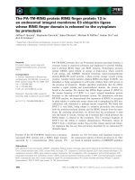
Fig. 1. Construction of Vps4p C-terminal mutants. (A) Alignment of
S. cerevisiae (S.c) Vps4p and human (H.s) VPS4B sequences using
LALIGN [50]. Conserved blocks deleted in individual mutants are
shown in bold, and residues that were substituted with alanine in
the RDE mutant are shown in bold italics. The secondary structure
of the corresponding region of VPS4B is also shown. (B) Schematic
representation of wild-type Vps4p with the domain organization
inferred from structural data of VPS4A and VPS4B [27,34]. (C) Locat-
ion of highly conserved residues in the human VPS4B structure
that were mutated in yeast Vps4p. The b domain is circled, and the
RKI, LTP and GAI motifs are shown in red, light blue and green,
respectively. The charged residues in the RDE motif are shown in
dark blue. The colour code for the nonmutated residues in the dif-
ferent domains is: b domain, orange; N-terminal AAA subdomain,
pink; C-terminal AAA subdomain, light brown; C-terminal a-helix,
brown. (D) Total cell lysates from RH2906 (vps4D) yeast cells carry-
ing plasmids expressing wild-type Vps4p, Vps4p-RDE, Vps4p-RKI,
Vps4p-LTP and Vps4p-GAI mutant proteins or carrying empty vector
(YCplac111) were subjected to western blotting using either a poly-
clonal antibody to Vps4p or a monoclonal antibody to actin.
P. R. Vajjhala et al. Role of the Vps4 b domain
FEBS Journal 273 (2006) 2357–2373 ª 2006 The Authors Journal compilation ª 2006 FEBS 2359
phenotypes of vps4D cells was assessed. Vacuolar accu-
mulation of a fluid-phase endocytic marker, MVB sort-
ing of a membrane protein to the vacuole lumen,
delivery of soluble vacuolar proteins to the vacuole and
growth at high temperature were examined.
Endocytosis and subsequent vacuolar accumulation
of the fluid-phase marker, Lucifer Yellow, was restored
by wild-type Vps4p but not by Vps4p-RKI, Vps4p-
GAI or Vps4p-LTP mutant forms of Vps4p when
compared with empty vector alone (Fig. 2A). In con-
trast, vacuolar accumulation of Lucifer Yellow was
efficiently restored by Vps4p-RDE. To examine MVB
sorting, we used the iron transporter homologue,
Fth1p, fused to ubiquitin (Fth1p-Ub) as a marker.
Fth1p normally resides on the limiting membrane of
the vacuole, but, when tagged with ubiquitin, it under-
goes ubiquitin-dependent MVB sorting and is delivered
to the vacuole lumen [31]. Green fluorescent protein
(GFP)-tagged Fth1p-Ub was mainly observed in a
class E compartment adjacent to the vacuole in vps4D
cells expressing Vps4p-RKI, Vps4p-GAI or Vps4p-
LTP similar to vps4D cells carrying the empty vector
(Fig. 2B). The small amount that reached the vacuole
was present on the limiting membrane. In contrast,
expression of wild-type Vps4p or Vps4p-RDE resulted
in delivery of GFP-tagged Fth1p-Ub to the vacuole
lumen. These data clearly demonstrate that the con-
served C-terminal motifs that are required for efficient
fluid-phase endocytosis are also critical for ubiquitin-
dependent MVB sorting.
To assess the ability of the mutant Vps4p con-
structs to function in vacuolar protein sorting, we
assayed their ability to correct the missorting and
secretion of carboxypeptidase Y (CPY) in vps4D
cells. CPY is a soluble resident vacuolar protein that
is translocated into the endoplasmic reticulum and
then transported to the Golgi where a receptor,
Vps10p, sorts it from secretory proteins destined for
the cell surface into a pathway that takes it via
endosomes to the vacuole. In vps4D cells, CPY is
missorted at the late Golgi into vesicles destined for
the cell surface and secreted [8]. Expression of wild-
type Vps4p but not Vps4p-RKI, Vps4p-GAI or
Vps4p-LTP mutant proteins restored vacuolar deliv-
ery of CPY compared with empty vector alone
(Fig. 2C). In contrast, CPY sorting to the vacuole is
restored by Vps4p-RDE.
Finally, we examined the ability of the various
mutant Vps4p constructs to function in cell growth at
high temperature. vps4D cells expressing wild-type or
mutant Vps4p proteins or carrying empty vector were
tested for growth on solid medium at high tempera-
ture. Expression of wild-type Vps4p but not
Vps4p-RKI, Vps4p-GAI and Vps4p-LTP or empty
vector rescued growth at 40 °C (Fig. 2D). In contrast,
Vps4p-RDE was able to significantly restore growth at
40 °C. However, Vps4p-RDE was reproducibly less
efficient than wild-type Vps4p in restoring growth at
40 °Ctovps4D cells. There was no obvious difference
in the growth rate at 24 °C between vps4D cells expres-
sing wild-type Vps4p, any of the Vps4p mutants or
those carrying empty vector.
We conclude that the conserved motifs adjacent to
and within the b domain that were deleted in the
Vps4p-RKI, Vps4p-GAI and Vps4p-LTP mutants are
essential for all Vps4p functions tested but not for pro-
tein stability. The charged residues in the RDE motif at
the end of the C-terminal helix of Vps4p are not essen-
tial for protein stability or for most functions, however,
they are required for full cellular growth at 40 °C.
Fig. 2. Conserved motifs within and adjacent to the b domain of Vps4p are essential for several Vps4p in vivo functions. (A) Lucifer Yellow
uptake and accumulation in the vacuole was measured in RH2906 (vps4D) yeast cells carrying plasmids expressing wild-type Vps4p or
Vps4p mutant proteins or carrying empty vector (YCplac111). The same fields of cells are shown visualized by fluorescence (left) and Nomar-
ski (right) optics. The vacuoles appear as indentations in the cell profile by Nomarski optics. Scale bar, 5 lm. (B) Ubiquitin-dependent MVB
sorting of Fth1p-GFP-Ub in AMY245 (vps4D) yeast cells carrying plasmids expressing wild-type Vps4p or Vps4p mutant proteins or carrying
empty vector (YCplac111). Cells were incubated in YPUAD medium containing 100 l
M bathophenanthroline disulfonic acid for 6 h to chelate
iron and induce Fth1p-GFP-Ub expression. Cells were then washed with buffer containing 1% sodium azide, 1% sodium fluoride, 100 m
M
phosphate, pH 8.0, to stop further transport. The same fields of cells are shown visualized by fluorescence (left) and Nomarski (right) optics.
Scale bar, 5 lm. (C) Vacuolar protein sorting in RH2906 (vps4D) yeast cells carrying plasmids expressing wild-type Vps4p or Vps4p mutant
proteins or carrying empty vector (YCplac111) or no vector. Cells were grown on YPUAD solid medium for 2 days at 24 °C in contact with a
nitrocellulose filter. RH1800 (wild-type) yeast cells without any vector (boxed in both panels) was included as a control. Cells were eluted
from the filter, and CPY on the filter was detected by immunoblotting with anti-CPY serum. To test for cell lysis, the blot was stripped and
reprobed with an antibody to a cytoplasmic protein (calmodulin). (D) Temperature-sensitive growth assay of RH2906 (vps4D) yeast cells
carrying plasmids expressing wild-type Vps4p or Vps4p mutant proteins or carrying empty vector (YCplac111). Cells were serially diluted
10-fold, and 7-lL aliquots were spotted on to YPUAD solid medium and incubated at 24 °C (left) or 40 °C (right). Plates were photographed
after 4 or 12 days, respectively.
Role of the Vps4 b domain P. R. Vajjhala et al.
2360 FEBS Journal 273 (2006) 2357–2373 ª 2006 The Authors Journal compilation ª 2006 FEBS
Conserved motifs adjacent to and within the
Vps4p b domain are required for interaction with
Vta1p, but not Vps20p or Did2p
A number of Vps4p-interacting proteins have previ-
ously been identified in a yeast two-hybrid screen
including the class E Vps proteins Vps20p and Vta1p
[30]. These interactions were confirmed by the demon-
stration that recombinant glutathione S-transferase
(GST)-Vps20p or GST-Vta1p bind GFP-tagged
Vps4p present in yeast cell lysates as well as to
recombinant His
6
-tagged Vps4p in vitro. Using yeast
Vps4p-GAI
Vps4p-L TP
Vps4p-RDE
Vps4p-WT
empt y
vector
Vps4p-RKI
Fluorescence Nomarski
Vps4p-GAI
Vps4p-LTP
Vps4p-RDE
Vps4p-WT
empty
vector
Vps4p-RKI
Fluorescence Nomarski
A
B
C
Vps4p-WT
Vps4p-GAI
Vps4p-RDE
no vector
empty vector
Vps4p-RK I
Vps4p- LT P
wild-type
Vps4p-WT
Vps4p-GAI
Vps4p-RDE
no vector
empty vector
Vps4p-RK I
Vps4p- LT P
wild-type
CPY blo t
Calmodulin blo t
D
Vps4p-WT
empty vector
Vps4p-RK I
Vps4p-GAI
Vps4p-L TP
Vps4p-RDE
40 C
O
24 C
O
P. R. Vajjhala et al. Role of the Vps4 b domain
FEBS Journal 273 (2006) 2357–2373 ª 2006 The Authors Journal compilation ª 2006 FEBS 2361
two-hybrid assays, Vps20p was shown to interact
strongly with the N-terminal region of Vps4p and
weakly with both full-length Vps4p and a C-terminal
region containing residues 351–437. Vta1p interacts
strongly with full-length Vps4p and with the C-ter-
minal region, but does not interact with the N-ter-
minal region. Did2p, also known as Chm1p ⁄
Fti1p ⁄ Vps46p, is also a class E Vps protein which
interacts with Vps4p [30,32], and this interaction is
also conserved in mammalian cells [33]. We used a
yeast two-hybrid assay to determine the region of
Vps4p that interacts with Did2p (Fig. 3A). Did2p
binds strongly to full-length Vps4p and, like Vps20p,
binds very strongly to the N-terminal region of Vps4p
and only weakly to the C-terminal region. This is
consistent with a recent finding that the MIT domain
of VPS4A interacts with CHMP1B [34].
Loss of interaction with these proteins may be
responsible for loss of Vps4p function caused by the
C-terminal mutations. We therefore tested whether the
conserved motifs in the C-terminal region of Vps4p are
required for two-hybrid interaction with each of these
proteins (Fig. 3B). Vta1p interacted with wild-type
Vps4p but not with the Vps4p-RKI, Vps4p-GAI or
Vps4p-LTP mutant proteins. In contrast with the other
mutant proteins, Vps4p-RDE retained the ability to
interact with Vta1p. As expected, all the Vps4p mutant
proteins retained the ability to interact with Vps20p.
This interaction, which is mediated by the N-terminal
region, should not be directly affected by C-terminal
mutations. Interestingly Vps4p-RKI, Vps4p-GAI and
Vps4p-LTP exhibited an apparent increase in interac-
tion with Vps20p. The interaction with Did2p was
unaffected by any of the mutations.
A
Vps4p
pLexA
Vps4p-RDE
Vps4p-RKI
Vps4p-GAI
Vps4p-LTP
pB42AD Did2p Vps20p Vta1p
B
pB42AD
Did2p
Vps20p
Vta1p
pLexA
Vps4p
N-Vps4p
C-Vps4p
AAA-Vps4p
C
GST-Vps20p
GST-Vta1p
GST
ATP + - +
++
-
bound
unbound
bound
unbound
bound
unbound
+-+-ATP
ATP
Vps4p-6His Vps4p-GAI-6His
Vps4p-6His Vps4p-GAI-6His
Vps4p-6His Vps4p-GAI-6His
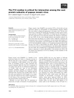
Fig. 3. Role of conserved Vps4p C-terminal motifs in interaction
with Vta1p, Vps20p and Did2p. (A) Yeast two-hybrid interaction anal-
ysis of Did2p (residues 41–204 ⁄ end) with full-length wild-type
Vps4p (residues 1–437 ⁄ end), the N-terminal region of Vps4p
(N-Vps4p; residues 1–128), the previously predicted AAA domain
(AAA-Vps4p; residues 129–350) [8] and C-terminal region (C-Vps4p;
residues 351–437 ⁄ end). The interaction analyses with Vta1p
(residues 108–333 ⁄ end) and Vps20p (residues 3–221 ⁄ end) were
included as controls. (B) Yeast two-hybrid interaction analysis of
wild-type Vps4p and Vps4p C-terminal mutants with the same frag-
ments of Vta1p, Vps20p and Did2p as in (A). In (A) and (B), EGY48
carrying pLexA-based bait plasmids and pB42AD-based prey plas-
mids as well as p8op-LacZ reporter plasmid were spotted on to
medium containing X-gal. Plates were photographed after overnight
incubation, and two-hybrid interaction was assessed by blue color-
ation. Two independent transformants are shown in (B). (C) In vitro
binding of His
6
-tagged wild-type Vps4p and Vps4p-GAI to GST-
Vps20p and GST-Vta1p. Equal amounts of full-length His
6
-tagged
proteins were incubated with glutathione ⁄ agarose bearing GST-
Vta1p, GST-Vps20p or GST alone in the presence or absence of
ATP. The unbound protein was recovered in the supernatants.
Bound protein was released with Laemmli sample buffer. The
bound and unbound fractions were subjected to SDS ⁄ PAGE and
immunoblotting with a polyclonal antibody to Vps4p. The amount of
wild-type Vps4p-His
6
or Vps4p-GAI-His
6
bound to GST-Vps20 was
quantified by densitometry. The shift in the relative positions of the
wild-type Vps4p-His
6
and Vps4p-GAI-His
6
bound to GST-Vps20 is
due to the presence of the GST-Vps20 protein, which migrates very
close to the His
6
-tagged proteins.
Role of the Vps4 b domain P. R. Vajjhala et al.
2362 FEBS Journal 273 (2006) 2357–2373 ª 2006 The Authors Journal compilation ª 2006 FEBS
Because yeast two-hybrid is an indirect measure
of binding strength, we used in vitro protein-binding
assays to confirm the effect of the Vps4p-GAI muta-
tion on the interaction with Vps20p and Vta1p. Both
wild-type Vps4p and Vps4p-GAI were expressed with
His
6
affinity tags in Escherichia coli and used in
binding assays with GST-Vta1p and GST-Vps20p
(Fig. 3C). Consistent with the yeast two-hybrid data,
the Vps4p-GAI-His
6
mutant protein did not interact
with Vta1p. Also consistent with our yeast two-hybrid
results, binding to GST-Vps20p was increased com-
pared with wild-type Vps4p-His
6
(both in the presence
or absence of ATP). In the presence of ATP, there was
a 35% decrease in the amount of Vps4p-GAI bound
to GST-Vps20p. In contrast, there was an 87%
decrease in the amount of wild-type Vps4p bound to
GST-Vps20p. Hence, the ATPase-dependent dissoci-
ation of GST-Vps20p is affected by deletion of the
conserved GAI motif in Vps4p.
We conclude that the RKI, GAI and LTP
motifs within and adjacent to the Vps4p b domain
are essential for interaction with Vta1p. The
increased interaction with Vps20p, when these motifs
are deleted may be due to defective ATPase-depend-
ent dissociation, which we showed directly for
Vps4p-GAI.
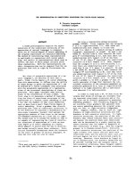
The Vps4p GAI motif in the b domain is not
required for Vps4p recruitment to endosomes
The Vps4p N-terminal domain has been shown to play
a key role in recruitment of Vps4p to endosomes, but
whether recruitment also requires the C-terminal
domain was not tested [24]. To test whether the C-ter-
minal GAI motif is important for Vps4p recruitment
to endosomes, we examined the subcellular localization
of GFP-tagged Vps4p-GAI and compared it with that
of GFP-tagged wild-type Vps4p (Fig. 4A). In vps4D
yeast cells, the fluorescence distribution of wild-type
Vps4p-GFP and Vps4p-GAI was both diffuse and
localized to punctate structures that are likely to be
endosomes. In contrast, the subcellular distribution of
GFP-tagged Vps4p-DCC, which lacks the N-terminal
MIT domain, was diffuse in the cytoplasm. This is
consistent with a critical role for the N-terminal MIT
domain in recruitment of Vps4p to endosomal mem-
branes that has previously been described [24]. Fur-
thermore, when the DCC mutation was introduced
into Vps4p-GAI, the subcellular distribution also
became diffuse in the cytoplasm. We conclude that
Vps4p-GAI, but not Vps4p-DCC-GAI, is recruited to
endosomal membranes. This suggests that the C-ter-
minal GAI motif is not essential for Vps4p recruitment
to endosomes.
The Vps4p GAI motif is not essential
for ATPase activity
To determine whether the GAI motif in the b domain is
important for Vps4p ATPase activity, perhaps via con-
formational effects on the AAA domain, we assayed the
ATPase activity of His
6
-tagged Vps4p-GAI and com-
pared it with that of His
6
-tagged wild-type Vps4p. The
affinity-purified Vps4p-GAI-His
6
mutant protein was
difficult to obtain as a full-length protein from bacteria
and most preparations contained some degradation
products. In the best preparations, 30% of the protein
was full-length as determined by densitometry. When
equivalent amounts of full-length protein were assayed
in the presence of 0.1 mm ATP, the Vps4p-GAI-His
6
protein had 52% of wild-type activity (Fig. 5). However,
when equal amounts of full-length protein were assayed
in the presence of 1 mm ATP, the Vps4p-GAI-His
6
pro-
tein had only 14% of wild-type activity. We conclude
that the Vps4p GAI motif in the b domain is important,
although not essential for Vps4p ATPase activity.
Fluorescence Nomarski
vps4∆/
Vps4p-GFP
vps4∆/
Vps4p-GAI-GFP
vps4∆/
Vps4p-∆CC-GFP
vps4∆/
Vps4p-∆CC-GAI-GFP
Fig. 4. The GAI motif in the b domain is not essential for localiz-
ation of Vps4p to endosomes. (A) RH2906 vps4D yeast cells
expressing GFP-tagged wild-type Vps4p, Vps4p-GAI, Vps4p-DCC or
Vps4p-DCC-GAI were grown in SD medium, and the GFP-tagged
proteins visualized by fluorescence microscopy. Scale bar, 5 lm.
P. R. Vajjhala et al. Role of the Vps4 b domain
FEBS Journal 273 (2006) 2357–2373 ª 2006 The Authors Journal compilation ª 2006 FEBS 2363
The phenotypes conferred by mutation of the
conserved motifs adjacent to and within the
Vps4p b domain are not dominant
To investigate whether mutation of the Vps4p RKI,
GAI and LTP motifs confers dominant phenotypes,
like most previously characterized vps4 mutants, we
expressed the mutant proteins in wild-type cells and
examined the effect on Vps4p function. Wild-type
cells expressing the Vps4p-RKI, Vps4p-GAI and
Vps4p-LTP mutants did not missort and secrete
CPY, grew well at 40 °C, and transported Fth1p-
Ub-GFP to the vacuole lumen (data not shown). In
contrast, wild-type cells expressing the dominant-neg-
ative Vps4p-E233Q mutant [24] showed the full
range of dominant-negative phenotypes. This indi-
cates that the defects conferred by mutation of the
Vps4p-RKI, Vps4p-GAI and Vps4p-LTP motifs are
not dominant.
An intact b domain is essential for Vps4p
self-association
Although wild-type Vps4p expressed in bacteria is a
dimer [24], previous studies using the yeast two-hybrid
technique did not reveal a homotypic interaction in
wild-type Vps4p [25]. We used a different yeast two-
hybrid system to determine whether we could detect
this homotypic interaction. The data obtained show
that wild-type Vps4p can interact with itself strongly
(Fig. 6). This is consistent with previous data showing
that Vps4p forms a dimer.
As the b-domain mutants are recessive, we consid-
ered the possibility that these mutants may not be able
to interact with wild-type Vps4p. To test this, we used
the yeast two-hybrid assay to study the interaction
between Vps4p-GAI and wild-type Vps4p. However,
the Vps4p-GAI mutant protein barely interacted with
wild-type Vps4p and did not interact with itself
(Fig. 6). The Vps4p-RKI and Vps4p-LTP mutant pro-
teins also did not interact with wild-type Vps4p (data
not shown). We surmise that an intact b domain is
required for Vps4p self-association.
An intact b domain is required for the
Vps4p-E233Q mutant to have a dominant-
negative phenotype
The E233Q mutation in Vps4p confers a dominant-
negative phenotype [8,28], and the equivalent muta-
tion in mammalian VPS4 isoforms has been widely
used to study the effect of VPS4 inactivation in
mammalian cells [35,19]. Vps4p-E233Q forms a 10–
12-mer [24,27], and it has been speculated that wild-
type Vps4p may also form a high-molecular-mass
oligomer that is transient in the presence of a func-
tional ATPase domain. The dominant-negative pheno-
type conferred by Vps4p-E233Q is believed to be due
to interaction of Vps4p-E233Q with wild-type Vps4p
in vivo based on the ability of Vps4p-E233Q to oligo-
1.0 0.1
GAI WT GAI WT
[ATP] m
M
0
5
10
15
20
25
nmol inorganic phosphate released
/h/µg full-length protein
Fig. 5. The Vps4p-GAI mutant has a diminished ATPase activity.
Affinity-purified His
6
-tagged wild-type Vps4p (WT) and Vps4p-GAI
(GAI) were assayed in vitro for ATPase activity at 30 °C. The
amount of protein assayed was normalized based on the level of
full-length wild-type Vps4p and Vps4p-GAI. ATPase activity was
assayed in the presence of 0.1 m
M ATP or 1 mM ATP and is
expressed as nmol inorganic phosphate releasedÆh
)1
Æ(lg full-length
protein)
)1
and shown graphically.
pLexA/ pB42AD
pLexA Vps4p/ pB42AD Vps4p
pLexA Vps4p/ pB42AD Vps4p-GAI
pLexA Vps4p-GAI/ pB42AD Vps4p
pLexA Vps4p-GAI/ pB42AD Vps4p-GAI
Fig. 6. The conserved GAI motif in the b domain of Vps4p is
required for homotypic interaction between wild-type Vps4p mole-
cules. The interaction between various combinations of wild-type
Vps4p and Vps4p-GAI was assessed using the yeast two-hybrid
technique. EGY48 carrying a p8op reporter plasmid and pLexA-
based bait plasmids and pB42AD-based prey plasmids were spot-
ted on to synthetic galactose medium containing X-gal. Plates
were photographed after 2 days, and two-hybrid interaction was
assessed by blue coloration. Three independent transformants are
shown.
Role of the Vps4 b domain P. R. Vajjhala et al.
2364 FEBS Journal 273 (2006) 2357–2373 ª 2006 The Authors Journal compilation ª 2006 FEBS
merize with Vps4p-DCC [24] in vitro. To demonstrate
a role for the b domain in Vps4p assembly in vivo,
we mutated the GAI motif in the dominant-negative
Vps4p-E233Q mutant. Unlike Vps4p-E233Q, the
resulting double mutant (Vps4p-E233Q-GAI) did not
confer dominant-negative phenotypes (Fig. 7A–C). As
the expression level of Vps4p-E233Q-GAI is similar
to that of Vps4p-E233Q (Fig. 7D), loss of the domin-
ant-negative phenotype is not due to decreased
expression of the double mutant. Our data strongly
suggest that the b domain is required for interaction
with wild-type Vps4p in vivo.
Vps20p and Vta1p both stimulate Vps4p ATPase
activity, but Vps20p stimulates its activity to a
greater extent
As the b domain of Vps4p is required for full ATPase
activity as well as for interaction with Vta1p, it is poss-
ible that binding of Vta1p to Vps4p might have an
effect on ATPase activity. To test this hypothesis, we
assayed the in vitro ATPase activity of wild-type His
6
-
tagged Vps4p in the presence of increasing concentra-
tions of GST-Vta1p. We also assayed the activity of
Vps4p in the presence of increasing concentrations of
GST and GST-Vps20p. We included GST-Vps20p
because Vps20p binds to Vps4p in an ATP-sensitive
manner, and substrate binding is known to increase
the activity of some AAA ATPases such as Hsp104
and Katanin [36,37]. The data in Fig. 8 show that both
Vta1p and Vps20p have a stimulatory effect on Vps4p
ATPase activity, but the effect of Vps20p is much
greater. It is interesting that, although the b domain is
required for assembly of Vps4p into a complex with
full ATPase activity, binding of Vta1p to the b domain
does not inhibit Vps4p ATPase activity.
Discussion
Here we identify conserved motifs in the C-terminal
region of Vps4p and provide evidence that those
within the b domain are critical for all Vps4p in vivo
A
D
Vps4p-E233Q
Vps4p-E233Q-GAI
Vps4p
actin
Vps4p-
E233Q
Vps4p-
E233Q-
GAI
Fluorescence
Nomarski
C
24
O
C
40
O
C
Vps4p-E233Q
Vps4p-E233Q-GAI
B
Vps4p-E233Q
Vps4p-E233Q-GAI
Vps4p-E233Q
Vps4p-E233Q-GAI
CPY blot
calmodulin blot
Fig. 7. The dominant-negative Vps4p-E233Q mutant becomes recessive upon mutation of the GAI motif. (A) The Vps4p-E233Q-GAI mutant
protein does not confer a dominant-negative MVB sorting defect. RH1800 (wild-type) yeast cells expressing Fth1p-GFP-Ub and either Vps4p-
E233Q or Vps4p-E233Q-GAI were grown in SD selective medium and assayed for MVB sorting as in Fig. 2. Scale bar, 5 lm. (B) The Vps4p-
E233Q-GAI mutant protein does not confer a dominant-negative vacuolar protein sorting defect. RH1800 (wild-type) yeast cells expressing
Vps4p-E233Q or Vps4p-E233Q-GAI were grown on selective SD solid medium at 24 °C in contact with a nitrocellulose filter. CPY missorting
was assayed as in Fig. 2. (C) The Vps4p-E233Q-GAI mutant protein does not confer a dominant-negative growth defect. Wild-type RH1800
yeast cells expressing Vps4p-E233Q or Vps4p-E233Q-GAI were assayed for growth at high temperature on solid SD selective medium as in
Fig. 2. (D) Total cell lysates from RH2906 (vps4D) yeast cells carrying plasmids expressing Vps4p-E233Q or Vps4p-E233Q-GAI were subjec-
ted to western blotting as in Fig. 1D.
P. R. Vajjhala et al. Role of the Vps4 b domain
FEBS Journal 273 (2006) 2357–2373 ª 2006 The Authors Journal compilation ª 2006 FEBS 2365
functions including fluid-phase endocytosis, MVB sort-
ing, vacuolar protein sorting and growth at high tem-
perature. Two of these motifs, LTP and GAI, are in
the b domain and the third, RKI, is partly within the
b domain and partly within the AAA domain. We pro-
vide evidence that the b domain is important for full
ATPase activity of Vps4p. We also show that the b
domain is required for two protein interactions. The
first is a homotypic interaction that may be important
for assembly of a fully catalytically active oligomer,
and the second is with Vta1p. Both of these interac-
tions are likely to be important for Vps4p function
in vivo. We also show that the charged residues in an
RDE motif at the end of the final C-terminal a-helix
are not required for most Vps4p functions but are
required for full growth at high temperature. More-
over, these charged residues are not required for Vta1p
interaction.
Several lines of evidence suggest that mutation of
these motifs have specific effects on Vps4p in vivo func-
tion. Deletion of the motifs did not compromise stable
expression, indicating that loss of in vivo function is
not merely due to lowered expression levels. The
Vps4p proteins carrying mutations in the b domain
retained at least part of their native structure, as they
were able to interact with Vps20p and Did2p, which
interact with the N-terminal region of Vps4p ([30]; this
study). This was shown by yeast two-hybrid assay and
confirmed in one b domain mutant using an in vitro
binding assay. In addition, the retention of native
structure in b domain mutant proteins is supported by
the ability of a b domain mutant protein to be recrui-
ted efficiently to endosomes in vivo as assessed visually
by microscopy. Hence it is possible to mutate the b
domain without grossly affecting Vps4p structure.
Moreover, based on the structure of the human VPS4B
monomer, the b domain is an independent domain
that is separated from the AAA domain by random
coils (Fig. 1E), thus mutations in this domain are unli-
kely to perturb the structure of the AAA domain.
However, we cannot exclude the possibility that dele-
tion of the conserved motifs within the b domain may
perturb the local structure of the b domain.
The requirement for an intact b domain for full
ATPase activity is clearly demonstrated in the in vitro
ATPase assays and further supported by interaction
analysis of the b domain mutant with Vps20p. The
Vps4p–Vps20p interaction is known to be sensitive to
ATP hydrolysis and is stabilized by the E233Q muta-
tion that perturbs Vps4p ATPase activity [30]. Consis-
tent with a decreased ATPase activity, interaction of
the Vps4p b domain mutant with Vps20p was
enhanced in the in vitro binding assay and in the
in vivo yeast two-hybrid assay.
Most AAA ATPases assemble into higher-order
oligomeric rings. Consistent with this, wild-type
Vps4p purified from bacteria forms a dimer [24], and
Vps4p forms a 10–12-mer when its ATPase activity
is compromised and it is locked in the ATP-bound
conformation, as in the Vps4p-E233Q mutant
[24,27]. Arg352 in the AAA domain of Vps4p has
recently been shown to be important for assembly of
Vps4p-E233Q into oligomers but not for dimer for-
mation [27]. The motifs of Vps4p required for dime-
rization have not previously been identified. Previous
studies using a yeast two-hybrid assay have demon-
10 23456789
0
2
4
6
8
ratio of GST alone or GST-fusion protein (µg): Vps4p (µg)
nmol inorganic phosphate released
/h/µg Vps4p
GST alone+Vps4p
GST alone
GST-Vps20p+Vps4p
GST-Vps20p
GST-Vta1p+Vps4p
GST-Vta1p
Fig. 8. Vps20p strongly stimulates Vps4p ATPase activity, but binding of Vta1p to the b domain of Vps4p has only a marginal stimulatory
effect. The ATPase activity of recombinant Vps4p-His
6
was assayed at 30 °C in the presence of 0.3 mM ATP and increasing amounts of
recombinant GST-Vta1p, GST-Vps20p or GST alone. ATPase activity is expressed as nmol inorganic phosphate releasedÆh
)1
Æ(lg Vps4p)
)1
.
The ATPase activities of GST alone and the GST fusion proteins without any Vps4p were also assayed and are shown.
Role of the Vps4 b domain P. R. Vajjhala et al.
2366 FEBS Journal 273 (2006) 2357–2373 ª 2006 The Authors Journal compilation ª 2006 FEBS
strated an interaction between the yeast Vps4p-
E233Q mutant and wild-type Vps4p. However, a
homotypic interaction in wild-type Vps4p was not
demonstrated [25]. Here, we detect a homotypic
interaction in wild-type Vps4p using a different yeast
two-hybrid system. In addition, we show that the b
domain plays an important role in this homotypic
interaction.
A model for the Vps4 oligomer has recently been
proposed. According to this model the Vps4 oligomer
comprises two stacked hexameric rings [27]. In this
proposed model, the b domain is not well placed to
mediate intersubunit interactions within a hexameric
ring. However, interactions between the b domains
may be important during the assembly. For example,
the homotypic interaction mediated by the b domain
may be required for formation of a dimer, and six of
these may subsequently assemble into a double hexa-
meric ring such that the protomers comprising a dimer
are present in each of the two rings. The b domains
would thus mediate the interaction between the two
stacked rings.
Homotypic interactions mediated by the b domain
may be required for formation of a fully catalytically
active enzyme in vivo. Three lines of evidence support
this hypothesis. First, most vps4 mutants described to
date that have an intact b domain are dominant-negat-
ive [8,17,28] and are thus likely to assemble with wild-
type Vps4p to form a functionally inactive complex. In
contrast, the b-domain mutants are not dominant-neg-
ative, suggesting that they are unable to interact with
wild-type Vps4p. Secondly, mutation of the b domain
abolishes the dominant-negative effects of Vps4p-
E233Q, further suggesting that the b domain is
required for association with wild-type Vps4p in vivo .
Thirdly, in the presence of an increased concentration
of ATP, we observed a substantial increase in the
ATPase activity of wild-type Vps4p. However, the
increase in activity of a b-domain mutant was not as
dramatic, suggesting that this mutant cannot assemble
into a fully active ATPase.
Our finding that all the three motifs that were within
the b domain or partially adjacent to it are required
for the Vta1p interaction suggests that the Vta1p-bind-
ing site includes several motifs. Interestingly, these
three highly conserved motifs cluster together in the
human VPS4B structure consistent with them having a
common function (Fig. 1C). Our data are consistent
with a recent report [27] showing that mutation of
Arg352 in the RKI motif abolished interaction with
Vta1p. This mutation (R352A) is in the AAA domain
and also prevents oligomerization of Vps4p dimers
into a higher-order oligomer. In addition, mutations in
the RKI and LTP motifs (D362A and S377A, respect-
ively) that are within the b domain also abolished the
Vta1p interaction. However, the effect of these b
domain mutants on Vps4p oligomeric assembly was
not addressed. The fact that our b domain mutant
affected homotypic interactions and perhaps assembly
suggests that loss of the Vta1p interaction may be
partially due to inability of the mutant to form a
higher-order oligomer.
Our data showing that both Vps20p and Vta1p sti-
mulate Vps4p ATPase activity are consistent with
recent data showing that Vta1p binding stimulates
Vps4p ATPase activity [38]. Vta1p has previously been
shown to stabilize Vps4p oligomers [27] and is sugges-
ted to modulate Vps4p ATPase activity [38]. However,
we find that Vps20p, which is a component of ESC-
RT-III, stimulates ATPase activity to a greater extent
than Vta1p, suggesting that it is a stronger modulator
of Vps4p activity.
The N-terminal region of Vps4p, which contains an
MIT domain, has been reported to be important for
Vps4p localization to endosomes [24]. However, a
requirement for the C-terminal region for Vps4p endo-
some recruitment was not investigated. Our findings
suggest that the b domain does not normally play a
role in Vps4p recruitment to endosomes. Moreover the
fact that the Vta1p interaction is compromised in the
b-domain mutants demonstrates that interaction with
Vta1p is not required for recruitment to endosomes.
Our suggestion that assembly of Vps4p into an
oligomeric complex is required for full ATPase activity
is consistent with previous findings. First, the forma-
tion of a higher-order complex from a Vps4p dimer is
proposed to be ATP-dependent, as the Vps4p-E233Q
mutant forms a 10–12-mer in the presence of ATP
[24,27]. ATP-dependent oligomerization is also
observed in several other AAA ATPases including
ClpB and PspF [39,40]. Secondly, oligomerization sti-
mulates ATPase activity in a number of AAA and
AAA+ ATPases including RuvB and FtsH [41,42].
This is achieved by allosteric stimulation of ATPase
activity by one or two conserved arginine residues in
the N-terminal subdomain of the AAA+ or AAA
domains (respectively) that contact the c-phosphate of
ATP bound to an adjacent subunit.
The role of the AAA+ domain in oligomerization
of AAA+ proteins is well established. Our study,
however, shows that for Vps4p, regions outside of the
AAA domain are also required for assembly. Although
this finding was unexpected, there is a precedent for
the role of regions outside of the AAA domain in
oligomerization. For example, the linker region
between the two AAA domains of p97 is required for
P. R. Vajjhala et al. Role of the Vps4 b domain
FEBS Journal 273 (2006) 2357–2373 ª 2006 The Authors Journal compilation ª 2006 FEBS 2367
hexamerization of the N-terminal AAA domain [43].
Furthermore, Katanin requires microtubule binding
for oligomeric assembly [44]. As AAA+ proteins are
studied, more examples of such proteins that require
regions outside the AAA+ domain for assembly may
become apparent.
The function of the C-terminal a-helix of Vps4p has
not been previously characterized. The finding that the
charged residues in a conserved motif at the end of this
helix are not required for most endocytic functions of
Vps4p but are required for full growth at high tempera-
ture is interesting. This suggests that Vps4p may have
an important role in growth at high temperature, dis-
tinct from its endocytic roles, which may depend on the
charged residues. Alternatively, this mutant may be only
partially functional endocytosis at high temperature.
In summary, the b domain of Vps4p is not required
for recruitment to endosomes. It appears to play a crit-
ical role in the formation of a Vps4p ATPase complex
that is functional in vivo. The formation of the latter
may be a prerequisite for interaction with Vta1p. Fur-
thermore, our data highlight the importance of Vps4p
assembly during MVB sorting and other endocytic
functions.
Experimental procedures
Media, reagents, strains and plasmids
YPUAD and SD minimal media were as described previ-
ously [30]. Lucifer Yellow carbohydrazide dilithium salt
was from Fluka AG (Buchs, Switzerland). Horseradish per-
oxidase-conjugated goat anti-rabbit IgG and bathophen-
anthroline disulfonic acid were from Sigma (St Louis, MO,
USA). Horseradish peroxidase-conjugated anti-mouse IgG
was from Bio-Rad Laboratories (Hercules, CA, USA), and
horseradish peroxidase-conjugated anti-goat IgG was from
Zymed (San Francisco, CA, USA). Ni ⁄ nitrilotriacetate ⁄
agarose and monoclonal anti-pentaHis IgG were from
Qiagen (Hilden, Germany). Immobilized glutathione on
agarose was from Pierce (Rockford, IL, USA). Prestained
protein molecular mass marker was from Fermentas (Han-
over, MD, USA). Poly(vinylidene difluoride) (PVDF) mem-
brane was from Millipore (Bedford, MA, USA). Polyclonal
anti-(carboxypeptidase Y) and anti-calmodulin sera were
gifts from H. Riezman (University of Geneva, Switzerland),
polyclonal antibody to Vps4p was from Santa Cruz Bio-
technology (Santa Cruz, CA, USA), and monoclonal anti-
body to actin was from Chemicon (Temecula, CA, USA).
EDTA-free protease inhibitor cocktail tablets were from
Roche Diagnostics (Basel, Switzerland).
Saccharomyces cerevisiae strains used in this study are lis-
ted in Table 1. Y15588 was obtained from EUROSCARF
(European Saccharomyces cerevisiae Archives for Func-
tional Analysis, Frankfurt, Germany). AMY245 was gener-
ated by transformation of RH1201 with the vps4D::KanMx
disruption cassette, which was amplified from Y15588.
Transformants were selected on medium containing G418
and then induced to sporulate. Tetrads were dissected using
a Singer MSM manual (Singer Instrument Co. Ltd, Wat-
chet, Somerset, UK). The vps4D disruption cassette segrega-
ted 2 : 2. Yeast was transformed with plasmid DNA using
a modified lithium acetate protocol [45].
PCR primers used for plasmid constructions were from
GeneWorks (Thebarton, Australia) and are listed in
Table 2. Plasmids used in this study are listed in Table 3.
The sequence of all constructs was confirmed by automated
DNA sequencing (Australian Genome Research Facility,
Brisbane, Australia).
Construction of VPS4 wild-type and mutant
constructs
Genomic DNA was prepared from S. cerevisiae as des-
cribed previously [46], and PCR was carried out using
the proof-reading DNA polymerases Platinum Pfx (Invi-
trogen, Carlsbad, CA, USA) or Pfu (Fermentas). A full-
length VPS4 construct, including 396 bp upstream
sequence and 122 bp downstream sequence, was first pre-
pared in which a SalI site was introduced between bases
700 and 705 of the coding sequence, such that it did not
alter the encoded amino acids. The 5¢ fragment of VPS4
was amplified using the Vps4 Upstr forward (F) and
Vps4 SalI reverse (R) primers, and the 3¢ fragment was
amplified using Vps4 SalI F and Vps4 Dstr R primers.
Table 1. Yeast strains used in this study.
Strain Genotype Source
Y15588 MATa vps4-D::KanMx his3 leu2 lys2 ura3 EUROSCARF
RH1201 MATa ⁄ MATa his4 ⁄ his4 leu2 ⁄ leu2
ura3 ⁄ ura3 lys2 ⁄ lys2 bar1 ⁄ bar1
Riezman lab strain
RH1800 MATa his4 leu2 ura3 bar1 Riezman lab strain
RH2906 MATa vps4-D::URA3 his4 leu2 ura3 lys2 bar1 [17]
AMY174 MATa vps20-D::KanMx his4 leu2 ura3 bar1 [30]
AMY245 MATa vps4-D::KanMx leu2 ura3 his4 lys2 bar1 This study
Role of the Vps4 b domain P. R. Vajjhala et al.
2368 FEBS Journal 273 (2006) 2357–2373 ª 2006 The Authors Journal compilation ª 2006 FEBS
To generate full-length VPS4 ,5¢ and 3¢ PCR products
were cut with PstI and SalIorBamHI and SalI, respect-
ively, and ligated into PstI ⁄ BamHI-digested YCplac111.
C-Terminal mutations were generated by site-directed
mutagenesis using the oligonucleotides listed in Table 2.
In each case, two separate PCRs were set up with either
Vps4 SalI F primer and a mutagenic reverse primer or a
mutagenic forward primer and Vps4 Dstr R primer. The
two PCR products were combined for a third PCR using
the SalI F and Vps4 Dstr R primers. The resulting PCR
product encoding the C-terminal region of Vps4p, with a
mutation, was ligated with the PCR product containing
the 5¢ fragment of VPS4 (described above). The VPS4
wild-type and mutant constructs were then cloned into
YCplac111.
To generate wild-type and mutant VPS4-pLexA or
VPS4-pB42 constructs, VPS4 was amplified without any
upstream sequence and with suitable restriction sites for
cloning. To express wild-type Vps4p and Vps4p-GAI with a
C-terminal GFP tag, both constructs were amplified with-
out a stop codon and cloned in-frame into a YCplac111-
based plasmid encoding yEGFP, which was cloned from
pYM12 [47]. To express Vps4p-GAI with a C-terminal His
6
tag in E. coli, VPS4-GAI was amplified with a primer-enco-
ded C-terminal His
6
tag and ligated downstream of the T7
promoter of pET11d (Novagen, Madison, WI, USA). The
YCplac111 plasmid expressing the Vps4p-DCC mutant was
generated as previously described [24], and the YCplac111
plasmid expressing the previously described Vps4p-E233Q
mutant [8] was generated by replacing a 0.6-kb AfeI–NcoI
fragment in wild-type VPS4 with the corresponding frag-
ment from pAM483 [30]. To generate pAM928, the PstI–
NcoI fragment in pAM819 was replaced with the corres-
ponding fragment from pAM922. To generate pAM931
and pAM932, the PstI–SalI fragments in pAM864 and
pAM863 were replaced with the corresponding fragment
from pAM927.
Phenotypic assays
Fluid-phase endocytosis was assayed using the membrane-
impermeable dye Lucifer Yellow as described previously
[48] except that the cells were incubated with Lucifer Yel-
low for 1 h at 30 °C. MVB sorting using Fth1-GFP-Ub
was performed as previously described [30] except that,
where stated, the cells were grown in SD medium to ensure
plasmid retention. CPY missorting was assayed as described
previously [48]. For temperature-sensitive growth, a single
transformant colony was resuspended in 1 mL water and
serially diluted by 10-fold. Aliquots (7 lL) were spotted on
solid medium and incubated at 24 °Cor40°C.
Yeast two-hybrid protein interaction analysis
Protein interactions were assayed using the Matchmaker
LexA yeast two-hybrid system from Clontech (Palo Alto,
CA, USA) as described previously [30]. Briefly, bait plas-
mids containing LexA fusion proteins were cotransformed
into the yeast strain EGY48 along with prey plasmids
encoding proteins fused to an activation domain and the
reporter plasmid p8op-LacZ. To test for interaction, trans-
formants were spotted on to synthetic galactose ⁄ raffinose
(SG) complete medium lacking Ura, Trp, and His and con-
taining X-gal. The strength of protein interactions was
assessed by blue coloration on this medium.
In vitro binding assay
In vitro binding assays were performed as previously des-
cribed [30]. The His
6
-tagged wild-type Vps4p and Vps4p-
Table 2. Primers used for mutagenesis.
Primer Sequence
Vps4 Upstr F 5¢-CGCTGCAGTAAGAGCAGTAAACCCG-3¢
Vps4 SalIR 5¢-GAGAATCAGTGTCGACTTCATCTATAAAAATAATAGAAGGTTTATT-3¢
Vps4 SalIF 5¢-GCCCATATTCGTCGACGCGCTAACAGGTACCAGAGGAGAAGGAGAGAGCGAAGCAAGTAG-3¢
Vps4 Dstr R 5¢-GGGCGGATCCTCTGCTTTTCTTTATC-3¢
Vps4 Ter1 F 5¢-GCGCTAATGCAACCGTAGTCAATTGATTAACGTGCT-3¢
Vps4 Ter1 R 5¢-AGCACGTTAATCAATTGACTACGGTTGCATTAGCGC-3¢
Vps4 Ter2 F 5¢-TTAAAAGAACCAGATTAGTCAATTGATTAACGTGCT-3¢
Vps4 Ter2 R 5¢-AGCACGTTAATCAATTGACTAATCTGGTTCTTTTAA-3¢
Vps4 RDE F 5¢-AAGCAAGAACAGTTCACTGCAGCTTTTGGTCAAGCAGGTAACTAGTCAATTGAT-3¢
Vps4 RDE R 5¢-ATCAATTGACTAGTTACCTGCTTGACCAAAAGCTGCAGTGAACTGTTCTTGCTT-3¢
Vps4 RKI F 5¢-GCGCTAATGCAACCGATAGATGTCTCTACGGAGGAC-3¢
Vps4 RKI R 5¢-GTCCTCCGTAGAGACATCTATCGGTTGCATTAGCGC-3¢
Vps4 LTP F 5¢-GACGACGAAACAAGAAAAGATGGCGCCATCGAGATG-3¢
Vps4 LTP R 5¢-CATCTCGATGGCGCCATCTTTTCTTGTTTCGTCGTC-3¢
Vps4 GAI F 5¢-TGCTCTCCAGGTGATGATATTGAAGCTGATGAATTA-3¢
Vps4 GAI R 5¢-TAATTCATCAGCTTCAATATCATCACCTGGAGAGCA-3¢
P. R. Vajjhala et al. Role of the Vps4 b domain
FEBS Journal 273 (2006) 2357–2373 ª 2006 The Authors Journal compilation ª 2006 FEBS 2369
GAI as well as GST-tagged Vps20p and Vta1p, and GST
alone were expressed in BL21-CodonPlus
tm
(DE3)–RIL
E. coli and affinity-purified on Ni ⁄ nitrilotriacetate ⁄ agarose
or glutathione ⁄ agarose, respectively. The His
6
-tagged pro-
teins were eluted from the resin using 250 mm imidazole and
buffer exchanged into binding buffer (20 mm Hepes, 200 mm
sorbitol, 100 mm potassium acetate, 1 mm EDTA, 1 mm
dithiothreitol, 20 mm MgCl
2
, 0.1% Triton X-100). The
Vps4p-GAI mutant protein was poorly expressed in E. coli
compared with wild-type Vps4p and gave additional bands
on SDS ⁄ PAGE, which reacted with an anti-Vps4p serum.
Full-length Vps4p-GAI comprised 30% of the total pro-
tein. For in vitro binding assays, 6 lg each full-length His
6
-
tagged protein was incubated with gluthathione ⁄ agarose,
bearing GST alone (560 lg), GST-Vps20 (50 lg), or GST-
Vta1p (30 lg) in the presence or absence of 1 mm ATP in a
total volume of 1 mL binding buffer. Samples were incubated
overnight at 4 °C. The resin was washed three times with
binding buffer, and the protein bound to 10 lL resin was
eluted by heating with Laemmli sample buffer. An aliquot of
the supernatant containing unbound protein was diluted
1 : 1 in Laemmli sample buffer. Bound and unbound pro-
teins were resolved by SDS ⁄ PAGE and transferred to a
PVDF filter. Wild-type Vps4p and Vps4p-GAI were detected
with an antibody to Vps4p and enhanced chemilumines-
cence.
Western blot analysis of total yeast cell lysates
For western blot analysis of total cell lysates, RH2906
(vps4D) yeast carrying expression plasmids were grown at
24 °C for 48 h. Cells from 2 mL culture were pelleted and
resuspended in 240 lL lysis solution (1.85 m NaOH, 1.06
m 2-mercaptoethanol) followed by incubation for 10 min
Table 3. Plasmids used in this study.
Plasmid Description Source
YCplac111 CEN4 ARS1 LEU2 E. coli ⁄ yeast shuttle vector [49]
pGEX5X-1 GST fusion expression vector GE Healthcare
pET11d T7 RNA polymerase-based gene expression vector Novagen
p8op-lacZ Two-hybrid reporter plasmid Clontech
pLexA Two-hybrid bait vector Clontech
pB42AD Two-hybrid prey vector Clontech
pPL1640 URA3 CEN plasmid expressing Fth1p-GFP-Ub [31]
pAM349 Original library clone of VPS20 in pB42AD (encoding Vps20p 3–221 ⁄ end) [30]
pAM377 pGEX5X-1 expressing Vps20p with an N-terminal GST tag [30]
pAM378 pGEX5X-1 expressing Vta1p with an N-terminal GST tag [30]
pAM398 Original library clone of VTA1 in pB42AD (encoding Vta1p 108–330 ⁄ end) [30]
pAM452 pLexA expressing LexA fused to N-terminal domain of Vps4p (residues 1–128) [30]
pAM453 pLexA expressing LexA fused to AAA domain of Vps4p (residues 129–350) [30]
pAM454 pLexA expressing LexA fused to C-terminal domain of Vps4p (351–437 ⁄ end) [30]
pAM482 pET11a E. coli expression vector expressing Vps4p with a C-terminal 6HIS tag [30]
pAM496 Original library clone of DID2 ⁄ CHM1 in pB42AD (encoding Did2p ⁄ Chm1p 41–204 ⁄ end) This study
pAM813 YCplac111 expressing Vps4p This study
pAM814 YCplac111 expressing Vps4p D401-437 ⁄ end (Vps4p-Ter2) This study
pAM815 YCplac111 expressing Vps4p D351-437 ⁄ end (Vps4p-Ter1) This study
pAM816 YCplac111 expressing Vps4p R430A, D431A, E435A (Vps4p-RDE) This study
pAM817 YCplac111 expressing Vps4p D373-380 (Vps4p-LTP) This study
pAM818 YCplac111 expressing Vps4p D352-361 (Vps4p-RKI) This study
pAM819 YCplac111 expressing Vps4p D382-390 (Vps4p-GAI) This study
pAM855 pLexA expressing LexA fused to Vps4p This study
pAM856 pLexA expressing LexA fused to Vps4p-RDE This study
pAM857 pLexA expressing LexA fused to Vps4p-LTP This study
pAM858 pLexA expressing LexA fused to Vps4p-RKI This study
pAM859 pLexA expressing LexA fused to Vps4p-GAI This study
pAM862 pET11d E. coli expression vector expressing Vps4p-GAI with a C-terminal 6HIS tag This study
pAM863 YCplac111 expressing Vps4p with a C-terminal yEGFP tag This study
pAM864 YCplac111 expressing Vps4p-GAI with a C-terminal yEGFP tag This study
pAM870 pB42AD expressing the activation domain fused to Vps4p This study
pAM871 pB42AD expressing the activation domain fused to Vps4p-GAI This study
pAM922 YCplac111 expressing Vps4p-E233Q This study
pAM928 YCplac111 expressing Vps4p-E233Q, D382-390 (Vps4p-E233Q-GAI) This study
pAM931 YCplac111 expressing Vps4p- D31-87, D382-390 (Vps4p-DCC-GAI) with a C-terminal yEGFP tag This study
pAM932 YCplac111 expressing Vps4p D31-87 (Vps4p-DCC) with a C-terminal yEGFP tag This study
Role of the Vps4 b domain P. R. Vajjhala et al.
2370 FEBS Journal 273 (2006) 2357–2373 ª 2006 The Authors Journal compilation ª 2006 FEBS
on ice. Proteins were precipitated by the addition of an
equal volume of trichloroacetic acid, and the pellet was
washed with ice-cold acetone. The pellet was resuspended
in 50 lL resuspension solution (5% SDS, 0.5 m Tris) and
mixed with an equal volume of 75% glycerol ⁄ 0.12 m dithio-
threitol ⁄ 0.05% bromophenol blue and boiled for 5 min.
Samples were subjected to SDS ⁄ PAGE, and proteins were
transferred to a PVDF filter that was then probed with a
goat polyclonal antibody to Vps4p. To assess sample load-
ing, the same samples were electrophoresed on a second
gel, transferred to a PVDF filter, and probed with a mono-
clonal antibody to actin.
Microscopy
Microscopy was performed using an Olympus AX70 with a
Nomarski filter (Olympus Australia Pty Ltd., Mount
Waverely, Victoria, Australia) for visualizing vacuoles and
the appropriate filters for viewing Lucifer Yellow or GFP
fluorescence.
Assays of ATPase activity
The His
6
-tagged wild-type Vps4p or Vps4p-GAI
were expressed in E. coli and purified as described for
the in vitro binding assay. To assay for ATPase activity,
50 lL of each protein containing 0.7 lg wild-type Vps4p
or the equivalent amount of full-length Vps4p-GAI in
ATPase assay buffer (0.1 m potassium acetate, 5 mm
magnesium acetate, 20 mm Hepes, pH 7.4) [8] was incu-
bated with 0.1 mm ATP or 1 mm ATP for 1 h at 30 °C.
Released inorganic phosphate was quantified using a
phosphate detection kit (R & D Systems, Minneapolis,
MN, USA). To test the effect of added GST-Vta1p,
GST-Vps20p or GST alone on Vps4p ATPase activity,
GST and the GST fusion proteins were purified as
described for the in vitro binding assays and eluted from
glutathione ⁄ agarose in assay buffer containing 5 mm
glutathione. Various amounts of GST or GST fusion
protein (0.07–1.4 lg) were incubated with 0.17 lg Vps4p
in 24.5 lL assay buffer containing 0.3 mm ATP for
30 min at 30 °C. Inorganic phosphate released was quan-
tified using the phosphate detection kit.
Acknowledgements
This work was made possible by funding from the
National Health and Medical Research Council of
Australia Project Grant 252750, from A*STAR (Singa-
pore), and from core support from the Queensland
State Government, all to A.L.M. The yeast two-hybrid
screen that showed the interaction of Vps4p with
Did2p [30] was carried out by Mahendra Wagle. We
thank Ellen Wren for technical assistance.
References
1 Bonaldi T, Talamo F, Scaffidi P, Ferrera D, Porto A,
Bachi A, Rubartelli A, Agresti A & Bianchi ME (2003)
Monocytic cells hyperacetylate chromatin protein
HMGB1 to redirect it towards secretion. EMBO J 22,
5551–5560.
2 Fevrier B & Raposo G (2004) Exosomes: endosomal-
derived vesicles shipping extracellular messages. Curr
Opin Cell Biol 16, 415–421.
3 Resh MD (2005) Intracellular trafficking of HIV-1 Gag:
how Gag interacts with cell membranes and makes viral
particles. AIDS Rev 7, 84–91.
4 Morita E & Sundquist WI (2004) Retrovirus budding.
Annu Rev Cell Dev Biol 20, 395–425.
5 Pelchen-Matthews A, Raposo G & Marsh M (2004)
Endosomes, exosomes and Trojan viruses. Trends
Microbiol 12, 310–316.
6 Prescianotto-Baschong C & Riezman H (2002) Ordering
of compartments in the yeast endocytic pathway. Traffic
3, 37–49.
7 Vida TA, Huyer G & Emr SD (1993) Yeast vacuolar
proenzymes are sorted in the late Golgi complex and
transported to the vacuole via a prevacuolar endosome-
like compartment. J Cell Biol 121, 1245–1256.
8 Babst M, Sato TK, Banta LM & Emr SD (1997)
Endosomal transport function in yeast requires a
novel AAA-type ATPase, Vps4p. EMBO J 16, 1820–
1831.
9 Rieder SE, Banta LM, Kohrer K, McCaffery JM &
Emr SD (1996) Multilamellar endosome-like compart-
ment accumulates in the yeast vps28 vacuolar protein
sorting mutant. Mol Biol Cell 7, 985–999.
10 Raymond CK, Howald-Stevenson I, Vater CA & Ste-
vens TH (1992) Morphological classification of the yeast
vacuolar protein sorting mutants: evidence for a preva-
cuolar compartment in class E vps mutants. Mol Biol
Cell 3, 1389–1402.
11 Davis NG, Horecka JL & Sprague GF Jr (1993) Cis-
and trans-acting functions required for endocytosis of
the yeast pheromone receptors. J Cell Biol 122, 53–65.
12 Piper RC & Luzio JP (2001) Late endosomes: sorting
and partitioning in multivesicular bodies. Traffic 2, 612–
621.
13 Cereghino JL, Marcusson EG & Emr SD (1995) The
cytoplasmic tail domain of the vacuolar protein sorting
receptor Vps10p and a subset of VPS gene products reg-
ulate receptor stability, function, and localization. Mol
Biol Cell 6, 1089–1102.
14 Babst M, Katzmann DJ, Snyder WB, Wendland B &
Emr SD (2002) Endosome-associated complex, ESCRT-
II, recruits transport machinery for protein sorting at
the multivesicular body. Dev Cell 3, 283–289.
15 Babst M, Katzmann DJ, Estepa-Sabal EJ, Meerloo T &
Emr SD (2002) ESCRT-III: an endosome-associated
P. R. Vajjhala et al. Role of the Vps4 b domain
FEBS Journal 273 (2006) 2357–2373 ª 2006 The Authors Journal compilation ª 2006 FEBS 2371
heterooligomeric protein complex required for mvb sort-
ing. Dev Cell 3, 271–282.
16 Riezman H, Munn A, Geli MI & Hicke L (1996) Actin-,
myosin- and ubiquitin-dependent endocytosis. Experien-
tia 52, 1033–1041.
17 Zahn R, Stevenson BJ, Schroder-Kohne S, Zanolari B,
Riezman H & Munn AL (2001) End13p ⁄ Vps4p is
required for efficient transport from early to late endo-
somes in Saccharomyces cerevisiae. J Cell Sci 114, 1935–
1947.
18 Wang P, Zhang Y, Li H, Chieu HK, Munn AL & Yang
H (2005) AAA ATPases regulate membrane association
of yeast oxysterol binding proteins and sterol metabo-
lism. EMBO J 24, 2989–2999.
19 Yoshimori T, Yamagata F, Yamamoto A, Mizushima
N, Kabeya Y, Nara A, Miwako I, Ohashi M, Ohsumi
M & Ohsumi Y (2000) The mouse SKD1, a homologue
of yeast Vps4p, is required for normal endosomal traf-
ficking and morphology in mammalian cells. Mol Biol
Cell 11, 747–763.
20 Fujita H, Yamanaka M, Imamura K, Tanaka Y, Nara
A, Yoshimori T, Yokota S & Himeno M (2003) A
dominant negative form of the AAA ATPase
SKD1 ⁄ VPS4 impairs membrane trafficking out of endo-
somal ⁄ lysosomal compartments: class E vps phenotype
in mammalian cells. J Cell Sci 116, 401–414.
21 Sachse M, Strous GJ & Klumperman J (2004) ATPase-
deficient hVPS4 impairs formation of internal endoso-
mal vesicles and stabilizes bilayered clathrin coats on
endosomal vacuoles. J Cell Sci 117, 1699–1708.
22 Garrus JE, von Schwedler UK, Pornillos OW, Morham
SG, Zavitz KH, Wang HE, Wettstein DA, Stray KM,
Cote M, Rich RL, et al. (2001) Tsg101 and the vacuolar
protein sorting pathway are essential for HIV-1 bud-
ding. Cell 107, 55–65.
23 von Schwedler U.K., Stuchell M, Muller B, Ward DM,
Chung HY, Morita E, Wang HE, Davis T, He GP,
Cimbora DM, Scott A, Krausslich HG, Kaplan J, Mor-
ham SG & Sundquist WI (2003) The protein network of
HIV budding. Cell 114, 701–713.
24 Babst M, Wendland B, Estepa EJ & Emr SD (1998)
The Vps4p AAA ATPase regulates membrane associa-
tion of a Vps protein complex required for normal
endosome function. EMBO J 17, 2982–2993.
25 Scheuring S, Rohricht RA, Schoning-Burkhardt B,
Beyer A, Muller S, Abts HF & Kohrer K (2001) Mam-
malian cells express two VPS4 proteins both of which
are involved in intracellular protein trafficking. J Mol
Biol 312, 469–480.
26 Fujita H, Umezuki Y, Imamura K, Ishikawa D, Uchim-
ura S, Nara A, Yoshimori T, Hayashizaki Y, Kawai J,
Ishidoh K, et al. (2004) Mammalian class E Vps pro-
teins, SBP1 and mVps2 ⁄ CHMP2A, interact with and
regulate the function of an AAA-ATPase SKD1 ⁄ Vps4B.
J Cell Sci 117, 2997–3009.
27 Scott A, Chung HY, Gonciarz-Swiatek M, Hill GC,
Whitby FG, Gaspar J, Holton JM, Viswanathan R,
Ghaffarian S, Hill CP, et al. (2005) Structural and
mechanistic studies of VPS4 proteins. EMBO J 24,
3658–3669.
28 Finken-Eigen M, Rohricht RA & Kohrer K (1997) The
VPS4 gene is involved in protein transport out of a
yeast pre-vacuolar endosome-like compartment. Curr
Genet 31, 469–480.
29 Ciccarelli FD, Proukakis C, Patel H, Cross H, Azam S,
Patton MA, Bork P & Crosby AH (2003) The identifi-
cation of a conserved domain in both spartin and spas-
tin, mutated in hereditary spastic paraplegia. Genomics
81, 437–441.
30 Yeo SC, Xu L, Ren J, Boulton VJ, Wagle MD, Liu C,
Ren G, Wong P, Zahn R, Sasajala P, et al. (2003)
Vps20p and Vta1p interact with Vps4p and function
in multivesicular body sorting and endosomal
transport in Saccharomyces cerevisiae. J Cell Sci 116,
3957–3970.
31 Urbanowski JL & Piper RC (2001) Ubiquitin sorts pro-
teins into the intralumenal degradative compartment of
the late-endosome ⁄ vacuole. Traffic 2, 622–630.
32 Bowers K, Lottridge J, Helliwell SB, Goldthwaite LM,
Luzio JP & Stevens TH (2004) Protein–protein interac-
tions of ESCRT complexes in the yeast Saccharomyces
cerevisiae. Traffic 5, 194–210.
33 Howard TL, Stauffer DR, Degnin CR & Hollenberg
SM (2001) CHMP1 functions as a member of a newly
defined family of vesicle trafficking proteins. J Cell Sci
114, 2395–2404.
34 Scott A, Gaspar J, Stuchell-Brereton MD, Alam SL,
Skalicky JJ & Sundquist WI (2005) Structure and
ESCRT-III protein interactions of the MIT domain of
human VPS4A. Proc Natl Acad Sci USA 102, 13813–
13818.
35 Bishop N & Woodman P (2000) ATPase-defective mam-
malian VPS4 localizes to aberrant endosomes and
impairs cholesterol trafficking. Mol Biol Cell 11, 227–
239.
36 Cashikar AG, Schirmer EC, Hattendorf DA, Glover JR,
Ramakrishnan MS, Ware DM & Lindquist SL (2002)
Defining a pathway of communication from the C-term-
inal peptide binding domain to the N-terminal ATPase
domain in a AAA protein. Mol Cell 9, 751–760.
37 Hartman JJ & Vale RD (1999) Microtubule disassembly
by ATP-dependent oligomerization of the AAA enzyme
katanin. Science 286 , 782–785.
38 Azmi I, Davies B, Dimaano C, Payne J, Eckert D,
Babst M & Katzmann DJ (2006) Recycling of ESCRTs
by the AAA-ATPase Vps4 is regulated by a conserved
VSL region in Vta1. J Cell Biol 172, 705–717.
39 Mogk A, Schlieker C, Strub C, Rist W, Weibezahn J &
Bukau B (2003) Roles of individual domains and con-
served motifs of the AAA+ chaperone ClpB in olig-
Role of the Vps4 b domain P. R. Vajjhala et al.
2372 FEBS Journal 273 (2006) 2357–2373 ª 2006 The Authors Journal compilation ª 2006 FEBS
omerization, ATP hydrolysis, and chaperone activity.
J Biol Chem 278, 17615–17624.
40 Schumacher J, Zhang X, Jones S, Bordes P & Buck
M (2004) ATP-dependent transcriptional activation by
bacterial PspF AAA+ protein. J Mol Biol 338, 863–
875.
41 Hishida T, Han YW, Fujimoto S, Iwasaki H & Shina-
gawa H (2004) Direct evidence that a conserved arginine
in RuvB AAA+ ATPase acts as an allosteric effector
for the ATPase activity of the adjacent subunit in a hex-
amer. Proc Natl Acad Sci USA 101, 9573–9577.
42 Karata K, Inagawa T, Wilkinson AJ, Tatsuta T &
Ogura T (1999) Dissecting the role of a conserved
motif (the second region of homology) in the AAA
family of ATPases. Site-directed mutagenesis of the
ATP-dependent protease FtsH. J Biol Chem 274,
26225–26232.
43 Wang Q, Song C, Yang X & Li CC (2003) D1 ring is
stable and nucleotide-independent, whereas D2 ring
undergoes major conformational changes during the
ATPase cycle of p97-VCP. J Biol Chem 278, 32784–
32793.
44 Hartman JJ, Mahr J, McNally K, Okawa K, Iwamatsu
A, Thomas S, Cheesman S, Heuser J, Vale RD &
McNally FJ (1998) Katanin, a microtubule-severing
protein, is a novel AAA ATPase that targets to the
centrosome using a WD40-containing subunit. Cell 93,
277–287.
45 Munn AL, Stevenson BJ, Geli MI & Riezman H (1995)
end5, end6, and end7: mutations that cause actin
delocalization and block the internalization step of
endocytosis in Saccharomyces cerevisiae. Mol Biol Cell
6, 1721–1742.
46 Adams A, Gottschling DE, Kaiser KA & Stearns T
(1997) Methods in Yeast Genetics. A Cold Spring Harbor
Laboratory Course Manual. Cold Spring Harbor
Laboratory Press, Cold Spring Harbor, New York.
47 Knop M, Siegers K, Pereira G, Zachariae W, Winsor B,
Nasmyth K & Schiebel E (1999) Epitope tagging of
yeast genes using a PCR-based strategy: more tags and
improved practical routines. Yeast 15, 963–972.
48 Munn AL, Heese-Peck A, Stevenson BJ, Pichler H &
Riezman H (1999) Specific sterols required for the inter-
nalization step of endocytosis in yeast. Mol Biol Cell.
10, 3943–3957.
49 Gietz RD & Sugino A (1988) New yeast–Escherichia
coli shuttle vectors constructed with in vitro mutagen-
ized yeast genes lacking six-base pair restriction sites.
Gene 74, 527–534.
50 Huang X & Miller W (1991) A time-efficient linear-
space local similarity algorithm. Adv Appl Math 12,
337–357.
P. R. Vajjhala et al. Role of the Vps4 b domain
FEBS Journal 273 (2006) 2357–2373 ª 2006 The Authors Journal compilation ª 2006 FEBS 2373