Báo cáo khoa học: Recombinant hemoglobinbG83C-F41Y An octameric protein pdf
Bạn đang xem bản rút gọn của tài liệu. Xem và tải ngay bản đầy đủ của tài liệu tại đây (338.47 KB, 12 trang )
Recombinant hemoglobin bG83C-F41Y
An octameric protein
Corinne Vasseur-Godbillon
1
, Sarata C. Sahu
2
, Elisa Domingues
1
, Christophe Fablet
1
,
Janel L. Giovannelli
2
, Tsuey Chyi Tam
2
, Nancy T. Ho
2
, Chien Ho
2
, Michael C. Marden
1
and Ve
´
ronique Baudin-Creuza
1
1 INSERM Unite
´
473, Le Kremlin-Bice
ˆ
tre, France
2 Department of Biological Sciences, Carnegie Mellon University, Pittsburgh, PA, USA
Considerable progress has been made in the develop-
ment of red blood cell substitutes, in particular with
hemoglobin (Hb) based oxygen carriers designed to
correct oxygen deficiency. Different problems are
encountered with acellular Hb in the plasma. The two
major problems that have been very well investigated
in the past years are the optimum oxygen affinity for
adequate oxygen delivery to tissues and the dissoci-
ation of Hb tetramers into dimers. Different molecules
have been developed to decrease the oxygen affinity
and to prevent tetramer dissociation, either by chem-
ical modification (HemAssist
TM
, Baxter Healthcare,
Deerfield, IL, USA)
1
[1] or protein engineering technology
(Optro
TM
, rHb1.1) [2]. These solutions theoretically
Keywords
blood substitute; disulfide bridge;
hemoglobin; octamer; oligomerization
kinetics
Correspondence
V. Baudin-Creuza, INSERM U 473,
78 rue du Ge
´
ne
´
ral Leclerc, 94275
Le Kremlin-Bice
ˆ
tre Cedex, France
Fax: +33 1 49 59 56 61
Tel: +33 1 49 59 56 84
E-mail: veronique.baudin-creuza@
kb.inserm.fr
(Received 4 July 2005, revised 20
September 2005, accepted 16 November
2005)
doi:10.1111/j.1742-4658.2005.05063.x
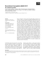
We have engineered a stable octameric hemoglobin (Hb) of molecular mass
129 kDa, a dimer of recombinant hemoglobin (rHb bG83C-F41Y) tetra-
mers joined by disulfide bonds at the b83 position. One of the major prob-
lems with oxygen carriers based on acellular hemoglobin solutions is
vasoactivity, a limitation which may be overcome by increasing the mole-
cular size of the carrier. The oxygen equilibrium curves showed that the
octameric rHb bG83C-F41Y exhibited an increased oxygen affinity and a
decreased cooperativity. The CO rebinding kinetics, auto-oxidation kinet-
ics, and size exclusion chromatography did not show the usual dependence
on protein concentration, indicating that this octamer was stable and did
not dissociate easily into tetramers or dimers at low concentration. These
results were corroborated by the experiments with haptoglobin showing no
interaction between octameric rHb bG83C-F41Y and haptoglobin, a
plasma glycoprotein that binds the Hb dimers and permits their elimination
from blood circulation. The lack of dimers could be explained if there are
two disulfide bridges per octamer, which would be in agreement with the
lack of reactivity of the additional cysteine residues. The kinetics of reduc-
tion of the disulfide bridge by reduced glutathione showed a rate of
1000 m
)1
Æh
)1
(observed time coefficient of 1 h at 1 mm glutathione) at
25 °C. Under air, the cysteines are oxidized and the disulfide bridge forms
spontaneously; the kinetics of the tetramer to octamer reaction displayed a
bimolecular reaction of time coefficient of 2 h at 11 lm Hb and 25 °C. In
addition, the octameric rHb bG83C-F41Y was resistant to potential redu-
cing agents present in fresh plasma.
Abbreviations
DSS, 2,2-dimethyl-2-silapentane-5-sulfonate; DTNB, 5,5¢-dithiobis(2-nitrobenzoic acid); DTT, dithiothreitol; GSH, reduced glutathione; HbA,
adult human hemoglobin; rHb, recombinant hemoglobin; DCL-Hb, diaspirin cross-linked hemoglobin; Hp, haptoglobin; SEC, size exclusion
chromatography; Tm, melting temperature.
230 FEBS Journal 273 (2006) 230–241 ª 2005 The Authors Journal compilation ª 2005 FEBS
allow correct oxygen delivery; however, clinical trials
have shown a vasoactivity of these molecules in
plasma. Increasing the molecular size of the carrier has
been proposed to reduce the undesirable vasoactive
properties. Different approaches have been developed,
such as surface modification of different Hbs by
poly(ethylene) glycol
2
conjugation [3–5]. These different
Hb derivatives, chemically modified by polymerization
or coupling to macromolecules, may overcome the ex-
travasation and vasoactive effects of acellular Hb.
We have produced the recombinant Hb b83Glyfi
Cys, b41PhefiTyr (rHb bG83C-F41Y), where the first
mutation provides an intertetramer disulfide bridge
and the second mutation [6] decreases the oxygen affin-
ity of the rHb. In this study, in addition to the basic
ligand binding properties and the thermal stability, we
explore the oxido-reduction kinetics of the disulfide
bridge; once formed, the octameric form (dimer of
tetramers) is stable whatever the protein concentration,
and does not interact with haptoglobin (Hp), a plasma
glycoprotein.
Results
The elution profile of rHb bG83C-F41Y obtained by
size exclusion chromatography (SEC) after purification
on Q-Sepharose XL anion exchanger, shows the pres-
ence of a major fraction eluted at a volume corres-
ponding to 129 kDa, as previously observed for the
singly mutated rHb bG83C [7]. This value is consistent
with a dimer of tetramers or an octamer [a
2
b
2
G83C-
F41Y]
2
. A minor peak eluting at the expected volume
for a tetramer was observed; this tetramer fraction
evolves to the octameric fraction over several days at
4 °C. In this study, we show that under specific experi-
mental conditions, the process of forming the octamers
can be much faster.
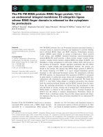
Stability of the bG83C-F41Y oligomers
Concentration dependent dissociation equilibrium
We have studied the concentration dependence of the
oligomers by SEC on Superose
TM
12 HR 10 ⁄ 300 GL
(Amersham Biosciences, Uppsala, Sweden). For the
control adult human hemoglobin (HbA), the eluted
peak profile shifts with decreasing concentration from
the tetrameric to dimeric form (Fig. 1A); the peak
position and width of the predominantly dimeric form
occurred at about 4 lm concentration (on a heme
basis) applied to the column, with typically a 60-fold
dilution of sample in the column. In contrast, irres-
pective of the applied concentration (from 150 to
6 lm), the octameric rHb bG83C-F41Y eluted at the
same volume (Ve ¼ 12.19 ± 0.02 mL), corresponding
to a molecular complex formed by 2 tetramers
(2.08 ± 0.055)
3
(Fig. 1B). The peak width at half
height remained small and constant (653 ± 10 lL),
indicating that the oligomer has a high degree of size
homogeneity and stability, and does not dissociate into
smaller species. The same results were obtained with
the singly mutated rHb bG83C [7].
Auto-oxidation
The sensitivity of the octamers in relation to heme
oxidation was determined by measuring the auto-
oxidation rates. The octameric rHb bG83C-F41Y and
octameric rHb bG83C were studied at 37 °C in phos-
phate buffer at low protein concentrations of 24 lm
and 10 lm, on a heme basis, respectively. The diaspirin
cross-linked hemoglobin (DCL-Hb) at 38 lm was used
A
B
Fig. 1. SEC profiles of HbA (A) and octameric rHb bG83C-F41Y (B)
at protein concentrations ranging from 150 to 6 l
M, on a heme
basis. Aliquots of 10 lL were applied on Superose
TM
12 HR
10 ⁄ 300 GL column and eluted at 0.4 mLÆmin
)1
flow rate.
C. Vasseur-Godbillon et al. Tetramer–octamer transition of Hb bG83C-F41Y
FEBS Journal 273 (2006) 230–241 ª 2005 The Authors Journal compilation ª 2005 FEBS 231
as a tetrameric Hb control. The time coefficient of
auto-oxidation kinetics was 17 h for DCL-Hb, 12 h
for rHb bG83C-F41Y and 15 h for rHb bG83C sam-
ples. In all cases, there was a small fraction having an
auto-oxidation rate nearly 10 times higher; note that
Hb dimers oxidize about 10 times more rapidly than
tetramers [9]; a polymer formed of a series of dimers
would probably show an enhanced oxidation rate. The
time coefficient of auto-oxidation of octameric bG83C-
F41Y was slightly decreased compared to that of
DCL-Hb but was much longer than other polymeric
mutants described recently [8].
Secondary structure and thermal stability
The far-UV CD spectrum (results not shown) deconvo-
luted using cdnn software (Bohm, Halle, Germany,
/>4
,
revealed that octameric rHb bG83C-F41Y and rHb
bG83C contained 72% and 77% a-helix, respectively.
These values were similar that those observed for HbA
and DCL-Hb (74% a-helix), suggesting that the disul-
fide bridge does not modify the secondary structure of
the molecule.
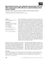
The stability of octameric rHb bG83C-F41Y was
investigated as a function of temperature. Figure 2
shows the first derivative of the ellipticity as a function
of temperature for HbA, DCL-Hb, and the octameric
forms of rHb bG83C and rHb bG83C-F41Y. The
calculated melting temperature (Tm) values are 72, 79,
78 and 77 °C, respectively. The Tm values for the
octamers exceed that for HbA, indicating that the oc-
tamers maintain good conformational stability.
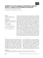
Reaction with Hp
Hp binds rapidly to Hb dimers but not to tetramers
[10] according to the reaction scheme:
a
2
b
2
þ Hp $ 2ab þ Hp ! HpðabÞ
2
We have studied the possible interaction between octa-
meric rHb bG83C-F41Y and Hp that would indicate
whether dimers might dissociate from the octamers. In
Fig. 3A, we show the elution profile by SEC of Hp
Fig. 2. First derivative of the fraction unfolded (f
u
) vs. temperature,
of native HbA (s), octameric rHb bG83C (–), octameric rHb bG83C-
F41Y (h) and DCL-Hb (m). Protein concentration was 18 l
M (on a
heme basis) in 2.5 m
M Na
2
HPO
4
, 37.5 mM NaCl buffer at pH 7.4.
The change in ellipticity was recorded at 222.6 nm from 25 to
100 °C with a heating rate of 1 °CÆmin
)1
. The peaks correspond to
the median melting temperature (Tm).
Fig. 3. SEC profiles after reaction of Hp with HbA (A) or octameric
rHb bG83C-F41Y (B). The reactions were achieved at 25 °Cin
150 m
M Tris ⁄ acetate buffer at pH 7.5 and after 15 min incubation,
the different species were analyzed on Superose
TM
12 HR
10 ⁄ 300 GL column. The mixtures of Hp with HbA or octameric rHb
bG83C-F41Y are represented by a solid line. The Hp is represented
with a dotted line. The control Hb samples are represented by
dash-dot line.
Tetramer–octamer transition of Hb bG83C-F41Y C. Vasseur-Godbillon et al.
232 FEBS Journal 273 (2006) 230–241 ª 2005 The Authors Journal compilation ª 2005 FEBS
(Ve ¼ 10.44 mL), HbA (Ve ¼ 13.28 mL) and the mix-
ture of HbA and Hp after a 15 min incubation at
room temperature. With the mixture, only one peak
was observed with Ve ¼ 10.06 mL corresponding to
the elution volume of the Hp:(dimer)
2
. When the same
experiment was performed by mixing octameric rHb
bG83C-F41Y with Hp (Fig. 3B), the elution profile of
this mixture shows the presence of two species (at
elution volumes of 10.45 mL and 11.63 mL) corres-
ponding to the elution volumes of Hp and octameric
rHb bG83C-F41Y, respectively. The same result was
obtained for Hp with the octameric rHb bG83C. The
same type of result (no interaction with Hp) was also
obtained with DCL-Hb, which does not dissociate into
dimers (data not shown). The lack of interaction with
Hp indicates that the two octamers do not dissociate
into dimers.
Stability in fresh plasma
The stability of octameric rHb bG83C-F41Y was tes-
ted in the presence of the reducing agents present in
blood. The octameric rHb bG83C-F41Y was incubated
in fresh human plasma at 37 °C. The analysis of the
relative populations of the disulfide species by SEC
showed only the octameric species for incubation times
as long as 24 h (data not shown).
These different results indicate a stable octameric
form for rHb bG83C-F41Y vs. temperature, protein
dilution, or in a physiological environment.
Study of disulfide bridge kinetics
It is important to determine if the formation of the
disulfide bridge is a reversible process. To test this
hypothesis, the octameric rHb bG83C-F41Y was
reduced by 100 molar excess of reduced glutathione
(GSH) or dithiothreitol (DTT), and then checked along
with time for the tetrameric species apparition from the
octameric ones. These experiments demonstrated that
the disulfide bond could be reduced, leading to the
dissociation into tetramers on the order of hours; the
process of forming the octamer could be repeated by
stripping the reducing agents and simply incubating the
sample under air, an oxidizing condition.
Octamer to tetramer transition
The disulfide bridge can be reduced by GSH provo-
king loss of the octameric form. In the first experi-
ment, we studied the reduction of octamers at various
concentrations of GSH during a 2 h incubation at
25 °C (Fig. 4). Two concentrations of octamers were
tested: 6 and 13 lm on a heme basis. For both concen-
trations, the curves are biphasic. Treating the rate
coefficients as a second-order reaction, the rapid phase
has a time coefficient of about 1 h at 1 mm GSH
whereas the second phase was nearly an order of mag-
nitude slower and concerns about 25% of octamers.
In the second experiment, we studied the disulfide
bridge reduction kinetics of octamers for fixed (1 or
25 mm) GSH concentrations (Fig. 5). The kinetics
show an initial phase, followed by a plateau; the rate
of the initial phase is 1000 m
)1
Æh
)1
for 1 mm of GSH.
At 25 mm GSH, the rapid phase was not fully
resolved. The experiment was achieved in the presence
of air, which explains why the octamers were not com-
pletely dissociated, because equilibrium is established
between air oxidation and GSH reduction. The reduc-
tion reaction is more complete at higher GSH concen-
trations or for samples under a nitrogen atmosphere.
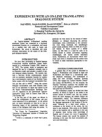
Disulfide bridge formation between rHb bG83C-F41Y
tetramers
The octameric rHb bG83C-F41Y was first reduced by
a 100 molar excess of GSH, taken up on Superose
TM
12 HR 10 ⁄ 300 GL, and then the tetrameric rHb
bG83C-F41Y at 11 lm on a heme basis was incubated
at 4 °Cor25°C. The kinetics of octamer formation at
4 °C and 25 °C are shown in Fig. 6. The kinetic curves
are not simple exponential, but show a form with
Fig. 4. Reduction of disulfide bridge in octameric rHb bG83C-F41Y
by GSH. The fraction of octamers vs. GSH concentration is shown
for samples after an incubation of 2 h at 25 °C. The initial concentra-
tions of bG83C-F41Y octamers were 6 l
M (d) and 13 lM (m)ona
heme basis. The experiments were performed at 25 °C in 150 m
M
Tris ⁄ acetate buffer at pH 7.5. The solution of GSH was prepared in
the same buffer. After incubation, a 70 lL aliquot of the mixture was
analyzed on Superose
TM
12 HR 10 ⁄ 300 GL column.
C. Vasseur-Godbillon et al. Tetramer–octamer transition of Hb bG83C-F41Y
FEBS Journal 273 (2006) 230–241 ª 2005 The Authors Journal compilation ª 2005 FEBS 233
decreasing rate vs. time, as expected for a dimerization
reaction. As the source of reactants (tetramers in this
case) is depleted, the overall bimolecular rate coeffi-
cient decreases. The curves were therefore simulated
with a dimerization model and a single bimolecular
rate coefficient of 2000 m
)1
Æh
)1
at 25 °C. The kinetics
showed observed rates of 0.0055 h
)1
and 0.025 h
)1
at
4 °C and 25 °C, respectively.
A reduction ⁄ oxidation cycle can thus be repeated to
form the disulfide bridges in rHb bG83C-F41Y. For
both the reduction and the oxidation processes, the
transition was faster at 25 °C than at 4 °C.
The octamer formation kinetics were slower at lower
protein concentrations; the kinetics at 0.3 lm (on a
heme basis) were too slow to be observed on the time-
scale used. These results confirm the bimolecular char-
acter of the kinetics of octamer formation.
Hybridization with HbA
HbA does not form disulfide bonds via the b93 cys-
teine residues; however, there was a question as to
whether Hb Tali could form octamers via a b93–b83
interaction. The experiments for the tetramer to
octamer transition of rHb bG83C-F41Y were per-
formed at 25 °C in the presence of DCL-Hb or HbA
to determine if the b93 cysteine residues could partici-
pate in the disulfide bridge formation (Fig. 7). For a
fixed concentration of rHb bG83C-F41Y, the initial
velocity of octamer formation and the amount of
octamer were similar in the presence or absence of
the other Hb (HbA or DCL-Hb), meaning that the
quantity of octamers formed was reduced by half,
Fig. 6. Disulfide bridge formation kinetics of tetrameric rHb bG83C-
F41Y. A stock of tetramers was prepared by incubating rHb
bG83C-F41Y in a 100 molar excess of GSH at 25 °C for 2 h. After
purification on Superose
TM
12 HR 10 ⁄ 300 GL column in 150 mM
Tris ⁄ acetate buffer at pH 7.5, the tetrameric fraction rHb bG83C-
F41Y (11 l
M on a heme basis) was incubated under air at 4 °C(d)
or at 25 °C(m). At different times, a 70 lL aliquot of mixture was
analyzed on Superose
TM
12 HR 10 ⁄ 300 GL column. The kinetics
show the shape typical of dimerization (lines are simulations),
where the effective rate constant decreases as the source of reac-
tants is depleted.
Fig. 7. Oxidation disulfide bridge kinetics of tetrameric rHb bG83C-
F41Y in the presence of HbA or DCL-Hb. The experiments were
performed at 25 °Cin150m
M Tris ⁄ acetate buffer at pH 7.5. The
tetrameric fraction of rHb bG83C-F41Y was purified on Superose
TM
12 HR 10 ⁄ 300 GL column, then the tetrameric rHb bG83C-F41Y, at
11 l
M on a heme basis, was incubated in the presence of the same
concentration of HbA (d) or of DCL-Hb (n) or in Tris ⁄ acetate buffer
as control sample (m). At different times, a 70 lL aliquot of the mix-
ture was analyzed on a Superose
TM
12 HR 10 ⁄ 300 GL column.
Fig. 5. Disulfide bridge reduction kinetics of octameric rHb bG83C-
F41Y by 1 m
M (m)or25mM (d) GSH. The experiments were per-
formed at 25 °C in 150 m
M Tris ⁄ acetate buffer at pH 7.5. The
solution of GSH was prepared in the same buffer. The final concen-
tration of octameric rHb bG83C-F41Y was 5 l
M on a heme basis.
At different times, a 70 lL aliquot of the mixture was analyzed on
Superose
TM
12 HR 10 ⁄ 300 GL column.
Tetramer–octamer transition of Hb bG83C-F41Y C. Vasseur-Godbillon et al.
234 FEBS Journal 273 (2006) 230–241 ª 2005 The Authors Journal compilation ª 2005 FEBS
considering the total Hb concentration. This indicates
that neither HbA nor DCL-Hb participated in the
intermolecular S–S cross-linking between the b93 and
b83 cysteine residues.
Functional studies
CO rebinding kinetics
The CO rebinding kinetics for rHb bG83C and the
double mutant rHb bG83C-F41Y were typical of
HbA, showing two phases characteristic of the oxy
(R-state) and deoxy (T-state) conformations of tetra-
meric Hb (Fig. 8). Unlike HbA, the octameric rHbs
did not dissociate into component dimers at low pro-
tein concentration. At low Hb concentration, HbA
shows a higher percentage of the rapid CO recombina-
tion, because dimers display kinetics that are similar to
the rapidly reacting R-state tetramer conformation.
However, the rHb bG83C-F41Y octamers did not
show a change in kinetics over the range 0.2–10 lm.
The CO rebinding kinetics for rHb bG83C-F41Y
showed more of the slow CO recombination, relative
to HbA and rHb bG83C (Fig. 8). This same effect
occurs for rHb bF41Y relative to HbA, indicating that
the shift towards the T-state conformation occurs for
both the tetrameric and octameric form. This mutation
at the b41 site can thus be useful to modulate the over-
all oxygen affinity [6].
The main difference between the octameric and tetra-
meric forms is in the ligand cooperativity. The octamers
do not show a full transition. At high CO photodissocia-
tion levels (50%), the CO recombination kinetics of
octameric rHb bG83C-F41Y were similar to HbA,
except for the increase in the amount of slow phase
mentioned above. As the laser photolysis energy is
decreased, the kinetics of HbA tend to show only the
rapid phase, because the main photoproduct is triply
liganded Hb which is predominantly R-like. However,
the octameric rHbs maintain a significant fraction slow
phase, even at low photodissociation levels.
Oxygen-binding properties of octameric rHbs
The oxygen-binding properties of rHb bG83C-F41Y,
rHb bG83C, and HbA are summarized in Fig. 9, for
Fig. 8. Recombination kinetics of CO to rHb bG83C-F41Y. At a
50% photodissociation level, the rHb bG83C-F41Y (n) shows two
phases, corresponding to two Hb allosteric states, as for HbA (d).
The rHb bG83C-F41Y shows more slow phase than rHb bG83C
(m), due to the b41 mutation.
Fig. 9. pH-dependence of the oxygen affinity (A) and the maximum
Hill coefficient (n
max
)(B):(d) HbA; (m)rHbbG83C; (n)rHbbG83C-
F41Y. Oxygen dissociation data were obtained at a concentration
of 0.1 m
M Hb (in terms of heme) in 0.1 M
14
sodium phosphate buffer
in the pH range 5.8–8.4 at 29 °C. P
50
(mmHg) and n
max
were deter-
mined from each curve.
C. Vasseur-Godbillon et al. Tetramer–octamer transition of Hb bG83C-F41Y
FEBS Journal 273 (2006) 230–241 ª 2005 The Authors Journal compilation ª 2005 FEBS 235
samples in 0.1 m sodium phosphate buffer at 29 °C.
Oxygen affinity (or P
50
) corresponds to the oxygen
partial pressure at 50% saturation of the hemes. The
rHb bG83C-F41Y has an oxygen affinity, and pH
dependence of P
50
, similar to that of HbA, although
there is a somewhat higher oxygen affinity at pH < 7
compared to that of HbA. The rHb bG83C has a
higher oxygen affinity than that of HbA (e.g. at
pH 7.4, P
50
¼ 4.6 mmHg vs. 9.3), and displays a wea-
ker pH dependence.
Both octamers show a cooperativity of oxygen bind-
ing, but there is a decrease in cooperativity relative to
the control HbA. The Hill coefficient for the rHbs is
slightly lower than that of HbA over the pH (e.g. n
max
2.6 for the double mutant or 2.3 for the single
mutant vs. 3.0 for HbA).
There is not much difference in the Bohr effect
(–Dlog P
50
⁄ log pH) between HbA and rHb bG83C-
F41Y over the pH range from 6.5 to 8.2. The rHb
bG83C shows a much lower Bohr effect.
1
H-NMR Studies
Figure 10 shows the 300 MHz NMR spectra of rHb
bG83C-F41Y and HbA in the CO and deoxy forms in
0.1 m sodium phosphate buffer at 29 °C. A general
feature of the NMR spectra of this rHb is that the
line-width is much broader than that of HbA due to
the oligomerization of this rHb. Figure 10A shows the
exchangeable proton resonance region of a Hb mole-
cule in the CO form. The exchangeable resonances at
12.9 and 12.1 p.p.m. from 2,2-dimethyl-2-silapentane-
5-sulfonate (DSS) are excellent markers for the a
1
b
1
subunit interface arising from the H-bonds between
a122His and b35Tyr and between a103His and
b131Gln, respectively [11–13]. There is a very slight
downfield shift ( 0.1 p.p.m.) for the resonance at 12.9
p.p.m. for the rHb compared to HbA and no observ-
able change in the resonance at 12.1 p.p.m. These
results indicate that the a
1
b
1
subunit interface remains
essentially intact in the rHb. The resonance at 10.7
p.p.m. has been assigned to the side chain of b37Trp
in the a
1
b
2
subunit interface of HbA in CO form
[12,14]. There is a very slight downfield shift ( 0.1
p.p.m.) for this resonance in rHb bG83C-F41Y.
Figure 10B shows the hyperfine-shifted and exchange-
able proton resonances for rHb bG83C-F41Y and
HbA in the deoxy form in 0.1 m sodium phosphate
buffer at 29 °C. The line-widths for the observed reso-
nances for the rHb are broader than those of HbA
due to the oligomerization of the rHb. The exchange-
able resonance at 14.2 p.p.m. has been assigned as
the H-bond between a42Tyr and b99Asp in the a
1
b
2
subunit interface of deoxy HbA [15], an important
quaternary structural marker of deoxy HbA [16].
There is a downfield shift of 0.2 p.p.m. in this reson-
ance in the rHb, indicating that there is a slight pertur-
bation in the a
1
b
2
subunit interface compared to that
in deoxy HbA. This is consistent with the observed
slight shift of the resonance assigned to b37Trp in the
a
1
b
2
subunit interface of the rHb in the CO form men-
tioned above. It is noted that the hyperfine-shifted pro-
ton resonances over the spectral region from 14 to 24
p.p.m. are different between rHb bG83C-F41Y and
HbA in the deoxy form. These resonances arise from
the protons of amino acid residues situated in the
vicinity of the heme groups and of the porphyrins of
both the a- and b-chains of hemoglobin. The amino
acid substitutions in this rHb could perturb the heme
environment, resulting in an alteration of the hyper-
fine-shifted proton resonances as observed. In addition,
Fig. 10.
1
H-NMR spectra (300 MHz) of 5% HbA and rHb bG83C-
F41Y in 0.1
M sodium phosphate
15
at pH 7.0 in H
2
O and at 29 °C:
exchangeable proton resonances in the CO form (A) and hyperfine-
shifted and exchangeable proton resonances in the deoxy form (B).
Tetramer–octamer transition of Hb bG83C-F41Y C. Vasseur-Godbillon et al.
236 FEBS Journal 273 (2006) 230–241 ª 2005 The Authors Journal compilation ª 2005 FEBS
the much broader resonances in the region from 14 to
24 p.p.m. are probably due to formation of met-Hb in
the rHb. In general, these broad resonances due to the
hyperfine-shifted proton resonances of met-Hb can be
removed by the addition of dithionite. However, we
have found that the presence of dithionite can break
S–S bonds in hemoglobin (results not shown) and
other proteins as reported in the literature [17,18].
Thus, we did not add dithionite to our deoxy-rHb
bG83C-F41Y for NMR measurements resulting from
the formation of met-Hb in the sample.
Discussion
In vivo and in vitro disulfide formation is catalyzed by
specialized enzymes. In vivo, disulfide formation was
achieved in the endoplasmic reticulum by enzymes
belonging to a thioredoxin superfamily, such as protein
disulfide isomerase in eukaryotes and disulfide bond
proteins in prokaryotes [19]. It has been shown that
the oxidative folding of different proteins in vitro is
accelerated by protein disulfide isomerase [20]. In
the case of the natural mutant Hb Porto Alegre
(Hb bS9C) [21,22] or the rHb Prisca (rHb
bS9C + C93A + C112G) [23] both of which carry in
position b9 an extra thiol group oriented towards the
exterior of the Hb molecule, the oligomerization pro-
cess was not observed immediately, either after lysis of
the red cells or after purification of the rHb. In the
case of rHb Prisca, the maximum oligomer was
obtained after a 110 day incubation at 25 °C [23].
Recently, another recombinant polymeric Hb was
described, the rHb Minotaur containing a-human and
b-bovine in which the b9Ala was replaced by Cys and
the b93Cys was replaced by Ala. The polymer of puri-
fied rHb Minotaur was obtained after 2 days at 30 °C
or 30 days at 4 °C [8]. The present study of rHb
bG83C-F41Y shows that the disulfide bridge forma-
tion in this recombinant mutant is a relatively fast pro-
cess that does not require any external reagents such
as the glutathione redox system. Contrary to the other
polymeric Hbs, the oligomerization process of rHb
bG83C-F41Y and of the single mutant rHb bG83C
was observed immediately at 4 °C after purification.
Number of disulfide bonds
In rHb bG83C-F41Y, as for rHb bG83C, disulfide
bonds between the b83 cysteine residues stabilize the
octameric structure. Once formed the octameric frac-
tion rHb bG83C-F41Y remained stable for several
months at 4 °C. There is still a question as to whether
the octamer is formed with one or two disulfide bonds.
A single disulfide bond would correspond to tethered
tetramers, each relatively free to make the allosteric
transition or tetramer-dimer reaction. By symmetry, if
each beta chain participates in the formation of a
disulfide bond (2 per octamer), the tetramers would be
more constrained. The octamer would be more stable as
well; even if one tetramer dissociated into dimers, each
dimer would be held via an S–S bond to the other tetra-
mer (see Fig. 5 in [7]).
After intravascular hemolysis, Hp binds the free Hb,
allowing the clearance of Hb from the plasma. The
monocyte ⁄ macrophage specific glycoprotein CD136
was recently described as a receptor that scavenges Hb
by mediating endocytosis of the Hp–Hb complex [24].
Accordingly, the Hp–Hb complex is eliminated from
the circulation.
Neither of the octameric rHbs (bG83C-F41Y and
rHb bG83C) react with Hp, confirming that these
octamers do not dissociate easily into dimers; the
absence of the formation of complexes of Hp with
these octamers would increase their useful lifetime for
oxygen delivery. The lack of any interaction with Hp
indicates that there are no free dimers and would sup-
port the model with two disulfide bonds.
A hypothetical diagram is shown in Fig. 11, based
on the crystallographic structure of the Hb tetramer.
By symmetry, both beta chains can form a disulfide
bond, provided there is no steric hindrance of other
protein residues. The b83 glycine (in red) would have
two additional atoms (C–S) for the bG83C mutation.
A disulfide bond is typically about 6 A
˚
between the C
a
atoms. As can be seen, the intervening residues (b79
and 80) would not require a greater distance. On the
other hand, forming two b83–b93 bonds would be
more difficult as the helix A would cause a larger hin-
drance.
As an alternative analysis, one could consider the
two tetramers, in a first approximation, as tangent
spheres. Based on the angle between vectors from the
center to each b83 residue, one can calculate the dis-
tance between the b83 C
a
of the opposing spheres. For
two disulfide bonds via the b83 residues, the distance
would be 6 A
˚
, which is quite compatible with a typical
disulfide bond. For a b83–b93 bonding, the distance
required would be 8 A
˚
and therefore not possible.
Note that the distance between certain residues
such as b83 between the b chains of the same tetra-
mer depend on the allosteric state. In going from
the deoxy to oxy conformation the b
1
83–b
2
83 dis-
tance would decrease from 24 to 19 A
˚
(for b
1
83–
b
2
93, the change would be from 31 to 25 A
˚
). This
would require the two tetramers forming the octamer
to make the allosteric transition together. This could
C. Vasseur-Godbillon et al. Tetramer–octamer transition of Hb bG83C-F41Y
FEBS Journal 273 (2006) 230–241 ª 2005 The Authors Journal compilation ª 2005 FEBS 237
explain the decreased cooperativity observed for the
octamer in the equilibrium and especially the kinetic
experiments.
Finally the experiments detecting the number of tit-
ratable cysteines confirm the hypothesis of two disul-
fide bonds. For unfolding condition, the mutant and
native Hb samples show the same signal amplitude,
indicating the same number of marked cysteines; this
implies that the new cysteines are not marked and
must therefore be part of a disulfide bond. There is
still the possibility of a b83–b93 bond, but the hybrid
(mutant + HbA) experiments, and the steric hin-
drance considerations, indicate that b83 is the most
probable site for formation of the two disulfide
bonds.
Heme oxidation
It has been shown that cross-linked or polymerized
Hb may show an acceleration of the auto-oxidation
process [25]. The study of auto-oxidation of octa-
meric rHb bG83C-F41Y shows that this octamer
does not modify the half time of heme auto-oxida-
tion, which remains close to that of DCL-Hb or
HbA, contrary to the other polymers such as rHb
Minotaur, which has a half time of auto-oxidation
of 3.2 h [8].
The oxygen equilibrium curves of the rHb bG83C
display a higher average affinity, and a lower cooper-
ativity (Fig. 9). Addition of the second mutation
decreases the oxygen affinity, but induces a further
decrease in the Hill coefficient. The results of the lig-
and binding kinetics and oxygen equilibrium curves
indicate some limitations in the allosteric transition.
One factor to consider is the double bridging of the
tetramers; that is, each beta chain might form a disul-
fide bond with the opposing tetramer, unless some
steric hindrance prevents the formation of the second
bond. With both bonds present, the two tetramers
must make the allosteric transition together, because
the distance between the b83 residues changes. This
could lead to new constraints on the synchronized
allosteric transition of both tetramers within the
octamer. With their stabilization in dilute solution,
the octameric rHb bG83C-F41Y and the octameric
rHb bG83C are both good model molecules to
develop hemoglobin-based oxygen carriers. The octa-
meric form of both recombinant Hbs (with or with-
out the additional mutation at the b41 site) showed a
high stability; there was no interaction with hapto-
globin and no dissociation provoked by incubation in
fresh plasma. These octameric Hbs are thus poten-
tially useful as blood substitutes. The clinical trial
of HemeAssist
TM
(Baxter Healthcare) revealed some
escape of the tetrameric Hb from the blood vessels
[26]; an octameric form is thus the logical extension
of research for a blood substitute based on Hb solu-
tions. The best choice between the two molecules tes-
ted here is not obvious. There was initially an attempt
to mimic the physiological oxygen affinity, which is
better approximated by the mutant b41. However,
lower oxygen affinities lead to higher oxidation rates,
and in the present case the double mutant displayed
less cooperativity. Current ideas suggest that a higher
oxygen affinity may still be useful and provide a better
oxygenation of the capillaries [27]. The single mutant
may thus be the better candidate molecule.
Experimental procedures
Hemoglobin expression and purification
The mutated Hbs were produced in JM 109 strains of
Escherichia coli using the expression plasmid pHE7 contain-
ing human a-, b-globin cDNAs and an E. coli methionine
aminopeptidase cDNA [28], after introduction of the
b41Phe fi Tyr from the pHE7 template containing the
b83Gly fi Cys mutation (Quick change
TM
site
Fig. 11. Proposed scheme for an octamer formed by disulfide
bonds between Hb tetramers via the b83 site (glycine in HbA;
shown as red spheres), where only residues b79 (orange) and b80
(blue) might interfere. The mutation bG83C would introduce addi-
tional atoms (–C–S) to bridge the distance (shown as a dotted
yellow line) between b83 sites of the opposing (top vs. bottom)
tetramers. Within a tetramer the b
1
83 to b
2
83 distance is 24 A
˚
16
for
the deoxy Hb conformation (but only 19 A
˚
for the oxy form; where
the analogous position for the b83 glycine is shown in green). This
large change in distance would imply that the two tetramers must
make the allosteric transition together. Note that the corresponding
distances for a b83 to b93 (yellow residue) bonding are higher and
would involve more steric hindrance.
Tetramer–octamer transition of Hb bG83C-F41Y C. Vasseur-Godbillon et al.
238 FEBS Journal 273 (2006) 230–241 ª 2005 The Authors Journal compilation ª 2005 FEBS
directed-mutagenesis kit, Stratagene Europe, Amsterdam,
the Netherlands) and verification of the a- and b-globin
coding sequences (MWG Biotech, Courtaboeuf, France).
The cells were harvested by centrifugation at 6000 g for
10 min at 4 °C
7
and stored frozen at )80 °C until needed
for purification. The rHb was isolated and purified as des-
cribed by Shen et al. [28,29] with minor modifications [7].
The oligomeric and tetrameric fractions were then separated
by SEC on a Superose
TM
12 HR 10 ⁄ 300 GL column
8
(Amersham Biosciences, Uppsala, Sweden) equilibrated at
25 °C with 150 mm Tris ⁄ acetate buffer at pH 7.5 [7].
Auto-oxidation kinetics
The kinetics of auto-oxidation of rHb bG83C-F41Y and
rHb bG83C were followed by absorption spectrophoto-
metry at 37 °C, for samples under air [9]. Hb solutions were
in 100 mm potassium phosphate at pH 7.0.
Thermal denaturation
Thermal denaturation was achieved with a Jasco J810
spectropolarimeter (Jasco, Tokyo, Japan), using a 0.5 mm
path-length cell, and the temperature in the cell was pro-
grammed using a Jasco PTC-423S thermoelectric tempera-
ture controller. The ellipticity at 222.6 nm was monitored
over a temperature range of 25–100 °C, using a bandwidth
of 1 nm, and a temperature gradient of 1 °C per min. The
Hb samples were in the CO form and at a concentration
of 18 lm (on a heme basis) in 2.5 mm Na
2
HPO
4
, 37.5 mm
NaCl buffer at pH 7.4. The Tm corresponds to 50%
unfolded molecule. The CD signal was normalized to
obtain the unfolded fraction: f
u
¼ (y
N
–y
obs
) ⁄ (y
N
–y
u
),
where y
obs
is the observed CD signal and y
N
and y
u
the
CD signal of the native and unfolded protein, respectively.
Interaction of octamers with haptoglobin
Reaction with Hp (Sigma Aldrich, Saint Quentin Fallavier,
France) was achieved at room temperature in 150 mm
Tris ⁄ acetate buffer at pH 7.5 containing Hp at 2.9 lm and
either 7 lm of HbA (control reaction) (on a heme basis), or
6 lm octameric rHb bG83C or rHb bG83C-F41Y (on a
heme basis). After a 15 min incubation, the presence of
different species was analyzed by SEC on a Superose
TM
12 HR 10 ⁄ 300 GL column.
Stability of octamers in fresh plasma
The octameric rHb bG83C-F41Y was mixed with fresh
human plasma in a ratio of 7 g Hb to 500 mL plasma
at 37 °C. At different times, an aliquot was withdrawn,
centrifuged at 3000 g at room temperature for 2 min
9
and
analyzed on Superose
TM
12 HR 10 ⁄ 300 GL column.
Disulfide reduction kinetics of the oligomeric rHb
bG83C-F41Y
In the first experiment, 100 lL aliquots of purified oligo-
meric fraction at 6 and 13 lm (heme basis) were incubated
at 25 °C in the presence of increased concentration of GSH
during 2 h and the relative populations of the disulfide spe-
cies obtained were analyzed by SEC on a Superose
TM
12
HR 10 ⁄ 300 GL column. In the second experiment, the puri-
fied oligomeric fraction at 6 lm (heme basis) was incubated
at 25 °C in the presence of 1 or 25 mm GSH. Aliquots of
100 lL were withdrawn at various times, and the relative
populations of the disulfide species of the mixture were ana-
lyzed by SEC [7].
Disulfide bridge formation of rHb bG83C-F41Y
The disulfide bridge of rHb bG83C-F41Y was first
reduced with a 100 molar excess of GSH for 2 h at 25 °C;
the GSH was removed by SEC. Then the tetramer to oc-
tamer formation of rHb bG83C-F41Y (11 lm on a heme
basis) was achieved at 25 °C and 4 °C. At various times,
an aliquot was withdrawn and the relative populations of
the disulfide species of the mixture were analyzed by SEC.
In the second experiment the re-oxidation kinetics of the
tetrameric rHb bG83C-F41Y (11 lm on a heme basis)
were achieved in the presence of the same concentration
of DCL-Hb or HbA.
CO recombination kinetics
Kinetics of CO recombination were obtained after flash
photolysis using 10 ns YAG laser pulses (Quantel
10
, Les Ulis,
France) providing 160 mJ at 532 nm. Samples were in 1 or
10 mm cuvettes equilibrated under 0.1 atm (100 lm) CO,
with observation at 436 nm. Measurements were made at
25 °C in 150 mm Tris ⁄ acetate buffer at pH 7.5 [30].
Oxygen-binding measurements
Oxygen-binding measurements were carried out using a
Hemox Analyzer (TCS Medical Products, Huntington
Valley, PA, USA). As previously described, the experi-
ments were run at 29 °C as a function of pH in 0.1 m
sodium phosphate buffer and contained 0.1 mm Hb (on a
heme basis) [28,29]. The maximum Hill coefficient, n
max
,
was determined from the maximum slope of the Hill plot
as a measure of cooperativity in the oxygenation process.
The P
50
values (in mmHg) are given with an accuracy of
±5%. The n
max
values are reported with an accuracy of
±7%. The Bohr effect was obtained from the P
50
values
as a function of pH using the linkage equation (DH
+
¼
–¶log P
50
ڦpH), which gives the number of the Bohr pro-
tons released upon oxygenation per heme [31,32].
C. Vasseur-Godbillon et al. Tetramer–octamer transition of Hb bG83C-F41Y
FEBS Journal 273 (2006) 230–241 ª 2005 The Authors Journal compilation ª 2005 FEBS 239
1
H-NMR spectra
1
H-NMR spectra were obtained on a Bruker
11
Avance DRX-
300 NMR spectrometer (Billerica, MA, USA) operating at
300 MHz for proton spectra. Samples consisted of a 5%
Hb concentration (2.5 mm on a heme basis) in 0.1 m
sodium phosphate buffer at pH 7.0 in 95% H
2
O and 5%
deuterium oxide, and were measured at 29 °C. A jump-and-
return pulse sequence was used to suppress the water signal
[33]. Proton chemical shifts were indirectly referenced to the
methyl proton resonance of the sodium salt of DSS
through the use of the internal reference of the water signal
at 4.76 p.p.m. downfield of DSS at 29 °C.
Cysteine reactivity
The reaction with the thiol reagents, 5,5¢-dithiobis(2-nitro-
benzoic acid) (DTNB) was used to determine the accessibil-
ity of the sulfhydryl groups in the native and mutant Hbs,
as described by Jocelyn [34]. The reaction was performed at
pH 7 in 100 mm potassium phosphate buffer and 100 lm
DTNB, with a protein concentration of 5 lm, determined
from the Soret band for the CO species using the extinction
coefficient of 190 mm
)1
Æcm
)1
12
at 420 nm. The absorption at
455 nm was monitored over 2 h and corrected against a
blank to which no protein was added. Under denaturing
conditions (6 m guanidinium chloride) these samples
showed the same signal amplitude per cysteine; this value
was taken as 100% reactivity.
Acknowledgements
We thank G. Caron for skilful technical assistance and
Dr Ming F. Tam for carrying out the Edman degrada-
tion measurements for our rHb sample. We are grate-
ful to the Baxter Healthcare Company for supplying
DCL-Hb. This work was supported by the Institut
National de la Sante
´
et de la Recherche Me
´
dicale, the
Association Recherche et Transfusion (contract no. 21-
2000) and by research grants from the National Insti-
tutes of Health (R01HL-24525 and P01HL-71064). C.
Fablet was supported by the De
´
le
´
gation Ge
´
nerale pour
l’Armement (Ministe
`
re de la De
´
fense, France).
References
1 Chatterjee R, Welty EV, Walder RY, Pruitt SL, Rogers
PH, Arnone A & Walder JA (1986) Isolation and char-
acterization of a new hemoglobin derivative cross-linked
between the alpha chains (lysine 99 alpha 1 fi lysine 99
alpha 2). J Biol Chem 261, 9929–9937.
2 Looker D, Abbott-Brown D, Cozart P, Durfee S, Hoff-
man S, Mathews AJ, Miller-Roehrich J, Shoemaker S,
Trimble S, Fermi G, Komiyama NH, Nagai K & Stetler
GL (1992) A human recombinant haemoglobin designed
for use as a blood substitute. Nature 35, 258–260.
3 Vandegriff KD, Malavalli A, Wooldridge J, Lohman J
& Winslow RM (2003) MP4, a new nonvasoactive
PEG-Hb conjugate. Transfusion 43, 509–516.
4 Barnikol WK, Burkhard O, Poetzschke H, Domack U,
Dinkelmann S, Guth S, Fiedler B & Manz B (2002) New
artificial oxygen carriers made of pegulated polymerised
pyridoxylated porcine haemoglobin (P(4)Hb). Comp
Biochem Physiol A Mol Integr Physiol 132, 185–191.
5 Manjula BN, Tsai A, Upadhya R, Perumalsamy K,
Smith PK, Malavalli A, Vandegriff K, Winslow RM,
Intaglietta M, Prabhakaran M, Friedman JM &
Acharya AS (2003) Site-Specific PEGylation of Hemo-
globin at Cys-93(beta): Correlation between the Colliga-
tive Properties of the PEGylated Protein and the Length
of the Conjugated PEG Chain. Bioconjug Chem 14,
464–472.
6 Baudin V, Pagnier J, Lacaze N, Bihoreau MT, Kister J,
Marden M, Kiger L & Poyart C (1992) Allosteric prop-
erties of haemoglobin beta 41 (C7) Phe fi Tyr: a stable,
low-oxygen-affinity variant synthesized in Escherichia
coli. Biochim Biophys Acta 1159, 223–226.
7 Fablet C, Marden MC, Green BN, Ho C, Pagnier J &
Baudin-Creuza V (2003) Stable octameric structure of
recombinant hemoglobin alpha (2) beta 2, 83
Gly fi Cys. Protein Sci 12, 690–695.
8 Bobofchak KM, Mito T, Texel SJ, Bellelli A, Nemoto
M, Traystman RJ, Koehler RC, Brinigar WS & Fronti-
celli C (2003) A recombinant polymeric hemoglobin
with conformational, functional, and physiological char-
acteristics of an in vivo O
2
transporter. Am J Physiol
Heart Circ Physiol 285, 549–561.
9 Griffon N, Baudin V, Dieryck W, Dumoulin A, Pagnier
J, Poyart C & Marden MC (1998) Tetramer-dimer equi-
librium of oxyhemoglobin mutants determined from
auto-oxidation rates. Protein Sci 7, 673–680.
10 Nagel RL & Gibson QH (1971) The binding of hemo-
globin to haptoglobin and its relation to subunit disso-
ciation of hemoglobin. J Biol Chem 246, 69–73.
11 Russu IM, Ho NT & Ho C (1987) A proton nuclear
Overhauser effect investigation of the subunit interfaces
in human normal adult haemoglobin. Biochim Biophys
Acta 914, 40–48.
12 Simplaceanu V, Lukin JA, Fang TY, Zou M, Ho NT &
Ho C (2000) Chain-selective isotopic labeling for NMR
studies of large multimeric proteins: application to hae-
moglobin. Biophys J 79, 1146–1154.
13 Chang CK, Simplaceanu V & Ho C (2002) Effects of
amino acid substitutions at beta 131 on the structure
and properties of hemoglobin: evidence for communica-
tion between alpha 1 beta 1- and alpha 1 beta 2-subunit
interfaces. Biochemistry 41, 5644–5655.
14 Fang TY, Simplaceanu V, Tsai CH, Ho NT & Ho C
(2000) An additional H-bond in the alpha 1 beta 2
Tetramer–octamer transition of Hb bG83C-F41Y C. Vasseur-Godbillon et al.
240 FEBS Journal 273 (2006) 230–241 ª 2005 The Authors Journal compilation ª 2005 FEBS
interface as the structural basis for the low oxygen affinity
and high cooperativity of a novel recombinant hemoglo-
bin (beta L105W). Biochemistry 39, 13708–13718.
15 Fung LWM & Ho C (1975) A proton nuclear magnetic
resonance study of the quaternary structure of human
hemoglobins in water. Biochemistry 14, 2526–2535.
16 Perutz MF (1970) Stereochemistry of cooperative effects
in haemoglobin. Nature 228, 726–739.
17 Hille R & Massey V (1982) The presence of a reducible
disulfide bond in milk xanthine oxidase. J Biol Chem
257, 8898–8901.
18 Wang PF, Veine DM, Ahn SH & Williams CH Jr
(1996) A stable mixed disulfide between thioredoxin
reductase and its substrate, thioredoxin: preparation
and characterization. Biochemistry 35, 4812–4819.
19 Noiva R (1994) Enzymatic catalysis of disulfide forma-
tion. Protein Expr Purif 5, 1–13.
20 Shin HC & Scheraga HA (2000) Catalysis of the oxida-
tive folding of bovine pancreatic ribonuclease A by pro-
tein disulfide isomerase. J Mol Biol 300, 995–1003.
21 Tondo CV (1971) Study of the polymerization of hae-
moglobin Porto Alegre tetramers by disulfide bridge
formation. An Acad Bras Cienc 43, 651–669.
22 Baudin-Creuza V, Fablet C, Zal F, Green BN, Prome
D, Marden MC, Pagnier J & Wajcman H (2002) Hemo-
globin Porto Alegre forms a tetramer of tetramers
superstructure. Protein Sci 11, 129–136.
23 Fronticelli C, Arosio D, Bobofchak KM & Vasquez GB
(2001) Molecular engineering of a polymer of tetrameric
hemoglobins. Proteins 44, 212–222.
24 Kristiansen M, Graversen JH, Jacobsen C, Sonne O,
Hoffman HJ, Law SK & Moestrup SK (2001) Identifi-
cation of the haemoglobin scavenger receptor. Nature
409, 198–201.
25 Nagababu E, Ramasamy S, Rifkind JM, Jia Y &
Alayash AI (2002) Site-specific cross-linking of human
and bovine hemoglobins differentially alters oxygen
binding and redox side reactions producing rhombic
heme and heme degradation. Biochemistry 41, 7407–
7415.
26 Friedman HI, Devenuto F, Kerwin A, Carson K &
Bynoe R (2000) Hemoglobin solutions as blood substi-
tutes. J Invest Surg 13, 79–94.
27 Winslow RM (2005) Targeted O
2
delivery by low-p50
hemoglobin: a new basis for hemoglobin-based oxygen
carriers. Artif Cells Blood Substit Immobil Biotechnol 33,
1–12.
28 Shen TJ, Ho NT, Zou M, Sun DP, Cottam PF,
Simplaceanu V, Tam MF, Bell DA & Ho C (1997)
Production of human normal adult and fetal
hemoglobins in Escherichia coli. Protein Eng 10,
1085–1097.
29 Shen TJ, Ho NT, Simplaceanu V, Zou M, Green BN,
Tam MF & Ho C (1993) Production of unmodified
human adult hemoglobin in Escherichia coli. Proc Natl
Acad Sci USA 90, 8108–8112.
30 Marden MC, Kister J, Bohn B & Poyart C (1988)
T-state hemoglobin with four ligands bound. Biochemis-
try 27, 1659–1664.
31 Wyman J (1948) Heme Proteins. Adv Protein Chem 4,
407–531.
13
32 Wyman J (1964) Linked functions and reciprocal effects
in hemoglobin: a second look. Adv Protein Chem 19,
223–286.
33 Plateau P & Gue
´
ron M (1982) J Am Chem Soc 104,
7310–7311.
34 Jocelyn PC (1987) Chemical reduction of disulfides.
Methods Enzymol 143, 246–256.
C. Vasseur-Godbillon et al. Tetramer–octamer transition of Hb bG83C-F41Y
FEBS Journal 273 (2006) 230–241 ª 2005 The Authors Journal compilation ª 2005 FEBS 241