Báo cáo khoa học: Recombinant bovine zona pellucida glycoproteins ZP3 and ZP4 coexpressed in Sf9 cells form a sperm-binding active hetero-complex ppt
Bạn đang xem bản rút gọn của tài liệu. Xem và tải ngay bản đầy đủ của tài liệu tại đây (416.89 KB, 16 trang )
Recombinant bovine zona pellucida glycoproteins ZP3 and
ZP4 coexpressed in Sf9 cells form a sperm-binding active
hetero-complex
Saeko Kanai
1
, Naoto Yonezawa
1
, Yuichiro Ishii
1
, Masaru Tanokura
2
and Minoru Nakano
1
1 Graduate School of Science and Technology, Chiba University, Japan
2 Graduate School of Agriculture and Life Science, The University of Tokyo, Japan
Mammalian oocytes are surrounded by the zona pellu-
cida (ZP), a transparent envelope that mediates several
critical aspects of fertilization, including species-selective
sperm recognition, blocking of polyspermy, and protec-
tion of the oocyte and embryo until implantation [1–3].
The ZP consists of three or four kinds of glycoproteins
(ZPGs). Human and rat ZPs consist of four ZPGs (ZP1,
ZP2, ZP3, and ZP4) [4,5], whereas porcine and bovine
ZPs comprise three ZPGs (ZP2, ZP3, and ZP4) that cor-
respond to ZPA, ZPC, and ZPB, respectively, in other
nomenclature [6]. Murine ZP also consists of three
ZPGs (ZP1, ZP2, and ZP3) [7]. Porcine, bovine and
murine ZPs have ZP2 and ZP3 in common, whereas
ZP1 and ZP4 are products of distinct genes [8]. All
ZPGs contain a domain that consists of 260 amino
acids and contains eight conserved Cys residues [9].
Keywords
baculovirus-Sf9; fertilization; glycoprotein;
zona pellucida; ZP domain
Correspondence
M. Nakano, Graduate School of Science,
Chiba University, 1-33 Yayoi-cho, Inage-ku,
Chiba 263-8522, Japan
Fax: +81 43 290 2874
Tel: +81 43 290 2794
E-mail:
(Received 18 April 2007, revised 27 July
2007, accepted 24 August 2007)
doi:10.1111/j.1742-4658.2007.06065.x
The zona pellucida (ZP) is a transparent envelope that surrounds the mam-
malian oocyte and mediates species-selective sperm–egg interactions. Por-
cine and bovine ZPs are composed of the glycoproteins ZP2, ZP3, and
ZP4. We previously established an expression system for porcine ZP glyco-
proteins (ZPGs) using baculovirus in insect Sf9 cells. Here we established a
similar method for expression of bovine ZPGs. The recombinant ZPGs
were secreted into the medium and purified by metal-chelating column
chromatography. A mixture of bovine recombinant ZP3 (rZP3) and rZP4
coexpressed in Sf9 cells exhibited inhibitory activity for bovine sperm–ZP
binding similar to that of a native bovine ZPG mixture, whereas neither
bovine rZP3 nor rZP4 inhibited binding. An immunoprecipitation assay
revealed that the coexpressed rZP3 ⁄ rZP4 formed a hetero-complex. We
examined the functional domain structure of bovine rZP4 by constructing
ZP4 mutants lacking the N-terminal domain or lacking both the N-termi-
nal and trefoil domains. When either of these mutant proteins was
coexpressed with bovine rZP3, the resulting mixtures exhibited inhibitory
activity comparable to that of the bovine rZP3 ⁄ rZP4 complex. Hetero-com-
plexes of bovine rZP3 and porcine rZP4, or porcine rZP3 and bovine
rZP4, also inhibited bovine sperm–ZP binding. Our results demonstrate
that the N-terminal and trefoil domains of bovine rZP4 are dispensable for
formation of the sperm-binding active bovine rZP3 ⁄ rZP4 complex and,
furthermore, that the molecular interactions between rZP3 and rZP4 are
conserved in the bovine and porcine systems.
Abbreviations
ACA, Amaranthus candatus agglutinin; BO, Brackett and Oliphant; FITC, fuorescein isothiocyanate; Fuc, fucose; GNA, Galanthus nivalis
agglutinin; LC, liquid chromatography; LCA, Lens culinaris agglutinin; Man, mannose; MOI, multiplicity of infection; PA, pyridylamino; PHA,
Phaseolus vulgaris agglutinin; PSA, Pisum sativum agglutinin; RCA
120
, Ricinus communis agglutinin; rZP2, recombinant ZP2; rZP3,
recombinant ZP3; rZP3
FLAG
, FLAG-tagged rZP3; rZP4, recombinant ZP4; rZP4
FLAG
, FLAG-tagged rZP4; rZPG, recombinant ZPG; ZP, zona
pellucida; ZPG, zona pellucida glycoprotein.
5390 FEBS Journal 274 (2007) 5390–5405 ª 2007 The Authors Journal compilation ª 2007 FEBS
In mice, ZP3 is thought to be involved in gamete
recognition [1–3]. ZP assembly is controlled by short,
hydrophobic sequences in the C-terminal propeptides
of ZPG precursors, and requires the ZP domains of
ZP2 and ZP3 [10,11]. The molar ratio of murine tran-
scripts is estimated at ZP1 ⁄ ZP2 ⁄ ZP3 ¼ 1 : 4 : 4 [12], a
ratio that is consistent with a suggested model in
which a ZP2 ⁄ ZP3 heterodimer forms filaments that are
crosslinked by a ZP1 dimer [13]. However, the molar
ratio of ZPGs in the murine ZP does not seem to
correspond to the molar ratio of their transcripts
[7]. In pigs, the estimated protein molar ratio of
ZP2 ⁄ ZP3 ⁄ ZP4 is 1 : 6 : 6 [14]. Although neither ZP3
nor ZP4 exhibits porcine sperm-binding activity by
itself, a high molecular mass ZP3 ⁄ ZP4 hetero-complex
does exhibit this activity [15,16].
When subjected to nonreducing SDS ⁄ PAGE, bovine
ZPGs form a band at an average apparent molecular
mass of 74 kDa, which is broad owing to heterogeneity
in glycosylation [17]. After endo-b-galactosidase-cata-
lyzed removal of N-acetyl-lactosamine repeats at the
nonreducing ends of carbohydrate chains, bovine ZP2,
ZP3 and ZP4 migrate as three distinct bands of appar-
ent molecular masses of 72, 45 and 58 kDa, respec-
tively, under nonreducing conditions [17]. Under
reducing conditions, the apparent molecular masses of
the endo-b-galactosidase-digested components shift to
76, 63 and 21 kDa for ZP2, to 47 kDa for ZP3, and to
68 kDa for ZP4 [17]. Processing of bovine ZP2 occurs
at a specific site upon fertilization, and yields disulfide-
bonded polypeptides of 63 and 21 kDa [17,18]. A large
fraction of ZP2 obtained from unfertilized eggs is
already processed, probably as an artefact of the prep-
aration, but the 76 kDa band of ZP2 completely dis-
appears upon fertilization [17,18].
The amino acid sequences of porcine and bovine
ZP2, ZP3, and ZP4, which were previously determined
by cDNA cloning and sequencing [6,19–21], are 77%,
85% and 75% identical, respectively. The mature por-
cine and bovine ZP4 polypeptides differ in that an
N-terminal region corresponding to residues 1–135 of
bovine ZP4 (with the translation initiation Met num-
bered 1) is lacking in the porcine protein [19,21,22]
(Fig. 1A). The estimated protein molar ratio of bovine
ZP2 ⁄ ZP3 ⁄ ZP4 is 1 : 2 : 1 [21], which differs signifi-
cantly from the porcine molar ratio, suggesting that
the structures of the bovine and porcine ZPs are differ-
ent.
In a previous study, we partially separated an endo-
b-galactosidase-digested bovine ZPG mixture into
three components by RP-HPLC [21]. Of the three
components, ZP4 exhibited the strongest sperm-bind-
ing activity. ZP2 and ZP3 exhibited much weaker
activity [21]. The components were not completely
resolved by HPLC, indicating cross-contamination;
thus, whether each bovine ZPG has sperm-binding
activity by itself is not yet clear. A previous report that
bovine sperm–egg binding is inhibited in the presence
of anti-porcine ZP3 or ZP4 suggests that both ZP3
and ZP4 are involved in sperm–ZP binding [23].
In mice, in vitro studies have proposed that sperm
ligands consist of O-linked carbohydrate chains linked
to Ser332 and Ser334 of ZP3 [24,25]. Nevertheless, a
recent structural analysis using MS did not show evi-
dence for glycosylation [26]. The in vivo studies per-
formed to date using transgenic mice lacking each
glycosyltransferase gene do not support the involve-
ment of carbohydrate chains of mouse ZP in sperm
binding [27–29]. In pigs, neutral tri-antennary and
tetra-antennary complex-type chains have the strongest
sperm-binding activity of the N-linked chains of ZP
[30], and O-linked chains also have sperm-binding
activity [31]. The nonreducing terminal b-galactosyl
A
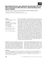
BC
Fig. 1. Recombinant bovine ZP proteins. (A) Schematic representa-
tion of the rZP2, rZP4, rZP4
136)464
, rZP4
182)464
and rZP3 polypep-
tides. These recombinant polypeptides were expressed with His-
and S-tags at their N-termini. Open square, region specific to ZP2,
ZP4, or ZP3; dotted square, trefoil domain; filled square, ZP
domain. Arrows indicate the putative furin cleavage sites that con-
stitute the C-termini of the expressed polypeptides. The calculated
molecular masses of the polypeptide moieties of the recombinants,
excluding extra peptides derived from the transfer vector, are
shown in kDa to the right of each polypeptide. (B, C) SDS ⁄ PAGE
and immunoblot analyses of rZP2 (lane 1), rZP4 (lane 2),
rZP4
136)464
(lane 3), rZP4
182)464
(lane 4), and rZP3 (lane 5). The pro-
teins were expressed in Sf9 cells, secreted into the culture med-
ium, isolated using metal-chelation column chromatography, and
detected by SDS ⁄ PAGE (B) or by immunoblot analysis using anti-
bodies specific for each of the ZPGs (C). Arrowheads indicate the
recombinant protein bands. Molecular mass markers are indicated
in kDa on the left of each panel.
S. Kanai et al. Recombinant bovine zona pellucida glycoproteins
FEBS Journal 274 (2007) 5390–5405 ª 2007 The Authors Journal compilation ª 2007 FEBS 5391
residues of the complex-type N-linked chains are
involved in sperm binding [32]. In cows, the major
neutral N-linked chain of ZP consists of only one
structure, a high-mannose-type chain containing five
mannose residues [33]. Thus, the structures of the por-
cine and bovine neutral chains are quite different.
a-Mannosyl residues at nonreducing termini are essen-
tial for the sperm-binding activity of bovine ZP [34],
although the participation of O-linked chains in sperm
binding has not yet been investigated. Recently, we
reported that porcine recombinant ZPGs (rZPGs)
expressed in insect Sf9 cells have pauci-mannose and
high-mannose-type chains and bind to bovine sperm
but not to porcine sperm [16]. This result supports a
significant role for a-mannosyl residues in bovine
sperm recognition and also demonstrates the utility of
rZPG expression in Sf9 cells.
In this study, we used the Sf9 expression system to
obtain each of the bovine rZPGs without the possibil-
ity of contamination by the other rZPGs and examined
the sperm-binding activity and complex formation of
these rZPGs. We also created deletion mutants of
recombinant (r)ZP4 to examine whether its N-terminal
region and trefoil domain are necessary for sperm–ZP
binding activity.
Results
Expression of bovine rZP2, rZP3, rZP4 and rZP4
mutants in Sf9 cells infected with recombinant
baculoviruses
Native ZPGs are synthesized as transmembrane
proteins, processed at a site N-terminal to their trans-
membrane regions, and then secreted as mature poly-
peptides without their transmembrane regions. Here,
His- and S-tagged recombinant polypeptides corre-
sponding to bovine ZP2 (Ile36 to Arg637), ZP3 (Arg32
to Arg348) and ZP4 (Lys25 to Arg464) were expressed
in Sf9 cells (Fig. 1A). The N-termini of these rZPGs
correspond to those previously reported for mature
native bovine ZPGs [17]. We presume that the N-ter-
mini of the native ZP3 and ZP4 polypeptides are
blocked and that the N-termini reported previously
might have been a result of degradation [17]. Thus, the
N-termini of rZP3 and rZP4 expressed here are likely
to closely correspond to the N-termini of their native
counterparts.
The C-termini of the mature bovine ZP2, ZP3 and
ZP4 polypeptides have not yet been determined. The
immature proteins have putative furin-processing sites
at Arg634 to Arg637, Arg345 to Arg348, and Arg461
to Arg464, respectively. Recent studies have revealed
that porcine, murine and human ZPGs are processed
at consensus sites for furin or furin-like processing
enzymes [35–38]. In at least three murine ZPGs and in
porcine ZP3 and ZP4, this processing is followed by
removal of the basic amino acid residues in the consen-
sus sites by a carboxypeptidase [26,35]. We presume
that bovine ZPGs are processed similarly.
Two N-terminal deletion mutants of bovine rZP4
were also expressed in this study. The rZP4
136)464
mutant lacks residues Lys25 to Pro135 and consists of
the trefoil and ZP domains of rZP4. The rZP4
182)464
mutant lacks residues Lys25 to Tyr181 and thus con-
sists only of the ZP domain (Fig. 1A).
The apparent molecular masses of the recombinant
proteins, as determined by SDS ⁄ PAGE, agreed with
the molecular masses predicted from their encoded
amino acid sequences, and immunoblots with specific
antibodies to ZPG confirmed the presence of the pro-
teins (Fig. 1B,C). The absorbance at 280 nm of the
eluted fractions was used to estimate the yield of the
recombinant proteins; about 15 lg of each rZPG was
obtained from 200 mL of culture medium.
Sperm-binding activity of bovine rZPGs
We examined the inhibitory activity of the bovine
rZPGs towards binding of bovine sperm to plastic
wells coated with solubilized bovine ZP (Method 1;
Fig. 2). In the presence of 2 lgÆmL
)1
of solubilized
bovine ZP, sperm binding to solubilized ZP-coated
wells was reduced to its plateau level, which was about
10% of the level observed in the absence of solubilized
ZP. In contrast, none of the bovine rZPGs significantly
inhibited binding.
Sf9 cells were coinfected with the appropriate re-
combinant viruses to form rZP3 ⁄ rZP4, rZP2 ⁄ rZP4,
rZP2 ⁄ rZP3 and rZP2 ⁄ rZP3 ⁄ rZP4 mixtures. Expression
of the mixtures was confirmed by SDS ⁄ PAGE (Fig. 3A)
and immunoblot analysis (data not shown). Bovine
sperm binding to solubilized bovine ZP-coated wells
was not significantly inhibited by rZP2, rZP3, or rZP4
(Fig. 3B; see also Fig. 2), but it was inhibited by the
rZP3 ⁄ rZP4 mixture. The mixture reduced binding to a
level similar to that observed with solubilized bovine ZP
(Fig. 3B). The rZP2 ⁄ rZP4 and rZP2 ⁄ rZP3 mixtures did
not significantly inhibit binding (Fig. 3B). When rZP3
and rZP4 were expressed separately in Sf9 cells and then
mixed, the mixture did not inhibit binding (Fig. 3B).
To assess the effect of rZP2 on the inhibitory activ-
ity of the rZP3 ⁄ rZP4 mixture, we compared the inhibi-
tory activity of the rZP3 ⁄ rZP4 mixture to that of the
rZP2 ⁄ rZP3 ⁄ rZP4 mixture. The total amount of rZP3
and rZP4 in the mixtures was the same and was equal
Recombinant bovine zona pellucida glycoproteins S. Kanai et al.
5392 FEBS Journal 274 (2007) 5390–5405 ª 2007 The Authors Journal compilation ª 2007 FEBS
to 0.2 or 0.4 lg (Fig. 3C). The inhibitory activity of
rZP2 ⁄ rZP3 ⁄ rZP4 was not significantly different from
that of rZP3 ⁄ rZP4.
In a previous study, we examined the inhibitory
activity of each bovine ZPG for the binding of sperm
to ZP-encased eggs using an in vitro competition assay
(Method 2 [21]). Recently, we established a competi-
tion assay using solubilized ZP-coated plastic wells
(Method 1 [16]). In Method 1, washing to remove
sperm loosely attached to ZP does not require mouth
pipetting; therefore, Method 1 is technically much eas-
ier and more reproducible than Method 2. The inhibi-
tory activity of a larger number of ZPGs can be
examined at one time in Method 1 than in Method 2.
However, Method 2 is an accepted assay system that
has been used to evaluate the inhibitory activity of
materials for sperm–ZP binding in many species,
including mouse, cow, and pig. Thus, we determined
whether Method 2 yields parallel results to Method 1.
In Method 2, bovine sperm binding to bovine eggs
was not inhibited by rZP3 or rZP4, whereas binding
was reduced by the rZP3 ⁄ rZP4 mixture to a level simi-
lar to that observed with solubilized native bovine ZP
(Fig. 4). Thus, the two competition assay systems gave
similar results.
We examined whether the incubation of bovine
sperm with solubilized bovine ZP or rZP3 ⁄ rZP4 induced
the acrosome reaction of the sperm by using fluorescein
isothiocyanate (FITC)-conjugated Pisum sativum agglu-
tinin (PSA) (FITC-PSA). This lectin binds to the acro-
somal area of acrosome-intact, acrosome-damaged and
AB C
Fig. 3. Inhibitory effects of various bovine rZPG mixtures on bovine sperm-solubilized ZP binding. (A) rZP2 ⁄ rZP4 (lane 1), rZP2 ⁄ rZP3 (lane 2),
rZP3 ⁄ rZP4 (lane 3), rZP3 ⁄ rZP4
136)464
(lane 4), rZP3 ⁄ rZP4
182)464
(lane 5) and rZP2 ⁄ rZP3 ⁄ rZP4 (lane 6) mixtures were expressed by simulta-
neous infection of Sf9 cells with the two or three corresponding recombinant viruses. The rZPGs were collected from the culture superna-
tant using metal-chelation column chromatography and detected by SDS ⁄ PAGE with silver staining. Arrowheads indicate the recombinant
protein bands. Molecular mass markers are indicated in kDa. (B) Bovine sperm were incubated with 0.2 lg of solubilized native ZP, 0.4 lg
of each rZPG, 0.27 lg of each bi-component rZPG coexpressed mixture, or a mixture of 0.4 lg of rZP3 and 0.4 lg of rZP4 that were sepa-
rately expressed, purified and mixed (rZP3 + rZP4) for 30 min, and the inhibitory effect of the proteins was determined by Method 1 as
described in the legend to Fig. 2. The number of sperm binding to the ZP in the absence of inhibitors is designated 100%. Assays were per-
formed at least three times, and the data shown represent means ± SD. (C) Bovine sperm were incubated for 30 min with a coexpressed
mixture of rZP3 and rZP4 or a coexpressed mixture of rZP2, rZP3, and rZP4. The total amount of rZP3 and rZP4 was 0.2 or 0.4 lg, and the
inhibitory effect of the rZPG mixtures was determined by Method 1 as described in the legend to Fig. 2.
Fig. 2. Inhibitory effects of rZP2, rZP3, rZP4 and solubilized bovine
ZP on bovine sperm-solubilized ZP binding. Solubilized native bovine
ZP was adsorbed to each well of a 96-well plate (0.2 lg per well;
Method 1). Bovine sperm (4 · 10
5
) were incubated with 0.2, 0.4 or
0.6 lg of solubilized ZP (·), rZP2 (r), rZP3 (m), or rZP4 (j) for
30 min, and then transferred to the coated wells. After incubation
for 2 h, the wells were washed and 50 lL of glycerol ⁄ NaCl ⁄ P
i
was
added to each well. The sperm that bound to the ZP were recov-
ered from the wells by vigorous pipetting, and the number of
sperm in 0.1 lL of the suspension was determined. The number of
sperm binding to the ZP in the absence of inhibitors is designated
100%. Assays were repeated at least three times, and the data
shown represent means ± SD.
S. Kanai et al. Recombinant bovine zona pellucida glycoproteins
FEBS Journal 274 (2007) 5390–5405 ª 2007 The Authors Journal compilation ª 2007 FEBS 5393
partially acrosome-reacted bovine sperm but not to
acrosome-reacted bovine sperm [39]. We performed this
experiment four times, and in each experiment, 100
sperm were observed for each incubation condition. The
percentages of sperm positively stained with FITC-PSA
were 97.8 ± 0.9% for the sperm before incubation with
the zona proteins, 93.8 ± 2.2% for the sperm after 3 h
of incubation in the absence of the zona proteins,
94.2 ± 3.6% for the sperm after 3 h of incubation with
solubilized bovine ZP, and 92.8 ± 1.3% for the sperm
after 3 h of incubation with rZP3 ⁄ rZP4. This indicates
that the percentages of acrosome-reacted sperm, which
were not stained with FITC-PSA, increased significantly
but only slightly after 3 h of incubation in the absence
and also in the presence of zona proteins, and therefore
neither solubilized bovine ZP nor rZP3 ⁄ rZP4 induced
the acrosome reaction of bovine sperm under the experi-
mental conditions used in this study. Neither solubilized
bovine ZP nor rZP3 ⁄ rZP4 affected sperm motility
as compared to the sperm incubated without the zona
proteins (data not shown).
The binding of sperm to rZPGs and to solubi-
lized ZP was compared by indirect immunofluores-
cence detection of rZPG-bound sperm. Solubilized,
native bovine ZP and the rZP3 ⁄ rZP4 mixture bound
to the acrosomal region of bovine sperm, as shown
by fluorescent staining, but rZP2, rZP3 and rZP4 did
not bind to sperm (Fig. 5). These results suggest that
the inhibition of sperm–ZP binding by the rZP3 ⁄ rZP4
mixture is due to specific binding of rZP3 ⁄ rZP4 to
the acrosomal area of sperm, but not due to
Fig. 4. Inhibitory effects of various bovine rZPGs on bovine sperm–
egg binding. Bovine sperm were incubated with 0.7 lg of solubilized
native ZP, rZP3, rZP4 or rZP3 ⁄ rZP4 mixture for 30 min and then
incubated with bovine eggs. The inhibitory effects of the proteins
were determined by Method 2. The number of sperm binding to
eggs in the absence of inhibitors is designated 100%. Assays were
performed six times, and the data shown represent means ± SD.
Fig. 5. Indirect immunofluorescence staining of sperm-bound bovine rZPGs. Suspensions of bovine sperm (50 lLat2· 10
6
mL
)1
) were
incubated with 0.2 lg of rZP2, rZP3, rZP4, rZP3 ⁄ rZP4, rZP3 ⁄ rZP4
136)464
, rZP3 ⁄ rZP4
182)464
or solubilized native ZP for 30 min. The proteins
that bound to sperm were detected using a mixture of anti-porcine ZP2, ZP3, and ZP4 as the primary antibodies, and Alexa Fluor 546-conju-
gated goat anti-(rabbit IgG) as the secondary antibody. The sperm were observed using fluorescence microscopy. As a control, the sperm
were incubated without solubilized native ZP or rZPGs and then treated with the antibodies. Insets, magnified fluorescence images of the
sperm head. Phase, phase-contrast image; fluorescence, fluorescence image.
Recombinant bovine zona pellucida glycoproteins S. Kanai et al.
5394 FEBS Journal 274 (2007) 5390–5405 ª 2007 The Authors Journal compilation ª 2007 FEBS
induction of the acrosome reaction of sperm by
rZP3 ⁄ rZP4.
Effect of N-terminal deletions of rZP4 on the
sperm-binding activity of rZP3
⁄
rZP4
Neither rZP4
136)464
nor rZP4
182)464
significantly inhib-
ited bovine sperm-solubilized ZP binding (data not
shown). Mixtures of rZP3 with each of these N-termi-
nal deletion mutants were prepared by coinfection of
Sf9 cells with the corresponding baculoviruses, and
protein expression was confirmed by SDS ⁄ PAGE
(Fig. 3A). The rZP3 ⁄ rZP4
136)464
mixture exhibited
inhibitory activity similar to that of solubilized native
ZP and rZP3 ⁄ rZP4 (Fig. 3B), indicating that residues
25–135 of rZP4 are not necessary for the sperm-bind-
ing activity of rZP3 ⁄ rZP4. The rZP3 ⁄ rZP4
182)464
mixture was slightly less inhibitory than the
rZP3 ⁄ rZP4
136)464
mixture. Although statistically signif-
icant, this difference was very small, indicating that the
trefoil domain of rZP4 is not essential for the sperm-
binding activity of rZP3 ⁄ rZP4.
The rZP3 ⁄ rZP4
136)464
and rZP3 ⁄ rZP4
182)464
mix-
tures exhibited significant binding to the acrosomal
region, as shown by fluorescent staining (Fig. 5), in a
manner similar to the rZP3 ⁄ rZP4 mixture, suggesting
that the inhibition of sperm-solubilized ZP binding by
the mixtures is due to specific binding of the mixtures
to the acrosomal area of sperm.
Complex formation of FLAG-tagged rZP3
(rZP3
FLAG
) with rZP4
To examine whether rZP3 associates with rZP4, we
prepared rZP3 whose N-terminal His-tag was changed
to FLAG-tag (rZP3
FLAG
) and investigated whether
rZP4 (without FLAG-tag) was coimmunoprecipitated
with rZP3
FLAG
using anti-FLAG M2 gels. rZP3
FLAG
expressed alone in Sf9 cells was precipitated with anti-
FLAG gels and detected by antibody to FLAG
(Fig. 6A, lane 6 in the right panel) but not by antibody
to His (Fig. 6A, lane 6 in the left panel). The bands
indicated by closed circles in Fig. 6 were detected in
the culture supernatants both in the absence and in the
presence of baculovirus infection, and therefore were
unrelated to rZPGs. rZP4 expressed alone was not pre-
cipitated by the anti-FLAG gels, as rZP4 was not
detected by antibody to His in the pellet (Fig. 6A,
lane 2 in the left panel), although the rZP4 was precip-
itated using S-protein agarose from the supernatant of
the immunoprecipitation from the anti-FLAG gels
(Fig. 6A, lane 3 in the left panel). When the coex-
pressed rZP3 ⁄ rZP4 mixture was subjected to the
immunoprecipitation, neither rZP3 nor rZP4 was pre-
cipitated by the anti-FLAG gels (Fig. 6A, lane 4 in the
left panel), but they were precipitated using S-protein
agarose from the supernatant of the immunoprecipita-
tion with anti-FLAG gels (Fig. 6A, lane 5 in the
left panel). Antibody to FLAG detected rZP3
FLAG
(Fig. 6A, lanes 6 and 7 in the right panel) but not
rZP3 or rZP4 (Fig. 6A, lanes 3 and 5 in the right
panel). When the rZP3
FLAG
⁄ rZP4 mixture coexpressed
in Sf9 cells was subjected to immunoprecipitation,
rZP4 and rZP3
FLAG
were coprecipitated and detected
by immunoblots with antibody to His (Fig. 6A, lane 7
in the left panel) and antibody to FLAG (Fig. 6A,
lane 7 in the right panel), respectively. These results
indicate that there was no nonspecific binding of rZP4
or rZP3 ⁄ rZP4 mixture to the anti-FLAG gels and that
rZP4 was pulled down by the anti-FLAG gels through
the FLAG-tag of rZP3
FLAG
. Thus, we found that the
immunoprecipitation assay using FLAG-tag is useful
for examining complex formation between rZPGs.
When rZP3
FLAG
and rZP4 were expressed separately
in Sf9 cells and the culture supernatants were mixed,
incubated overnight, and subjected to immunoprecipi-
tation using anti-FLAG gels, rZP3
FLAG
was pulled
down, as revealed by the detection with antibody to
FLAG (Fig. 6B, lane 4 in the right panel), but rZP4
was not coprecipitated with rZP3
FLAG
(Fig. 6B, lane 4
in the left panel). This result indicates that the sepa-
rately expressed rZP3 and rZP4 did not form a com-
plex.
When the rZP3
FLAG
⁄ rZP4
182)464
mixture coex-
pressed in Sf9 cells was subjected to immunoprecipi-
tation, rZP4
182)464
and rZP3
FLAG
were coprecipitated
by anti-FLAG gels and detected by antibody to His
(Fig. 6C, lane 4 in the left panel) and antibody to
FLAG (Fig. 6C, lane 4 in the right panel), respec-
tively. When the coexpressed rZP3 ⁄ rZP4
182)464
mix-
ture was subjected to immunoprecipitation, neither
rZP3 nor rZP4
182)464
was detected in the pellet
(Fig. 6C, lane 1 in the left panel) but both were
pulled down by S-protein agarose from the superna-
tant of the immunoprecipitation (Fig. 6C, lane 2 in
the left panel), indicating that rZP3
FLAG
and
rZP4
182)464
formed a complex and that the complex
was pulled down through the FLAG-tag of
rZP3
FLAG
.
These results of the immunoprecipitation assay indi-
cate that complex formation between rZP3 and rZP4
is correlated with the inhibitory activity of the
rZP3 ⁄ rZP4 mixture for sperm–ZP binding. In addition,
these results indicate that the N-terminal and trefoil
domains of rZP4 are dispensable for complex forma-
tion of rZP4 with rZP3.
S. Kanai et al. Recombinant bovine zona pellucida glycoproteins
FEBS Journal 274 (2007) 5390–5405 ª 2007 The Authors Journal compilation ª 2007 FEBS 5395
Glycosylation of rZPGs
The carbohydrate moieties of the rZPGs were analyzed
by digestion with glycopeptidase F. The mobility of
rZP3 on SDS ⁄ PAGE increased as digestion progressed
(Fig. 7A), and three bands with higher mobilities
appeared, indicating that rZP3 has three N-linked
chains. Although the mobilities of rZP2 and rZP4 also
increased after digestion with glycopeptidase F, indi-
cating that rZP2 and rZP4 have N-linked chains
(Fig. 7A), the resulting bands were not sufficiently
resolved to deduce the number of N-linked chains in
these proteins. Native bovine ZP2 has three N-linked
chains [40], but the numbers of N-linked chains in
native bovine ZP3 and ZP4 have not been reported.
Therefore, whether the N-linked glycosylation charac-
teristics of the recombinant proteins are similar to
those of their native counterparts cannot be deter-
mined at present.
We examined the carbohydrate structures of
rZP4
136)464
by liquid chromatography (LC) ⁄ MS analy-
sis of its pyridylaminated chains. This protein was cho-
sen for MS analysis because its yield was the highest
among the bovine rZPGs described here. Only one
major peak was observed by LC, and was assigned
as Man
3
-GlcNAc-(Fuc-)GlcNAc-pyridylamino (PA)
(Man, mannose; Fuc, fucose) from m ⁄ z ¼ 1135.5
([M +H]
+
) [41–43]. Two minor peaks were also
observed by LC, and were assigned as Man
2
-GlcNAc-
(Fuc-)GlcNAc-PA and Man
3
-GlcNAc-GlcNAc-PA
from m ⁄ z ¼ 973.3 ([M +H]
+
) and 989.4 ([M +H]
+
),
respectively [41–43]. The calculated m ⁄ z ([M +H]
+
)
values of these structures were 1135.4, 973.4, and 989.4,
respectively.
We also compared the carbohydrate structures of
the recombinant and native ZPGs using five different
lectins. The two ZP4 deletion mutants and all three
rZPGs were recognized by Galanthus nivalis agglutinin
(GNA) and Lens culinaris agglutinin (LCA) (Fig. 7B),
but not by Ricinus communis agglutinin (RCA
120
),
Phaseolus vulgaris agglutinin (PHA-L
4
), or Amaranthus
A
B
C
Fig. 6. Complex formation between rZP3
FLAG
and rZP4. (A) Immu-
noprecipitation of the coexpressed mixture of rZP3
FLAG
⁄ rZP4. Cul-
ture supernatants without rZPGs (lane 1 in each panel), containing
rZP4 expressed alone (lanes 2 and 3 in each panel), containing
coexpressed rZP3 ⁄ rZP4 mixture (lanes 4 and 5 in each panel), con-
taining rZP3
FLAG
expressed alone (lane 6 in each panel), or contain-
ing coexpressed rZP3
FLAG
⁄ rZP4 mixture (lane 7 in each panel), as
indicated above each panel, were subjected to anti-FLAG immuno-
precipitation. The rZPGs pulled down by the anti-FLAG gels (F)
were detected by immunoblotting with antibody to His (left panel)
and with antibody to FLAG (right panel). The rZP3 and rZP4 remain-
ing in the supernatant after the immunoprecipitation were sub-
jected to pull-down by S-protein agarose (S) to examine the
expression of the rZPGs. (B) Immunoprecipitation of rZP3
FLAG
⁄ rZP4
mixture individually expressed and then combined. Culture superna-
tants containing rZP4 expressed alone (lanes 1 and 2 in each
panel), rZP3
FLAG
expressed alone (lane 3 in each panel), or a mix-
ture of rZP3
FLAG
and rZP4 individually expressed, mixed, and incu-
bated overnight (lane 4 in each panel), as indicated above each
panel, were subjected to anti-FLAG immunoprecipitation. The
rZPGs pulled down by anti-FLAG gels (F) were detected by immu-
noblotting with antibody to His (left panel) and with anti-FLAG M2
(right panel). rZP4 remaining in the supernatant after the immuno-
precipitation was subjected to pull-down by S-protein agarose (S)
to examine the expression of rZP4. (C) Immunoprecipitation of
rZP3
FLAG
⁄ rZP4
182)464
mixture coexpressed in Sf9 cells. Culture
supernatants containing coexpressed rZP3 ⁄ rZP4
182)464
(lanes 1 and
2 in each panel), rZP3
FLAG
expressed alone (lane 3 in each panel),
or coexpressed rZP3
FLAG
⁄ rZP4
182)464
(lane 4 in each panel), as indi-
cated above each panel, were subjected to anti-FLAG immunopre-
cipitation. The rZPGs pulled down by anti-FLAG gels (F) were
detected by immunoblotting with antibody to His (left panel) and
with anti-FLAG M2 (right panel). rZP3 and rZP4
182)464
remaining in
the supernatant after the immunoprecipitation were subjected to
pull-down by S-protein agarose (S) to examine the expression of
the rZPGs (lane 2 in each panel). The rZP3 and rZP3
FLAG
bands are
indicated by arrowheads in (A), (B), and (C). The rZP4 band is indi-
cated by an arrow in (A) and (B). The ZP4
182)464
band is indicated
by an asterisk in (C). Bands detected by the antibodies but unre-
lated to rZPGs are indicated by closed circles in (A), (B), and (C).
Molecular mass markers are indicated in kDa on the left of each
panel in (A), (B), and (C). IB, immunoblot.
Recombinant bovine zona pellucida glycoproteins S. Kanai et al.
5396 FEBS Journal 274 (2007) 5390–5405 ª 2007 The Authors Journal compilation ª 2007 FEBS
candatus agglutinin (ACA) (data not shown). In con-
trast, all tested lectins recognized native bovine ZP2,
ZP3, and ZP4. This latter result is consistent with the
known structures of the bovine ZP [33]; a native
bovine ZPG mixture has a high-mannose-type chain
and acidic di-antennary, tri-antennary, and tetra-anten-
nary complex-type chains. The lectin staining results
for rZP4
136)464
are consistent with the above MS
assignments. N-linked chains of similar structure to
those of rZP4
136)464
; i.e. pauci-mannose-type chains
with or without fucose, may be abundant in rZPGs,
and these chains were recognized by GNA and LCA.
Since rZPGs were not recognized by RCA or PHA-L
4
,
complex-type chains may not be abundant in rZPGs.
The lectin-staining results for rZPGs and the MS
results for rZP4
136)464
are consistent with the major
structures of N-linked chains found in recombinant
glycoproteins expressed in Sf9 cells, i.e. pauci-man-
nose-type chains with or without fucose residues linked
to the innermost GlcNAc residue [41–43].
Sperm-binding activity of interspecific mixtures
of porcine and bovine rZP3 and rZP4
Recently, we reported that a porcine rZP3 ⁄ rZP4 mix-
ture coexpressed in Sf9 cells binds bovine, but not
porcine, sperm, owing to the presence of pauci-
mannose-type and high-mannose-type chains on por-
cine rZP3 ⁄ rZP4 [16]. In this study, we obtained inter-
specific rZP3 ⁄ rZP4 mixtures by coinfection of Sf9 cells
with baculoviruses encoding either bovine ZP3 and
porcine ZP4, or porcine ZP3 and bovine ZP4. We
examined these mixtures for inhibitory activity towards
bovine sperm-solubilized ZP binding after confirming
expression by immunoblotting (Fig. 8A). Both of the
interspecific rZP3 ⁄ rZP4 mixtures inhibited binding to
an extent similar to that observed for the bovine
rZP3 ⁄ rZP4 mixture (Fig. 8B). None of the interspecific
rZP3 ⁄ rZP4 mixtures coexpressed in Sf9 cells was
immunoprecipitated by anti-FLAG gels (Fig. 8C,D,
lane 1 in the left panels), whereas both interspecific
rZP3 ⁄ rZP4 mixtures were precipitated by S-protein
agarose from the supernatants of the immunoprecipita-
tion assays (Fig. 8C,D, lane 2 in the left panels). When
bovine rZP4 whose N-terminal His-tag was changed to
FLAG-tag (rZP4
FLAG
) and porcine rZP3 were coex-
pressed and subjected to the immunoprecipitation
using anti-FLAG gels, porcine rZP3 and bovine
rZP4
FLAG
were coprecipitated and detected by anti-
body to His (Fig. 8C, lane 3 in the left panel) and anti-
body to FLAG (Fig. 8C, lane 3 in the right panel),
respectively. When bovine rZP3
FLAG
and porcine rZP4
were coexpressed and subjected to immunoprecipita-
tion, bovine rZP3
FLAG
and porcine rZP4 were copre-
cipitated and detected by antibody to FLAG (Fig. 8D,
lane 3 in the right panel) and antibody to His
(Fig. 8D, lane 3 in the left panel), respectively. These
results indicate that porcine rZP3 ⁄ bovine rZP4
FLAG
and bovine rZP3
FLAG
⁄ porcine rZP4 complexes were
formed and immunoprecipitated through FLAG-tag.
In the interspecific rZP3 ⁄ rZP4 mixtures, complex for-
mation was parallel to sperm-binding activity.
AB
Fig. 7. N-glycans of bovine rZPGs. (A) The rZP2, rZP3 and rZP4 proteins were digested with glycopeptidase F for 0 min or 24 h (for rZP2
and rZP4), or for 0, 1 or 5 min or 24 h (for rZP3), and the mobility shifts of the rZPGs on SDS ⁄ PAGE (8% separating gel) were examined.
After 1 min of digestion, the rZP3 sample yielded three bands (indicated by bars) of higher mobility than undigested rZP3 (0 min), indicating
that rZP3 contains three N-linked chains. After 24 h of digestion, rZP2 and rZP4 also migrated faster than undigested rZP2 and rZP4 (0 min),
indicating that rZP2 and rZP4 contain N-linked chain(s) as well. The bands were not sufficiently resolved, however, to allow determination of
the number of N-linked chains. Molecular mass markers are indicated in kDa on the left of each panel. (B) GNA and LCA recognized the
endo-b-galactosidase-digested native bovine ZPGs (lane 1 in each panel), as expected from the reported structures of the major N-linked
chains [33]. rZP2 (lane 2), rZP4 (lane 3), rZP4
136)464
(lane 4), rZP4
182)464
(lane 5) and rZP3 (lane 6) were also recognized by GNA and LCA.
Molecular mass markers are indicated in kDa on the left of each panel.
S. Kanai et al. Recombinant bovine zona pellucida glycoproteins
FEBS Journal 274 (2007) 5390–5405 ª 2007 The Authors Journal compilation ª 2007 FEBS 5397
Discussion
We previously reported that native bovine ZP3 and
ZP4 partially purified by RP-HPLC each has sperm-
binding activity, although the activity of ZP3 is much
weaker [21]. Native ZP2 also has weak sperm-binding
activity, but whether this activity is significant is
unknown. We also reported that a mixture of native
ZP3 and native ZP4 proteins has sperm-binding activ-
ity that is slightly stronger than that of ZP4 alone, sug-
gesting that ZP3 promotes binding of ZP4 to sperm
[21].
In this study, we found that none of the bovine
rZPGs bound to sperm when assayed alone, as
revealed by two kinds of in vitro competitive inhibition
assays and indirect immunofluorescence staining. Of
the three possible dual combinations of the three
rZPGs, only the rZP3 ⁄ rZP4 mixture bound to sperm.
rZP3 and rZP4 coexpressed in Sf9 cells formed a het-
ero-complex. When rZP3 and rZP4 were expressed
separately in Sf9 cells and then mixed, the mixture did
not inhibit sperm–ZP binding, and an interaction
between rZP3 and rZP4 was not detected. As complex
formation between rZP3 and rZP4 was parallel to the
sperm-binding activity of the rZP3 ⁄ rZP4 mixture,
sperm binding to the bovine ZP in vitro is mediated by
a hetero-complex of rZP3 and rZP4. This conclusion
obtained using the rZPGs further suggests that the pre-
viously reported sperm-binding activity of partially
purified native ZP4 [21] was due to contamination with
ZP3. The weak sperm-binding activities that we
reported for native ZP2 and ZP3 [21] may be also
ascribed to contamination with both ZP3 and ZP4
or with ZP4, respectively. In pigs, native ZP4
AB C
D
Fig. 8. Inhibitory effect of heterospecific porcine ⁄ bovine rZP3 ⁄ rZP4 mixtures on bovine sperm-solubilized ZP binding. (A) Mixtures of porcine
rZP3 and bovine rZP4 (rpZP3 ⁄ rbZP4, lane 1 in each panel) or of bovine rZP3 and porcine rZP4 (rbZP3 ⁄ rpZP4, lane 2 in each panel) were
expressed by simultaneous infection of Sf9 cells with the two corresponding recombinant viruses. The rZPGs were collected from the cul-
ture supernatant using metal-chelation column chromatography and detected by SDS ⁄ PAGE (left panel) and immunoblotting (right panel)
using a mixture of antibodies specific for each ZPG. Arrowheads indicate the rZPG bands. Molecular mass markers are indicated in kDa on
the left of each panel. (B) Bovine sperm were incubated with 0.4 lg of the rbZP3 ⁄ rbZP4, rpZP3 ⁄ rbZP4 or rbZP3 ⁄ rpZP4 mixtures for 30 min.
The assay (Method 1) was performed as described in the legend to Fig. 2. The number of sperm binding to the solubilized ZP in the absence
of inhibitors was designated 100% (without inhibitors). Assays were performed at least three times, and the data shown represent
means ± SD. (C) Immunoprecipitation of rpZP3 ⁄ rbZP4
FLAG
mixture coexpressed in Sf9 cells. Culture supernatants containing coexpressed
rpZP3 ⁄ rbZP4 (lanes 1 and 2 in each panel) or coexpressed rpZP3 ⁄ rbZP4
FLAG
(lane 3 in each panel), as indicated above each panel, were sub-
jected to anti-FLAG immunoprecipitation. rZPGs pulled down by anti-FLAG gels (F) were detected by immunoblotting with antibody to His
(left panel) and with anti-FLAG M2 (right panel). The rpZP3 and rbZP4 remaining in the supernatant after the immunoprecipitation were sub-
jected to pull-down by S-protein agarose (S) to examine the expression of the rZPGs (lane 2 in each panel). (D) Immunoprecipitation of
rbZP3
FLAG
⁄ rpZP4 mixture coexpressed in Sf9 cells. Culture supernatants containing coexpressed rbZP3 ⁄ rpZP4 (lanes 1 and 2 in each panel)
or coexpressed rbZP3
FLAG
⁄ rpZP4 (lane 3 in each panel), as indicated above each panel, were subjected to anti-FLAG immunoprecipitation.
The rZPGs pulled down by anti-FLAG gels (F) were detected by immunoblotting with antibody to His (left panel) and with anti-FLAG M2
(right panel). The rbZP3 and rpZP4 remaining in the supernatant after the immunoprecipitation were subjected to pull-down by S-protein aga-
rose (S) to examine the expression of the rZPGs (lane 2 in each panel). The rpZP3, rbZP3
FLAG
and rbZP3 bands are indicated by arrowheads
in (C) and (D). The rbZP4, rbZP4
FLAG
and rpZP4 bands are indicated by arrows in (C) and (D). The bands detected by the antibodies but unre-
lated to rZPGs are indicated by closed circles in (C) and (D). Molecular mass markers are indicated in kDa on the left of each panel in (C)
and (D). IB, immunoblot.
Recombinant bovine zona pellucida glycoproteins S. Kanai et al.
5398 FEBS Journal 274 (2007) 5390–5405 ª 2007 The Authors Journal compilation ª 2007 FEBS
uncontaminated with ZP3 exhibits no sperm-binding
activity, and only the ZP3 ⁄ ZP4 hetero-complex has
sperm-binding activity [15]. Recently, we reported a
parallel result for porcine rZPGs; neither rZP3 nor
rZP4 has physiologically significant sperm-binding
activity, but rZP3 ⁄ rZP4 coexpressed in Sf9 cells does
have activity [16]. Thus, in both the porcine and
bovine systems, sperm binding to the ZP is mediated
by a ZP3 ⁄ ZP4 hetero-complex. Furthermore, all three
ZPGs are shared in the porcine and bovine systems.
The molecular mechanisms by which sperm interact
with the ZP appear to be similar for pigs and cows.
Neither solubilized bovine ZP nor rZP3 ⁄ rZP4 signif-
icantly induced the acrosome reaction of bovine sperm
in this study. However, this does not mean that solubi-
lized bovine ZP does not have acrosome reaction-
inducing activity. Previous reports have shown that
30–35% of bovine sperm complete the acrosome reac-
tion after incubation with 50 ngÆlL
)1
of solubilized
bovine ZP as compared to about 10% after incubation
with unrelated glycoproteins [44,45]. The induction of
the acrosome reaction is only 3–4% in those reports at
9ngÆlL
)1
of solubilized bovine ZP, however, which is
the concentration examined in the present study. As
the concentrations of the zona proteins examined
in the competitive inhibition assays in the present
study were lower than 9 ngÆlL
)1
, it could be concluded
that the acrosome reaction of bovine sperm was not
significantly induced under the experimental conditions
used in this study. Because in mice a recent report sug-
gested that an intact porous structure of ZP is neces-
sary for mechanical induction of the acrosome reaction
of mouse sperm [46], it remains to be clarified whether
solubilization of bovine ZP reduces its acrosome reac-
tion-inducing activity for sperm. According to previous
reports, 4 h of incubation is necessary for complete
capacitation of bovine sperm [44,45]. Then, it is also
possible that the bovine sperm used in this study were
not completely capacitated after 30 min of incubation,
and therefore the acrosome reaction was not induced
significantly by incubation with the zona proteins.
Native bovine, porcine and murine ZP2 are pro-
cessed at a specific site by an unidentified enzyme upon
fertilization [17,47,48]. This processing plays a role in
blocking polyspermy by the ZP [49]. Specific proteo-
lysis of bovine ZP2, together with formation of intra-
molecular and intermolecular disulfide linkages, is
involved in ZP hardening [18], but the role of ZP2 in
sperm binding is not yet clear. Because, here, a bovine
rZP2 ⁄ rZP3 ⁄ rZP4 mixture coexpressed in Sf9 cells
inhibited bovine sperm–ZP binding at a level similar to
that of rZP3 ⁄ rZP4, we conclude that rZP2 does not
affect the sperm-binding activity of rZP3 ⁄ rZP4.
Neither rZP2 ⁄ rZP4 nor rZP2 ⁄ rZP3 coexpressed in Sf9
cells exhibited sperm-binding activity. Thus, we found
no evidence for involvement of ZP2 in sperm–ZP bind-
ing. In mice, a ZP consisting of mouse ZP1, human
ZP2 and mouse ZP3 was made using transgenic mice
[49]. Human ZP2 in the chimeric ZP remained unc-
leaved after fertilization, and mouse sperm continued
to bind to the ZP. On the basis of these observations,
a model was proposed in which mouse sperm recognize
the supramolecular structure of the ZP but not the car-
bohydrate structure of the ZP [3,49]. Additionally,
sperm cannot recognize the supramolecular structure
modulated by ZP2 processing. Considering this model,
it remains to be clarified whether processed ZP2 inhib-
its the sperm-binding activity of the ZP3 ⁄ ZP4 complex
in cows.
The mature bovine ZP4 polypeptide consists of a
unique N-terminal region, a trefoil domain, and a ZP
domain. Although porcine and bovine ZP4 are homol-
ogous, the mature porcine ZP4 polypeptide lacks the
N-terminal region found in the bovine protein [21,22].
The trefoil domain was first discovered in proteolysis-
resistant trefoil factor peptides that play roles in muco-
sal defense and healing [50]. As trefoil factor peptides
are expressed in association with mucins, they are
likely to interact with mucins through carbohydrate or
polypeptide moieties [50]. The roles of the N-terminal
region and trefoil and ZP domains of bovine ZP4 have
not yet been clarified; however, in mouse, the ZP
domain is essential for the assembly of ZP2 and ZP3
[10]. In this study, both coexpressed rZP3 ⁄ rZP4
136)464
and coexpressed rZP3 ⁄ rZP4
182)464
mixtures showed
sperm-binding activity similar to that of the rZP3⁄
rZP4 mixture, as revealed by a competitive inhibition
assay (Method 1) and indirect immunofluorescence
staining. Moreover, rZP3 and rZP4
182)464
formed het-
ero-complexes. These data indicate that the N-terminal
region and trefoil domain of rZP4 are not necessary
for the sperm-binding activity and hetero-complex for-
mation of rZP3 ⁄ rZP4.
a-Mannosyl residues at the nonreducing termini of
high-mannose-type chains of the bovine ZP are essen-
tial for sperm binding, as previously shown by the fact
that a-mannosidase treatment greatly reduces the
inhibitory activity of native ZP against sperm–egg
binding [34]. Porcine rZPGs expressed in Sf9 cells have
pauci-mannose-type and high-mannose-type chains
with or without fucose at the innermost GlcNAc, and
do not have detectable amounts of complex-type
chains [16]. Porcine rZP3 ⁄ rZP4, which binds to bovine
sperm but not to porcine sperm, loses most of its
inhibitory activity towards bovine sperm–ZP binding
upon a-mannosidase treatment [16]. Here, MS and
S. Kanai et al. Recombinant bovine zona pellucida glycoproteins
FEBS Journal 274 (2007) 5390–5405 ª 2007 The Authors Journal compilation ª 2007 FEBS 5399
lectin blot analyses indicated that the major N-linked
chains of bovine and porcine rZPGs are similar. Thus,
bovine and porcine rZPGs have nonreducing terminal
a-mannosyl residues that are essential for bovine
sperm binding. This study further suggests that the
presence of nonreducing terminal a-mannosyl moieties
is insufficient for bovine sperm binding, which addi-
tionally requires a specific three-dimensional inter-
action between bovine rZP3 and rZP4.
Studies using transgenic mice have demonstrated
that coexpression of mouse ZP1, human ZP3 and
either mouse or human ZP2 leads to successful forma-
tion of the ZP matrix [49,51]. The interactions between
ZP2 and ZP3, and between ZP1 and the ZP2 ⁄ ZP3
complex, are conserved in humans and mice. As por-
cine rZPGs expressed in Sf9 cells bind to bovine
sperm, we examined the bovine sperm-binding activity
of heterospecific bovine ⁄ porcine rZP3 ⁄ rZP4 mixtures.
Both porcine rZP3 ⁄ bovine rZP4 and bovine rZP3 ⁄ por-
cine rZP4 inhibited bovine sperm–ZP binding at a level
similar to that of the bovine ⁄ bovine mixture. The het-
erospecific mixtures formed complexes. These results
suggest that the polypeptide regions involved in the
interaction between ZP3 and ZP4 are highly conserved
in cows and pigs.
The porcine and bovine rZPGs may be useful for
obtaining new insights into structure–function relation-
ships of ZPGs, although at present it is uncertain how
closely the rZPGs resemble their native counterparts.
Further experimental work is necessary to determine
whether the rZPGs can represent the native ZPGs in
an in vitro functional analysis of ZPGs.
Experimental procedures
Construction of recombinant baculovirus transfer
plasmids for bovine rZPGs and rZP4 mutants
RT-PCR was used to obtain cDNA encoding the secreted,
mature bovine ZP3 polypeptide. Bovine ovary poly(A)
+
RNA, isolated according to the methods of Chomczynski
& Sacchi [52], was used as the template. The cDNAs encod-
ing the secreted, mature polypeptides for bovine ZP2 and
ZP4 were obtained by PCR using their previously described
cDNAs [21] as templates. The resulting constructs encoded
regions Ile36 to Arg637 of ZP2, Arg32 to Arg348 of ZP3,
and Lys25 to Arg464 of ZP4, with the translation initiation
Met residues numbered 1. The 5¢-sense primers for ZP2,
ZP3 and ZP4 contained EcoRI, BamHI and SmaI sites,
respectively. The 3¢-antisense primers contained XhoI,
BamHI and HindIII sites, respectively, in addition to a stop
codon. Preparation of cDNAs encoding N-terminal deletion
mutants of bovine ZP4 was performed by PCR using
bovine ZP4 cDNA as template. The ZP4 mutants
rZP4
136)464
and rZP4
182)464
correspond to regions Asp136
to Arg464 and Gly182 to Arg464, respectively. The 5¢-sense
primers contained SmaI sites, and the 3¢-antisense primer
used for rZP4 was also used for both ZP4 mutants.
The PCR products were electrophoresed on 1% agarose
gels, bands of expected sizes were excised from the gels,
and the recovered DNA was ligated to pGEM-T Easy vec-
tor (Promega, Madison, WI, USA). The DNA sequences of
the PCR products were confirmed by DNA sequencing and
then subcloned into the baculovirus transfer vector pBAC-
gus-6 (Novagen, Madison, WI, USA). The resulting recom-
binant proteins had N-terminal His- and S-tags and were
secreted into the medium.
Construction of recombinant baculovirus transfer
plasmids for bovine rZP3
FLAG
and rZP4
FLAG
The baculovirus transfer vector pBACgus-6 was digested
with NcoI and SacII to remove the region encoding His-
tag. Two synthetic DNA oligomers, sense oligomer 5¢-CAT
GGATTACAAGGACGACGATGACAAGTCCGC-3¢ and
antisense oligomer 5¢-GGACTTGTCATCGTCGTCCTTG
TAATC-3¢, were annealed and ligated to the digested plas-
mid to insert the sequence encoding FLAG-tag in place of
His-tag. The cDNAs encoding bovine ZP3 and ZP4 were
inserted into the plasmid as described above. The DNA
sequences including the region encoding FLAG-tag of the
plasmid and the 5¢- and 3¢-terminal restriction sites of ZP3
and ZP4 cDNAs ligated to the plasmid were confirmed by
DNA sequencing. The resulting rZP3
FLAG
and rZP4
FLAG
had N-terminal FLAG- and S-tags, but did not have His-
tag, and were secreted into the medium.
Expression and purification of rZPGs, mutant
rZP4s, and rZPG
FLAG
s
Plasmid DNA preparations containing individual cDNAs
(0.25 lg) were transfected along with 0.1 lg of BacVector-
2000 Virus DNA (Novagen) into Sf9 cells using Eufectin
(Novagen), according to the manufacturer’s protocol.
Recombinant plaques were identified and purified by the
plaque assay protocol supplied with the BacVector-2000
kit. Sf9 cells were routinely propagated in Sf-900II serum-
free medium (Invitrogen, Groningen, the Netherlands).
Several purified plaques were examined for expression and
secretion of the recombinant proteins.
Sf9 cells (1.8 · 10
6
) were attached to flasks, infected with
recombinant virus from each purified plaque at a multiplic-
ity of infection (MOI) of 5–10, and cultured in 2.5 mL of
Sf-900II serum-free medium for 48 h at 27 °C. The culture
supernatant fraction was recovered, and 500 lL was mixed
with 10 lL of S-protein agarose suspension (Novagen) that
had been prewashed with NaCl ⁄ P
i
(pH 7.4). The mixture
Recombinant bovine zona pellucida glycoproteins S. Kanai et al.
5400 FEBS Journal 274 (2007) 5390–5405 ª 2007 The Authors Journal compilation ª 2007 FEBS
was shaken gently at room temperature for 30 min to allow
binding of the recombinant proteins to the S-protein agarose
through their N-terminal S-tags. The agarose beads were
washed three times with NaCl ⁄ P
i
, followed each time by
centrifugation at 2000 g using a Sprout minicentrifuge
(Heathrow Scientific, Vernon Hills, IL, USA). The pellet that
contained the agarose beads was prepared for SDS ⁄ PAGE.
For large-scale protein production, Sf9 cells (200 mL at
1.0 · 10
6
cellsÆmL
)1
) were infected with recombinant virus
at an MOI of 10. When two or three proteins were coex-
pressed, each virus was used at an MOI of 5. After 48 h of
culture in suspension, the medium was centrifuged at 800 g
for 10 min using an SCT5B centrifuge (Hitachi Koki Co.
Ltd., Ibaraki, Japan) with RT5S2 swing-type rotor, and the
supernatant fraction was filtered through a 0.45 lm filter.
The filtrate was sonicated and then stored at 4 °C.
For purification of His-tagged proteins, the filtered and
sonicated supernatants were subjected to metal-chelation
column chromatography using His-Bind resin (Novagen)
equilibrated with column buffer (20 mm Tris ⁄ HCl, pH 7.9,
0.5 m NaCl) containing 5 mm imidazole at a flow rate of
0.5 mLÆmin
)1
at 4 °C. The column was washed with 10 col-
umn volumes of the equilibration buffer, and the bound
protein was eluted with six column volumes of column
buffer containing 60 mm imidazole followed by six column
volumes of column buffer containing 1 m imidazole.
rZP3
FLAG
and rZP4
FLAG
were not purified, but the su-
pernatants were directly used in a pull-down assay using
anti-FLAG gels as described below or S-protein agarose as
described above.
Electrophoresis, immunoblot analysis and lectin
blot analysis of rZPGs
SDS ⁄ PAGE was performed under reducing conditions
according to the Laemmli protocol [53]. The proteins were
separated on gels of various polyacrylamide concentrations
(8–13%), and either visualized by silver staining or trans-
ferred to Immobilon-P membranes (Millipore, Bedford,
MA, USA) according to the method of Towbin for immu-
noblot and lectin blot analyses [54]. The membranes were
blocked with 3% BSA in Tris-buffered saline (NaCl ⁄ Tris;
20 mm Tris ⁄ HCl, pH 7.5, 500 mm NaCl) for 1 h. The
membranes were then incubated for 2 h with rabbit poly-
clonal antibodies against porcine ZP2, ZP3 and ZP4 [17,30]
that were diluted 1 : 200, 1 : 2000, and 1 : 2000, respec-
tively, in NaCl ⁄ Tris containing 1% BSA. The membranes
were washed three times for 15 min each with NaCl ⁄ Tris
containing 0.05% Tween-20 (T-NaCl ⁄ Tris) and then incu-
bated for 1.5 h with horseradish peroxidase-conjugated goat
anti-(rabbit IgG) that was diluted to 1 lgÆmL
)1
in NaCl ⁄
Tris containing 1% BSA. The membranes were again
washed three times with T-NaCl ⁄ Tris, and the blots were
developed using an Immunostain Kit (Seikagaku Kogyo,
Tokyo, Japan).
For lectin blots, membranes were blocked with T-NaCl ⁄
Tris for 1 h and then incubated for 2 h with 1 lgÆmL
)1
of
either horseradish peroxidase-conjugated or biotin-conju-
gated lectin in T-NaCl ⁄ Tris containing 1 mm each MgCl
2
and CaCl
2
. The horseradish peroxidase-conjugated lectins
were LCA and RCA
120
. The biotin-conjugated lectins were
PHA-L
4
, ACA, and GNA. ACA and GNA were purchased
from EY Laboratories (San Mateo, CA, USA), and the
remaining lectins were from Seikagaku Kogyo. For the per-
oxidase-conjugated lectins, membranes were washed three
times for 15 min each with T-NaCl ⁄ Tris containing 1 mm
each MgCl
2
and CaCl
2
, and developed as described above.
For the biotin-conjugated lectins, membranes were incu-
bated for an additional hour with 0.5 lgÆmL
)1
of horserad-
ish peroxidase-conjugated streptavidin (Sigma, St Louis,
MO, USA) in T-NaCl ⁄ Tris containing 1 mm each MgCl
2
and CaCl
2
before washing and color development as
described above.
Glycopeptidase F digestion of rZPGs
Glycopeptidase F was obtained from Takara (Kyoto,
Japan). Each rZPG (0.4 lg) was mixed with 10 lLof1%
SDS solution and boiled for 2 min. The resulting solutions
were then mixed with 40 lL of reaction buffer (50 mm
sodium phosphate, pH 8.0, 12.5 m m EDTA, 5 mm sodium
azide, 1.25% Nonidet P-40), boiled for 2 min, and cooled
on ice. Aliquots corresponding to 0 min of digestion were
taken from the solutions, and enzymatic digestion was initi-
ated with 1 mU of glycopeptidase F. Additional aliquots
were taken at 1 and 5 min and at 24 h, and digestion was
terminated by boiling.
Competitive inhibition assay ) Method 1
Solubilized bovine ZP (0.2 lgin50lL of NaCl ⁄ P
i
) was
added to each well of a 96-well plate (Nalge Nunc, Roches-
ter, NY, USA), which was then incubated at 4 °C overnight.
The wells were rinsed with NaCl ⁄ P
i
and then blocked with
NaCl ⁄ Tris containing 3% BSA at 37 °C for 2 h. Frozen
bovine sperm were thawed and then washed twice in pre-
warmed (38.5 °C) Brackett and Oliphant (BO) solution
without BSA [21,55]. The bovine sperm were then capaci-
tated by incubation in BO solution containing BSA for
30 min. Capacitation and subsequent incubations were car-
ried out at 38.5 °C under 2% CO
2
. Aliquots (50 lL) con-
taining 4 · 10
5
capacitated sperm were mixed with 50 lLof
BO solution containing solubilized bovine ZP or each rZPG
and then incubated for 30 min. The amounts of protein used
are indicated in the legends for Figs 2, 3 and 8. The wells
were rinsed three times with NaCl ⁄ P
i
, the preincubated
sperm solutions were transferred into the wells, and the
plates were incubated for 2 h. The wells were then washed
three times with BO solution, 50 lL of 70% glycerol in
NaCl ⁄ P
i
was added to each well, and the sperm bound to
S. Kanai et al. Recombinant bovine zona pellucida glycoproteins
FEBS Journal 274 (2007) 5390–5405 ª 2007 The Authors Journal compilation ª 2007 FEBS 5401
the wells were recovered by 20 strokes of vigorous pipetting.
The number of sperm in 0.1 lL of suspension was deter-
mined using a hemocytometer. The average number of
sperm in control experiments without inhibitors was 60.4.
Competitive inhibition assay: Method 2
Method 2 was performed according to our previous report
[21]. Frozen bovine sperm were thawed and capacitated as
described in Method 1. Several drops (50 lL each) of BO
solution containing the rZPGs to be examined were pre-
pared under paraffin oil. The amounts of protein used are
indicated in the legend for Fig. 4. A 10 lL aliquot of the
capacitated sperm was added to the drop to give a sperm
concentration of 1.0 · 10
6
cellsÆmL
)1
. The suspension was
incubated at 38.5 °C for 30 min in 2% CO
2
. Then,
10–13 bovine eggs prepared from ovaries were added to
each drop, and the mixture was incubated at 38.5 °C for
another 3 h. After the eggs were washed 10 times to
remove the loosely attached sperm by transfer to fresh BO
solution using a pipette with a bore size of around
200 lm, the eggs were fixed with 3% glutaraldehyde in
NaCl ⁄ P
i
. The sperm heads binding to ZP were counted
after staining with 4¢,6-diamidino-2-phenylindole. The
average number of sperm bound to one egg in control
experiments without inhibitors was 70.0.
Indirect immunofluorescence staining of rZPGs
bound to sperm
Frozen bovine sperm were washed and capacitated as
described above, and 50 lL aliquots (2 · 10
6
spermÆmL
)1
)
were incubated with 0.2 lg of solubilized bovine ZP or
rZPGs in BO solution at 38.5 °C for 30 min under 2% CO
2
.
The sperm were washed three times by centrifugation at
2000 g for 1 min using a Sprout minicentrifuge (Heathrow
Scientific), suspended in NaCl ⁄ P
i
, transferred onto cover
glasses, and fixed with 3.7% formaldehyde in NaCl ⁄ P
i
at
37 °C for 30 min. The cover glasses were then rinsed with
NaCl ⁄ P
i
and blocked with 3% BSA in NaCl ⁄ Tris at 37 °C
for 1 h. The proteins that bound to sperm were detected
using a mixture of anti-porcine ZP2, ZP3, and ZP4 (diluted
1 : 25, 1 : 100 and 1 : 1000 with 1% BSA in NaCl ⁄ Tris,
respectively) as primary antibodies, and Alexa Fluor 546-
conjugated goat anti-(rabbit IgG) antibody (diluted to
1 lgÆmL
)1
with 1% BSA in NaCl ⁄ Tris (Mole-
cular Probes, Eugene, OR, USA) as the secondary antibody.
The sperms were observed under a fluorescence microscope.
Staining of bovine sperm with FITC-PSA
Frozen bovine sperm were washed and capacitated as
described above, and 50 lL aliquots containing
4 · 10
5
sperm were mixed with 50 lL of BO solution
without zona proteins, or BO solution containing 0.9 lgof
solubilized bovine ZP or 1.0 lg of rZP3 ⁄ rZP4 at 38.5 °C for
3 h under 2% CO
2
. The sperm were washed three times by
centrifugation at 2000 g for 1 min using a Sprout mini-
centrifuge (Heathrow Scientific), suspended in NaCl ⁄ P
i
,
transferred onto cover glasses, and fixed with 3.7% formal-
dehyde in NaCl ⁄ P
i
at 37 °C for 30 min. The cover glasses
were then rinsed with NaCl ⁄ P
i
and blocked with 3% BSA
in NaCl ⁄ Tris at 37 °C for 1 h. Sperm were incubated in
NaCl ⁄ P
i
containing 10 l gÆmL
)1
of 4¢,6-diamidino-2-phenyl-
indole and 1 lgÆ mL
)1
of FITC-PSA (Sigma) at 37 °C for
1 h. The cover glasses were rinsed with NaCl ⁄ P
i
and
mounted on slide glasses. Sperm heads stained with
4¢,6-diamidino-2-phenylindole (total sperm), and the sperm
stained with FITC-PSA (acrosome-intact, acrosome-
damaged or partially acrosome-reacted sperm [39]) were
counted under a fluorescence microscope. This experiment
was repeated four times, and 100 sperm were observed for
each incubation condition of each experiment.
Detection of complex formation between rZP3
and rZP4
Culture media containing rZP3
FLAG
or rZP4 expressed
alone, and containing a coexpressed mixture described in
the legends for Figs 6 and 8, were prepared by infection of
Sf9 cells (40 mL at 1.0 · 10
6
cellsÆmL
)1
) with a correspond-
ing baculovirus at an MOI of 10 or a mixture of corre-
sponding baculoviruses each at an MOI of 5 and sub
sequent culture for 48 h at 27 °C. Culture medium without
rZPGs was prepared by culturing Sf9 cells without infec-
tion. Culture medium for Fig. 6B was prepared by combin-
ing the culture medium (20 mL) containing rZP3
FLAG
and
the culture medium (20 mL) containing rZP4 and incubat-
ing the mixture overnight at 4 °C. The culture media were
centrifuged at 800 g for 10 min using an SCT5B centrifuge
(Hitachi Koki Co. Ltd.) with RT5S2 swing-type rotor, and
each supernatant was divided into two 20 mL aliquots.
Each aliquot was mixed with 14 lL of a 50% suspension of
anti-FLAG M2 gels (Sigma), and the mixture was gently
shaken for 1 h at room temperature. The anti-FLAG gels
were recovered as pellets by centrifugation at 800 g for
3 min using an SCT5B centrifuge (Hitachi Koki Co. Ltd.)
with RT5S2 swing-type rotor, and the supernatants were
used to check the expression of each rZPG using S-protein
agarose as described above. The pellets were then washed
three times with NaCl ⁄ P
i
containing 1% Nonidet P-40,
0.5% sodium deoxycholate, and 0.1% SDS, followed each
time by centrifugation at 2000 g for 1 min using a Sprout
minicentrifuge (Heathrow Scientific). The pellet that con-
tained the anti-FLAG gels was prepared for SDS ⁄ PAGE.
Immunoblots were performed as described above. His-
tagged rZPGs and FLAG-tagged rZPGs were detected with
antibody to His (diluted 1 : 3000; GE Healthcare, Chalfont
Recombinant bovine zona pellucida glycoproteins S. Kanai et al.
5402 FEBS Journal 274 (2007) 5390–5405 ª 2007 The Authors Journal compilation ª 2007 FEBS
St Giles, UK) and anti-FLAG M2 (diluted to 4.8 lgÆmL
)1
;
Sigma), respectively, as primary antibodies, and horseradish
peroxidase-conjugated anti-mouse IgG (diluted 1 : 2000;
Sigma) as a secondary antibody.
Analysis of carbohydrate chains by MS
Forty micrograms of rZP4
136)464
was purified from the Sf9
culture supernatant fraction as described above, desalted by
dialysis against water, and lyophilized. N-linked carbohy-
drate chains were released by hydrazinolysis [56], and the
carbohydrate chains were fluorescently labeled with
2-aminopyridine, as described previously [56]. LC ⁄ MS anal-
ysis of pyridylaminated chains was commercially performed
by the Asahi KASEI Analysis & Simulation Center (Fuji,
Japan). Pyridylaminated chains were separated using Asahi-
pak NH
2
P-50 4E (Showa Denko K.K., Kawasaki, Japan).
The eluents were 0.05% formic acid in water (eluent A)
and acetonitrile (eluent B). The pyridylaminated chains
were eluted at a flow rate of 0.5 mLÆmin
)1
with a gradient
of eluent B from 80% to 50% in 40 min at 40 °C. Mass
spectra were recorded on an LCQ mass spectrometer
(Thermo Electron, Waltham, MA, USA). The mass spec-
trometer was operated in positive ion mode. Ions in the
range of m ⁄ z 500–2000 were acquired.
Statistical analysis
Welch’s t-test was used to determine whether rZPGs had
inhibitory activity against sperm-solubilized ZP or sperm–
egg binding, and whether the inhibitory activities of the
solubilized bovine ZP and the rZPG mixtures were
significantly different. Differences were considered to be
significant at P < 0.05.
Acknowledgements
We thank Dr Atsushi Tanaka and Dr Kazunori Toma
for the LC ⁄ MS analysis of sugar chains. We also
thank Naoto Yoda and Ai Mariko for technical assis-
tance. This work was supported in part by Grants-in-
Aid for Scientific Research and the National Project
on Protein Structural and Functional Analyses from
the Ministry of Education, Culture, Sports, Science,
and Technology of Japan.
References
1To
¨
pfer-Petersen E (1999) Carbohydrate-based interac-
tions on the route of a spermatozoon to fertilization.
Hum Reprod Update 5, 314–329.
2 Wassarman PM, Jovine L & Litscher ES (2001) A
profile of fertilization in mammals. Nat Cell Biol 3,
59–64.
3 Hoodbhoy T & Dean J (2004) Insights into the molecu-
lar basis of sperm–egg recognition in mammals. Repro-
duction 127, 417–422.
4 Lefievre L, Conner SJ, Salpekar A, Olufowobi O, Ash-
ton P, Pavlovic B, Lenton W, Afnan M, Brewis IA,
Monk M et al. (2004) Four zona pellucida glycoproteins
are expressed in the human. Hum Reprod 19, 1580–
1586.
5 Hoodbhoy T, Joshi S, Boja ES, Williams SA, Stanley P
& Dean J (2005) Human sperm do not bind to rat
zonae pellucidae despite the presence of four homolo-
gous glycoproteins. J Biol Chem 280, 12721–12731.
6 Harris JD, Hibler DW, Fontenot GK, Hsu KT,
Yurewicz EC & Sacco AG (1994) Cloning and charac-
terization of zona pellucida genes and cDNAs from a
variety of mammalian species: the ZPA, ZPB and ZPC
gene families. DNA Seq 4, 361–393.
7 Bleil JD & Wassarman PM (1980) Structure and func-
tion of the zona pellucida: identification and character-
ization of the proteins of the mouse oocyte’s zona
pellucida. Dev Biol 76, 185–202.
8 Hughes DC & Barratt CLR (1999) Identification of the
true human orthologue of the mouse Zp1 gene: evidence
for greater complexity in the mammalian zona pellu-
cida? Biochim Biophys Acta 1447 , 303–306.
9 Jovine L, Darie CC, Litscher ES & Wassarman PM
(2005) Zona pellucida domain proteins. Annu Rev Bio-
chem 74, 83–114.
10 Jovine L, Qi H, Williams Z, Litscher E & Wassarman
PM (2002) The ZP domain is a conserved module for
polymerization of extracellular proteins. Nat Cell Biol 4,
457–461.
11 Jovine L, Qi H, Williams Z, Litscher ES & Wassarman
PM (2004) A duplicated motif controls assembly of
zona pellucida domain proteins. Proc Natl Acad Sci
USA 101, 5922–5927.
12 Epifano O, Liang LF, Familari M, Moos MC Jr &
Dean J (1995) Coordinate expression of the three zona
pellucida genes during mouse oogenesis. Development
121, 1947–1956.
13 Greve JM & Wassarman PM (1985) Mouse egg extra-
cellular coat is a matrix of interconnected filaments pos-
sessing a structural repeat. J Mol Biol 181, 253–264.
14 Nakano M, Yonezawa N, Hatanaka Y & Noguchi S
(1996) Structure and function of the N-linked carbohy-
drate chains of pig zona pellucida glycoproteins.
J Reprod Fertil Suppl 50, 25–34.
15 Yurewicz EC, Sacco AG, Gupta SK, Xu N & Gage DA
(1998) Hetero-oligomerization-dependent binding of pig
oocyte zona pellucida glycoproteins ZPB and ZPC to
boar sperm membrane vesicles. J Biol Chem 273, 7488–
7494.
16 Yonezawa N, Kudo K, Terauchi H, Kanai S, Yoda N,
Tanokura M, Ito K, Miura K, Katsumata T & Nakano
S. Kanai et al. Recombinant bovine zona pellucida glycoproteins
FEBS Journal 274 (2007) 5390–5405 ª 2007 The Authors Journal compilation ª 2007 FEBS 5403
M (2005) Recombinant porcine zona pellucida glycopro-
teins expressed in Sf9 cells bind to bovine sperm but not
to porcine sperm. J Biol Chem 280, 20189–20196.
17 Noguchi S, Yonezawa N, Katsumata T, Hashizume K,
Kuwayama M, Hamano S, Watanabe S & Nakano M
(1994) Characterization of the zona pellucida glycopro-
teins from bovine ovarian and fertilized eggs. Biochim
Biophys Acta 1201, 7–14.
18 Iwamoto K, Ikeda K, Yonezawa N, Noguchi S, Kudo
K, Hamano S, Kuwayama M & Nakano M (1999)
Disulfide formation in bovine zona pellucida
glycoproteins during fertilization: evidence for the
involvement of cystine cross-linkages in hardening of
the zona pellucida. J Reprod Fertil 117, 395–402.
19 Yurewicz EC, Hibler D, Fontenot GK, Sacco AG &
Harris J (1993) Nucleotide sequence of cDNA encod-
ing ZP3a, a sperm-binding glycoprotein from zona
pellucida of pig oocyte. Biochim Biophys Acta 1174,
211–214.
20 Taya T, Yamasaki N, Tsubamoto H, Hasegawa A &
Koyama K (1995) Cloning of a cDNA coding for por-
cine zona pellucida glycoprotein ZP1 and its genomic
organization. Biochem Biophys Res Commun 207, 790–
799.
21 Yonezawa N, Fukui N, Kuno M, Shinoda M, Goko S,
Mitsui S & Nakano M (2001) Molecular cloning of
bovine zona pellucida glycoproteins ZPA and ZPB and
analysis for sperm-binding component of the zona. Eur
J Biochem 268, 3587–3594.
22 Yurewicz EC, Sacco AG & Subramanian MG (1987)
Structural characterization of the Mr ¼ 55,000 antigen
(ZP3) of porcine oocyte zona pellucida. Purification and
characterization of a- and b-glycoproteins following
digestion of lactosaminoglycan with endo-b-galactosi-
dase. J Biol Chem 262, 564–571.
23 Topper EK, Kruijt L, Calvete J, Mann K, To
¨
pfer-Peter-
sen E & Woelders H (1997) Identification of bovine
zona pellucida glycoproteins. Mol Reprod Dev 46, 344–
350.
24 Florman HM & Wassarman PM (1985) O-linked oligo-
saccharides of mouse egg ZP3 account for its sperm
receptor activity. Cell 41, 313–324.
25 Chen J, Litscher ES & Wassarman PM (1998) Inactiva-
tion of the mouse sperm receptor, mZP3, by site-direc-
ted mutagenesis of individual serine residues located at
the combining site for sperm. Proc Natl Acad Sci USA
95, 6193–6197.
26 Boja ES, Hoodbhoy T, Fales HM & Dean J (2003)
Structural characterization of native mouse zona pellu-
cida proteins using mass spectrometry. J Biol Chem 278,
34189–34202.
27 Lu Q & Shur BD (1997) Sperm from b 1,4-galactosyl-
transferase-null mice are refractory to ZP3-induced
acrosome reactions and penetrate the zona pellucida
poorly. Development 124, 4121–4131.
28 Shi S, Williams SA, Seppo A, Kurniawan H, Chen W,
Ye Z, Marth DJ & Stanley P (2004) Inactivation of the
Mgat1 gene in oocytes impairs oogenesis, but embryos
lacking complex and hybrid N-glycans develop and
implant. Mol Cell Biol 24, 9920–9929.
29 Thall AD, Maly P & Lowe JB (1995) Oocyte Gal a
1,3Gal epitopes implicated in sperm adhesion to the
zona pellucida glycoprotein ZP3 are not required for
fertilization in the mouse. J Biol Chem 270 , 21437–
21440.
30 Kudo K, Yonezawa N, Katsumata T, Aoki H &
Nakano M (1998) Localization of carbohydrate chains
of pig sperm ligand in the glycoprotein ZPB of egg zona
pellucida. Eur J Biochem 252, 492–499.
31 Yurewicz EC, Pack BA & Sacco AG (1991) Isolation,
composition, and biological activity of sugar chains of
porcine oocyte zona pellucida 55K glycoproteins. Mol
Reprod Dev 30, 126–134.
32 Yonezawa N, Amari S, Takahashi K, Ikeda K, Imai
FL, Kanai S, Kikuchi K & Nakano M (2005) Participa-
tion of the nonreducing terminal b-galactosyl residues
of the neutral N-linked carbohydrate chains of porcine
zona pellucida glycoproteins in sperm–egg binding. Mol
Reprod Dev 70, 222–227.
33 Katsumata T, Noguchi S, Yonezawa N, Tanokura M &
Nakano M (1996) Structural characterization of the
N-linked carbohydrate chains of the zona pellucida gly-
coproteins from bovine ovarian and fertilized eggs. Eur
J Biochem 240, 448–453.
34 Amari S, Yonezawa N, Mitsui S, Katsumata T, Hama-
no S, Kuwayama M, Hashimoto Y, Suzuki A, Takeda
Y & Nakano M (2001) Essential role of the nonreduc-
ing terminal a-mannosyl residues of the N-linked carbo-
hydrate chain of bovine zona pellucida glycoproteins in
sperm–egg binding. Mol Reprod Dev 59, 221–226.
35 Yonezawa N & Nakano M (2003) Identification of the
carboxyl termini of porcine zona pellucida glycoproteins
ZPB and ZPC. Biochem Biophys Res Commun 307, 877–
882.
36 Williams Z & Wassarman PM (2001) Secretion of
mouse ZP3, the sperm receptor, requires cleavage of its
polypeptide at a consensus furin cleavage-site. Biochem-
istry 40, 929–937.
37 Zhao M, Gold L, Ginsberg AM, Liang LF & Dean J
(2002) Conserved furin cleavage site not essential for
secretion and integration of ZP3 into the extracellular
egg coat of transgenic mice. Mol Cell Biol 22, 3111–3120.
38 Kiefer SM & Saling P (2002) Proteolytic processing of
human zona pellucida proteins. Biol Reprod 66, 407–
414.
39 Cross NL & Watson SK (1994) Assessing acrosomal
status of bovine sperm using fluoresceinated lectins.
Theriogenology 42, 89–98.
40 Ikeda K, Yonezawa N, Naoi K, Katsumata T, Hamano
S & Nakano M (2002) Localization of N-linked
Recombinant bovine zona pellucida glycoproteins S. Kanai et al.
5404 FEBS Journal 274 (2007) 5390–5405 ª 2007 The Authors Journal compilation ª 2007 FEBS
carbohydrate chains in glycoprotein ZPA of the bovine
egg zona pellucida. Eur J Biochem 269, 4257–4266.
41 Voss T, Ergulen E, Ahorn H, Kubelka V, Sugiyama K,
Maurer-Fogy I & Glossl J (1993) Expression of human
interferon omega 1 in Sf9 cells. No evidence for com-
plex-type N-linked glycosylation or sialylation. Eur
J Biochem 217, 913–919.
42 Hooker AD, Green NH, Baines AJ, Bull AT, Jenkins
N, Strange PG & James DC (1999) Constraints on the
transport and glycosylation of recombinant IFN-gamma
in Chinese hamster ovary and insect cells. Biotechnol
Bioeng 63, 559–572.
43 Charlwood J, Dingwall C, Matico R, Hussain I, Johan-
son K, Moore S, Powell DJ, Skehel JM, Ratcliffe S,
Clarke B et al. (2001) Characterization of the glycosyla-
tion profiles of Alzheimer’s b-secretase protein Asp-2
expressed in a variety of cell lines. J Biol Chem 276 ,
16739–16748.
44 Florman HM & First NL (1988) The regulation of acro-
somal exocytosis. I. Sperm capacitation is required for
the induction of acrosomal reactions by the bovine zona
pellucida in vitro. Dev Biol 128, 453–463.
45 Florman HM & First NL (1988) Regulation of acroso-
mal exocytosis. II. The zona pellucida-induced acrosome
reaction of bovine spermatozoa is controlled by extrin-
sic positive regulatory elements. Dev Biol 128, 464–473.
46 Baibakov B, Gauthier L, Talbot P, Rankin TL & Dean
J (2007) Sperm binding to the zona pellucida is not suf-
ficient to induce acrosome exocytosis. Development 134,
933–943.
47 Hasegawa A, Koyama K, Okazaki Y, Sugimoto M &
Isojima S (1994) Amino acid sequence of a porcine
zona pellucida glycoprotein ZP4 determined by pep-
tide mapping and cDNA cloning. J Reprod Fertil 100,
245–255.
48 Moller CC & Wassarman PM (1989) Characterization
of a proteinase that cleaves zona pellucida glycoprotein
ZP2 following activation of mouse eggs. Dev Biol 132,
103–112.
49 Rankin TL, Coleman JS, Epifano O, Hoodbhoy T,
Turner SG, Castle PE, Lee E, Gore-Langton R & Dean
J (2003) Fertility and taxon-specific sperm binding
persist after replacement of mouse sperm receptors with
human homologs. Dev Cell 5, 33–43.
50 Wright NA, Hoffmann W, Otto WR, Rio MC & Thim
L (1997) Rolling in the clover: trefoil factor family
(TFF)-domain peptides, cell migration and cancer.
FEBS Lett 408, 121–123.
51 Rankin TL, Tong ZB, Castle PE, Lee E, Gore-Langton
R, Nelson LM & Dean J (1998) Human ZP3 restores
fertility in Zp3 null mice without affecting order-specific
sperm binding. Development 125, 2415–2424.
52 Chomczynski P & Sacchi N (1987) Single-step method
of RNA isolation by acid guanidinium thiocyanate-
phenol-chloroform extraction. Anal Biochem 162, 156–
159.
53 Laemmli UK (1970) Cleavage of structural proteins
during the assembly of the head of bacteriophage T4.
Nature 227, 680–685.
54 Towbin H, Staehelin T & Gordon J (1979) Electropho-
retic transfer of proteins from polyacrylamide gels to
nitrocellulose sheets: procedure and some applications.
Proc Natl Acad Sci USA 76, 4350–4354.
55 Brackett BG & Oliphant G (1975) Capacitation of rab-
bit spermatozoa in vitro. Biol Reprod 12, 260–274.
56 Noguchi S, Hatanaka Y, Tobita T & Nakano M
(1992) Structural analysis of the N-linked carbohy-
drate chains of the 55-kDa glycoprotein family (PZP3)
from porcine zona pellucida. Eur J Biochem 204,
1089–1100.
S. Kanai et al. Recombinant bovine zona pellucida glycoproteins
FEBS Journal 274 (2007) 5390–5405 ª 2007 The Authors Journal compilation ª 2007 FEBS 5405