Báo cáo khoa học: Molecular mechanisms regulating molting in a crustacean Halagowder Devaraj and Ayithan Natarajan ppt
Bạn đang xem bản rút gọn của tài liệu. Xem và tải ngay bản đầy đủ của tài liệu tại đây (341.83 KB, 8 trang )
Molecular mechanisms regulating molting in a crustacean
Halagowder Devaraj and Ayithan Natarajan
Unit of Biochemistry, Department of Zoology, University of Madras, Tamil Nadu, India
Periodic shedding of exoskeleton is associated with
growth in crustaceans. The mechanisms that control
this phenomenon may also control the growth and dif-
ferentiation processes. Eyestalk ablation has been tra-
ditionally carried out to shorten the duration of the
molt cycle and to influence the growth, reproduction
and other metabolic activities of crustaceans [1]. Lack
of adequate knowledge on the molecular physiology
of eyestalk ablation, excessive loss of hemolymph,
labor intensiveness, increased incidence of shrimp mor-
tality and reduction in life span restrict the applicabil-
ity of the eyestalk ablation technique to modern
aquaculture practices. Efforts therefore have been
made to understand the molecular physiology of mol-
ting and its ramifications in aquaculture practices.
As the growth in crustaceans is not continuous
because of the rigid exoskeleton, it is often shed to
allow periodic growth [2]. Molting is controlled by
a complex interplay of hormones, in particular, the
negative regulation of molt-inhibiting hormone (MIH)
from X-organ sinus gland (XO-SG) complex which
suppresses the synthesis or secretion of molting hor-
mones (ecdysteroids) from the Y-organ [3,4].
As crustacean growth consists of tandem prolifera-
tive and growth arrest phases, we investigated the
expression of the growth arrest-specific protein (Gas7)
in the molting process of crustaceans. Gas7 was pri-
marily characterized in NIH 3T3 cultured fibroblast
cells which enter a quiescent state following serum
deprivation [5]. It promotes G
0
arrest and is preferen-
tially expressed in differentiated neuronal cells and
peripheral nerves in mouse and other animals [6]
Individual Gas7 genes have been implicated in a vari-
ety of biological functions, including the control of
Keywords
crustaceans; ecdysteroid; growth arrest-
specific protein (Gas7); molt-inhibiting
hormone; X-organ sinus gland complex
Correspondence
H. Devaraj, Unit of Biochemistry,
Department of Zoology, University of
Madras, Life Sciences Building, Guindy
Campus, Chennai 600 025, Tamil Nadu,
India
Fax: +91 44 22301003
Tel: +91 44 22200574
E-mail:
(Received 20 September 2005, revised
15 December 2005, accepted 22 December
2005)
doi:10.1111/j.1742-4658.2006.05117.x
Crustacean growth and development is characterized by periodic shedding
(ecdysis) and replacement of the rigid exoskeleton. Secretions of the
X-organ sinus gland complex control the cellular events that lead to growth
and molting. Western blot and ELISA results showed a progressive
increase in growth arrest-specific protein (Gas7) from early postmolt stage
to a maximum at late postmolt stage. Phosphorylation of ERK2, a down-
stream signaling protein, was also identified in the subsequent stages.
ERK2 phosphorylation resulted in the expression of molt-inhibiting hor-
mone (MIH). Specific ERK inhibitors (PD98059 and UO126) exhibited the
ability to reduce the molting duration of Fenneropenaeus indicus from
12–14 days to 7–8 days, suggesting that the ERK1 ⁄ 2 signaling pathway is
responsible for the expression of MIH, which controls the molt cycle. We
have identified the stage-specific expression of Gas7 ( 48 kDa) in the
X-organ sinus gland complex of eyestalk which is involved in the down-
stream signaling of the ERK1 ⁄ 2 pathway regulating the expression of MIH
during the molt cycle of the white shrimp, F. indicus. These are the first
data showing an association between the Gas7 signal-transduction process
and regulation of the molt cycle and provides an alternative molecular
intervention mechanism to the traditional eyestalk ablation in crustaceans.
Abbreviations
ERK, extracellular signal-regulated kinase; Gas7, growth arrest-specific protein 7; MIH, molt-inhibiting hormone; XO-SG complex, X-organ
sinus gland complex.
FEBS Journal 273 (2006) 839–846 ª 2006 The Authors Journal compilation ª 2006 FEBS 839
microfilament organization [6], nerve cell growth or
differentiation [7], tyrosine kinase receptor activity [7],
and the negative [8] and positive [7] control of cell
cycling in human Schwann cells.
Receptor tyrosine kinases play a central role in
transducing the external signals across cell membranes
into the intracellular signaling systems, which in turn
lead to cell proliferation, differentiation, and other
responses in human Schwann cells [7]. Receptor tyro-
sine kinases transduce the signals via extracellular
signal-regulated kinases (ERKs), the serine ⁄ threonine
protein kinases belonging to the family of mitogen-
activated protein kinases in cardiac myocytes [9].
ERKs play an important role in the downstream regu-
lation of several cellular processes, such as prolifer-
ation and differentiation, and directly modulate
cellular functions that influence gene transcription and
translation in cancer cells [10].
The role of Gas7 has not been studied so far in crus-
taceans. As the XO-SG complex is a structural and
functional homologue of vertebrate hypothalamus,
which is involved in the secretion of neurohormones
that control growth and differentiation processes such
as molting [11], we focused on the expression of Gas7
in the XO-SG complex and its role in the regulation of
MIH expression.
Therefore this study aimed to illustrate the signal-
transduction pathway in the regulation of the molt
cycle in Fenneropenaeus indicus and to devise a mole-
cular intervention technology as an alternative to the
traditional eyestalk ablation process.
Results
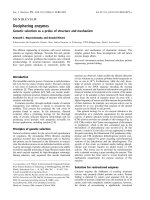
Western blot analysis
Western blot analysis of extracts prepared from the
eyestalk of F. indicus during different molting stages
showed a single dominant band of Gas7 only in the
early postmolt (A) and late postmolt (B) stages
(Fig. 1A). The immunoreactive band of Gas7 had a
molecular mass of 48 kDa. It was prominently detec-
ted in the XO-SG complex using rabbit polyclonal
anti-Gas7 serum, but was not observed in other stages
of molting (Fig. 1A).
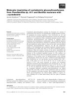
Immunohistochemical localization
Immunohistochemical analysis of eyestalk neural gan-
glia sections showed a highly positive immunoreactive
expression of Gas7 in the XO-SG complex of the eye-
stalk of F. indicus only in the early postmolt (A) and
late postmolt (B) stages but not in the intermolt (C),
early premolt (D
0
and D
1
) and late premolt (D
2-3
) sta-
ges using antibody to Gas7 (Fig. 2). The immunoreac-
tivity of Gas7 was observed prominently as a cluster
of neurosecretory cell bodies or group of cell mass
exclusively over the neurosecretory centers of the
XO-SG complex. Most of the Gas7-expressing neuro-
secretory cell bodies were found in the peripheral
regions and the neuropile regions of eyestalk neural
ganglia and more adjacent to the medulla terminalis
X-organ and sinus gland.
A
B
Fig. 1. Western blot analysis of proteins from the eyestalk of
F. indicus. (A) The supernatant from the XO-SG complex of the
eyestalk of F. indicus was subjected to SDS ⁄ PAGE (10% gel) and
electroblotted on to nitrocellulose membrane. The 48-kDa Gas7
was detected only in the early postmolt and late postmolt stages
by western blot analysis using rabbit Gas7 polyclonal antibody. (B)
Protein samples were extracted from the eyestalk of different
molting stages of F. indicus and subjected to SDS ⁄ PAGE (10%
gel); proteins were transferred on to nitrocellulose membrane.
Phosphorylation of ERK2 protein was found in the intermolt and
early premolt stages by a shift in electrophoretic mobility on west-
ern blot analysis using rabbit ERK1 ⁄ 2 antibody, goat anti-rabbit
IgG–horseradish peroxidase conjugate and developed with 3,3¢-di-
aminobenzidine tetrahydrochloride substrate. KD, kDa; M, broad
range of standard protein markers run on SDS ⁄ 10% polyacryl-
amide gel, electroblotted on to nitrocellulose membrane, and
stained with Ponceau S red; A, early postmolt; B, late postmolt;
C, intermolt; D
0
, early premolt; D
1
, early premolt; D
2-3
, late
premolt.
Gas7 and the ERK1 ⁄ 2 signaling pathway in MIH expression H. Devaraj and A. Natarajan
840 FEBS Journal 273 (2006) 839–846 ª 2006 The Authors Journal compilation ª 2006 FEBS
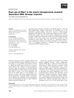
ELISA
Gas7 concentrations were determined by direct ELISA
using antibody to Gas7. A progressive increase from
the early postmolt stage to a maximum at the late
postmolt stage followed by a sudden fall in the sub-
sequent stages (Fig. 3) was observed. The results are
consistent with the results of western blot and immu-
nohistochemical analysis.
Concentrations of the immunoreactively expressed
MIH protein were also quantified by direct ELISA
from the extracts prepared from the XO-SG complex
of the eyestalk from control groups as well as groups
treated with specific inhibitors (PD98059 and UO126).
In control stages, MIH concentrations were increased
significantly from early postmolt to a maximum at
intermolt, but were dramatically reduced to a mini-
mum at early premolt and late premolt stages. In the
case of the PD98059-treated and UO126-treated stages
(intermolt and early premolt), MIH concentrations
were reduced to a basal level (Fig. 4A,B).
Phosphorylation of ERK2
Phosphorylation or activation of ERK2 protein was
found only in intermolt and early premolt by a shift
in mobility on western blot analysis (Fig. 1B). No
shift in electrophoretic mobility was observed in early
postmolt, late postmolt, early premolt and late
premolt stages.
Fig. 2. Immunohistochemical localization of Gas7 expression was
observed only in (A) early postmolt and (B) late postmolt stages in
the eyestalk tissue of F. indicus using rabbit polyclonal antibody to
Gas7. (N), Negative control treated with normal goat serum not
showing expression of Gas7; ME, medulla externa; MI, medulla
interna; MT, medulla terminalis; SG, sinus gland; XO, X-organ; NSC,
neurosecretory cell bodies. Scale bar represents 100 lm.
Fig. 3. Quantitative determination of Gas7 concentrations in
extracts prepared from the XO-SG complex of F. indicus by direct
ELISA using rabbit polyclonal antibody to Gas7. The line diagram
represents high concentrations of Gas7 immunoreactivity only in
early postmolt, maximum at late postmolt, and basal level in subse-
quent stages. Values are expressed as mean ± SD (n ¼ 5).
H. Devaraj and A. Natarajan Gas7 and the ERK1 ⁄ 2 signaling pathway in MIH expression
FEBS Journal 273 (2006) 839–846 ª 2006 The Authors Journal compilation ª 2006 FEBS 841
Treatment with inhibitors and blocking
of ERK2 activation
Phosphorylation of ERK2 and expression of MIH were
studied in both control and inhibitor-treated (PD98059
and UO126) stages. Phosphorylation of ERK2 was
detected as a shift in mobility during the intermolt
and early premolt stages of control shrimps, whereas
PD98059-treated stages (intermolt and early premolt)
showed no shift in ERK2 protein mobility using rabbit
anti-ERK1 ⁄ 2 IgG (Fig. 5A). Mouse anti-phospho-
ERK1 ⁄ 2 IgG specifically detected only phosphorylated
ERK2 protein in control groups of intermolt and early
premolt stages, but the antibody could not detect the
ERK2 protein in UO126-treated stages (Fig. 5B). Like-
wise, the immunoreactive band of MIH was also detec-
ted only in the control stages, i.e. early postmolt, late
postmolt, intermolt and early premolt, but not in the
PD98059-treated and UO126-treated stages (intermolt
and early premolt) (Fig. 6A,B). The results are consis-
tent with the results of direct ELISA.
Fig. 4. Effect of (A) PD98059 and (B) UO126 on MIH concentra-
tions of F. indicus. MIH concentrations were determined by direct
ELISA in extracts prepared from control (Untreated) and PD98059
or UO126 treated stages of F. indicus using anti-r-Pej-MIH IgG.
The bar diagram shows the highly significant concentrations
(P<0.0001; analysis of variance) of MIH in the intermolt stage of
the control samples, but PD98059 and UO126 treated stages (inter-
molt and early premolt) show very low concentrations of MIH.
Values are expressed as mean ± SD (n ¼ 5).
A
B
Fig. 5. Effect of (A) PD98059 and (B) UO126 on the phosphoryla-
tion of the ERK2 protein from the XO-SG complex. (A) Protein sam-
ples from the XO-SG complex of different molting stages were run
on SDS ⁄ 10% polyacrylamide gel and electroblotted on to nitrocellu-
lose membrane. Phosphorylation of the ERK2 protein was detected
by a shift in electrophoretic mobility on western blot analysis only
in control stages (intermolt and early premolt), but PD98059-treated
stages did not show ERK2 phosphorylation using rabbit antibody to
ERK1 ⁄ 2. (B) Mouse anti-phosphoERK1 ⁄ 2 IgG specifically detected
only phosphorylated ERK2 protein in control groups of intermolt
and early premolt stages, but the antibody could not detect the
ERK2 protein in UO126-treated stages. KD, kDa; M, broad range
of standard protein markers run on SDS ⁄ 10% polyacrylamide gel,
electroblotted on to nitrocellulose membrane, and stained with
Ponceau S red; A, early postmolt; B, late postmolt; C, intermolt;
D
0
, early premolt; D
1
, early premolt; D
2-3
, late premolt.
Gas7 and the ERK1 ⁄ 2 signaling pathway in MIH expression H. Devaraj and A. Natarajan
842 FEBS Journal 273 (2006) 839–846 ª 2006 The Authors Journal compilation ª 2006 FEBS
Molting duration of F. indicus
The inhibitor-treated (PD98059 and UO126) stages
showed an absence of MIH, which reduces the molting
duration of F. indicus. The shrimps in the control
groups required 12–14 days to complete the molt
cycle, whereas in those injected with either PD98059 or
UO126 the duration of the molt cycle was reduced to
7–8 days (Fig. 7). The available data also show a
correlative and significant change in the shortening of
the molt cycle which leads to frequent molting and
growth of shrimps.
Discussion
Gas7 is a multifunctional protein involved in the mat-
uration of neurons and release of neurotransmitters
[5]. The stage-specific expression of the Gas7 gene
using RT-PCR of Gas7 mRNA and coimmunoprecipi-
tation of Sky receptor has been identified in the early
and late postmolt stages of Penaeus monodon, suggest-
ing that Gas7 may act as a ligand for tyrosine kinase
Sky receptor (unpublished work). ERK2 is part of the
Ras ⁄ Raf ⁄ MEK downstream signaling pathway for
many growth factors that act by binding to tyrosine
kinase receptors in cardiac myocytes [19]. The mito-
gen-activated protein kinase pathway is involved in a
number of cellular changes inducing growth and differ-
entiation in human Schwann cells [7]. In cardiac myo-
cytes, protein kinase C generates a positive feedback
loop to ERK2 phosphorylation [19], and this effect
has also been demonstrated in P. monodon (unpub-
lished work).
In decapod crustaceans, the growth and differenti-
ation processes are controlled by a complex interplay
of neuropeptides that are stage specific and inducible
[3,4]. To elucidate the functional necessity of Gas7
in MIH expression, highly selective ERK inhibitors
(PD98059 and UO126) were used, which completely
inhibited the Gas7-dependent downstream signaling
during ERK2 phosphorylation. ERK2 is active only
when it is phosphorylated [7]. Inhibition of ERK2 with
ERK ⁄ MEK inhibitors (PD98059 and UO126) appears
A
B
Fig. 6. Effect of (A) PD98059 and (B) UO126 on 7–8-kDa MIH
from the XO-SG complex. Protein samples prepared from the
XO-SG complex of the eyestalk of F. indicus were run on
SDS ⁄ 15% polyacrylamide gel. Immunoblotting was performed
using anti-r-Pej-MIH IgG in different molting stages. The 7–8-kDa
MIH protein was detected only in the control stages (early post-
molt, late postmolt, intermolt and early premolt), whereas shrimps
treated with PD98059 and UO126 (intermolt and early premolt sta-
ges) show an absence of MIH immunoreactivity. KD, kDa; M, broad
range of standard protein markers run on SDS ⁄ 15% polyacrylamide
gel, electroblotted on to nitrocellulose membrane, and stained with
Ponceau S red; A, early postmolt; B, late postmolt; C, intermolt;
D
0
, early premolt; D
1
, early premolt; D
2-3
, late premolt.
Fig. 7. Molt cycle duration of F. indicus in controls and shrimps
injected with specific inhibitors (PD98059 and UO126) on day 3
after the first molt was recorded as hours between the first molt
and the second molt. Values are mean ± SD (n ¼ 5). The highly
significant (P < 0.0001, analysis of variance) reduction in molt cycle
duration was observed only in the D
1
stage.
H. Devaraj and A. Natarajan Gas7 and the ERK1 ⁄ 2 signaling pathway in MIH expression
FEBS Journal 273 (2006) 839–846 ª 2006 The Authors Journal compilation ª 2006 FEBS 843
to inhibit ERK phosphorylation, which can be seen as
shift in the mobility of ERK2 protein during inter-
molt and early premolt stages, resulting in the down-
regulation of MIH expression. The data show that
phosphorylation of ERK2 results in activation of tran-
scription factors inside the nucleus that modulate MIH
gene transcription and translation.
The shrimps treated with ERK inhibitors showed a
shortening of the intermolt and early premolt periods,
which suggests changes in the periodicity of the mol-
ting process. In addition, the molt cycle duration of
F. indicus was reduced dramatically from 12 ± 2 days
to 7 ± 1 days when the shrimps were injected with
ERK inhibitors. Thus, application of these inhibitors
significantly reduced the molt cycle duration, as they
inhibit MIH by interfering with downstream signals
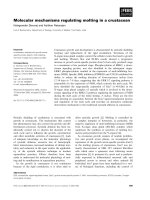
from Gas7. Hence, these data clearly indicate that
transcription of Gas7 occurs through co-ordinated
events involving activation of the ERK signal (Fig. 8).
These are the first data to show an association
between the Gas7 gene and molting processes in crus-
taceans mediated by the expression of MIH through
the ERK1 ⁄ 2 signaling pathway. Reduction of the molt
cycle by ERK inhibitors has an application potential
in the aquaculture industry.
Experimental procedures
Experimental animals
The Indian white shrimp F. indicus was selected for this
study because of its nonseasonal availability and easy main-
tenance. They were obtained from the coastal region of
Kovalam, Chennai, India. They were maintained in the
laboratory at a temperature of 28 ± 2 °C under natural
photoperiod (12 h light ⁄ 12 h darkness) in plastic tanks con-
taining filtered, continuously aerated seawater (26–28%
salinity). The stocking density was 10 shrimps per tank in
accordance with the water quality. The filtered seawater
was changed everyday, and the animals were fed every day
ad libitum with commercial feed pellets.
Identification of molting stages
The different molting stages of F. indicus were determined
from morphological changes in the setae of the uropod and
pleopod, carapace and exoskeleton, on the basis of criteria
established by Vijayan et al. [12]. The pleopod and uropod
of the same animal were removed and placed on a clean
micro slide; the slide was then covered with a rectangular
coverslip. The slide was kept under a light microscope and
the various molting stages such as early postmolt (A), late
Gas7
Gas7 binding to Sky receptor
(Stage A and B)
PD98059
UO126
Inhibition of ERK2
Phosphorylation (Stage C and D
0
)
Inhibition of MIH expression
(Stage C and D
0
)
Phosphorylation of ERK2
(Stage C and D
0
)
P
P
Ras
Raf
MEK1
ERK1/2
MIH
MIHGene
MIHGene
Expression of MIH
(Stage C and D
0
)
Sky receptor phosphorylation
(Stage B and C)
Fig. 8. Role of Gas7 in the molt cycle of
F. indicus. Sky receptor dimerization and
autophosphorylation is mediated by Gas7
during early postmolt (A) and late postmolt
(B) stages. Activation of the Ras ⁄ Raf ⁄ MEK
pathway subsequent to Sky receptor activa-
tion results in ERK2 phosphorylation and
expression of MIH during intermolt (C) and
early premolt (D
0
) which suppresses the
ecdysteroid concentrations. Inhibition of
ERK2 phosphorylation by PD98059 and
UO126 suppresses MIH expression and
increases ecdysteroid concentrations, which
results in shortening of the molt cycle.
Gas7 and the ERK1 ⁄ 2 signaling pathway in MIH expression H. Devaraj and A. Natarajan
844 FEBS Journal 273 (2006) 839–846 ª 2006 The Authors Journal compilation ª 2006 FEBS
postmolt (B), intermolt (C), early premolt (D
o
and D
1
) and
late premolt (D
2-3
) were identified.
Extraction of eyestalk neural ganglia
The eyestalks were clipped from different molting stages of
live shrimps. The eyestalks containing whole peduncular
neural ganglia (XO-SG complex) were dissected from the
surrounding exoskeleton of the eyestalk under a Carl Zeiss
(Go
¨
ttingen, Germany) Stereo Zoom dissection microscope.
They were homogenized in lysis buffer containing 135 mm
NaCl, 20 mm Tris ⁄ HCl, 2 mm EDTA and 1 mm phenyl-
methanesulfonyl fluoride, pH 7.4, as described by Watson
et al. [13], with slight modifications. The total homogenate
was microfuged at 8000 g,4°C for 10 min, and the super-
natant was recovered in different eppendorf tubes. They
were stored at )20 °C until subsequent analysis.
Western blot analysis
The proteins were separated by SDS ⁄ PAGE (10% gel) for
the detection of Gas7 [14] and electroblotted [15] on a
nitrocellulose membrane at 25 V ⁄ 130mA for 4 h at 4 °C.
The membrane was incubated overnight at 4 °C with rabbit
polyclonal antibody to Gas7 (1 : 1000 dilution; gift from S.
Lin-Chao, Institute of Molecular Biology, Academia Sinica,
Taiwan) followed by incubation for 2 h at room tempera-
ture with goat anti-rabbit IgG (1 : 2000 dilution) conju-
gated with horseradish peroxidase. Finally, Gas7 was
detected by incubation with 3,3¢-diaminobenzidine tetra-
hydrochloride as chromogenic substrate by the method
of Ju et al. [5].
Immunohistochemistry
Paraffin sections (15 lm) were prepared by conventional
methods [16]. The tissue sections were dewaxed in xylene and
rehydrated in descending alcohol series and washed in
NaCl ⁄ P
i
(pH 7.4). After being blocked with 3% BSA ⁄ Tris ⁄
NaCl ⁄ Tween, the sections were incubated separately with
Gas7 antibody (1 : 1000 dilution) for 16–18 h at 4 °C. After
being washed, goat anti-rabbit IgG–horseradish conjugate
(1 : 2000 dilutions) was applied to the sections for 2 h at
room temperature. Subsequently, Tris ⁄ NaCl (pH 7.6) con-
taining 0.05% 3,3¢-diaminobenzidine tetrahydrochloride and
0.01% H
2
O
2
were added as substrate for color development.
The reaction was stopped with Tris ⁄ NaCl, dehydrated in an
ascending alcohol series, cleared in xylene, and mounted with
DPX permount for observation [17].
ELISA
ELISA was performed to analyze the concentrations
of Gas7 present in the eyestalk as well as to study the
concentrations of MIH in the presence of inhibitors
(PD98059 and UO126) in the different stages of F. indicus as
described by Shih et al. [17]. In this assay, the specific pro-
tein samples were diluted with coating buffer and coated on
to 96-well microtiter plates. The primary antiserum used was
anti-Gas7 (1 : 1000 dilution). Goat anti-rabbit IgG–horse-
radish peroxidase conjugate (1 : 2000 dilution) was used as
secondary antibody. Thereafter, 3,3¢,5,5¢-tetramethylbenzi-
dine was used for color development. The reaction was
stopped with 1 m H
2
SO
4
, and the absorbance of the protein
was measured spectrophotometrically at 450 nm.
Phosphorylation of ERK2
Phosphorylation of ERK2 was detected by a shift in elec-
trophoretic mobility as described by Li et al. [7]. The protein
samples of different molting stages of F. indicus were run on
SDS ⁄ 10% polyacrylamide gels and transferred to nitrocellu-
lose membrane (25 V, 130mA for 4 h). The membrane was
treated with antibodies raised in rabbit against ERK1 ⁄ 2 pro-
tein (primary antibody, 1 : 1000 dilution; Chemicon Inter-
national, Temecula, CA, USA) followed by incubation with
goat anti-rabbit IgG (1 : 2000 dilution) coupled with horse-
radish peroxidase. The membrane was later incubated with
3,3¢-diaminobenzidine tetrahydrochloride substrate.
Treatment with inhibitors (PD98059 and UO126)
and blocking of ERK2 activation
The cultivable white shrimps (F. indicus) were purchased,
and two groups of animals at different molting stages were
maintained in the laboratory (salinity 26–28%, pH 8.1, and
temperature 28 ± 2 °C). Each group contained 10–15
shrimps, of which, one group from the intermolt and early
premolt stages were injected with specific inhibitors, and
the other served as control. PD98059 and UO126 (Chem-
icon International) are highly selective in vivo inhibitors of
the ERK kinase cascade. A 50 lm solution was diluted in
dimethyl sulfoxide and injected into test shrimps via the
arthrodial joints. The control groups were injected with
dimethyl sulfoxide only [18].
The injected shrimps were maintained until they reached
the second molt of the same stage. Then, the neural ganglia
were dissected and homogenized in lysis buffer on ice.
The supernatant was recovered after centrifugation at
10 000 r.p.m. for 10 min. The extracts prepared from the
XO-SG complex of inhibitor-injected and control shrimps
were run on SDS ⁄ 10% polyacrylamide gel and electroblot-
ted on to nitrocellulose membrane. Then, the membranes
were probed separately with rabbit anti-ERK1 ⁄ 2, mouse
anti-phosphoERK1 ⁄ 2 and anti-r-Pej-MIH IgG. In addition,
the molting behaviors and molting duration were observed
in the control and inhibitor-treated shrimps, and the data
were recorded.
H. Devaraj and A. Natarajan Gas7 and the ERK1 ⁄ 2 signaling pathway in MIH expression
FEBS Journal 273 (2006) 839–846 ª 2006 The Authors Journal compilation ª 2006 FEBS 845
Statistical analysis
Mean and standard deviation were calculated. Differences
between molting stages were analyzed using one-way analy-
sis of variance on spss version 10.0 software (SPSS Inc.,
Chicago, IL, USA).
Acknowledgements
This work was supported by grants from the Depart-
ment of Science and Technology (DST), UGC-DRS
program, COSIST program and Department of Bio-
technology (DBT) (BT PR3992 ⁄ AA2 ⁄ 03 ⁄ 202 ⁄ 2003),
New Delhi. We gratefully acknowledge Dr Sue Lin-
Chao, Institute of Molecular Biology, Academia Sinica,
Taiwan, for the gift of Gas7 antibody and Dr Tsuyoshi
Ohira, Department of Applied Biological Chemistry,
University of Tokyo, Japan, for the gift of MIH anti-
body. We also acknowledge Mr A. Anand Kumar for
his help in the preparation of the manuscript.
References
1 Chang ES (1995) Physiological and biochemical changes
during the molt cycle in decapod crustaceans: an over-
view. J Exp Mar Biol Ecol 193, 1–14.
2 Keller R (1992) Crustacean neuropeptides: structures,
function and comparative aspects. Experientia 48, 439–
447.
3 Soumoff C & O’Connor JD (1982) Repression of
Y-organ secretory activity by molt-inhibiting hormone
in the crab, Pachygrapsus crassipes. Gen Comp Endocri-
nol 48, 432–439.
4 Mattson MP & Spaziani E (1985) Characterization of
molt-inhibiting hormone (MIH) action on crustacean
Y-organ segments and dispersed cells in culture and a
bioassay for MIH activity. J Exp Zool 236, 93–101.
5 Ju YT, Chang ACY, She BR, Tsaur ML, Hwang HM,
Chao CCK, Cohen SN & Lin-Chao S (1998) gas7 :a
gene expressed preferentially in growth-arrested fibro-
blasts and terminally differentiated Purkinje neurons
affects neurite formation. Proc Natl Acad Sci USA 95,
11423–11428.
6 Brancolini C, Bottega S & Schneider C (1992) Gas 2, a
growth arrest-specific protein, is a component of the
microfilament network system. J Cell Biol 117, 1251–
1261.
7 Li R, Chen J, Hammonds G, Phillips H, Armanini M,
Wood P, Bunge R, Godowski PJ, Sliwkowski MX &
Mather JP (1996) Identification of gas6 as a growth factor
for human Schwann cells. J Neuro Sci 16, 2012–2019.
8 Del Sal G, Collavin L, Ruaro ME, Saccone S, Della
Valle G & Schneider C (1994) Structure, function, and
chromosome mapping of the growth suppressing human
homologue of the murine gas1 genes. Proc Natl Acad
Sci USA 91, 1848–1852.
9 Bogoyevitch MA, Glennon PE, Andersson MB, Clerk
A, Lazou A, Marshall CJ, Parker P & Sugden PH
(1994) Endothelin-1 and fibroblast growth factors sti-
mulate the mitogen-activated protein kinase signaling
cascade in cardiac myocytes. J Biol Chem 269, 1110–
1119.
10 Osborn MT & Chambers TC (1996) Role of stress-acti-
vated ⁄ c-Jun NH
2
-terminal protein kinase pathway in
the cellular response to adriamycin and other chemo-
therapeutic drugs. J Biol Chem 271, 30950–30955.
11 Cooke IM & Sullivan RE (1982) Hormones and neuro-
secretion. In The Biology of Crustacea: Structure and
Function (Atwood HL & Sandeman DC, eds), vol 3, pp
205–290. Academic Press, New York.
12 Vijayan KK, Sunil Kumar MK & Diwan AD (1997)
Studies on molt staging, molting duration and molting
behavior in Indian white shrimp, Penaeus indicus Milne
Edwards (Decapoda: Penaeidae). J Aqua Trop 12,
53–64.
13 Watson RD, Lee KJ, Borders KJ, Dircksen H & Lilly
KY (2001) Molt-inhibiting hormone immunoreactive
neurons in the eyestalk neuroendocrine system of the
blue crab, Callinectes sapidus. Arthropd Struct Dev 30,
69–76.
14 Laemmli UK (1970) Cleavage of structural proteins
during the assembly of the head of bacteriophage T
4
.
Nature 227, 680–686.
15 Towbin H, Staehelin T & Gordon J (1979) Electro-
phoretic transfer of proteins from polyacrylamide gel to
nitrocellulose sheets. Procedure and some applications.
Proc Natl Acad Sci USA 76, 4350–4354.
16 Presnell JK & Screibman MP (1997) Humason’s Animal
Tissue Techniques. Johns Hopkins University Press,
Baltimore.
17 Shih TW, Suzuki Y, Nagasawa H & Aida K (1998)
Immunohistochemical identification of hyperglycemic
hormone and molt-inhibiting hormone-producing cells
in the eyestalk of the kuruma prawn, Penaeus japonicus.
Zool Sci 15, 389–397.
18 Strohm C, Barancik T, Bruhl ML, Kilian SA & Schaper
W (2000) Inhibition of the ER Kinase cascade by
PD98059 and UO126 counteracts ischemic precondition-
ing in pig myocardium. J Cardiovasc Pharmacol 36,
218–229.
19 Kometiani P, Li J, Gnudi L, Kahn BB, Askari A & Xie
Z (1998) Multiple signal transduction pathways link
Na
+
⁄ K
+
-ATPase to growth-related genes in cardiac
myocytes. The role of Ras and mitogen-activated pro-
tein kinases. J Biol Chem 273, 15249–15256.
Gas7 and the ERK1 ⁄ 2 signaling pathway in MIH expression H. Devaraj and A. Natarajan
846 FEBS Journal 273 (2006) 839–846 ª 2006 The Authors Journal compilation ª 2006 FEBS