Báo cáo khoa học: EmbR, a regulatory protein with ATPase activity, is a substrate of multiple serine⁄threonine kinases and phosphatase in Mycobacterium tuberculosis doc
Bạn đang xem bản rút gọn của tài liệu. Xem và tải ngay bản đầy đủ của tài liệu tại đây (493.06 KB, 11 trang )
EmbR, a regulatory protein with ATPase activity, is a
substrate of multiple serine⁄threonine kinases and
phosphatase in Mycobacterium tuberculosis
Kirti Sharma1, Meetu Gupta1, Ananth Krupa2,*, Narayanaswamy Srinivasan2 and Yogendra Singh1
1 Institute of Genomics and Integrative Biology, Delhi, India
2 Molecular Biophysics Unit, Indian Institute of Science, Bangalore
Keywords
ATPase; kinase; phosphatase;
Mycobacterium; tuberculosis
Correspondence
Y. Singh, Institute of Genomics and
Integrative Biology, Mall Road, Delhi
110 007, India
Fax: +91 11 27667471
Tel: +91 11 27666156
E-mail:
*Present address
Cancer Research UK, Clare Hall
Laboratories, UK
(Received 23 March 2006, revised 19 April
2006, accepted 24 April 2006)
doi:10.1111/j.1742-4658.2006.05289.x
Phosphorylation of the mycobacterial transcriptional activator, EmbR, is
essential for transcriptional regulation of the embCAB operon encoding cell
wall arabinosyltransferases. This signaling pathway eventually affects the
resistance to ethambutol (a frontline antimycobacterial drug) and the cell
wall Lipoarabinomannan ⁄ Lipomannan ratio (an important determinant for
averting the host immune response). In this study, further biochemical
characterization revealed that EmbR, as a transcriptional regulator, interacts with RNA polymerase and possesses a phosphorylation-dependent
ATPase activity that might play a role in forming an open complex
between EmbR and RNA polymerase. EmbR was recently shown to be
phosphorylated by the cognate mycobacterial serine ⁄ threonine (Ser ⁄ Thr)
kinase, PknH. Using bioinformatic analysis and in vitro assays, we identified additional novel regulators of the signaling pathway leading to EmbR
phosphorylation, namely the Ser ⁄ Thr protein kinases PknA and PknB. A
previously unresolved question raised by this signaling scheme is the fate of
phosphorylated kinases and EmbR at the end of the signaling cycle. Here
we show that Mstp, a mycobacterial Ser ⁄ Thr phosphatase, antagonizes
Ser ⁄ Thr protein kinase–EmbR signaling by dephosphorylating Ser ⁄ Thr
protein kinases, as well as EmbR, in vitro. Additionally, dephosphorylation
of EmbR reduced its ATPase activity, interaction with Ser ⁄ Thr protein kinases and DNA-binding activity, emphasizing the antagonistic role of Mstp
in the EmbR–Ser ⁄ Thr protein kinase signaling system.
Serine ⁄ threonine (Ser ⁄ Thr) protein kinases (STPKs)
have emerged as crucial players for environmental
sensing and physiological signaling in prokaryotes.
These kinases have been implicated in diverse control
mechanisms, including stress responses, developmental
changes and host–pathogen interactions, in several
microorganisms. The genome of Mycobacterium tuberculosis, the causative agent for tuberculosis, has shown
the presence of 11 genes that code for putative STPKs
and one gene (Mstp) that codes for Ser ⁄ Thr phospha-
tase [1]. These STPKs have been proposed to mediate
signaling between mycobacteria and host cells to establish an environment that is favorable for the replication and survival of mycobacteria [2]. Recent reviews
have highlighted the importance of such signaling
mediated by mycobacterial STPKs and identified them
as potential drug targets [1,3,4]. To date, eight of these
STPKs or kinase domains have been expressed, purified
and shown to be active in vitro [5–12]. The mycobacterial STPKs regulate diverse processes by phosphorylating
Abbreviations
GST, glutathione S-transferase; RNAP, RNA polymerase; LAM ⁄ LM, Lipoarabinomannan ⁄ Lipomannan; SARP, Streptomyces coelicolor
antibiotic regulatory gene; Ser ⁄ Thr, serine ⁄ threonine; STPKs, serine ⁄ threonine protein kinases.
FEBS Journal 273 (2006) 2711–2721 ª 2006 The Authors Journal compilation ª 2006 FEBS
2711
Regulation of EmbR activity by STPKs and Mstp
K. Sharma et al.
120
100
80
EmbR
EmbR∆N
EmbR Heat inactivated
60
40
20
Results
Interaction of EmbR with RNA polymerase and
its phosphorylation dependent ATPase activity
EmbR belongs to the Streptomyces coelicolor antibiotic
regulatory gene (SARP) family of proteins, which are
known to regulate genes involved in the biosynthesis
of secondary metabolites through DNA binding to
specific gene sequences. Our previous results have
demonstrated the positive regulatory effect of EmbR
2712
on transcription of the embCAB operon after its phosphorylation by PknH in vivo [18]. Until very recently,
little was known about the mechanism by which
SARPs exerted their effect on gene expression. Bioinformatic analysis revealed that the SARP family
shares sequence homology with the OmpR ⁄ PhoB
family, a large family of transcription factors that bind
DNA through their winged helix-turn-helix motifs [23].
In agreement, the recently reported structure of EmbR
revealed that the structural elements relevant for function in OmpR are conserved in EmbR, including the
transactivation loop, which mediates interactions with
RNA polymerase (RNAP), the DNA recognition-helix
and the ‘wing’ [24]. Based on the presence of a
transactivation loop, termed as the ‘a loop’ in the
members of the OmpR ⁄ PhoB family [23], the possible
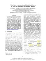
interaction of EmbR with RNAP holoenzyme (holoRNAP) was experimentally investigated by ELISA,
whereupon EmbR was found to interact with RNAP
in a concentration-dependent manner (Fig. 1). This
observation corroborates the function of EmbR as a
transcriptional activator of embCAB genes in view of
the fact that OmpR family members are known to
interact productively with RNAP for transcriptional
activation of their target genes [23]. EmbRDN, a deletion mutant lacking DBD and thus the ‘a loop’, failed
to interact with holo-RNAP, thereby suggesting that
the EmbR–RNAP interaction was specific (Fig. 1).
Relative ELISA signal
distinct substrates, including proteins implicated in
regulating cell division and morphology [13–15], an
ABC transporter [10,16], mediators of glutamate ⁄ glutamine metabolism [17] and a transcriptional regulator,
EmbR [9,18]. In addition, the Ser ⁄ Thr phosphatase,
Mstp, dephosphorylates two Ser ⁄ Thr kinases (PknA
and PknB) and has been implicated in regulating the
cell division of M. tuberculosis [19,20].
One of the major gaps in our knowledge concerns
identification of the key substrates of protein kinases
and phosphatases and how their phosphorylation ⁄
dephosphorylation contributes to the changes in cell
physiology evoked in response to particular signals.
PknH, a mycobacterial Ser ⁄ Thr kinase unique to the
members of M. tuberculosis complex [7], has been shown
to phosphorylate the cognate regulatory protein, EmbR
[9]. Recently, we reported that phosphorylated EmbR
serves as a transcriptional activator for arbinosyltransferases encoded by embCAB genes [18]. embCAB is a
gene cluster involved in arabinan synthesis and represents ethambutol targets in M. tuberculosis [21]. Our
results also revealed that EmbR phosphorylation affects
two important physiological phenomena, namely the
Lipoarabinomannan ⁄ Lipomannan (LAM ⁄ LM) ratio,
which is an important determinant of mycobacterial
virulence and resistance to ethambutol (a frontline antituberculosis drug) [18]. Concomitantly, AvenueGay and co-workers have shown that deletion of pknH
results in a hypervirulent phenotype and also suggested
a role of PknH in mediating NO toxicity [22]. Thus,
part of the signal transduction by PknH ⁄ EmbR has
been elucidated.
This study shows that EmbR is a substrate for
multiple STPKs, as well as a substrate for Mstp. In
addition, we show, for the first time, that a phosphorylation-dependent ATPase activity is associated with
EmbR. Dephosphorylation of EmbR by Mstp reduces
its ATPase activity, interaction with STPKs and
DNA-binding activity towards promoter regions of
embCAB genes, revealing the antagonistic role of the
phosphatase in the EmbR–STPK signaling system.
0
0.5
1
EmbR (mg/ml)
2
Fig. 1. EmbR–RNA polyerase (RNAP) interaction, as investigated by
ELISA. RNAP holoenzyme containing the principal sigma factor,
sigA, was purified from Mycobacterium smegmatis. Holo-RNAP
(100 ngỈwell)1) coated in wells was incubated with EmbR ⁄ EmbRDN
at graded concentrations. Unbound EmbR was removed and holoRNAP bound EmbR was quantified using anti-EmbR Ig.
FEBS Journal 273 (2006) 2711–2721 ª 2006 The Authors Journal compilation ª 2006 FEBS
K. Sharma et al.
Regulation of EmbR activity by STPKs and Mstp
A comparison of EmbR with its closest homologue,
AfsR, a transcriptional activator of Streptomyces [25],
revealed the absence of any defined ATPase domain in
EmbR. Besides, no such domain was identified in the
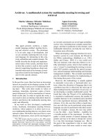
recently published 3D structure of EmbR [24]. However, after a closer examination of its amino acid
sequence, certain altered nucleotide-binding consensus
sequences were identified in EmbR (Fig. 2A). Therefore, the ability of EmbR to bind and hydrolyze nucleotide triphosphates (NTPs) was investigated by three
methods (Fig. 2). Interestingly, EmbR showed distinct
ATPase and GTPase activities, with ATP preferred
over GTP as a substrate (Fig. 2B). No phosphate was
released when ADP was used as a substrate, indicating
A
ATP
that EmbR is not a phosphatase. These results showed
that despite the absence of consensus nucleotide-binding motifs, EmbR exhibits ATPase and GTPase activities.
STPK-mediated phosphorylation of a transcriptional
activator whose function depends on ATP hydrolysis
is emerging as a central theme in prokaryotic signal
transduction systems [25,26]. Therefore, the effect of
PknH-mediated phosphorylation on the in vitro ATPase activity of EmbR was analyzed. While purified
EmbR showed an ATPase activity of 0.040 nmol of
phosphatmin)1Ỉlg)1 EmbR protein, the phosphorylated form of EmbR displayed an ATPase activity of
0.257 nmol of phosphatmin)1Ỉlg)1 EmbR (i.e. about
ATP + EmbR
1
10 20 30
(min)
Percent of original ATP hydrolyzed
Pi
ATP
Nucleotide binding motif
Consensus
EmbR
237
40
20
0
GXXXXGKT
226 GAYRRVKT
Consensus
EmbR
60
0
10
20
Time (min)
DXXG
DDLG
a
B
b
C
5
ATP + EmbR
6
GTP + EmbR
4
nmoles of Pi per mg protein
nmol of Pi per ug EmbR
30
ATP + heat inactivated EmbR
GTP + heat inactivated EmbR
3
2
1
0
EmbR
5
Phosphorylated EmbR
4
3
2
1
0
0
20
40
60
80
100
0
Time (min)
20
40
60
80
100
Time (min)
Fig. 2. (A) ATPase activity of EmbR. (a) EmbR was incubated with [32P]ATP[cP] for various time periods, and the release of 32Phosphate
(32Pi) was monitored by TLC. Also shown are altered nucleotide-binding motifs in EmbR. (b) EmbR was incubated with [32P]ATP[cP] for various time intervals (0–30 min). The filter binding assay was performed as described in the Experimental procedures. The ATP hydrolyzed at
each time point is shown as a percentage of the original [32P]ATP[cP] before incubation at 37 °C. (B) Time courses of ATP and GTP hydrolysis by EmbR. The release of Pi was measured, using the malachite green method, at various time points. The Pi release was assayed
when ATP or GTP was used as a substrate of EmbR. Each time point is the average of the values obtained from three independent experiments. (C) Effect of phosphorylation on ATPase activity. ATPase activities of EmbR and phosphorylated EmbR were compared. The phosphorylated EmbR sample was prepared by in vitro phosphorylation as described in the Experimental procedures.
FEBS Journal 273 (2006) 2711–2721 ª 2006 The Authors Journal compilation ª 2006 FEBS
2713
Regulation of EmbR activity by STPKs and Mstp
K. Sharma et al.
For further characterization of EmbR and the associated phosphorelay in M. tuberculosis, the ability of
mycobacterial cell lysate to phosphorylate purifed
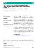
EmbR was analyzed. Resin-bound EmbR was incubated with whole cell lysate of M. tuberculosis in the
presence of [32P]ATP[cP] and it was observed that
the cell lysate of M. tuberculosis harbors the ability
to phosphorylate EmbR (Fig. 3A). Boiling of cell
lysate, or treatment with a kinase inhibitor, staurosporine, resulted in the complete loss of its ability to
phosphorylate EmbR (Fig. 3A). These observations
indicated that the EmbR phosphorylating activity in
the whole cell lysate of M. tuberculosis is caused by
the presence of STPK(s). Recently, it has been
shown that EmbR is phosphorylated in vitro by
PknH [9]. Therefore, it can be anticipated that mycobacterial cell lysate devoid of PknH should not
phosphorylate EmbR. Interestingly, whole cell lysate
of M. tuberculosis, pre-incubated with anti-PknH Ig
and thus neutralized for PknH, was also able to
phosphorylate EmbR (Fig. 3A). At the same time,
the anti-PknH Ig was able to prevent the phosphorylation of EmbR by purified PknH. All these observations suggested the presence of additional STPK(s)
that can phosphorylate EmbR.
An important clue towards other STPK(s) capable of
phosphorylating EmbR came from cross-genomic comparisons of bacterial protein kinases to identify homologues of kinases with known substrates. This study
revealed PknB of M. tuberculosis as the closest homolog of AfsK, an STPK-phosphorylating AfsR. Considering the homology of AfsR with EmbR, as well as the
significant sequence similarity observed between catalytic domains of AfsK and PknB (38% identity), it was
interesting to study EmbR as a possible substrate of
PknB. The in vitro assays revealed that autophosphorylated PknB phosphorylates EmbR, whereas heatinactivated PknB does not (Fig. 3B). In fact, EmbR
has previously been suggested as one of the targets for
a signal transduction pathway mediated by PknA and
PknB. If so, this pathway could link cell division and
peptidoglycan synthesis with arabinogalactan synthesis,
another process essential for growth [27]. PknA, an
STPK present in the same operon as PknB, was also
2714
sp
ori
ne
kn
H
kn
H
uro
+A
Pk
nH
nH
Pk
Ly
s
ate
+A
nti
-P
nti
-P
st a
mM
at e
+1
ate
Ly
s
Ly
s
Bo
ile
d
te
sa
Ly
Co
ntr
ol
Phosphorylation of EmbR by multiple Ser ⁄ Thr
kinases in M. tuberculosis
A
EmbR
1
B
2
3
4
1
2
3
4
PknA
EmbR
Autoradiogram
SDS-PAGE
PknB
EmbR
Autoradiogram
SDS-PAGE
(a)
Bound Radioactivity (cpm)
sixfold higher) (Fig. 2C). The ATPase activity of
EmbR probably provides energy to catalyze the isomerization of the closed complex between EmbR and
RNAP to a transcriptionally competent open complex,
as is proposed for AfsR [25].
80000
PknA
PknB
60000
40000
20000
0
0.5
10
20
30
Kinase
30
30 (min)
+
Heat
inactivated 20mM
EDTA
Kinase
(b)
Fig. 3. EmbR, a substrate for multiple serine ⁄ threonine protein kinases (STPKs). (A) Phosphorylation of EmbR with Mycobacterium
tuberculosis cell lysate. Resin-bound EmbR was incubated in the
presence of [32P]ATP[cP] and under the indicated experimental conditions, as described in the Experimental procedures. After elution,
EmbR was run on SDS ⁄ PAGE and its phosphorylation was visualized by autoradiography. (B) Phosphorylation of EmbR by PknA and
PknB. (a) In vitro kinase assays were performed to examine the
ability of PknA (upper half) and PknB (lower half) to phosphorylate
EmbR in the presence of [32P]ATP[cP]. The labeled proteins were
separated by SDS ⁄ PAGE and visualized by autoradiography or Coomassie Blue staining. Lane 1, EmbR; lane 2, PknA (upper half) or
PknB (lower half); lane 3, PknA or PknB incubated with EmbR; lane
4, heat-inactivated PknA or PknB incubated with EmbR. (b) For
resin-bound assays, EmbR bound to Ni-nitrilotriacetic acid resin
was incubated with purified PknA ⁄ PknB in the presence of
[32P]ATP[cP] for the indicated time periods and conditions. Shown
is the bound radioactivity in counts per minute.
FEBS Journal 273 (2006) 2711–2721 ª 2006 The Authors Journal compilation ª 2006 FEBS
K. Sharma et al.
tested for its ability to phosphorylate EmbR and it
came as an expected finding that PknA also phosphorylated EmbR (Fig. 3B). Incubation of EmbR alone in
the presence of [32P]ATP[cP], as a negative control, did
not yield any phosphorylated product.
The phosphorylation of EmbR by these kinases was
found to be specific, as other mycobacterial kinases,
such as PknG and PknI, could not phosphorylate EmbR
under similar conditions (data not shown). Thus, it was
confirmed that EmbR acts as a substrate of three mycobacterial STPKs, viz. PknH, PknA and PknB.
Regulation of EmbR activity by STPKs and Mstp
A
1
2
1
PknH
Mstp
SDS-PAGE
SDS-PAGE
Mstp
EmbR
B
100
Remaining radioactivity (%)
EmbR is phosphorylated by three STPKs, which themselves are believed to autophosphorylate in response
to environmental perturbations. This kinase-mediated
signaling should be ‘switched off’ when it is not
required. Returning to the inactive ⁄ resting state would
require either the synthesis of new proteins or the dephosphorylation of the existing phosphorylated species.
The only reported Ser ⁄ Thr phosphatase of M. tuberculosis, Mstp, is known to dephosphorylate PknA and
PknB, thereby acting as a regulator of these kinases
[19,20]. Therefore, it was tempting to examine whether
Mstp could dephosphorylate PknH in addition to
PknA and PknB, all of which are involved in EmbR
phosphorylation. We also examined the ability of Mstp
to dephosphorylate EmbR directly to ‘switch off’ signaling at the effector level.
As described in the Experimental procedures, two
methods were employed to examine dephosphorylation
of PknH by Mstp, namely resin-bound and in-solution
dephosphorylation assays. The prephosphorylated substrates for dephosphorylation assays were prepared
using resin-based phosphorylation reactions in the
presence of [32P]ATP[cP]. In-solution phosphatase
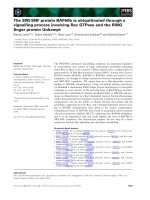
assays revealed that incubation with Mstp led to a
decrease in the intensity of bands corresponding to
prephosphorylated substrates, namely, PknH and
EmbR, thus confirming that PknH and EmbR are
substrates of Mstp in vitro. A reaction set with heatinactivated Mstp served as a negative control (Fig. 4A).
Incubation with heat-inactivated Mstp had no effect
EmbR phosphorylation status when compared with
the control phosphorylated EmbR with no addition of
Mstp (data not shown).
For resin-bound dephosphorylation assays, autophosphorylated PknH and phosphorylated EmbR
bound to resin were incubated with purified Mstp (or
heat-inactivated Mstp) in phosphatase buffer. Incubation with Mstp resulted in 73% and 79% dephospho-
Autoradiogram
Autoradiogram
Phosphorylated forms of PknH and EmbR are
substrates of Mstp in vitro
2
PknH
EmbR
80
60
40
20
0
0
5
10
+ Mstp
30
30
−
Mstp
30
(min)
Heat
inactivated
Mstp
Fig. 4. Dephosphorylation of EmbR and PknH by Mstp. (A) In vitro
dephosphorylation was analyzed by incubating prephosphorylated
PknH (upper half) or EmbR (lower half) with Mstp (lane 2) or heatinactivated Mstp (lane 1). The reaction products were resolved by
SDS ⁄ PAGE and the loss of labeling was visualized by autoradiography. Right, Coomassie Blue staining; left, corresponding autoradiogram. It is unclear why there are protein double bands of EmbR
and Mstp on SDS ⁄ PAGE. (B) For resin-bound assays, PknH or
EmbR bound to beads was prephosphorylated in the presence of
[32P]ATP[cP], as described in the Experimental procedures. Mstpmediated dephosphorylation of resin-bound prephosphorylated
PknH or EmbR was assessed by measuring the reduction in the
substrate-bound radioactivity after incubation for the indicated time
periods and under the experimental conditions described. Shown is
the residual PknH- and EmbR-associated radioactivity. Each value is
the average of two individual reactions and representative of three
experiments.
rylation of PknH and EmbR, respectively, in 30 min
(Fig. 4B). Our earlier studies, characterizing Mstp,
revealed that PknA and PknB, the endogenous substrate kinases present in an operon with Mstp, were
75% and 79% dephosphorylated after incubation with
Mstp for 60 min, respectively [20]. In concordance,
in this study, we observed that Mstp dephosphorylates
PknH and EmbR at comparable levels.
FEBS Journal 273 (2006) 2711–2721 ª 2006 The Authors Journal compilation ª 2006 FEBS
2715
Regulation of EmbR activity by STPKs and Mstp
K. Sharma et al.
Dephosphorylation of phosphorylated EmbR
decreases its DNA-binding activity
Our earlier results showed that EmbR is physically
and functionally engaged as a mediator of embCAB
activation by PknH in vivo [18]. The embA and embB
genes can be expressed from their own individual promoters [28]; however, synthesis of a polycistronic
mRNA encoding the three Emb proteins has also been
reported in M. tuberculosis [29]. PknH-mediated phosphorylation of EmbR is crucial for its interaction with
upstream regions of emb genes [18]. In view of our
observation that phosphorylated EmbR is a substrate
of Mstp, the modulation of its DNA-binding activity
upon dephosphorylation by this phosphatase was
examined. The dephosphorylated form of EmbR was
prepared by in vitro dephosphorylation of prephosphorylated EmbR, and the binding of phosphorylated ⁄
dephosphorylated EmbR to the upstream region of
embCAB genes was examined by the gel mobility shift
assay. Following its dephosphorylation, the strength of
DNA binding by EmbR decreased many fold, with
2 lg of dephosphorylated protein bringing about a
similar mobility shift as seen for 0.3 lg of phosphorylated EmbR (Fig. 5A).
Thus, while phosphorylation enhances the DNAbinding activity of EmbR, the dephosphorylated form
of EmbR was incapable of binding the promoter
regions of emb genes at low concentrations, in agreement with the belief that EmbR itself in the phosphorylated form interacts with upstream regions of emb
genes. Furthermore, it suggests that Mstp acts as an
antagonist of the STPK–EmbR signal relay. Moreover,
as one would anticipate, the dephosphorylation of
phosphorylated EmbR also reduces the level of
ATPase activity equivalent to that of unphosphorylated protein (data not shown).
Mstp-mediated dephosphorylation of
PknB ⁄ PknA ⁄ PknH inhibits their interaction with
EmbR
To further comprehend the role of Mstp, the effect of
Mstp-mediated dephosphorylation of kinases on their
specific interaction with endogenous substrate, EmbR,
was examined using a glutathione S-transferase (GST)
pull-down assay. To analyze the PknA–EmbR interaction (Fig. 5B), the soluble fraction containing His–
EmbR was incubated with either prephosphorylated
GST–PknA (lane 2) or dephosphorylated (using Mstp)
GST–PknA (lane 3). As controls, EmbR was incubated
with glutathione–Sepharose, either with GST (lane 4)
or alone (lane 5), in NaCl ⁄ Pi buffer. The binding assay
2716
was performed as described in the Experimental procedures. When pre-incubated with phosphorylated PknA,
EmbR was recovered in the soluble fraction eluted
from glutathione–Sepharose (lane 2). EmbR was not
recovered in control experiments when it was incubated either alone (lane 5) or in the presence of GST
(lane 4). Therefore, the complex was formed only via
the phosphorylated form of PknA. The absence of
recovery of EmbR upon pre-incubation with dephosphorylated PknA in this assay (lane 3) indicated,
by comparison with lane 2, that the Mstp-mediated
dephosphorylation abrogates the interaction of EmbR
with PknA (Fig. 5B).
Similar assays were performed with PknB and PknH
to show that Mstp inhibits the interaction of STPKs–
EmbR by directly dephosphorylating these kinases
(data not shown). In accordance with our observations,
previous reports have shown that the PknH–EmbR
interaction does not take place when a kinase mutant,
incapable of autophosphorylation, was used [9].
Discussion
Phosphorylation-dependent signal transduction between PknH and its cognate DNA-binding transcription regulator, EmbR, triggers the regulation of
mycobacterial embCAB genes and consequently influences ethambutol resistance and the LAM ⁄ LM ratio.
The present study shows that EmbR serves as a substrate of multiple STPKs. If each of the kinases senses
its own signal amongst a plethora of environmental
cues, as is known for eukaryotic protein kinases,
EmbR makes an integrator of the signals (Fig. 6). The
idea of one response regulator protein communicating
with multiple sensor kinases is not unusual, as exemplified by the PhoR ⁄ PhoM ⁄ PhoB system in Escherichia coli [30] and the AfsK ⁄ PkaG ⁄ PknL ⁄ AfsR system
in S. coelicolor [25]. Shared communication links help
the organism to integrate diverse signals into a global
response.
In analogy with its closest homolog – AfsR of
S. coelicolor [25] – EmbR also possesses a phosphorylation-dependent ATPase activity despite the absence
of any conserved nucleotide-binding motifs in its
amino acid sequence. The enhancement of DNA-binding activity of EmbR upon phosphorylation [18], and
its ability to interact with RNAP, is similar to that of
many other OmpR family members [23]. The energy
supplied by the intrinsic low ATPase activity of
unphosphorylated EmbR is thought to be insufficient
to overcome the activation energy barrier to open
complex formation. From the present study, together
with the similarity of EmbR with AfsR and other
FEBS Journal 273 (2006) 2711–2721 ª 2006 The Authors Journal compilation ª 2006 FEBS
K. Sharma et al.
Regulation of EmbR activity by STPKs and Mstp
A
P-Em bR
0
0.3
P-EmbR
deP-EmbR
1
2
5
( µg)
0
0.3
embA
-189/+135
deP-EmbR
1
2
5
P-EmbR
( µg)
0
0.3
deP-EmbR
1
2
5
( µg)
embC
-205/+112
embB
-194/+128
B
LANE
1
Dephosphorylated PknA
GST
EmbR
Mo l Weig ht Marker
Prephosphorylated PknA
3
4
5
6
7
+
Glutathione Sepharose
2
+
+
+
+
-
+
-
-
-
+
-
-
+
-
-
-
-
-
-
+
-
-
-
+
+
+
+
-
+
Anti His Blot
EmbR
Fig. 5. (A) Dephosphorylation of phosphorylated EmbR decreases its DNA-binding activity towards the promoter region of embCAB genes. A
total of 0.3 lg of phosphorylated EmbR (P-EmbR), or an increasing amount of dephosphorylated EmbR, was incubated with 32P-labeled
upstream regions of embC, embA and embB genes at 4 °C for 30 min. After incubation, complexes and free DNA were separated by nondenaturing polyacrylamide gels and subjected to autoradiography. The positions of EmbR-bound (solid arrow) and free (open arrow) probes are
shown. The numbers represent nucleotides relative to the translation start codon of the specific emb gene. (B) Mstp-mediated dephosphorylation of PknB ⁄ PknA ⁄ PknH inhibits their interaction with EmbR. The interaction of PknA with EmbR was analyzed using a pull-down assay.
The presence of either a protein or glutathione–Sepharose is indicated by ‘+’ and the absence by ‘–’. The soluble fraction of Escherichia coli
cells expressing recombinant His–EmbR was incubated with either prephosphorylated glutathione S-transferase (GST)–PknA (lane 2) or
dephosphorylated (using Mstp) GST–PknA (lane 3), each bound to glutathione–Sepharose resin. As controls, prephosphorylated GST–PknA
was incubated with glutathione–sepharose in the absence of EmbR (lane 6). In addition, EmbR was incubated with glutathione–Sepharose,
either with GST (lane 4) or alone (lane 5). GST complexes were pulled down with glutathione–Sepharose, separated by SDS ⁄ PAGE, and
transferred onto a nitrocellulose membrane before detection of recombinant poly histidine-tagged EmbR fusion protein (lower part). Purified
EmbR was run as a positive control (lane 7) and lane 1 represents the molecular weight marker.
OmpR family members, we infer that the association
of phosphorylation-modulated ATPase activity and
DNA binding ensures that phosphorylation of EmbR
is primarily coupled to the formation of site-specific
open complex during transcriptional initiation. Conversely, Mstp antagonizes this signaling by individually
dephosphorylating all three kinases as well as EmbR.
The phosphorylation-dependent enhancement in DNA
binding and ATPase activity of EmbR is reversed as a
consequence of its dephosphorylation by Mstp. Moreover, Mstp-mediated dephosphorylation of kinases
abrogates their interaction with EmbR, thus emphasizing the antagonistic role played by Mstp in the
EmbR–STPK signaling cascade.
In conclusion, by demonstrating multiple STPKmediated phosphorylation and Mstp-mediated dephosphorylation of EmbR, our findings add other
upstream effectors to the EmbR-mediated signaling
FEBS Journal 273 (2006) 2711–2721 ª 2006 The Authors Journal compilation ª 2006 FEBS
2717
Regulation of EmbR activity by STPKs and Mstp
K. Sharma et al.
SIGNAL
X
PknA
SIGNAL
A
SIGNAL
Y
PknB
SIGNAL
Z
PknH
(Sensor Kinases)
EXTR
ACE
LLUL
AR
Autophosphorylation
LAM / LM
Ratio
EMB
Resistance
Mstp
Dephosphorylation
INTR
ACE
LLUL
AR
P
EmbR
Arabinan
Metabolism
Dephosphorylation
P
EmbR
RNA
Polymerase
Increased transcription
of embCAB genes
Closed complex
P
ATP hydrolysis
RNA
Polymerase
P
Open complex
Fig. 6. A hypothetical scheme for the regulation of arabinan metabolism by the Mstp serine ⁄ threonine protein kinases (STPKs) ⁄ EmbR ⁄ embCAB system in Mycobacterium tuberculosis. By analogy with eukaryotic signal transduction, we speculate that multiple STPKs autophosphorylate on sensing certain external stimuli and transfer the signal to EmbR by means of phosphorylation. EmbR phosphorylation triggers the
transcriptional activation of embCAB genes and consequently influences ethambutol resistance and the LAM ⁄ LM ratio. On the contrary, the
Ser ⁄ Thr phosphatase, Mstp, antagonizes the STPK ⁄ EmbR signaling system. The environmental stimuli that activate PknH ⁄ PknB ⁄ PknA and
Mstp have yet to be identified.
network. Mediated by the action of STPKs and Mstp,
we demonstrate the modulation of ATPase activity
and DNA-binding activity of EmbR as a possible
physical mechanism to modulate its regulatory effect
on emb genes. On the basis of the results obtained so
far, we present a hypothetical model for the regulation
of arabinan metabolism by the Mstp ⁄ STPKs ⁄ EmbR ⁄
embCAB system in M. tuberculosis (Fig. 6). Collectively, these observations provide another example for
the mutual regulation of protein Ser ⁄ Thr kinases and
protein Ser ⁄ Thr phosphatases.
In vivo studies and further functional characterization to comprehend the role of these merging pathways in mycobacterial pathogenicity are in progress
and are expected to provide intriguing insights into
the significance of corresponding signaling events in
M. tuberculosis.
2718
Experimental procedures
Bacterial culture and growth conditions
Mycobacterial strains were grown in Middlebrook 7H9
broth supplemented with 0.5% glycerol and 10% albumin ⁄
dextrose ⁄ catalase at 37 °C, with shaking at 220 r.p.m., for
3–4 weeks. The E. coli strains were grown in Luria–Bertani
(LB) broth or on LB agar plates at 37 °C with shaking at
220 r.p.m.
Plasmid construction, mutagenesis and protein
purification
GST-tagged PknH and PknHK45M mutant protein were
used from previous studies [7]. EmbR and EmbDN were
expressed as His-tagged fusion proteins and purified under
FEBS Journal 273 (2006) 2711–2721 ª 2006 The Authors Journal compilation ª 2006 FEBS
K. Sharma et al.
denaturing conditions using Ni-nitrilotriacetic acid resin, as
per the manufacturer’s instructions and as described previously [18]. PknA, PknB and Mstp were also purified under
denaturing conditions as described in previous studies [20].
ELISA
The M. smegmatis RNAP holoenzyme, containing the principal sigma factor, sigA, was kindly provided by Prof. Anil
K. Tyagi. Purified Holo-RNAP was coated on a 96-well
ELISA plate (100 ngỈwell)1). His–EmbR or His–EmbRDN
fusion proteins were incubated, at three concentrations,
with the coated protein overnight at 4 °C in buffer comprising 10 mm Tris HCl, 150 mm NaCl, pH 7.5, 0.5% Tween
20 (TBS-T), after which the plates were washed and developed as described previously [31].
In vitro kinase assay
The in vitro kinase reactions routinely contained 500 ng of
the enzyme in the kinase buffer (25 mm Tris, pH 7.4,
10 mm MgCl2, 1 mm dithiothreitol) with 1 lg of EmbR
and 5 lCi of [32P]ATP[cP] and incubated for 30 min at
37 °C. The reactions were stopped by the addition of SDS
sample buffer, and proteins were separated by 1D gel electrophoresis, electroblotted onto nitrocellulose membranes
and visualized by autoradiography.
For resin-bound kinase assays, purified EmbR was phosphorylated by PknH in the kinase buffer, as described previously [20]. The counts associated with resin-bound EmbR
are a measure of its phosphorylation by the kinase. To
prepare phosphorylated substrates for dephosphorylation
reactions, the phosphorylated EmbR was eluted from
Ni-nitrilotriacetic acid beads using elution buffer (200 mm
imidazole in 50 mm Na phosphate, pH 7.0, 100 mm NaCl
and 10% glycerol). Similarly, GST–PknH was autophosphorylated and eluted from glutathione–Sepharose 4B, as
described previously for PknA and PknB [20]. After elution,
phosphorylated EmbR and PknH were dialyzed against
buffer (40 mm Tris, pH 7.6, and 10% glycerol) and stored
at )20 °C until further use.
To monitor phosphorylation of EmbR by mycobacterial
lysate, 1 lg of resin-bound EmbR was incubated with 10 lg
of whole cell lysate of M. tuberculosis in the presence of
15 lCi of [32P]ATP[cP] in 25 mm Tris, pH 7.4, 10 mm MgCl2,
1 mm dithiothreitol (TMD) buffer and 50 mm sodium fluoride (Ser ⁄ Thr phosphatase inhibitor) for 30 min at room temperature. The effects of boiling the whole cell lysate on its
ability to phosphorylate EmbR was examined by boiling the
whole cell lysate for 10 min, before incubation with resinbound EmbR. The effect of kinase inhibitor was investigated
by pre-incubating the whole cell lysate with 1 mm staurosporine. The effect of anti-PknH Ig was examined by pre-incubating the whole cell lysate with anti-PknH Ig (1 : 500 dilution)
for 20 min before incubation with resin-bound EmbR.
Regulation of EmbR activity by STPKs and Mstp
In vitro phosphatase assay
For resin-bound assays, dephosphorylation of phosphorylated PknH and EmbR by Mstp was examined by measuring the release of 32Phosphate (32Pi). Glutathione–
Sepharose 4B beads bound to phosphorylated GST–PknH,
or Ni-nitrilotriacetic acid beads bound to His-EmbR,
(2.5 lg each) were incubated with Mstp (1 lg) for different
time periods. After incubation, the beads were washed twice
with wash buffer to remove liberated 32Pi and the proteins
were eluted at 65 °C using elution buffer (1% SDS and
50 mm EDTA) for 10 min, as reported previously [20].
Radioactivity was measured using a scintillation counter. A
decrease in the counts of phosphorylated PknH ⁄ EmbR in
the presence of Mstp is a measure of the dephosphorylation
activity of Mstp.
The in vitro dephosphorylation of PknH ⁄ EmbR by Mstp
was also analyzed by using phosphorylated PknH ⁄ EmbR
that was eluted from affinity resin. Phosphorylated PknH ⁄
EmbR (2 lg) was incubated with Mstp (1 lg) in 50 mm TrisHCl, pH 8.0, 5 mm MnCl2 and 0.5 mm dithiothreitol, for
30 min at 30 °C, the mixtures were resolved by SDS ⁄ PAGE
and the loss of labeling was visualized by autoradiography.
ATPase activity measurements
The malachite green ATPase assay
The reaction buffer contained 10 lL of 10· TMD buffer,
10 lL of 5 mgỈmL)1 BSA, 4 lL of 100 mm ATP ⁄ GTP and
71 lL of H2O. Five microlitres of purified dephosphorylated or phosphorylated EmbR (1 mgỈmL)1) was added to the
reaction buffer and incubated at 37 °C. At various time
points (0, 5, 10, 20, 40 and 80 min), 10 lL of the reaction
mixture was added to 80 lL of freshly prepared malachite
green-ammonium molybdate reagent [three volumes of
0.045% malachite-green hydrochloride, one volume of
4.2% ammonium molybdate tetrahydrate in 4 m HCl and
0.02 volume of 1% Triton X-100]. After 1 min at room
temperature, 10 lL of 34% citric acid was added to stop
the colour development and the absorbance at 660 nm was
measured. The amounts of enzymatically released inorganic
phosphate in triplicate samples were measured photometrically by referring to a standard curve, which was prepared
with dilutions of a standard solution.
The ATPase activity of purified EmbR was also assayed
by polyethyleneimine-TLC, as described previously [32].
ATPase activity was also determined, as described previously, by a filter binding assay [32].
Gel mobility shift assay
The protein–DNA binding assay was performed as described previously [18]. 32P-labeled probe DNA was
prepared by end labeling using polynucleotide kinase, and
FEBS Journal 273 (2006) 2711–2721 ª 2006 The Authors Journal compilation ª 2006 FEBS
2719
Regulation of EmbR activity by STPKs and Mstp
K. Sharma et al.
labeled PCR products, representing different promoter
regions, were incubated with various amounts of phosphorylated and dephosphorylated EmbR at 4 °C for 30 min.
After incubation, complexes and free DNA were resolved
by 5% nondenaturing polyacrylamide gels. Gels were dried
and subjected to autoradiography.
PknA ⁄ PknB –EmbR interaction assay (GST pulldown assays)
The resin-bound phosphorylated GST–PknA, or the
dephosphorylated (using Mstp) form of PknA (10 lg
each), was incubated with a soluble fraction (5 lg of protein) of E. coli cells expressing EmbR, at 25 °C for 4 h
in 1 mL of NaCl ⁄ Pi buffer. The protein–resin complex
was washed six times with 1 mL of NaCl ⁄ Pi each wash.
The proteins thus retained on the beads were eluted with
elution buffer (50 mm Tris ⁄ HCl, pH 8.0, 5 mm MgCl2,
1 mm dithiothreitol, 15 mm glutathione). Eluted fractions
were precipitated with trichloroacetic acid, resuspended in
30 lL of SDS-loading buffer and boiled for 3 min. The
proteins were then resolved on a 10% SDS–polyacrylamide gel, electroblotted onto a poly(vinylidene difluoride)
membrane and probed with anti-His Ig conjugated to
horseradish peroxidase (HRP) to detect the poly histidine-tagged EmbR fusion protein. As a control, 5 lg of
EmbR was incubated either with 10 lg of GST bound to
resin or to resin alone in NaCl ⁄ Pi buffer. Similar assays
were performed to study the interaction of EmbR with
PknB and PknH.
Acknowledgements
Financial support was provided by CSIR (SMM 0003).
RNAP was a kind gift from Prof. Anil K. Tyagi,
University of Delhi, Delhi. Studentships of KS, MG
and AK were supported by CSIR, India. NS is an
International Senior Fellow of the Wellcome Trust,
UK.
References
1 Av-Gay Y & Everett M (2000) The eukaryotic-like
Ser ⁄ Thr protein kinases of Mycobacterium tuberculosis.
Trends Microbiol 5, 238–244.
2 Walburger A, Koul A, Ferrari G, Nguyen L, Prescianotto-Baschong C, Huygen K, Klebl B, Thompson C,
Bacher G & Pieters J (2004) Protein kinase G from
pathogenic mycobacteria promotes survival within
macrophages. Science 304, 1800–1804.
3 Koul A, Herget T, Klebl B & Ullrich A (2004) Interplay
between mycobacteria and host signalling pathways.
Nat Rev Microbiol 2, 189–202.
2720
4 Sharma K, Chopra P & Singh Y (2004) Recent
advances towards identification of new drug targets for
Mycobacterium tuberculosis. Expert Opin Ther Targets
8, 79–93.
5 Av-Gay Y, Jamil S & Drews SJ (1999) Expression and
characterization of the Mycobacterium tuberculosis
serine ⁄ threonine protein kinase PknB. Infect Immun 67,
5676–5682.
6 Chaba R, Raje M & Chakrabarty PK (2002) Evidences
that a eukaryotic-type serine ⁄ threonine protein kinase
from Mycobacterium tuberculosis regulates morphological changes associated with cell division. Eur J Biochem
269, 1078–1085.
7 Sharma K, Chandra H, Gupta PK, Pathak M, Narayan
A, Meena LS, D’Souza RC, Chopra P, Ramachandran
S & Singh Y (2004) PknH, a transmembrane Hank’s
type serine ⁄ threonine kinase from Mycobacterium tuberculosis, is differentially expressed under stress conditions. FEMS Microbiol Lett 233, 107–113.
8 Molle V, Girard-Blanc C, Kremer L, Doublet P,
Cozzone AJ & Prost JF (2003) Protein PknE, a novel
transmembrane eukaryotic-like serine ⁄ threonine kinase
from Mycobacterium tuberculosis. Biochem Biophys Res
Commun 308, 820–825.
9 Molle V, Kremer L, Girard-Blanc C, Besra GS, Cozzone
AJ & Prost JF (2003) An FHA phosphoprotein recognition domain mediates protein EmbR phosphorylation by
PknH, a Ser ⁄ Thr protein kinase from Mycobacterium
tuberculosis. Biochemistry 42, 15300–15309.
10 Molle V, Soulat D, Jault JM, Grangeasse C, Cozzone
AJ & Prost JF (2004) Two FHA domains on an ABC
transporter, Rv1747, mediate its phosphorylation by
PknF, a Ser ⁄ Thr protein kinase from Mycobacterium
tuberculosis. FEMS Microbiol Lett 234, 215–223.
11 Koul A, Choidas A, Tyagi AK, Drlica K, Singh Y &
Ullrich A (2001) Serine ⁄ threonine protein kinases PknF
and PknG of Mycobacterium tuberculosis: characterization and localization. Microbiology 147, 2307–2314.
12 Gopalaswamy R, Narayanan PR & Narayanan S (2004)
Cloning, overexpression, and characterization of a
serine ⁄ threonine protein kinase pknI from Mycobacterium tuberculosis H37Rv. Protein Expr Purif 36, 82–89.
13 Villarino A, Duran R, Wehenkel A, Fernandez P, England P, Brodin P, Cole ST, Zimny-Arndt U, Jungblut
PR, Cervenansky C et al. (2005) Proteomic identification of M. tuberculosis protein kinase substrates: PknB
recruits GarA, a FHA domain-containing protein,
through activation loop–mediated interactions. J Mol
Biol 350, 953–963.
14 Dasgupta A, Datta P, Kundu M & Basu J (2006) The
serine ⁄ threonine kinase PknB of Mycobacterium tuberculosis phosphorylates PBPA, a penicillin-binding protein
required for cell division. Microbiology 152, 493–504.
FEBS Journal 273 (2006) 2711–2721 ª 2006 The Authors Journal compilation ª 2006 FEBS
K. Sharma et al.
15 Kang CM, Abbott DW, Park ST, Dascher CC, Cantley
LC & Husson RN (2005) The Mycobacterium tuberculosis serine ⁄ threonine kinases PknA and PknB: substrate
identification and regulation of cell shape. Genes Dev
19, 1692–1704.
16 Deol P, Vohra R, Saini AK, Singh A, Chandra H, Chopra P, Das TK, Tyagi AK & Singh Y (2005) Role of
Mycobacterium tuberculosis Ser ⁄ Thr kinase PknF: implications in glucose transport and cell division. J Bacteriol
187, 3415–3420.
17 Cowley S, Ko M, Pick N, Chow R, Downing KJ,
Gordhan BG, Betts JC, Mizrahi V, Smith DA, Stokes
RW et al. (2004) The Mycobacterium tuberculosis protein serine ⁄ threonine kinase PknG is linked to cellular
glutamate ⁄ glutamine levels and is important for growth
in vivo. Mol Microbiol 52, 1691–1702.
18 Sharma K, Gupta M, Pathak M, Gupta N, Koul A,
Sarangi S & Singh Y (2006) Transcriptional control of
mycobacterial embCAB operon by PknH through a regulatory protein, EmbR in vivo. J Bacteriol 188, 2936–2944.
´
19 Boitel B, Ortiz-Lombardı´ a M, Duran R, Pompeo F,
Cole ST, Cervenansky C & Alzari PM (2003) PknB
kinase activity is regulated by phosphorylation in two
Thr residues and dephosphorylation by PstP, the cognate phospho-Ser ⁄ Thr phosphatase in Mycobacterium
tuberculosis. Mol Microbiol 49, 1493–1508.
20 Chopra P, Singh B, Singh R, Vohra R, Koul A, Meena
LS, Koduri H, Ghildiyal M, Deol P, Das TK et al.
(2003) Phosphoprotein phosphatase of Mycobacterium
tuberculosis dephosphorylates serine-threonine kinases
PknA and PknB. Biochem Biophys Res Commun 311,
112–120.
21 Briken V, Porcelli SA, Besra GS & Kremer L (2004)
Mycobacterial lipoarabinomannan and related lipoglycans: from biogenesis to modulation of the immune
response. Mol Microbiol 53, 391–403.
22 Papavinasasundaram KG, Chan B, Chung JH, Colston
MJ, Davis EO & Av-Gay Y (2005) Deletion of the
Mycobacterium tuberculosis pknH gene confers a higher
bacillary load during the chronic phase of infection in
BALB ⁄ c mice. J Bacteriol 16, 5751–5760.
23 Martinez-Hackert E & Stock AM (1997) Structural relationships in the OmpR family of winged-helix transcription factors. J Mol Biol 269, 301–312.
Regulation of EmbR activity by STPKs and Mstp
24 Alderwick LJ, Molle V, Kremer L, Cozzone AJ, Dafforn TR, Besra GS & Futterer K (2006) Molecular structure of EmbR, a response element of Ser ⁄ Thr kinase
signaling in Mycobacterium tuberculosis. Proc Natl Acad
Sci USA 103, 2558–2563.
25 Horinouchi S (2003) AfsR as an integrator of signals
that are sensed by multiple serine ⁄ threonine kinases in
Streptomyces coelicolor A3 (2). J Ind Microbiol Biotechnol 30, 462–467.
26 Kroos L (2005) Eukaryotic-like signaling and gene regulation in a prokaryote that undergoes multicellular
development. Proc Natl Acad Sci USA 102, 2681–2682.
27 Belaner AE & Inamine JM (2000) Genetics of cell wall
biosynthesis. In Molecular Genetics of Mycobacteria
(Hatfull G & Jacobs WR, eds), pp. 191–202. ASM
Press, Washington DC.
28 Escuyer VE, Lety MA, Torrelles JB, Khoo KH, Tang JB,
Rithner CD, Frehel C, McNeil MR, Brennan PJ & Chatterjee D (2001) The role of the embA and embB gene products in the biosynthesis of the terminal
hexaarabinofuranosyl motif of Mycobacterium smegmatis
arabinogalactan. J Biol Chem 52, 48854–48862.
29 Telenti A, Philipp WJ, Sreevatsan S, Bernasconi C,
Stockbauer KE, Wieles B, Musser JM & Jacobs WR Jr
(1997) The emb operon, a gene cluster of Mycobacterium tuberculosis involved in resistance to ethambutol.
Nat Med 3, 567–570.
30 Amemura M, Makino K, Shinagawa H & Nakata A
(1990) Cross talk to the phosphate regulon of Escherichia coli by PhoM protein: PhoM is a histidine protein
kinase and catalyzes phosphorylation of PhoB and
PhoM-open reading frame 2. J Bacteriol 172, 6300–
6307.
31 Li J, Smith GP & Walker JC (1999) Kinase interaction
domain of kinase-associated protein phosphatase, a
phosphoprotein-binding domain. Proc Natl Acad Sci
USA 96, 7821–7826.
32 Chopra P, Singh A, Koul A, Ramachandran S, Drlica
K, Tyagi AK & Singh Y (2003) Cytotoxic activity of
nucleoside diphosphate kinase secreted from Mycobacterium tuberculosis. Eur J Biochem 270, 625–634.
FEBS Journal 273 (2006) 2711–2721 ª 2006 The Authors Journal compilation ª 2006 FEBS
2721