- Trang chủ >>
- Khoa Học Tự Nhiên >>
- Vật lý
A simple large scale synthesis of very long aligned silica nanowires
Bạn đang xem bản rút gọn của tài liệu. Xem và tải ngay bản đầy đủ của tài liệu tại đây (250.35 KB, 5 trang )
A simple large-scale synthesis of very long aligned
silica nanowires
J.Q. Hu
1
, Y. Jiang, X.M. Meng, C.S. Lee, S.T. Lee
*
Department of Physics and Materials Science, Center of Super-Diamond and Advanced Films (COSDAF),
City University of Hong Kong, 83 Tat Chee Avenue, Kowloon, Hong Kong SAR, China
Received 13 August 2002; in final form 11 October 2002
Abstract
A simple method based on the thermal oxidation of Si wafers has been discovered to provide a large-scale synthesis
of very long, aligned silica nanowires. The as-grown product was characterized by scanning electron microscopy,
transmission electron microscopy, energy-dispersive X-ray spectroscopy, and photoluminescence. The obtained SiO
2
nanowires had no metal contaminations, ultralong lengths of millimeters, and most diameters of $50 nm. The PL
spectra of the SiO
2
nanowires showed a strong and stable green emission at 540 nm. The nucleation and growth of the
SiO
2
nanowires were investigated.
Ó 2002 Elsevier Science B.V. All rights reserved.
1. Introduction
In the development of nanotechnology, nano-
scale optical wires are of both scientific and tech-
nological interest because of their potential appli-
cations for localization of light, low-dimensional
waveguides, and scanning near-field optical mi-
croscopy (SNOM) [1]. As an important candidate
material, silica (SiO
2
), particularly its synthesis
and optical properties, has been actively studied
for a long time. The photoluminescence (PL) band
of bulk SiO
2
or SiO
2
films has a peak around 1.9–
4.3 eV [2,3]. Yu et al. [1] have synthesized SiO
2
nanowires using an excimer laser ablation method
and investigated their intense blue light emission.
Other methods, such as carbothermal reduction
[4], catalyzed thermal decomposition [5], and
sublimation of SiC in an O
2
flow [6], have also
been applied for the synthesis of SiO
2
nano-
wires. However, the obtained SiO
2
nanowires by
these routes were randomly distributed on the
substrates. The lack of alignment in the SiO
2
nanowires has hampered their experimental char-
acterization and applications for high-resolution
optical heads of SNOM and as nanointerconnects
in integrated optical device. Thus, it is of interest
to synthesize aligned and long SiO
2
nanowires
that can be explored for further applications.
Wang et al. [7] have observed a variety of silica
Chemical Physics Letters 367 (2003) 339–343
www.elsevier.com/locate/cplett
*
Corresponding author. Fax: + 852-2784-4696.
E-mail address: (S.T. Lee).
1
Present address: National Institute for Materials Science,
Advanced Materials and Nanomaterials Laboratory, Namiki
1-1, Tsukuba, Ibaraki 305-0044, Japan.
0009-2614/02/$ - see front matter Ó 2002 Elsevier Science B.V. All rights reserved.
PII: S 0 009-2614( 0 2)016 9 7 - 4
nanostructures including SiO
2
nanofiber ÔbundledÕ
arrays produced by pyrolysis of mixture of Si and
SiO powders. Recently, Pan et al. [8] have devel-
oped a molten gallium-catalyzed vapor–liquid–
solid (VLS) process for the growth of bundles of
highly aligned and packed SiO
2
nanowires. In this
Letter, we report the production of large-quanti-
ties of high-purity (no metal catalysis contamina-
tion) and ultralong (millimeters) SiO
2
nanowires
(most of the wires have uniform diameters of $50
nm, while some of them have thinner diameters of
5–10 nm) using a simple thermal oxidation route
and silicon wafers as a source material. We further
investigate the optical properties of the SiO
2
nanowires and their growth mechanisms.
2. Experimental
The synthesis of aligned SiO
2
nanowires was
carried out in a high-temperature tube-furnace.
Briefly, an alumina tube (outer diameter: 42 mm,
length: 80 cm) was mounted horizontally inside the
tube furnace. More than 10 strip-like polished Si
(1 0 0) wafers (about 10 mm in width and 50 mm in
length) were ultrasonically cleaned in acetone for
20 min and then placed one by one on a long alu-
mina plate (35 cm in length and 30 mm in width) to
act as the starting material and growth substrate.
After transferring these wafers together with the
alumina plate into the tube (one end of the plate
was at the center of the tube and the other end was
near the tubeÕs downstream end), the tube was
evacuated by a mechanical rotary pump to a base
pressure of 6 Â 10
À2
Torr. The furnace was heated
at a rate of 10 °C/min to 800 °C and kept at this
temperature for 30 min, and then further heated to
and kept at 1300 °C for 5 h. During the experiment,
high-purity argon (99.99%, H
2
< 1 ppm, H
2
O <
or ¼ 20 ppm, O
2
< or ¼ 20 ppm, hydrocarbon <
or ¼ 6 ppm) was kept flowing through the tube at a
rate of 50 sccm and a pressure of 300 Torr. The
temperature at the deposition position was mea-
sured by a movable thermocouple mounted inside a
thinner alumina tube that was inserted into the
larger tube. One end of the thinner tube was closed
and located at the center of the furnace, while the
other end was open and extended outside the
furnace. After the furnace was cooled naturally to
room temperature, the grown material was col-
lected and characterized by scanning electron mi-
croscopy (SEM; Philips XL 30 FEG), transmission
electron microscopy (TEM; Philips, CM200/FEG,
at 200 kV), energy-dispersive X-ray spectroscopy
(EDAX) (attached to the TEM), and photolumi-
nescence (PL) spectroscopy. The PL spectra were
measured at room temperature in the spectral
range of 300–800 nm using a He–Cd laser with a
wavelength of 325 nm as the excitation source.
3. Results and discussio n
After the synthesis, a large quantity of white
wool-like product covering approximately a 6 cm
region was formed on the silicon wafers and alu-
mina plate in the temperature range of 1100–1200
°C. For SEM investigations, the Si wafers were
directly transferred to the SEM chamber, without
disturbing the original nature of the products on
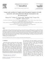
the wafers. Fig. 1a is a low-magnification cross-
sectional SEM image of the tilted sample. The
image shows the entire wire length from their
growth roots (lower edge of this image), which is
the location of the wafer (indicated by a two-way
arrow). It can be seen that the as-grown nanowires
on the wafer display well-aligned nature and have
length of up to several millimeters. A high-mag-
nification SEM image (Fig. 1b) clearly reveals the
diameter distribution of the nanowires. As seen
from this image, most of the wires have uniform
diameters of $50 nm, while some of them have
thinner diameters of 5–10 nm. A high-magnifica-
tion TEM image (Fig. 1c) shows that the nano-
wires are remarkably clean and smooth, and there
are no particles at its surface. An SAED pattern
(Fig. 1c, upper inset) of this wire reveals only dif-
fusive rings and no diffraction spots, showing the
amorphous nature of the synthesized nanowires.
The corresponding elemental composition is con-
firmed by EDAX (Fig. 1c, lower inset) to be Si and
O with an approximate atomic ratio of 1:2 (Cu
signal comes from TEM grids). Therefore, the
nanowires are identified as amorphous SiO
2
.In
contrast to the previous growth routes [1,4,5,8], no
metal catalytic particles (contamination) have been
340 J.Q. Hu et al. / Chemical Physics Letters 367 (2003) 339–343
found attached to the tips of the SiO
2
nanowires
(observed from SEM and TEM images).
The PL spectrum of the synthesized SiO
2
nanowires measured at room temperature is
shown in Fig. 2. The as-synthesized nanowires
have a stable (even after exposure to air for about
1 year), strong green emission band centered at 540
nm, which has been ascribed to neutral oxygen
vacancies [3]. Compared to the previous PL results
of SiO
2
nanowires, which show an intense main
peak with at least one shoulder [1,4], the present
PL curve is nearly symmetrical and appears not to
have any shoulder peaks. The exact nature of the
PL of the synthesized aligned SiO
2
nanowires re-
mains unclear and requires more detailed system-
atic investigations.
To study the growth processes of the SiO
2
nanowires, we placed several Si wafers in the re-
gion (on the alumina plate) where SiO
2
nanowire
growth would occur, and heated them in the tube
for different periods (1, 2, and 3 h). Fig. 3a shows
the Si wafer heated for 1 h, revealing the initial
nucleation stage of the SiO
2
nanowires. It seems
that at the given temperature the Si wafer surface
reacted with oxygen (the source of oxygen will be
discussed later) and formed numerous SiO
2
nanoparticles through homogeneous nucleation in
a suitable temperature region. The quantity of
these SiO
2
nanoparticles was so large that they
appeared as islands and covered partially the sur-
face of the wafer. Fig. 3b, c show the Si wafers
heated for 2 and 3 h, respectively, revealing the
different growth stages of SiO
2
nanowires. The
growth of SiO
2
nanowires appeared to start from
Fig. 2. Room temperature PL spectrum of SiO
2
nanowires.
Fig. 1. (a) Low-magnification cross-sectional SEM image with
the arrow indicating the wafer. (b) High-magnification SEM
image, and (c) TEM image (the insets show the SAED and
EDAX pattern, respectively), of the as-grown aligned SiO
2
nanowires.
J.Q. Hu et al. / Chemical Physics Letters 367 (2003) 339–343 341
the formed SiO
2
nanoparticles. The high density of
SiO
2
nanoparticles would lead to the concurrent
growth of a large number of SiO
2
nanowires, re-
sulting in the congested growth of SiO
2
nanowires.
The overcrowding effect would confine the prop-
agation of nanowires predominantly in the vertical
direction. As a result, SiO
2
nanowires emerged as
aligned bundles perpendicular to the Si wafer
surface, except for those formed at the exposed
edges where the SiO
2
nanoparticle islands could
grow freely. The nanowire alignment due to
overcrowding effect is somewhat similar to the
production of aligned carbon nanotubes [9,10]. In
comparison with the observation by Wang et al.
[7,8], the present growth of aligned SiO
2
nanowires
was based on a simple thermal oxidation of the
silicon wafer. In our case, the aligned SiO
2
nano-
wires grew in large area (the dense SiO
2
nanowires
covered the whole surface of the wafer) and had
ultralong lengths approaching several millimeters.
In addition, since no metal catalyst was involved,
the product was free of metal catalysis contami-
nation.
The source of oxygen that contributed to the
formation of SiO
2
nanowires may have several
origins. The most likely source of oxygen may
come from the low content of H
2
O($20 ppm) and
O
2
($20 ppm) in the carrier gas of Ar, which can
supply a constant oxygen source during the growth
of SiO
2
nanowires. Another likely source is the
oxygen adsorbed on the Si wafer due to air expo-
sure during the processing. The residual oxygen
may also be a source, as the base pressure
(6 Â 10
À2
Torr) of the vacuum system was rela-
tively high.
4. Conclusions
A simple method based on thermal oxidation of
Si wafers has been suggested for the large-scale
synthesis of very long aligned silica nanowires. The
SiO
2
nanowires were highly pure (no metal catal-
ysis contamination), ultralong (millimeters). Most
of wires had uniform diameters of $50 nm, while
some of them had thinner diameters of 5–10 nm.
Room-temperature PL spectra of the synthesized
SiO
2
nanowires showed a strong and stable green
emission peaking at 540 nm. By selecting suitable
gas source, e.g., NH
3
or CH
4
, it is reasonable to
expect that the aligned SiO
2
nanowires (acting as a
template or solid source material) can be converted
to other important material aligned nanowires,
e.g., SiC or Si
3
N
4
.
Acknowledgements
The authors express their gratitude to Dr. Q. L.
Liu (from Institute of Physics, Center for Con-
densed Matter Physics, Chinese Academy of Sci-
ences) for making available photoluminescence
measurements. The work was supported by a grant
Fig. 3. The Si wafers heated for different periods: (a) 1 h,
(b) 2 h, and (c) 3 h.
342 J.Q. Hu et al. / Chemical Physics Letters 367 (2003) 339–343
from the Research Grants Council of the Hong
Kong SAR, China [Project No. CityU 3/01C
(8730016)] and a Strategic Research Grant of the
City University of Hong Kong [Project No.
7001175].
References
[1] D.P. Yu, Q.L. Hang, Y. Ding, H.Z. Zhang, Z.G. Bai, J.J.
Wang, Y.H. Zou, Appl. Phys. Lett. 73 (1998) 3076.
[2] L.S. Liao, X.M. Bao, X.Q. Zheng, N.S. Li, N.B. Min,
Appl. Phys. Lett. 68 (1996) 850.
[3] H. Nishikawa, T. Shiroyama, R. Nakamura, Y. Ohiki, K.
Nagasawa, Y. Hama, Phys. Rev. B 45 (1992) 586.
[4] X.C. Wu, W.H. Song, K.Y. Wang, T. Hu, B. Zhao, Y.P.
Sun, J.J. Du, Chem. Phys. Lett. 336 (2001) 53.
[5] Z.Q. Liu, S.S. Xie, L.F. Sun, D.S. Tang, W.Y. Zhou, C.Y.
Wang, W. Liu, Y.B. Li, X.P. Zou, G. Wang, J. Mater. Res.
16 (2001) 683.
[6] H. Takikawa, M. Yatsuki, T. Sakakibara, Jpn. J. Appl.
Phys. 38 (1999) L401.
[7] Z.L. Wang, R.P. Gao, J.L. Gole, J.D. Stout, Adv. Mater.
12 (2000) 1938.
[8] Z.W. Pan, Z.R. Dai, C. Ma, Z.L. Wang, J. Am. Chem.
Soc. 124 (2002) 1817.
[9] W.Z. Li, S.S. Xie, L.X. Qian, B.H. Chang, B.S. Zou, W.Y.
Zhou, R.A. Zhao, G. Wang, Science 274 (1996) 1701.
[10] M. Terrones, N. Grobert, J. Olivares, J.P. Zhang, H.
Terrones, K. Kordatos, W.K. Hsu, J.P. Hare, P.D.
Townsend, K. Prassides, A.K. Cheetham, H.W. Kroto,
D.R.M. Walton, Nature 388 (1997) 52.
J.Q. Hu et al. / Chemical Physics Letters 367 (2003) 339–343 343