- Trang chủ >>
- Khoa Học Tự Nhiên >>
- Vật lý
Highly efficient and stable photoluminescence from silicon nanowires coated with sic
Bạn đang xem bản rút gọn của tài liệu. Xem và tải ngay bản đầy đủ của tài liệu tại đây (625.08 KB, 4 trang )
Highly ecient and stable photoluminescence from silicon
nanowires coated with SiC
X.T. Zhou, R.Q. Zhang, H.Y. Peng, N.G. Shang, N. Wang, I. Bello, C.S. Lee,
S.T. Lee
*
Department of Physics and Materials Science, Center of Super-Diamond and Advanced Films (COSDAF),
City University of Hong Kong, Hong Kong, People's Republic of China
Received 23 June 2000
Abstract
A reaction of silicon nanowires (SiNW) with methane and hydrogen has been performed to produce a thin coating
layer of cubic silicon carbide (b-SiC) using an ion beam deposition technique. High resolution transmission electron
microscopy (HRTEM) showed that silicon oxide shells originally cladding the as-grown SiNW were removed and
replaced by a thin layer of nano-sized crystals of b-SiC. This has led to stable photoluminescence (PL) observed from
the SiC-coated SiNW with high eciency almost tripled as compared with that before SiC coating. Ó 2000 Elsevier
Science B.V. All rights reserved.
In the past decade, intensive studies on porous
silicon related nano-technology have been stimu-
lated by its potential applications in silicon-based
optoelectronic devices [1±12]. One-dimensional
silicon nanowire in the same family is considered
more promising for its inherent quantum con®ne-
ment eect in the other two dimensions [13].
Large-scale synthesis, which is always an essential
requirement for wide applications, of silicon
nanowire (SiNW) has recently been achieved rou-
tinely [14,15]. However, problems of degradation
[2,16,17] and low photoluminescence (PL) e-
ciency [18] from the silicon nano-structures remain
unsolved. The PL degradation, usually accompa-
nied with a blue-shift of the PL peak position, is
generally believed to originate from the chemical
instability of the surfaces [2,16,17]. Upon exposure
to the atmosphere, the thickness of the surface
silicon oxide layer increases, and leads to increased
absorption and re¯ection of the incident light. This
eventually leads to degradation of the PL intensity
[16,17,19]. At the same time, the oxidation process
reduces the size of the core Si nano-crystallites and
thus results in the blue shift [16,17]. Before large-
scale application is possible, this instability prob-
lem has to be solved. One possible solution is to
passivate the silicon surface. Among the existing
passivation methods, a nitridation is generally
believed to be an advantage [20]. However, there is
a technological complication involving the re-
moval of the surface oxide layer in order to achieve
a satisfactory nitriding eect. In addition, a dia-
mond-like carbon ®lm has been used to coat po-
rous silicon [21]. Nevertheless, the problem of
stability has not yet been satisfactorily solved and
further eort is still needed to make the promising
22 December 2000
Chemical Physics Letters 332 (2000) 215±218
www.elsevier.nl/locate/cplett
*
Corresponding author. Fax: +852-2784-4696.
E-mail address: (S.T. Lee).
0009-2614/00/$ - see front matter Ó 2000 Elsevier Science B.V. All rights reserved.
PII: S 0 0 0 9 - 2 6 1 4 ( 0 0 ) 0 1 145-3
silicon nano-structures applicable for the widely
desired nano-technology.
Silicon carbon (SiC) is a semiconductor with
various merits including a wide energy band gap, a
high breakdown electric ®eld, and a high satura-
tion electron drift velocity. Furthermore, it is du-
rable at high temperature and possesses high
thermal conductivity and chemical stability. In
addition, it has good mechanical properties, such
as high tensile strength and a high Young's mod-
ulus which facilitate its application as a protective
coating material. Such a material should be ideal
for the integration with silicon in optoelectronic
devices. In this Letter, we show that SiNW can be
coated with b-SiC so as to integrate the various
advantages of the SiC with the important nano-
scaled materials in order to advance its applica-
tions in photophysics and device physics.
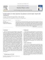
SiNW were synthesized by thermal evapora-
tion from a mixture of silicon and silicon dioxide
powder [14,15]. The SiNWs deposited on a silicon
wafer have an average diameter of about 20 nm,
as shown in Fig. 1. High resolution transmission
electron microscopy (HRTEM) shows that the
SiNW consists of a core of crystalline silicon
clothed by a shell of silicon oxide with a thickness
up to half of the wire radius (Fig. 1b). A micro-
Raman spectrometer (Ranishaw 2000, wavelength
of laser: 514 nm) was used to characterize the PL
properties of the sample. Very weak PL intensity
was obtained from the as-grown SiNW sample
(Fig. 2a). The PL peak centers at around 630 nm
(2.0 eV). No PL peak relating to the presence of
the silicon oxide was found. The low PL intensity
is likely due to the presence of the silicon oxide
shell, which covers the SiNWs. The silicon oxide
can absorb and re¯ect both the excitation inci-
dent light as well as the emitted PL light. Al-
though some kinds of silicon oxide can also give
visible PL, it is dicult to obtain a suitable
composition of silicon oxide required for ecient
luminescence. Therefore, it is desirable to mini-
mize or eliminate the silicon oxide in order to
obtain silicon nano-structures with ecient PL.
To address such a need, the following experiment
has been done.
The as-grown SiNW sample was transferred to
an ion beam deposition chamber with a base
vacuum of 2 Â 10
À7
Torr. A broad-beam Kaufman
ion source fed with a mixture of methane
(>99.9%), hydrogen (>99.999%) and argon
(>99.995%) was used to deposit thin ®lms onto the
Fig. 1. Typical TEM images of SiNW: (a) morphology at low
magni®cation; (b) morphology at higher magni®cation. Inset
shows the corresponding electron diraction pattern.
Fig. 2. PL from: (a) as-grown; (b) ion beam treated silicon
nanowire sample.
216 X.T. Zhou et al. / Chemical Physics Letters 332 (2000) 215±218
SiNW sample. The three gases were mixed in a
ratio of CH
4
:H
2
:Ar 1:50:173. The total ¯ow rate
was 2 sccm and the deposition pressure was kept at
about 5 Â 10
À4
Torr. Ions from the Kaufman
source were accelerated to bombard the SiNW
with an accelerating voltage of 200 V. The ion dose
as measured by a Faraday cup placed next to the
SiNW sample was 3 Â 10
19
cm
À2
. The substrate
temperature measured by an infrared pyrometer
was 700°C.
The TEM image and selected area electron
diraction (SAED) pattern as shown in Fig. 3a
indicate that a cubic silicon carbide layer has been
formed just outside the silicon nanowire. The
HRTEM images (Fig. 3b,c) show that a few b-SiC
nanoparticles contact the core of the SiNW di-
rectly for some nanowires (Fig. 3b), and that a
very thick outer layer composed of b-SiC nano-
particles exists for the other nanowires (Fig. 3c).
There is no clear evidence of the presence of the
silicon oxide layer in the ion bombarded SiNW
after the ion beam deposition (Figs. 2b and 3c). It
would be of great importance that the previously
observed silicon oxide layer covering the silicon
nanowire has been removed after the coating of
the silicon carbide.
It is of particular interest that the undesirable
oxide layer was replaced by a thin layer of stable,
wide band gap semiconductor. This new shell
would allow the high transmission of both the ex-
citation light and the PL emission with much less
loss. It is believed that during the deposition pro-
cess, hydrocarbon and hydrogen ions react with
silicon oxide to form carbon oxide and silicon car-
bide. The carbon oxide gas was pumped out while
silicon carbide remained on the silicon nanowire.
Fig. 2b shows the PL spectrum of the SiC-
coated SiNW sample. It can be seen that the PL
intensity has been increased by about three times.
The broadening of the PL peak towards higher
energy is probably due to the reduction of the di-
ameters of the SiNW upon ion bombardment. The
PL peak is in a similar region to that of the porous
silicon, showing no apparent change dierence
when the dimension of the silicon nano-structure is
increased from zero to one.
It is well known that SiC is relatively inert to air
and thus can prevent the silicon nanowire core
from further oxidation. Therefore, the coated
SiNW are likely to be very stable. We have mea-
sured PL of the coated SiNW after prolonged
exposure to a laser and exposure to the atmo-
sphere (75 days). No obvious PL degradation was
Fig. 3. Typical TEM images of SiNW after ion beam treat-
ment: (a) morphology at lower magni®cation; (b) morphology
at higher magni®cation.
X.T. Zhou et al. / Chemical Physics Letters 332 (2000) 215±218 217
observed. The present results indicate that the
ion beam SiC coating process may ®nd practical
usage in fabricating silicon nano-devices or photo-
devices.
Although there have been reports on the PL
from SiC nanostructures [22,23], the nano-scale
SiC layer on the silicon nanowire shows no no-
ticeable PL. The absence of PL relating to SiC
nano-structures may be due to insucient excita-
tion in our PL measurement.
In conclusion, silicon carbide coating of SiNW
can be achieved by ion beam deposition. The
coated SiNW show enhanced performance in
terms of both the intensity and stability of PL.
Acknowledgements
Financial support from the Research Grant
Council of Hong Kong under Grant No. 9040365
is gratefully acknowledged.
References
[1] L.T. Canham, Appl. Phys. Lett. 57 (1990) 1046.
[2] D.W. Cooke, B.L. Bennett, E.H. Farnum, W.L. Hults,
K.E. Sickafus, J.F. Smith, J.L. Smith, T.N. Taylor,
P. Tiwari, Appl. Phys. Lett. 68 (1996) 1663.
[3] A.G. Cullis, L.T. Canham, Nature 353 (1991) 335.
[4] T.K. Sham, D.T. Jiang, I. Couithard, J.W. Lorimer,
X.H. Feng, K.H. Tan, S.P. Frigo, R.A. Rosenberg,
D.C. Houghton, B. Bryskiewicz, Nature 363 (1993) 331.
[5] L. Tsybeskov, Ju.V. Vandyshev, P.M. Fauchet, Phys. Rev.
B 49 (1994) 7821.
[6] F. Buda, J. Kohano, M. Parrinello, Phys. Rev. Lett. 69
(1992) 1272.
[7] B. Delley, E.F. Steigmeier, Phys. Rev. B 47 (1993) 1397.
[8] J.P. Proot, C. Delerue, G. Allan, Appl. Phys. Lett. 61
(1992) 1948.
[9] A. Loni, A.J. Simons, T.I. Cox, P.D.J. Calcott, L.T.
Canham, Electron. Lett. 31 (1995) 1288.
[10] S. Lazarouk, P. Jaguiro, S. Katsouba, G. Masini, S. La
Monica, G. Maiello, A. Ferrari, Appl. Phys. Lett. 68 (1996)
2058.
[11] L. Tsybeskov, S.P. Duttagupta, K.D. Hirschman, P.M.
Fauchet, Appl. Phys. Lett. 68 (1996) 2058.
[12] K.D. Hirschman, L. Tsybeskov, S.P. Duttagupta, P.M.
Fauchet, Nature 384 (1996) 338.
[13] A.M. Morales, C.M. Lieber, Science 279 (1998) 208.
[14] N. Wang, Y.H. Tang, Y.F. Zhang, C.S. Lee, S.T. Lee,
Phys. Rev. B 58 (1998) R16024.
[15] N. Wang, Y.H. Tong, Y.F. Zhang, C.S. Lee, S.T. Lee,
Chem. Phys. Lett. 299 (1998) 237.
[16] L.T. Canham, M.R. Houlton, W.Y. Leong, C. Pickering,
J.M. Keen, J. Appl. Phys. 70 (1991) 442.
[17] M.A. Tischler, R.T. Collins, J.H. Stathis, J.C. Tsang, Appl.
Phys. Lett. 60 (1992) 639.
[18] D.P. Yu, Z.G. Bai, J.J. Wang, Y.H. Zou, W. Qian, J.S. Fu,
H.Z. Zhang, Y. Ding, G.C. Xiong, L.P. You, J. Xu,
S.Q. Feng, Phys. Rev. B 59 (1999) R2498.
[19] R.Q. Zhang, J. Costa, E. Bertran, Phys. Rev. B 53 (1996)
7847.
[20] A. Daami, G. Bremond, L. Stalmans, J. Poortmans,
J. Lumin. 80 (1998) 169.
[21] Y.P. Piryatinskii, V.A. Semenovich, N.I. Klyui, A.G.
Rozhin, Tech. Phys. 43 (1998) 423.
[22] H.W. Shim, K.C. Kim, Y.H. Seo, K.S. Nahm, E.K. Suh,
H.J. Lee, Y.G. Hwang, Appl. Phys. Lett. 70 (1997) 1757.
[23] O. Jessensky, F. Muller, U. Gosele, Thin Solid Films 297
(1997) 224.
218 X.T. Zhou et al. / Chemical Physics Letters 332 (2000) 215±218