- Trang chủ >>
- Khoa Học Tự Nhiên >>
- Vật lý
Ultrafast growth of single crystalline si nanowires
Bạn đang xem bản rút gọn của tài liệu. Xem và tải ngay bản đầy đủ của tài liệu tại đây (1.21 MB, 4 trang )
Ultrafast growth of single-crystalline Si nanowires
J.B. Chang, J.Z. Liu, P.X. Yan
⁎
, L.F. Bai, Z.J. Yan, X.M. Yuan, Q. Yang
Institute for Plasma and Metal Materials, Lanzhou University, Lanzhou 730000, China
Received 25 September 2005; accepted 22 December 2005
Available online 31 January 2006
Abstract
Silicon nanowires (SiNWs) have been catalytically synthesized by heat treatment of Si nanopowder at 980 °C. The SiNWs comprise crystalline
Si nanoparticles interconnected with metal catalyst. The formation mechanism of nanowires generally depends on the presence of Fe catalysts in
the synthesis process of solid–liquid–solid (SLS). Although gas phase of vapor–liquid–solid (VLS) method can be used to produce various of
different nanowire materials, growth model based on the SLS mechanism by heat treatment is more ascendant for providing ultrafast growth of
single-crystalline Si nanowires and controlling the diameter of them easily. The growth of single-crystalline SiNWs and morphology were
discussed.
© 2006 Elsevier B.V. All rights reserved.
Keywords: SiNWs
1. Introduction
Crystalline nanostructures offer unique access to low-
dimensional physics, and they can be used as nanotechnology
building blocks to reach higher device integration densities than
conventional fabrication methods and have more singularity
character. One-dimensional (1D) structure with nanometer
diameters, such as carbon nanotubers and semiconductor
nanowires, has great potential for testing and understanding
fundamental concepts about the roles of dimensionality and
size, for example, optical, electrical, and mechanical properties
and for their potential applications in research and electronic
nanodevices [1]. It's known that Si nanowires (SiNWs) have the
strong ability to confine photoenergy from visible light [2].
SiNWs are partic ularly attractive due to the central role of the
silicon semiconductor industry, which would allow SiNWs to
be implemented using existing technologies. Because silicon
turns into a direct band-gap semiconductor at nanometer size
due to quantum confinement [3],itcouldbeusedin
optoelectronics. SiNWs can be synthesized by laser ablation
[1], thermal evaporation of solid sources [4–6] and chemical
vapor deposition (CVD) [7]. The various directional features of
these techniques were reported and the model proposed for
preferred SiNWs growth directions [8]. These methods are often
based on the vapor–liquid–solid (VLS idea [9] using various
metals as catalysts, such as Au, Fe, Ti and Ga, to enhance the
growth of SiNWs. In this work, we demonstrate a simple
method of growing SiNWs. Si nanopowder was used in our
work instead of the dangerous gas of silane as the Si source.
The synthesis of SiNWs was carried out using a mixture of Si
nanoparticles and iron nitrate by thermal treatment at 980 °C in
an evacuated sealed quartz tube. The key parameter necessary to
induce nanowires formation is the tem perature and catalyst.
Materials Letters 60 (2006) 2125 – 2128
www.elsevier.com/locate/matlet
⁎
Corresponding author. Tel.: +86 931 8912661; fax: +86 931 8913554.
E-mail address: (P.X. Yan).
Fig. 1. XRD spectrum of Si nanoparticles prepared by cathode arc plasma.
0167-577X/$ - see front matter © 2006 Elsevier B.V. All rights reserved.
doi:10.1016/j.matlet.2005.12.085
2. Experiment
Before the preparation of producing SiN Ws, Si nanoparticles
were synthesized by cathode arc plasma which is one of the
most powerful met hods because of uniform particles and high
efficiency, using a mixture of SiO
2
and C with a molar
proportion (1 :1) in argon atmosphere. The discharge voltage is
20 V and the current is 120 A. The flux of hydrogen as
protective gas was 15 standard cubic centimeters per minute.
Then the Si nanopowder was ultrasonically dispersed in the
alcohol solution containing Fe(NO
3
)
3
, which was subsequently
evaporated and the dried samples were calcined in an H
2
flow at
980 °C for 1 h. X-ray diffraction (XRD) measurements and
transmission electron microscope (TEM) were employed to
Fig. 2. TEM image of Si nanoparticles by cathode arc plasma.
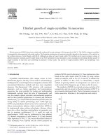
Fig. 3. (a) TEM image of Si nanoparticles after heat treatment at 980 °C without Fe catalysts and (b) nanowires with Fe, (c) TEM image showing nanopartical catalysts
at the end of nanowires. (d) TEM image of an individual smooth nanowire and corresponding SAED pattern.
2126 J.B. Chang et al. / Materials Letters 60 (2006) 2125–2128
investigate the structure and morphology of the Si nanoparticles
and nanowires.
3. Results and discussion
The phase composition and phase structure of the as-synthesized
products were examined by X-ray diffraction (XRD, Siemens D-500
with Cu Ka radiation and a normal 2θ scan ). Fig. 1 shows a typical
XRD spectrum of the Si nanoparticles on different crystal planes
synthesized by cathode plasma discharge. It can be seen from the
dominant diffraction peaks, as indexed in the spectrum, and originated
from cubic-structure Si, which can be readily indexed to face-centered
cell of Si (Joint Committee on Powder Diffraction Standard (JCPDS)
Card, No. 05-0565). Their average diameter of 21.6 nm was calculated
using Scherrer equation, which is in good accordance with the TEM
observation (Fig. 2).
During the heat treatment at 980 °C, Si nanoparticles couldn't be
transformed into nanowires if there was no iron catalysts as shown in
Fig. 3(a). When introduced iron catalysts, Si nanowires came out. Fig.
3(b) shows the TEM images of SiNWs with tens of nanometer in
diameter and several hundreds of micrometers in length. Fe
nanoparticles as catalysts are embedded in SiNWs as shown in Fig. 3
(c). Selected area electron diffraction (SAED) indicates that the
nanowires are made of crystalline silicon in Fig. 3(d).
After heat treatment, when H
2
flow was closed and air was
introduced into the quartz tube during the cooling procedure, SiO
x
nanotubes would be produced deriving from SiNWs oxidated (Fig. 4).
The ring in the SAED image inserted in Fig. 4(b) is from the reflection
of SiO
2
(400).
The mechanism of SiNWs growth is explained now. At high
temperature, Fe(NO
3
)
3
deposited into iron oxide and momentarily
deoxidized into iron nanodroplets in H
2
atmosphere. In the metal-
catalyzed SLS technique, a liquid metal cluster or catalyst acts as the
energetically favored site for the adsorption fusing Si nanoparticles of
solid-phase reactants, to function as Si reservoir by eutectic liquid
formation, and to become supersaturated with Si. The present of a
nanopartical catalyst at one end of the nanowires is the essential feature
of SLS growth. As shown by the arrow in Fig. 3(c), SiNWs terminated
at one end in a nanoparticle with a diameter 1∼1.2 times that of the
connected nanowire. Fig. 3(d) shows the TEM image of an individual
smooth nanowire and the corresponding selected area diffraction
(SAED) pattern. The d-spacings of the nanocrystals calculated from the
two diffraction dots of the SAED pattern are consistent with those of Si
(200) and (400). It is well established that Si nanowires grown by the
metal-catalyzed SLS technique usually have a growth direction along
(200) and are single-crystalline [10]. Therefore, the (200) growth
direction may be regarded as a typical feature of the metal-catalyzed
SLS process. We surprisedly find that nanotubes appear in this
experiment in Fig. 4(a). It is obvious that nanopartical catalysts are at
the end of nanotubes as shown by the arrow. Fig. 4(b) shows TEM
image of an individual nanotube and the corresponding SAED pattern
(diffraction ring), in which the ring is from reflection of SiO
2
(400).
When inpouring air into the evacuated sealed quartz tube at high
temperature, the surface of SiNWs was immediately oxided [11] and
meanwhile the Si inside was melted and subsequently evaporated
leaving a SiO
2
tube [2].
4. Propose mechanism
The formation mechanism of nanowires generally depends
on the presence or absence of metal catalysts in the synthesis
process, i.e., SLS and VLS except for oxide-assisted growth
[12]. Unlike the well-developed VLS, the detail of SLS process
for silicon nanowires is not expatiated. In this paper, we
Fig. 4. (a) TEM image showing catalysts at the end of nanotubes, (b) TEM image of an individual nanotube and SAED pattern.
Fig. 5. Schematic figure of nanowires growth process.
2127J.B. Chang et al. / Materials Letters 60 (2006) 2125–2128
expound SiNWs formati on mechanism. The solid–liquid– solid
(SLS) nanowires growth mechanism is illustrated in the case of
nanowires growth process in Fig. 5. The SiNWs have been
synthesized by heat treatment of Si nanoparticles. In the metal-
catalyzed SLS technique, a liquid metal cluster or catalyst acts
as the energetically favored site for the adsorption of liquid-
phase reactants. The heat treatment can be acted as a kind of
sinter. In this experiment, the prim ary growth mode is that
agglomeration incorporates particles into nanowires. In heat
treatment process, particles begin to melt and the adsorbability
gradually augments with the temperature increasing. Because of
the driving force of thermodynamics, the Si atoms dissolve in
the Fe nanocrystal to form a liquid FeSi seed droplet.
Thereupon, a tiny cervi x between Fe particle and Si particle
comes into being. The cervix is filled and leveled up through
diffusion of the surface. The seed droplet reaches the eutectic
composition. Si diffuses from the liquid molten alloy phase ball
and grows epitaxially at the liquid/solid interface. Simultaneity,
Fe catalysts remove ahead and continue to absorb other Si
nanoparticles resulting in the production of long SiNWs.
The nanowires are able to grow when the FeSi alloy eutectic
temperature and the concentration of crystallizing material can
be exceeded. The current interest in the physics and possible
applications of Si nanostructures and the need to develop
techniques to fabricate such structures made it appropriate to
take another look at the SLS technique as a means of Si
nanowires ultrafast growth of fabrication.
5. Conclusion
In summary, Si nanowires were catalytically synthesized by
calcining Si nanopowder containing Fe(NO
3
)
3
in an H
2
.A
pathway of the growth of SiNWs was presented based on SLS
mechanism. Under the conditions used to grow the nanowires,
diffusion of Si through or around a solid FeSi nanoparticle
appears to be rapid enough to transport Si away from the surface
to the growing wires. In addition, rapid oxidation of SiNWs
could lead to SiO
x
nanotubes. We further discussed the growth
mechanism.
Acknowledgements
We thank for Engineer Shuang Wang and Youxiang Li,
Testing and analytic center, Gansu Academy of Science, who
give a lot of help.
References
[1] Alfredo M. Morales, Charles M. Lieber, Science 279 (1998) 208.
[2] N. Wang, B.D. Yao, Y.F. Chan, X.Y. Zhang, Nano Lett. 3 (2003) 475.
[3] Xin yuan Zhao, M. Wei, L. Yang, M.Y. Choul, Phys. Rev. Lett. 92 (2004)
236805.
[4] N. Wang, Y.H. Tang, Y.F. Zhang, D.P. Yu, C.S. Lee, I. Bello, S.T. Lee,
Chem. Phys. Lett. 283 (1998) 368.
[5] Y.F. Zhang, Y.H. Zhang, N. Wang, D.P. Yu, C.S. Lee, I. Bello, S.T. Lee,
Appl. Phys. Lett. 72 (1998) 1835.
[6] D.P. Yu, et al., Appl. Phys. Lett. 72 (1998) 3458.
[7] S. Hofmann, C. Ducati, R.J. Neill, S. Piscanec, A.C. Ferrari, J. Appl. Phys.
94 (2003) 6005.
[8] T.Y. Tan, S.T. Lee, U. Gosele, Appl. Phys., A Mater. Sci. Process. 74
(2002) 423.
[9] R.S. Wagner, W.C. Ellis, Appl. Phys. Lett. 4 (1964) 89.
[10] Junjie Niu, Jian Sha, Xiangyang Ma, Chem. Phys. Lett. 367 (2003) 528.
[11] J.O. Hu, Y. Jing, X.M. Meng, C.S. Lee, Chem. Phys. Lett. 367 (2003) 339.
[12] C.N.R. Rao, F.L. Deepak, Gautam Gundiah, A. Govindaraj, Prog. Solid
State Chem. 31 (2003) 5–147.
2128 J.B. Chang et al. / Materials Letters 60 (2006) 2125–2128